

Animal models in spine research: applications, advantages, limitations, and future perspectives
 0
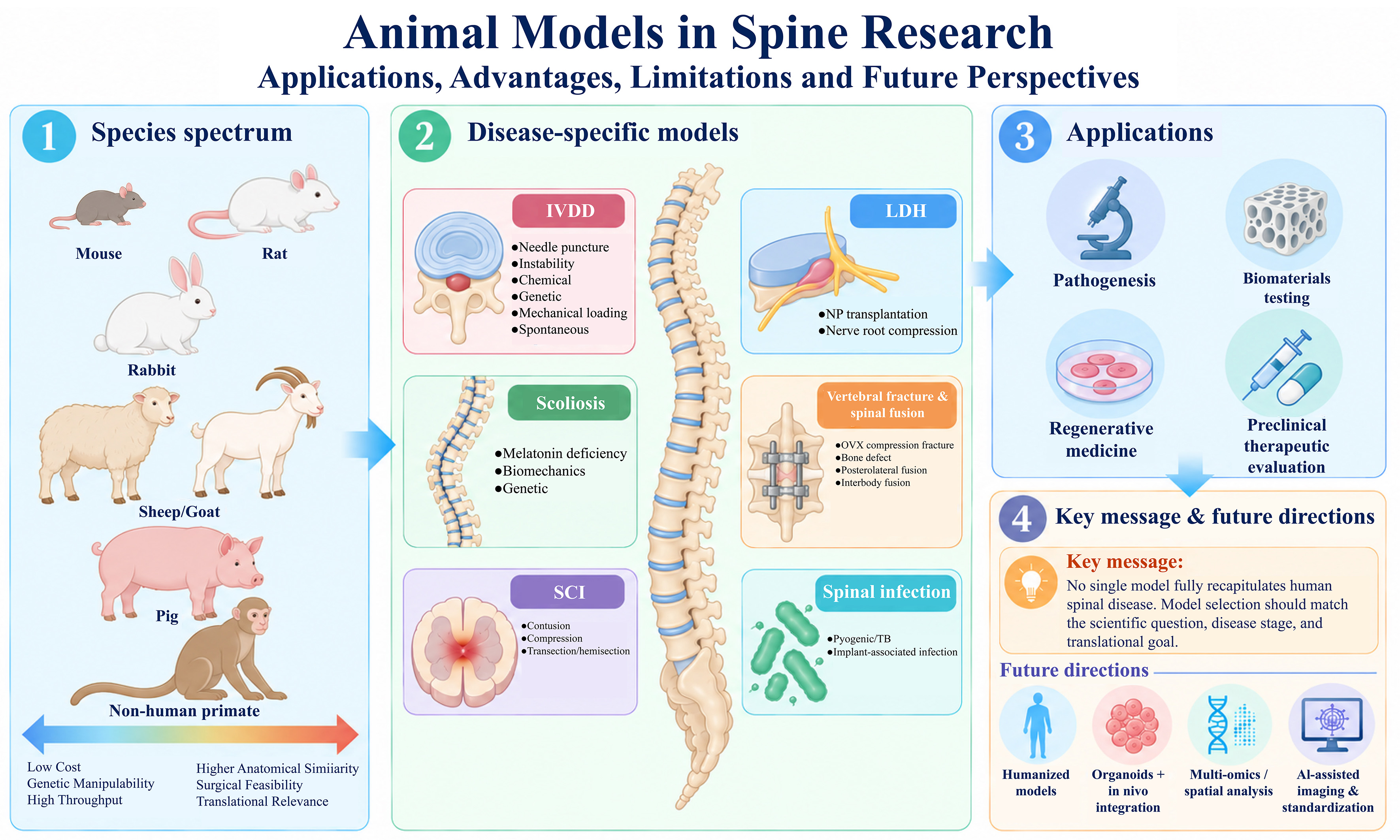
0 Graphical Abstract
Abstract
Animal models are indispensable for elucidating the pathophysiological mechanisms of spinal disorders and facilitating the translation of basic research into clinical practice. Given the anatomical complexity, distinctive biomechanical environment, and heterogeneous pathological features of the spine, no single model can fully recapitulate the spectrum of human spinal diseases. Careful selection of animal species and modeling approaches is therefore essential to ensure experimental validity, reproducibility, and translational relevance. In this review, we systematically summarize the major animal models used in spinal research, including those for intervertebral disc degeneration, lumbar disc herniation, scoliosis, vertebral fracture and spinal fusion, spinal cord injury, and spinal infection. For each disease category, we describe representative modeling approaches, key pathological features, major experimental applications, and their respective strengths and limitations. We further compare the suitability and translational relevance of commonly used species, ranging from rodents to large animals, and discuss their applications in mechanistic investigations, biomaterial testing, regenerative medicine, and preclinical therapeutic evaluation. In addition, we highlight the major limitations of current models and emerging advances in disease modeling technologies. Rather than serving as interchangeable experimental platforms, different animal models are best suited to distinct scientific questions and stages of translational research. This review provides a structured framework for understanding the characteristics of currently available spinal animal models and may facilitate more rational model selection in both basic and translational spine research.
Keywords


 Download PDF
INTRODUCTION
Spinal disorders are major contributors to chronic pain, functional impairment, and reduced quality of life, and account for a substantial and rapidly increasing proportion of the global musculoskeletal disease burden[1]. With population aging and profound changes in lifestyle and occupational patterns, the health and socioeconomic burden associated with spinal disorders continues to grow. Data from the Global Burden of Disease (GBD) Study consistently identify low back pain and related spinal conditions among the leading causes of disability worldwide, with their overall burden expected to increase further as populations expand and age[2]. Among degenerative spinal disorders, advancing age, obesity, sedentary behavior, and abnormal mechanical loading are recognized as important risk factors for the development and progression of intervertebral disc degeneration (IVDD) and lumbar disc herniation (LDH)[3]. Epidemiological and experimental evidence further indicates that age, elevated body mass index (BMI), lifestyle-related factors, and occupational mechanical loading are closely associated with disc degeneration and herniation. In particular, obesity may accelerate IVDD through a combination of increased axial spinal loading, chronic low-grade inflammation, and disruption of metabolic homeostasis within intervertebral disc cells[4]. Beyond degenerative conditions, scoliosis, spinal fractures, spinal cord injury (SCI), and spinal infections also represent important public health challenges whose burden is influenced by demographic shifts, changes in activity patterns, and healthcare-related factors[5-12]. Despite substantial advances in spinal surgery, diagnostic imaging, and biologically based therapies, the pathogenesis of many spinal disorders remains incompletely understood, and effective disease-modifying treatments remain limited. Elucidating the biological mechanisms underlying disease initiation and progression and identifying new therapeutic targets therefore remain major priorities in spine research.
Animal models are indispensable for bridging mechanistic investigation and clinical translation. Direct investigation in humans is often constrained by limited access to diseased tissues, the prolonged natural history of many spinal disorders, ethical considerations, and the difficulty of controlling experimental variables. Animal models provide a controllable in vivo platform to reproduce and systematically study disease initiation and progression, enabling investigation of pathological mechanisms, validation of therapeutic targets, and preclinical assessment of drugs, biomaterials, and other emerging interventions. Their importance is particularly evident in spinal research, where disease processes frequently involve coordinated alterations in bone, cartilage, intervertebral disc tissue, neural structures, vascular components, and the immune microenvironment. Such multicellular, biomechanically coupled interactions cannot be fully reproduced in vitro, underscoring the continued importance of well-designed animal models.
Spinal disorders are also distinguished from many other musculoskeletal conditions by their complex biomechanical and neuroanatomical environment. In addition to providing structural support and enabling movement, the spine protects the spinal cord and exiting nerve roots while continuously responding to dynamic mechanical loads. Consequently, the onset and progression of spinal disease often involve reciprocal interactions among abnormal mechanical stress, biomechanical instability, extracellular matrix (ECM) remodeling, inflammation, and neurological dysfunction. During IVDD, for example, excessive or aberrant mechanical loading can promote cellular senescence, inflammatory signaling, and ECM degradation through the activation of mechanosensitive pathways[13]. Similarly, following SCI, the initial mechanical insult initiates a secondary cascade characterized by inflammation, glial scar formation, and impaired neural regeneration[14]. An ideal spinal animal model should therefore reproduce not only the structural and histopathological features of the disease, but also, as far as possible, its biomechanical environment and key pathophysiological processes. Preservation of these features is essential for improving the biological validity and clinical relevance of experimental findings.
Over the past several decades, a wide range of animal models has been developed to study degenerative, traumatic, deformity-related, and infectious spinal disorders. Needle puncture, mechanical loading, and enzyme-induced models are widely used to investigate IVDD[15,16]. For LDH, autologous nucleus pulposus transplantation and nerve root compression models are commonly employed because they reproduce key pathological features, including interactions between herniated nucleus pulposus tissue and nerve roots, local inflammatory responses, and mechanical compression[17-19]. Bipedal models and pinealectomy-based models have contributed to investigations of the pathogenesis of adolescent idiopathic scoliosis[20], whereas contusion and compression models remain fundamental experimental approaches in SCI research[21]. In parallel with advances in regenerative medicine and tissue engineering, animal models are increasingly used to evaluate stem cell-based therapies, biomaterial scaffolds, hydrogel systems, and three-dimensional bioprinting strategies for spinal tissue repair and regeneration.
The selection of animal species is equally important because substantial interspecies differences exist in spinal anatomy, biomechanics, tissue composition, cellular phenotype, and patterns of disease progression. Rodents are widely used in mechanistic studies because of their relatively low cost, short reproductive cycles, and suitability for genetic manipulation. By contrast, medium- and large-sized animals, including rabbits, sheep, and pigs, more closely approximate certain aspects of human spinal dimensions, biomechanical properties, and surgical conditions, making them particularly valuable for preclinical validation and translational studies. Nevertheless, no species reproduces all features of the human spine, and the suitability of a given model depends strongly on the specific biological question, disease stage, therapeutic intervention, and outcome measure being investigated. Rational selection of both animal species and modeling strategy is therefore essential for improving experimental rigor, reproducibility, and translational relevance[22].
In this review, we summarize the current landscape of animal models used in spinal research, with particular emphasis on IVDD, LDH, scoliosis, vertebral fracture and spinal fusion, SCI, and spinal infection. We describe representative modeling approaches, key pathological features, experimental applications, and the main strengths and limitations of each model. We also compare the suitability of commonly used animal species and discuss the expanding application of these models in regenerative medicine, biomaterial evaluation, and tissue engineering. Finally, we highlight current limitations in spinal disease modeling and emerging directions that may improve physiological fidelity and translational value. By providing a structured comparison of available models, this review aims to support more rational model selection and facilitate the development of experimental systems that more closely reflect clinically relevant spinal pathology.
COMMONLY USED EXPERIMENTAL ANIMALS IN SPINE RESEARCH
The selection of an appropriate animal species is a critical consideration in the design of spinal research. Ideally, an experimental model should resemble the human spine in anatomy, biomechanical environment, disease progression, and therapeutic response, while also offering adequate experimental feasibility and reasonable cost. However, no single species can reproduce the full spectrum of pathological features observed in human spinal disorders. Animal selection should therefore be guided by the specific scientific question, disease model, intervention, and study endpoint.
The animals most commonly used in spinal research can be broadly categorized as small, medium-sized, and large species. Small animals primarily include mice and rats, whereas large animals include sheep or goats, pigs, and non-human primates (NHPs). Rabbits occupy an intermediate position in terms of body size and are therefore classified here as medium-sized animals [Figure 1]. These species differ substantially in spinal anatomy, intervertebral disc structure and composition, skeletal maturity and growth characteristics, biomechanical properties, experimental accessibility, and cost.
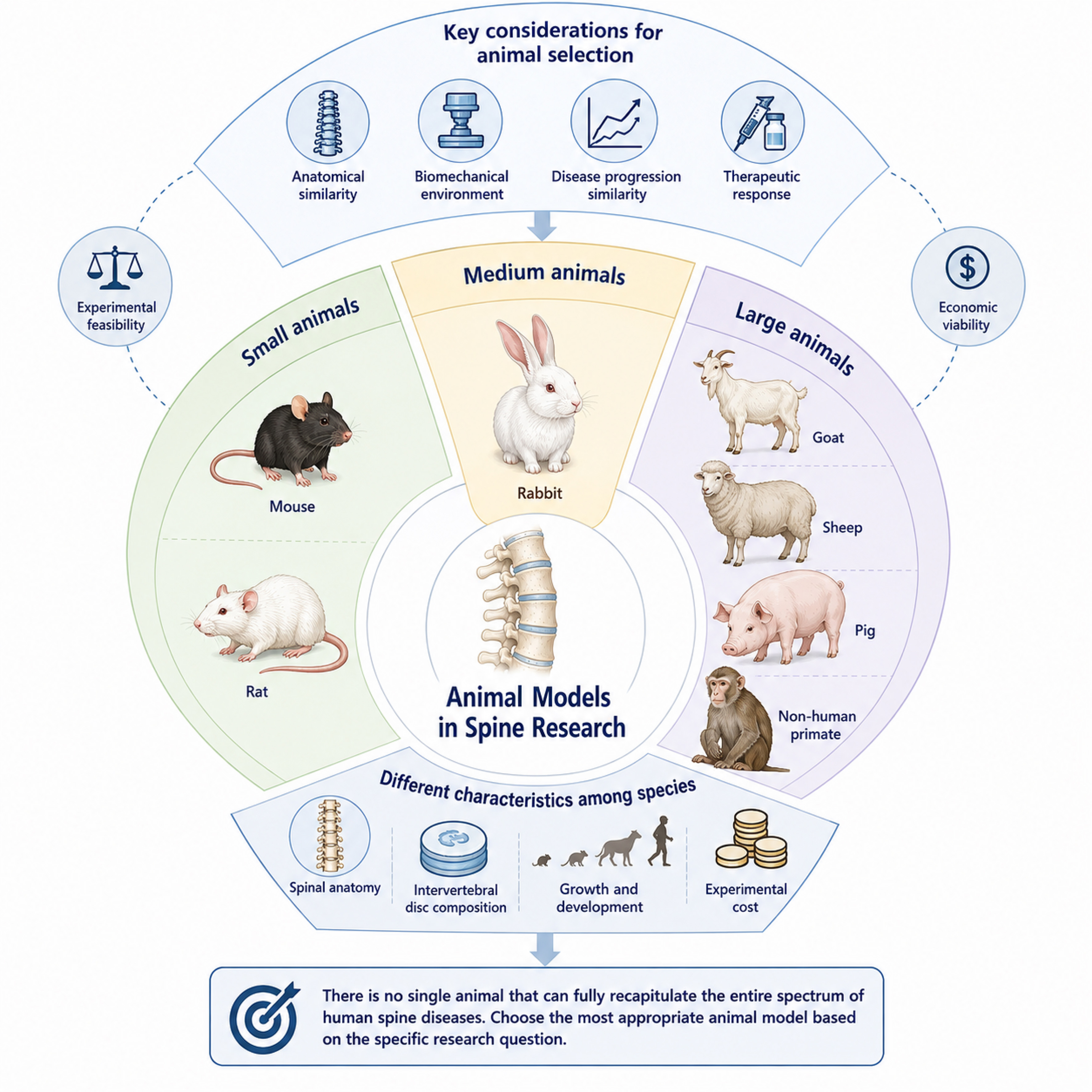
Figure 1. Commonly used experimental animals in spine research.
Mouse models
Mice are among the most widely used experimental animals in biomedical research because of their well-characterized genetic backgrounds, short reproductive cycles, relatively low maintenance costs, and extensive availability of genetic engineering tools. A wide range of transgenic, knockout, and conditional gene-modified mouse strains has been developed, providing a powerful experimental platform for investigating gene function and disease mechanisms.
In spine research, mice are extensively used to study IVDD, SCI, adolescent idiopathic scoliosis (AIS), and other spinal disorders[23-25]. Their greatest advantage lies in the availability of sophisticated genetic manipulation strategies. Cre-LoxP-based conditional gene targeting, clustered regularly interspaced short palindromic repeats/CRISPR-associated protein 9 (CRISPR/Cas9)-mediated genome editing, and lineage-tracing approaches enable cell type-specific interrogation of gene function, cell fate, and signaling pathways involved in spinal development and disease progression[26-29]. These features make mouse models particularly valuable for dissecting molecular and cellular mechanisms that are difficult to investigate in larger species.
Nevertheless, several anatomical and biological differences limit their direct translational relevance. Mouse intervertebral discs are very small, which restricts surgical manipulation, local therapeutic delivery, and repeated tissue sampling. In addition, notochordal cells remain relatively abundant within the nucleus pulposus during early life and young adulthood and gradually decline with age, differing from the cellular composition of adult human intervertebral discs. The quadrupedal posture of mice also produces loading patterns and spinal biomechanics that differ substantially from those of the human spine[30,31]. Accordingly, mouse models are particularly well suited to mechanistic and genetic studies, whereas their utility is more limited for technically demanding spinal procedures, device testing, and preclinical evaluation of interventions that require close anatomical or biomechanical similarity to humans.
Rat models
Rats are among the most commonly used experimental animals in spinal disease research. Compared with mice, their larger spinal dimensions facilitate surgical manipulation, local intervention, and tissue collection, while they retain practical advantages such as relatively low cost, straightforward husbandry, and short experimental cycles.
Rat models are extensively used for IVDD, LDH, and SCI, particularly in needle puncture-induced IVDD, nucleus pulposus-related LDH, and traumatic SCI paradigms[32-34]. In SCI research, the Basso, Beattie, and Bresnahan (BBB) locomotor rating scale is one of the most widely adopted methods for assessing hindlimb motor recovery in rats[35]. Rats also benefit from well-established experimental protocols for pain-related behavioral testing, pharmacological evaluation, imaging, and histological analysis, making them particularly useful for mechanistic validation and early-stage therapeutic assessment[36,37].
Despite these advantages, several limitations should be considered when extrapolating findings from rats to humans. Differences in intervertebral disc size, cellular composition, spinal loading patterns, and the rate and characteristics of experimentally induced degeneration may not fully reflect the slowly progressive and multifactorial nature of human disc degeneration. Accordingly, rat models are highly valuable for mechanistic studies and proof-of-concept evaluation but are less suitable for applications requiring close replication of human spinal anatomy, biomechanics, or long-term disease progression.
Rabbit models
Rabbits occupy an intermediate position between small rodents and large animal models and are therefore widely used as medium-sized experimental animals in spine research. Their relatively large intervertebral discs facilitate intradiscal injection, needle puncture, surgical manipulation, and imaging-based assessment compared with rodent models.
New Zealand White rabbits are among the most commonly used species for establishing needle puncture-induced IVDD models[38]. These models typically produce progressive and reproducible degenerative changes that can be readily evaluated by magnetic resonance imaging (MRI), histology, and other outcome measures, making rabbits particularly useful for studies of disc regeneration and the preclinical evaluation of biomaterials[39]. Rabbit models are also frequently employed in investigations of spinal fusion and spinal infection[40,41].
Despite these advantages, several limitations should be recognized. Adult rabbits may retain notochordal cells within the nucleus pulposus, and their intervertebral discs differ from those of adult humans in cellular composition and biological characteristics. In addition, rabbits have higher housing and experimental costs and require more demanding perioperative and husbandry management than rodents. Thus, although rabbit models offer greater anatomical accessibility and translational potential than small rodents for certain applications, species-specific differences should be carefully considered when extrapolating experimental findings to human spinal disease.
Goats and sheep models
Goats and sheep are widely used as large-animal models in translational spine research. Their spinal dimensions, intervertebral disc height, and biomechanical properties more closely approximate those of humans than small-animal models, making them particularly valuable for studies of spinal fusion, artificial disc replacement, and tissue-engineering strategies.
In IVDD research, goat and sheep models are well suited to studies requiring relatively long observation periods and can support longitudinal assessment using MRI, computed tomography (CT), histological analysis, and biomechanical testing[42]. Their larger vertebral dimensions also allow the use of clinically relevant implants, fixation systems, and surgical techniques, which is a major advantage in spinal fusion and device-related research[43]. These characteristics make ovine and caprine models particularly useful for evaluating the feasibility, safety, and therapeutic performance of regenerative strategies and spinal implants under conditions that more closely resemble clinical practice.
Nevertheless, these models have substantial limitations, including higher costs, greater requirements for housing and perioperative management, longer experimental periods, and fewer available genetic manipulation tools than rodent models. Accordingly, goats and sheep are most commonly used in late-stage preclinical studies, particularly when anatomical scale, biomechanical fidelity, and clinically relevant surgical procedures are important considerations.
Pig models
Pigs are increasingly used as large-animal models in translational spine research because their spinal anatomy, intervertebral disc structure, and biomechanical properties share several clinically relevant features with those of humans. The relatively large size of porcine intervertebral discs facilitates surgical manipulation, implantation procedures, and imaging-based assessment, while their endplate architecture and nucleus pulposus-annulus fibrosus organization provide a useful platform for evaluating disc degeneration and repair strategies. Accordingly, porcine models have been applied in studies of intervertebral disc regeneration, tissue engineering, artificial disc development, and spinal fusion techniques[44,45].
Miniature pigs have attracted particular interest in recent years because they preserve many of the anatomical and biomechanical advantages of conventional porcine models while offering improved practicality for long-term experimental studies. Their smaller body size generally facilitates housing, handling, perioperative management, and repeated imaging, while also reducing some of the logistical and economic demands associated with full-sized pigs[44,46]. These characteristics make miniature pigs particularly valuable for preclinical evaluation of biomaterials, regenerative therapies, and implantable devices requiring clinically relevant anatomical dimensions and surgical procedures.
NHPs models
NHPs share a number of anatomical, physiological, and biomechanical features with humans and therefore represent some of the most clinically relevant animal models available for translational spine research. Macaque models have been used to investigate naturally occurring or experimentally induced IVDD and SCI, particularly in studies aimed at evaluating the safety, efficacy, and translational potential of advanced therapeutic strategies[47-50].
Despite their translational advantages, the use of NHPs is substantially restricted by ethical considerations, high experimental and maintenance costs, limited availability, and stringent requirements for housing, handling, and experimental management. Their use is therefore generally reserved for selected late-stage preclinical studies in which close anatomical, physiological, or functional resemblance to humans is considered essential.
Considerations for model selection
Different animal models offer distinct strengths and limitations, and no single species is universally suitable for all research questions. Model selection should therefore be guided by the specific scientific objective, disease context, experimental duration, technical requirements, available resources, and intended stage of translation. In general, mice and rats are particularly well suited to mechanistic studies and early proof-of-concept experiments, whereas rabbits, goats, sheep, and pigs are more frequently used for biomaterial testing, device evaluation, surgical feasibility studies, and preclinical validation.
Rather than relying on a single species throughout the entire research process, a staged and complementary modeling strategy may provide greater translational value. Small-animal models can define molecular mechanisms and identify therapeutic targets, whereas medium- and large-animal models can provide more clinically relevant anatomical and biomechanical settings for validating therapeutic efficacy, safety, and procedural feasibility before clinical translation. With continued advances in gene editing, multi-omics technologies, humanized models, and other emerging experimental platforms, future spine research will likely adopt integrated, multi-model approaches that connect mechanistic discovery with preclinical validation and clinical translation.
DISEASE-SPECIFIC ANIMAL MODELS IN SPINE RESEARCH
Spinal disorders encompass a broad spectrum of degenerative, deformity-related, traumatic, and infectious conditions, each characterized by distinct pathogenic mechanisms, histopathological features, and clinical manifestations. Accordingly, disease-specific animal models are required to reproduce key biological and structural features most relevant to the scientific question being addressed.
Models of IVDD and LDH are among the most extensively used experimental systems for investigating degenerative spinal disorders. Scoliosis models are primarily employed to study abnormal spinal growth, asymmetric biomechanical loading, and the mechanisms underlying the initiation and progression of spinal deformity. Models of vertebral fracture, spinal fusion, and SCI are widely used to investigate tissue repair, bone regeneration, neural recovery, and the performance of biomaterials and implantable devices. Spinal infection models, including those designed to reproduce pyogenic, tuberculous, and implant-associated infections, provide additional platforms for investigating pathogen-host interactions, inflammatory responses, and infection-related tissue destruction. As experimental techniques and disease-modeling strategies advance, spinal animal models are becoming increasingly refined, reproducible, and clinically relevant [Figure 2].
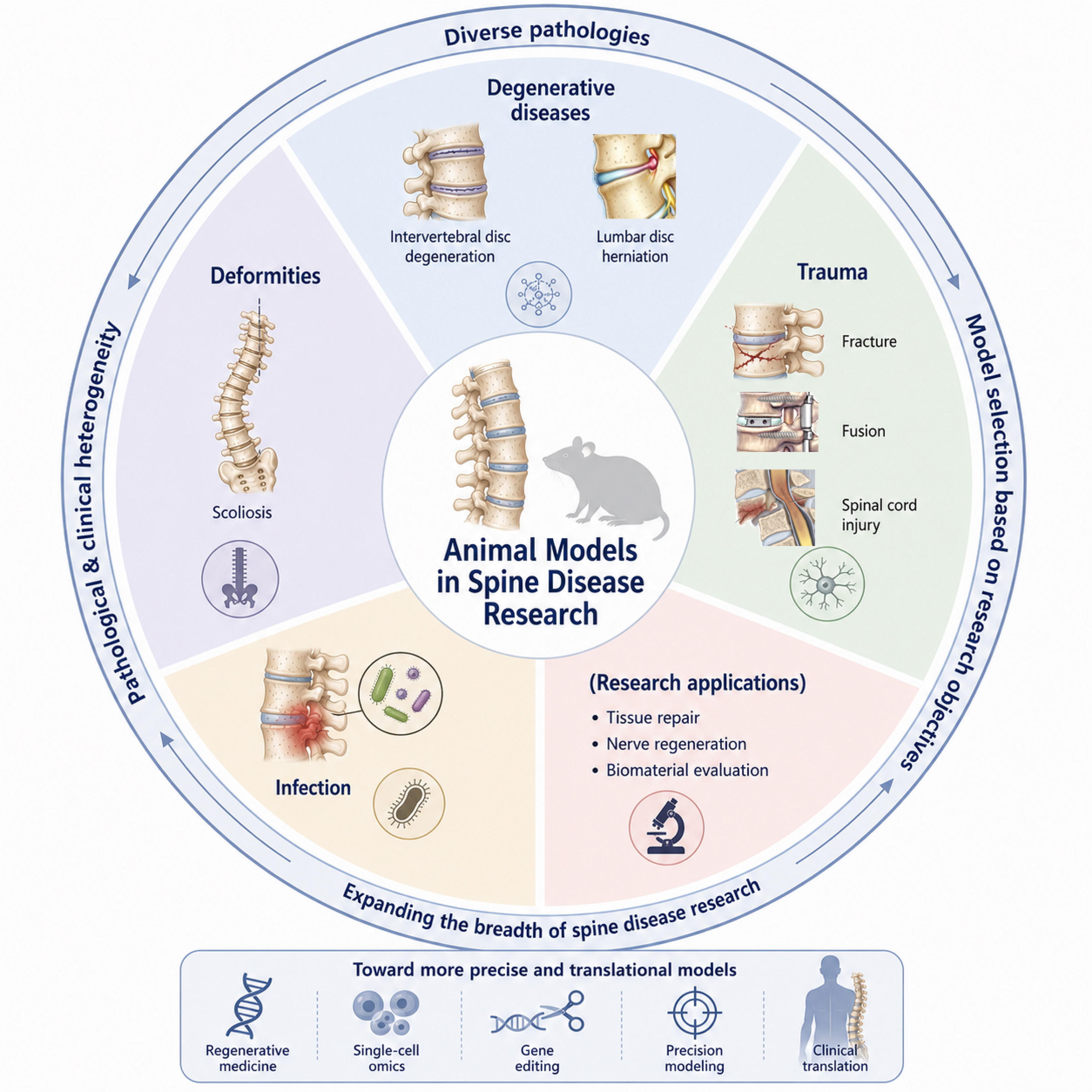
Figure 2. Disease-specific animal models in spine research.
To facilitate direct comparison and rational model selection, the representative models discussed in this review are summarized in Supplementary Table 1 according to the commonly used species, modeling strategies, typical experimental duration, reported reproducibility or model establishment rate, principal pathological and evaluation endpoints, and major limitations.
Animal models of IVDD
IVDD is a major pathological contributor to low back pain, LDH, and other degenerative spinal disorders. Its principal pathological features include loss and phenotypic alteration of nucleus pulposus cells, ECM degradation, endplate sclerosis, persistent inflammation, and disruption of intervertebral disc homeostasis[51-54]. In humans, IVDD develops gradually over years to decades and is influenced by multiple interacting factors, including genetic predisposition, aging, abnormal mechanical loading, metabolic dysfunction, and inflammatory processes. This complex and multifactorial nature makes it difficult for any single animal model to reproduce the full natural history and pathological heterogeneity of human IVDD[55,56]. Accordingly, a wide range of animal models has been developed to address different experimental objectives, ranging from mechanistic investigation and target validation to drug screening and assessment of regenerative therapies.
Based on the method of induction and underlying disease mechanism, commonly used IVDD models can be broadly classified into needle puncture-induced, mechanical loading-induced, chemically induced, genetically engineered, and spontaneous degeneration models, together with emerging multimodal models that combine two or more pathological stimuli.
Needle puncture-induced degeneration models
The needle puncture-induced degeneration model is one of the most widely used and standardized approaches for experimental IVDD[15,16]. By mechanically disrupting the annulus fibrosus, needle puncture alters intradiscal pressure and tissue integrity, leading to nucleus pulposus injury, ECM breakdown, and progressive structural degeneration. This approach has been successfully applied in multiple species, including rats, rabbits, goats, and pigs, and is widely used to investigate degenerative mechanisms and evaluate regenerative interventions[57].
A major advantage of the needle puncture model is its high degree of experimental controllability and reproducibility. The severity of degeneration can be modulated by varying needle diameter, puncture depth, rotation, and retention time[58]. In particular, the ratio of needle diameter to disc dimensions is an important determinant of the extent of degeneration[59]. In mouse models, the target caudal intervertebral disc is typically exposed or localized after anesthesia, followed by insertion of a fine needle through the annulus fibrosus into the nucleus pulposus. Depending on the experimental protocol, the needle may be rotated and retained briefly before withdrawal. This procedure induces progressive degenerative changes that can be monitored by imaging and histological assessment [Figure 3A]. Typical findings include reduced T2-weighted MRI signal intensity, loss of disc height, decreased proteoglycan content, depletion of nucleus pulposus cells, and disruption of annulus fibrosus organization[60,61].
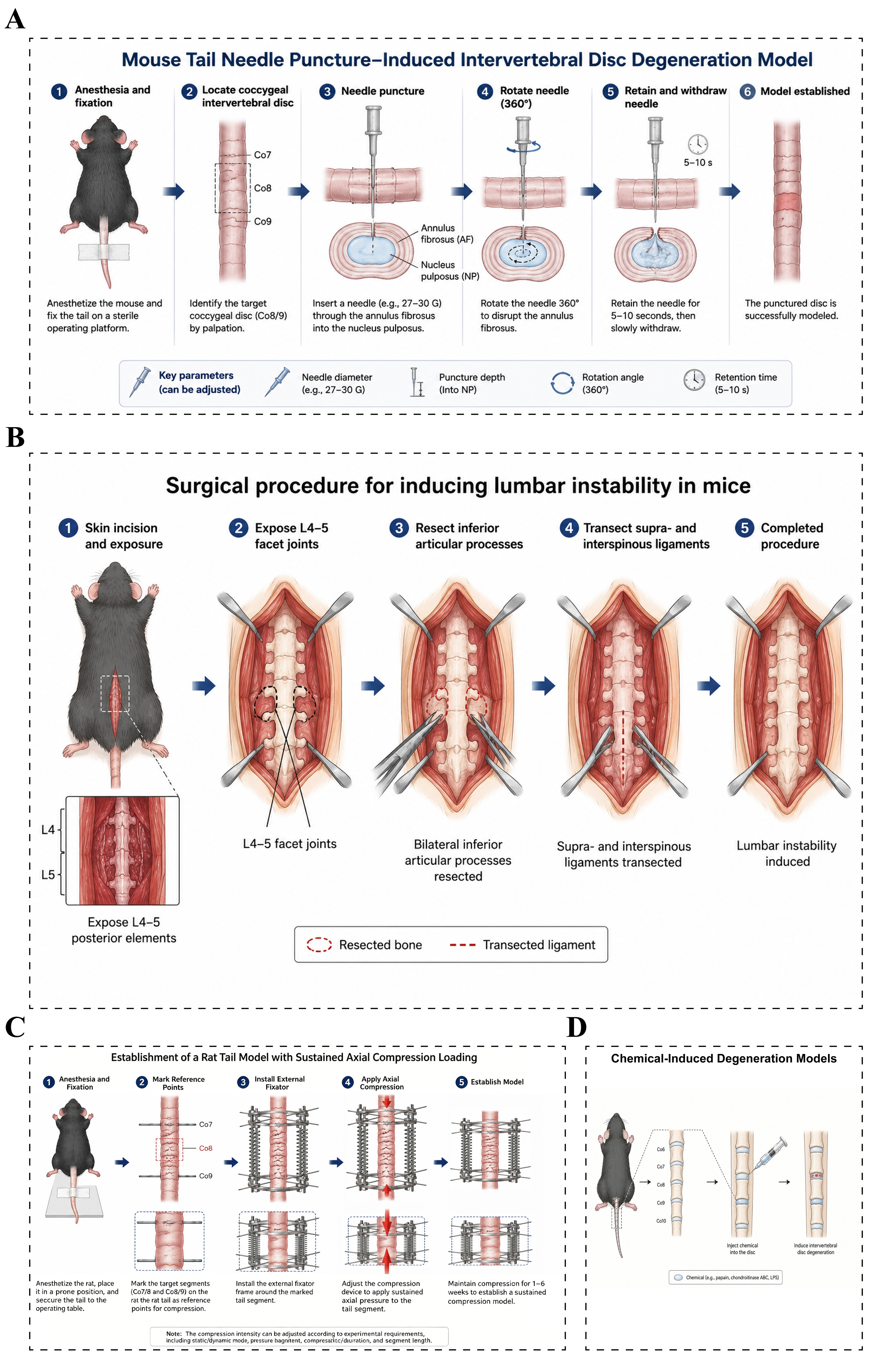
Figure 3. Animal models of intervertebral disc degeneration. (A) Needle puncture-induced intervertebral disc degeneration models: After anesthesia and fixation, the target caudal intervertebral disc in mice is identified, and a 27-30 G needle is inserted through the annulus fibrosus into the center of the nucleus pulposus. The needle is then rotated, retained briefly, and withdrawn, thereby causing annulus fibrosus injury and nucleus pulposus disruption to induce intervertebral disc degeneration; (B) Lumbar spinal instability models: After anesthesia, the posterior lumbar structures of the experimental mouse are exposed through a posterior midline approach. Under a microscope, the bilateral inferior articular processes are resected, and the supraspinous and interspinous ligaments are transected to disrupt posterior column stability and establish a lumbar motion segment instability model; (C) Mechanical loading models: Continuous axial loading is applied using spring-loading devices, Ilizarov external fixators, or customized compression systems to simulate a chronic mechanical overload environment; (D) Chemical-induced intervertebral disc degeneration models: Specific enzymes, inflammatory cytokines, or chemical reagents are injected into the intervertebral disc to disrupt the local microenvironment and rapidly induce intervertebral disc degeneration. NP: Nucleus pulposus.
At the molecular level, puncture-induced injury is accompanied by substantial remodeling of the intradiscal microenvironment. Expression of matrix-associated molecules such as aggrecan and collagen type II alpha 1 chain (COL2A1) is typically reduced, whereas catabolic enzymes, including matrix metallopeptidase 3 (MMP3), MMP13, and members of the a disintegrin and metalloproteinase with thrombospondin motifs (ADAMTS) family, are increased during disease progression[62]. Needle puncture models have also been widely used to investigate different forms of regulated cell death and stress responses, including apoptosis, autophagy dysregulation, pyroptosis, and ferroptosis[63]. Studies using these models have linked reduced glutathione peroxidase 4 (GPX4) activity, lipid peroxidation, and disturbances in iron metabolism to IVDD progression, thereby supporting further investigation of ferroptosis-related therapeutic strategies[64,65].
Despite their reproducibility and practical advantages, needle puncture models primarily represent an injury-induced form of disc degeneration. They therefore differ from the naturally evolving course of human IVDD, which typically develops over years under the combined influence of aging, genetic susceptibility, abnormal mechanical loading, metabolic dysfunction, and chronic inflammation. In contrast, puncture models initiate degeneration through an acute structural insult and subsequently reproduce many downstream degenerative changes. They are therefore particularly suitable for studying disease progression after structural injury and for evaluating therapeutic efficacy, but are less appropriate for investigating the earliest etiological events underlying spontaneous human IVDD.
Because of their technical simplicity, relatively short experimental duration, and consistent degenerative phenotype, needle puncture models remain widely used in studies of inflammation, matrix remodeling, cell death, and regenerative therapy[65]. They are especially valuable for proof-of-concept evaluation of interventions such as stem cell transplantation, biomaterial implantation, exosome-based therapy, and gene delivery[66-69]. However, the suitability of a specific species depends on the therapeutic strategy and experimental endpoint. Small-animal models, particularly mice and rats, are advantageous for mechanistic studies, genetic manipulation, dose exploration, and early efficacy assessment, whereas rabbits and larger animals offer greater anatomical accessibility for intradiscal delivery, longitudinal imaging, tissue-level repair assessment, and longer-term evaluation. These advantages must be balanced against higher costs, longer study periods, and greater technical demands. Accordingly, model selection for exosome- and gene-based therapies should be guided by the intended mechanistic, anatomical, and translational objectives rather than by reliance on a single universally preferred model.
Instability-induced degeneration models
Spinal stability is essential for maintaining the biomechanical environment and tissue homeostasis of the intervertebral disc. Together, the intervertebral disc, facet joints, spinal ligaments, and paraspinal muscles form the functional spinal unit (FSU), which collectively accommodates axial compression, flexion, extension, rotation, and shear forces while preserving segmental motion and structural integrity[70]. Disruption of this coordinated mechanical system, whether through facet joint degeneration, ligamentous injury, or damage to posterior stabilizing structures, can result in segmental instability and abnormal loading of the intervertebral disc. Sustained biomechanical disturbance may subsequently promote cellular dysfunction, ECM degradation, and progressive structural deterioration of disc tissues. Spinal instability is therefore considered an important contributor to the development and progression of degenerative lumbar spinal disorders.
Based on this concept, instability-induced models have been developed to reproduce disc degeneration secondary to chronic alterations in the spinal mechanical environment[71]. Among these, the mouse lumbar spine instability (LSI) model has been increasingly used in mechanobiological studies. The model is typically established through posterior disruption of stabilizing structures, most commonly bilateral resection of the inferior articular processes combined with transection of the supraspinous and interspinous ligaments [Figure 3B]. This procedure destabilizes the lumbar motion segment without directly injuring the intervertebral disc. Over the following weeks, progressive degenerative changes may develop, including loss of disc height, depletion or phenotypic alteration of nucleus pulposus cells, annulus fibrosus disorganization, and endplate remodeling.
A key distinction between the LSI model and needle puncture-based approaches is that degeneration arises indirectly from altered spinal biomechanics rather than from direct structural injury to the disc[72]. This feature makes the LSI model particularly useful for investigating how sustained mechanical instability contributes to degenerative changes across the spinal motion segment. It may therefore more closely reproduce aspects of clinically relevant degeneration associated with abnormal loading, posterior element dysfunction, and progressive disruption of spinal homeostasis, although it does not fully reproduce the multifactorial natural history of human IVDD.
Recent studies using LSI models have provided important insights into the molecular mechanisms through which abnormal mechanical loading drives disc degeneration. Spinal destabilization has been associated with activation of mechanosensitive and inflammatory signaling pathways, including piezo-type mechanosensitive ion channel component 1 (PIEZO1), Yes-associated protein/transcriptional coactivator with PDZ-binding motif (YAP/TAZ), Wingless-related integration site (Wnt), and nuclear factor kappa B (NF-κB) signaling[73-76]. These pathways may contribute to inflammatory mediator production, cellular senescence, altered cell fate, and increased expression of matrix-degrading enzymes, thereby accelerating degenerative remodeling. Importantly, instability-induced changes are not confined to the intervertebral disc. Endplate remodeling, facet cartilage degeneration, osteophyte formation, and alterations in paraspinal muscle composition have also been reported, supporting the concept that lumbar degeneration can involve multiple components of the FSU rather than the disc alone.
Because it reproduces biomechanical disturbance together with degenerative changes in several spinal tissues, the LSI model provides a valuable platform for studying mechanobiological regulation, inter-tissue crosstalk, and coordinated degeneration of the FSU. It is particularly useful for investigating how altered spinal mechanics influence cell fate, matrix homeostasis, and tissue remodeling over time.
Mechanical loading-induced degeneration models
Although IVDD is influenced by multiple factors, including genetic susceptibility, aging, metabolic dysfunction, and inflammation, abnormal mechanical loading is widely recognized as an important contributor to disease initiation and progression[77]. Intervertebral discs are continuously exposed to complex mechanical forces, including compression, tension, torsion, and shear. Within the physiological range, these stimuli are essential for maintaining ECM homeostasis and normal disc function. Excessive or prolonged loading, however, can disrupt cellular mechanotransduction, promote matrix catabolism and inflammatory signaling, and impair cell viability, ultimately contributing to progressive structural and functional deterioration of the disc[78]. Compared with models based on direct structural injury, mechanical loading models are particularly valuable for investigating how sustained biomechanical disturbance contributes to IVDD development[79,80].
Mechanical loading models were initially developed using rodent caudal intervertebral discs[81]. The caudal spine of rats and mice is readily accessible and can be subjected to controlled loading using external fixation systems, making it suitable for studies of load magnitude, duration, and frequency[82]. Axial compression is commonly applied using spring-based devices, Ilizarov-type external fixators, or customized loading systems to reproduce conditions of sustained mechanical overload [Figure 3C]. After prolonged loading, the affected discs typically exhibit progressive degenerative changes, including reduced disc height, decreased T2-weighted MRI signal intensity, and ECM degradation[83]. Histological findings may include proteoglycan loss within the nucleus pulposus, disruption of annulus fibrosus organization, and endplate remodeling or sclerosis[84]. Together, these changes reproduce several important features of mechanically driven disc degeneration.
The development of mechanobiology has further expanded the use of these models for investigating how disc cells sense and respond to abnormal mechanical cues. Rather than acting as passive load-bearing cells, nucleus pulposus, annulus fibrosus, and endplate cells express a variety of mechanosensitive molecules that convert physical stimuli into intracellular biochemical signals[85]. Mechanotransduction pathways involving PIEZO1, PIEZO2, transient receptor potential vanilloid 4 (TRPV4), integrins, and YAP/TAZ have therefore received increasing attention in IVDD research[86]. Among these, PIEZO1 has emerged as an important mechanosensitive ion channel capable of mediating calcium influx in response to excessive mechanical stimulation. Sustained PIEZO1 activation has been associated with mitochondrial dysfunction, reactive oxygen species (ROS) accumulation, inflammatory signaling, cellular senescence, and altered cell survival[86-90]. These findings show how mechanical loading models can link abnormal biomechanical stimuli to downstream molecular and cellular responses.
Mechanical loading models have also been widely applied to investigate stress-related cell injury and death during IVDD. In addition to apoptosis, excessive mechanical stress has been linked to ferroptosis and pyroptosis, as well as to dysregulation of autophagy and mitochondrial homeostasis[91]. Ferroptosis has attracted particular attention because of its close association with oxidative stress, lipid peroxidation, and iron metabolism[92]. Sustained compression has been reported to disturb iron homeostasis, increase lipid peroxide accumulation, reduce GPX4 activity, and promote oxidative damage in disc cells[93]. Studies focusing on regulatory molecules such as solute carrier family 7 member 11 (SLC7A11) and GPX4 further suggest that ferroptosis may contribute to nucleus pulposus cell loss under mechanically adverse conditions. Mechanical loading models therefore provide a useful platform for examining how biomechanical stress interacts with oxidative injury and regulated cell-death pathways during degeneration.
Beyond cell injury, these models have also contributed to the study of progenitor cell homeostasis and regenerative capacity within the intervertebral disc[94]. Advances in single-cell RNA sequencing have identified heterogeneous cell populations with progenitor-like characteristics and potential roles in tissue maintenance and repair[95]. Abnormal mechanical environments may compromise the maintenance, differentiation, and reparative capacity of these cell populations[96]. Mechanosensitive signaling pathways, including Hippo-YAP/TAZ, Wnt/β-catenin, and Notch signaling, have been implicated in the regulation of progenitor cell fate and tissue remodeling under mechanical stress[97,98]. Mechanical loading models are therefore useful not only for studying degenerative mechanisms but also for examining how the biomechanical environment influences endogenous repair potential.
Overall, mechanical loading models provide a controllable experimental system for investigating the contribution of abnormal biomechanics to IVDD. Their main strength is the ability to manipulate the magnitude, duration, and pattern of mechanical stimulation while avoiding direct injury to the disc itself. This makes them particularly suitable for studies of mechanotransduction, stress-induced cellular responses, cell fate regulation, and tissue homeostasis. However, the loading conditions generated by external devices remain simplified representations of the complex and dynamic mechanical environment of the human spine. Accordingly, these models should be regarded as complementary platforms for dissecting mechanically driven mechanisms of degeneration rather than as complete reproductions of the natural course of human IVDD.
Chemical-induced degeneration models
In addition to mechanical injury and abnormal loading, inflammation and dysregulated ECM metabolism play important roles in the initiation and progression of IVDD[99,100]. On this basis, a variety of chemically induced degeneration models have been developed to reproduce specific pathological features of the degenerative disc microenvironment. These models typically involve intradiscal administration of enzymes, inflammatory cytokines, or other bioactive agents to perturb matrix homeostasis, alter cellular function, and induce relatively rapid degenerative changes[100,101] [Figure 3D].
Enzyme-induced models represent some of the earliest chemically induced approaches used to reproduce IVDD. These models rely on intradiscal administration of proteolytic or matrix-degrading enzymes to disrupt ECM integrity and induce structural deterioration of the disc[102]. Papain, trypsin, and chondroitinase ABC (C-ABC) are among the most commonly used reagents. Papain rapidly degrades proteoglycans and collagenous matrix components, resulting in dehydration and volume loss of the nucleus pulposus. Experimental studies have shown that papain injection can produce reduced disc height, decreased T2-weighted MRI signal intensity, and increased histological degeneration scores within several weeks[57,102]. However, the relatively rapid and extensive tissue destruction induced by papain differs substantially from the gradual progression of human IVDD, limiting its use in studies aimed at reproducing chronic degeneration.
Compared with papain, C-ABC has gained increasing attention because it preferentially degrades chondroitin sulfate-containing glycosaminoglycans that are abundant within the nucleus pulposus[57]. This results in progressive proteoglycan depletion and loss of matrix hydration, thereby reproducing an important component of disc degeneration[103]. The severity of degeneration can be modulated by adjusting the concentration and dose of C-ABC, allowing the generation of graded degenerative phenotypes. Because matrix depletion can be induced in a relatively controlled manner while preserving the overall architecture of the disc, C-ABC models are particularly useful for evaluating regenerative strategies, including cell-based therapies, injectable hydrogels, and other tissue-engineering approaches[104,105].
With increasing recognition of the role of inflammation in IVDD, cytokine-induced models have also become widely used experimental systems[106,107]. Clinical and experimental evidence indicates that degenerative discs are characterized by persistent inflammatory activity, with cytokines such as tumor necrosis factor alpha (TNF-α), interleukin-1 beta (IL-1β), IL-6, and IL-17 contributing to matrix catabolism, cellular dysfunction, and disease progression[107,108]. Based on these observations, intradiscal administration of pro-inflammatory cytokines, particularly TNF-α and IL-1β, has been used to reproduce selected features of the inflammatory microenvironment associated with IVDD[109,110]. In contrast to enzyme-induced models, which primarily target matrix integrity, cytokine-based approaches better suit investigation of inflammatory signaling, cellular stress responses, and inflammation-driven remodeling of the disc microenvironment.
IL-1β and TNF-α are among the most frequently used cytokines in these models. IL-1β activates signaling pathways such as NF-κB and mitogen-activated protein kinase (MAPK), increases the expression of matrix-degrading enzymes including MMP3, MMP13, and ADAMTS5, and suppresses matrix-associated molecules such as aggrecan and COL2A1[111]. TNF-α similarly promotes inflammatory signaling and matrix catabolism and has also been associated with increased expression of neurotrophic mediators such as nerve growth factor (NGF) and brain-derived neurotrophic factor (BDNF), making TNF-α-based models useful for studies of inflammation-associated discogenic pain. Combined cytokine stimulation has also been explored as a means of generating a more sustained and complex inflammatory environment than that produced by a single cytokine alone[108].
Chemically induced models are also useful for investigating stress responses and regulated cell-death pathways associated with IVDD. Pro-inflammatory stimulation can promote oxidative stress, mitochondrial dysfunction, lipid peroxidation, and disturbances in iron metabolism. In IL-1β-based models, for example, increased ROS production and impaired antioxidant defense have been associated with ferroptosis-related changes[112,113]. Inflammatory stimulation may also activate the NOD-like receptor family pyrin domain containing 3 (NLRP3) inflammasome, promote caspase-1 activation and gasdermin D (GSDMD) cleavage, and thereby contribute to pyroptotic cell death[114,115]. These models therefore provide useful experimental platforms for studying the relationships among inflammation, oxidative stress, ferroptosis, pyroptosis, autophagy dysregulation, and cellular senescence.
Despite their experimental flexibility, chemically induced models have several inherent limitations. Most rely on artificial, simplified stimuli and therefore do not reproduce the multifactorial etiology of human IVDD. Some reagents induce rapid matrix destruction or intense inflammatory responses that differ from the chronic and progressive nature of naturally occurring degeneration. In addition, differences in reagent type, concentration, injection volume, treatment frequency, and experimental species can substantially influence disease severity and reduce comparability across studies. Chemically induced models are therefore best suited to investigating defined pathological processes or testing specific therapeutic mechanisms rather than reproducing the entire natural history of IVDD.
Overall, chemically induced models provide efficient and controllable approaches for studying matrix degradation, inflammation, oxidative stress, and related cellular responses in IVDD. Enzyme-based models are particularly useful for reproducing matrix depletion and evaluating regenerative interventions, whereas cytokine-based models are better suited to studies of inflammatory signaling, cell fate regulation, and discogenic pain. Future strategies that combine chemical stimulation with other disease-relevant factors, such as abnormal mechanical loading, aging, or genetic susceptibility, may provide more physiologically representative models for studying the complex pathogenesis of IVDD.
Genetically engineered models
Although needle puncture, mechanical loading, and chemically induced models can reproduce selected pathological features of IVDD, these exogenously induced models are less suitable for directly investigating the genetic determinants and cell-specific regulatory mechanisms involved in disease initiation and progression. With advances in genetic engineering, particularly Cre-LoxP-based conditional targeting and CRISPR/Cas9-mediated genome editing, genetically engineered models have become increasingly important for studying intervertebral disc development, tissue homeostasis, and degeneration. Unlike conventional induction-based approaches, these models enable targeted manipulation of specific genes in defined cell populations or developmental stages, providing a powerful way to dissect gene function and validate candidate therapeutic targets.
Early genetically engineered IVDD studies focused largely on genes involved in ECM organization and maintenance. Proteoglycans and collagen fibers are major structural components of the intervertebral disc, and their balanced synthesis and turnover are essential for preserving tissue hydration and biomechanical function[116]. Deficiency of aggrecan (ACAN) can lead to impaired disc development and premature degenerative changes, including reduced nucleus pulposus hydration, proteoglycan depletion, and loss of disc height[117]. Similarly, disruption or mutation of COL2A1 compromises type II collagen production and may result in abnormal annulus fibrosus organization and impaired matrix integrity[118]. These findings provided early genetic evidence that disruption of ECM homeostasis can directly contribute to disc degeneration.
Transcriptional regulators are equally important for maintaining disc cell phenotype and matrix homeostasis. SRY-box transcription factor 9 (SOX9), for example, is a key regulator of chondrogenic differentiation and ECM synthesis and controls the expression of cartilage-associated genes, including COL2A1[119,120]. Conditional loss of SOX9 has been associated with reduced ACAN and COL2A1 expression, altered cellular phenotype, and progressive disruption of intervertebral disc architecture[121]. Such models illustrate that IVDD may arise not only from external injury or mechanical stress but also from disruption of intrinsic developmental and homeostatic regulatory programs.
Despite their mechanistic value, genetically engineered models have several limitations. Global gene deletion may cause severe developmental abnormalities or embryonic lethality, making it difficult to distinguish developmental defects from adult-onset degeneration. Conditional and inducible systems are therefore increasingly preferred because they allow greater spatial and temporal control of gene manipulation. In addition, single-gene perturbations cannot fully reproduce the multifactorial nature of human IVDD, which reflects interactions among genetic susceptibility, aging, mechanical stress, metabolic disturbance, and inflammation. Generating and maintaining genetically modified animals can also be time-consuming and costly, limiting their use in large-scale or routine studies.
Overall, genetically engineered models have substantially expanded the ability to investigate the molecular and cellular basis of IVDD. They are particularly valuable for defining cell-specific gene functions, identifying causal regulatory pathways, and distinguishing developmental abnormalities from degenerative processes. Integration of conditional genetic models with lineage tracing, single-cell omics, and spatial transcriptomics is expected to provide a more detailed understanding of the cellular dynamics and regulatory networks that shape disc degeneration and repair.
Spontaneous degeneration models
Although experimentally induced models have contributed substantially to IVDD research, most rely on external interventions, such as structural injury, abnormal mechanical loading, chemical stimulation, or genetic manipulation, to accelerate degenerative changes. By contrast, human IVDD typically develops gradually over many years under the combined influence of aging, genetic susceptibility, mechanical stress, metabolic alterations, chronic inflammation, and declining endogenous repair capacity. Spontaneous degeneration models are therefore of particular interest because they allow age-associated degenerative changes to develop without direct experimental induction and may reproduce aspects of the natural history of human IVDD more closely than acute injury-based models.
A defining feature of spontaneous degeneration models is the progressive development of disc degeneration with age in the absence of deliberate local injury. These models enable longitudinal investigation of the transition from early disturbances in tissue homeostasis to progressive matrix loss, cellular dysfunction, and structural deterioration. They are particularly useful for studying aging-related mechanisms, chronic changes in the disc microenvironment, and the long-term effects of therapeutic interventions. However, their translational relevance remains dependent on species-specific differences in disc biology, lifespan, biomechanics, and patterns of age-related degeneration.
Sand rats are among the best-established spontaneous models used in IVDD research. These animals develop progressive age-associated changes in the intervertebral disc, including reduced nucleus pulposus cellularity, ECM deterioration, and endplate calcification[122]. Their gradual degenerative phenotype makes them useful for investigating mechanisms associated with aging and chronic disc degeneration[123]. Nevertheless, limited availability, specialized breeding requirements, and relatively restricted experimental resources have constrained their broader use.
Aged rodents have also become increasingly important for studying naturally occurring, age-associated disc degeneration. Wild-type mice and rats develop progressive structural and molecular changes in the intervertebral disc with advancing age, including loss of disc height, proteoglycan depletion, increased inflammatory activity, and accumulation of senescent cells[116,124]. These alterations are generally more pronounced in older animals and provide a useful platform for examining processes such as cellular senescence, mitochondrial dysfunction, chronic low-grade inflammation, and impaired regenerative capacity during IVDD progression[125]. Compared with rapidly induced models, aged rodents are particularly valuable for studying mechanisms linked to the gradual decline in disc homeostasis over time.
Despite these advantages, spontaneous degeneration models have several practical limitations. The development of stable degenerative phenotypes requires prolonged observation, resulting in substantially longer experimental timelines than those of induced models. Disease severity and progression can also vary among individual animals, reducing experimental consistency and increasing sample-size requirements. In addition, long-term housing, maintenance costs, and ethical considerations may further limit their use, particularly in larger species. For these reasons, spontaneous models are often used as complementary systems alongside induced models rather than as replacements.
Overall, spontaneous degeneration models provide valuable opportunities to investigate age-associated changes, chronic tissue remodeling, and progressive loss of disc homeostasis under conditions that do not require direct experimental injury. Their greatest strength lies in modeling the temporal evolution of degeneration, whereas their major limitations include long study duration, inter-individual variability, and species-specific differences. Combining spontaneous models with appropriately selected induced models may therefore provide a more comprehensive strategy for studying IVDD mechanisms and evaluating long-term therapeutic responses.
Animal models of LDH
LDH is a common degenerative spinal disorder and a major cause of sciatica and low back pain worldwide. Mechanical compression of the nerve root by herniated disc tissue has traditionally been regarded as a principal mechanism underlying LDH-associated pain and neurological dysfunction. However, accumulating evidence indicates that the pathophysiology of LDH extends beyond mechanical compression and also involves inflammatory responses, immune cell infiltration, neovascularization, and spontaneous resorption of herniated nucleus pulposus tissue. Clinical studies have shown that a substantial proportion of herniated disc material can undergo partial or complete spontaneous resorption without surgical intervention. This observation has broadened the focus of LDH research from nerve root compression alone to the dynamic interactions among herniated disc tissue, the immune microenvironment, vascular ingrowth, and tissue clearance. Accordingly, animal models that reproduce distinct components and stages of LDH are essential for investigating disease mechanisms and evaluating emerging therapeutic strategies.
Compared with IVDD models, LDH models are designed not only to reproduce disc disruption but, more importantly, to mimic the pathological interactions between herniated nucleus pulposus tissue and adjacent neural and immune structures. Among the most commonly used approaches are autologous nucleus pulposus transplantation models and nerve root compression models, which respectively emphasize the inflammatory and immune responses to exposed nucleus pulposus tissue and the mechanical consequences of neural compression.
Autologous nucleus pulposus transplantation models
The autologous nucleus pulposus transplantation model is a well-established experimental approach for investigating the inflammatory and neurobiological mechanisms associated with LDH. Early studies by Olmarker et al. demonstrated that exposure of nucleus pulposus tissue to neural structures can induce inflammatory changes and nerve root dysfunction even in the absence of substantial mechanical compression[126]. In this model, autologous nucleus pulposus tissue is typically harvested from the caudal or lumbar intervertebral discs and placed adjacent to the lumbar nerve root or dural sac [Figure 4A][127,128]. Following transplantation, local inflammatory responses may develop rapidly, characterized by macrophage recruitment, increased production of pro-inflammatory mediators, nerve root edema, and pain-related behavioral changes. Because sustained mechanical compression is absent, this model is particularly useful for isolating the biological effects of exposed nucleus pulposus tissue on neural and immune tissues.
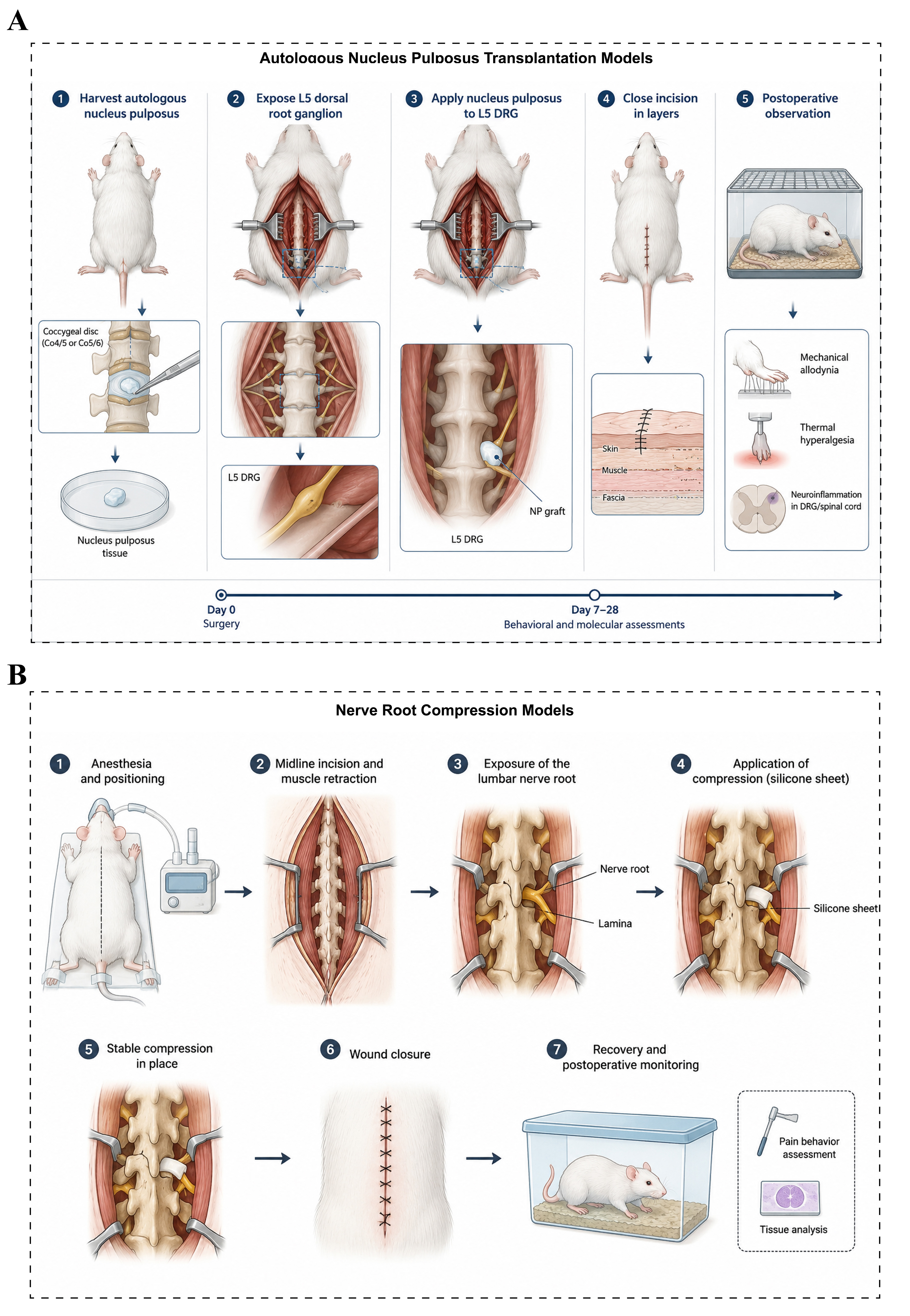
Figure 4. Animal models of lumbar disc herniation. (A) Autologous nucleus pulposus transplantation models: After anesthesia, the experimental animal is placed in the prone position, and autologous nucleus pulposus tissue is harvested from the caudal or lumbar intervertebral disc. The target lumbar nerve root (L4-L5 or L5-L6) is then exposed, and the harvested nucleus pulposus tissue is transplanted around the nerve root or dural sac. The incision is subsequently closed, followed by postoperative observation and relevant assessments; (B) Nerve root compression models: After anesthesia, the experimental animal is placed in the prone position, and the lamina and lumbar nerve root are exposed through a posterior midline lumbar incision. A silicone sheet, polyethylene tube, or other compression device is then placed around the nerve root to establish a sustained mechanical compression model. The incision is closed after surgery, followed by postoperative observation and evaluation of relevant outcome measures. NP: Nucleus pulposus; DRG: dorsal root ganglion.
Under physiological conditions, the nucleus pulposus is largely separated from systemic immune surveillance by the surrounding annulus fibrosus and cartilaginous endplates. Disruption of this anatomical isolation during disc herniation exposes nucleus pulposus components to the perineural environment and can initiate innate immune activation and recruitment of inflammatory cells. Studies using transplantation models have implicated mediators such as TNF-α, IL-1β, IL-6, and C-C motif chemokine ligand 2 (CCL2) in the development of neuroinflammation, nerve root sensitization, and pain-related responses following nucleus pulposus exposure[129]. These findings have helped establish inflammation as an important component of LDH pathophysiology and have provided an experimental basis for evaluating anti-inflammatory therapeutic strategies.
The principal strengths of the autologous nucleus pulposus transplantation model include good experimental reproducibility, relatively standardized surgical procedures, and the ability to examine inflammation-driven neural responses independently of persistent mechanical compression. However, because the model does not reproduce the complete process of annular rupture, tissue extrusion, and sustained nerve root compression observed in many patients with LDH, it is best regarded as a model of the biological and inflammatory consequences of nucleus pulposus exposure rather than a complete representation of clinical disc herniation.
Nerve root compression models
Nerve root compression models are designed to reproduce the mechanical component of LDH-associated nerve injury[130]. In these models, researchers apply sustained compression to the lumbar nerve root using materials or devices such as silicone sheets, vascular clips, suture ligation, or customized compression systems [Figure 4B]. These approaches have been widely used to investigate the consequences of persistent mechanical compression on neural function and pain behavior. Experimental studies have shown that nerve root compression can induce ischemia, edema, demyelination, and axonal injury, together with behavioral manifestations such as mechanical allodynia and thermal hyperalgesia[131-133].
However, mechanical compression alone does not fully explain the heterogeneity of symptoms observed in patients with LDH. The severity of radiographic nerve root compression does not always correlate closely with pain intensity or neurological dysfunction, indicating that additional biological factors contribute to symptom development[134]. Increasing evidence supports an important role for inflammation and immune activation in modulating nerve root sensitization and pain after disc herniation. Accordingly, combined models that integrate mechanical compression with exposure to autologous nucleus pulposus tissue have been developed to reproduce both the mechanical and inflammatory components of LDH. These models may provide a more representative platform for investigating the complex interactions between neural compression, local inflammation, and pain-related responses.
Animal models of scoliosis
AIS is one of the most common spinal deformities and is characterized by a three-dimensional alteration of spinal alignment that develops during adolescence, including coronal curvature, abnormalities in sagittal alignment, and vertebral rotation. AIS affects approximately 2%-4% of adolescents, yet its etiology and pathophysiological mechanisms remain incompletely understood. Current evidence suggests that AIS is a multifactorial disorder involving complex interactions among genetic susceptibility, neuroendocrine regulation, growth imbalance, and abnormal biomechanical loading. Because no single pathogenic mechanism has been established, no existing animal model can fully reproduce the spontaneous onset and progressive three-dimensional deformity observed in human AIS.
Accordingly, scoliosis models have been developed to investigate specific components of the proposed disease mechanisms. Common approaches include models based on melatonin-related disturbances, biomechanical asymmetry, altered growth regulation, and genetic manipulation. Although each model reproduces only selected features of AIS, together they provide complementary experimental platforms for investigating the mechanisms of curve initiation and progression and for evaluating potential preventive or therapeutic strategies.
Melatonin deficiency-based and biomechanics-based models
Melatonin-related abnormalities and altered spinal biomechanics represent two historically important hypotheses in scoliosis research. Early studies showed that pinealectomy in juvenile chickens can induce progressive spinal curvature with several radiographic features resembling those of human AIS, providing experimental support for a potential role of melatonin signaling in scoliosis development[135,136]. However, subsequent studies demonstrated that melatonin deficiency alone does not consistently induce scoliosis across species, suggesting that impaired melatonin signaling is unlikely to act as an independent cause of AIS and may instead interact with other developmental or biomechanical factors[137].
Biomechanical models have therefore been developed to investigate how altered loading influences spinal growth and deformity progression. Bipedal models, for example, are established by modifying normal quadrupedal posture to increase axial loading and reproduce selected features of the upright mechanical environment of the human spine[138]. Neither pinealectomy nor bipedalization alone consistently produces scoliosis with high penetrance, whereas combining melatonin-related abnormalities with altered mechanical loading can increase the incidence and stability of scoliosis-like deformities[138-140]. C57BL/6J mice, which exhibit low endogenous melatonin production because of defects in melatonin-synthesis pathways, have been used in combination with bipedalization to investigate interactions between melatonin signaling, spinal growth, and biomechanical loading [Figure 5A]. These models are particularly useful for exploring how neuroendocrine and mechanical factors may interact during scoliosis development, although they do not reproduce the full complexity of human AIS.
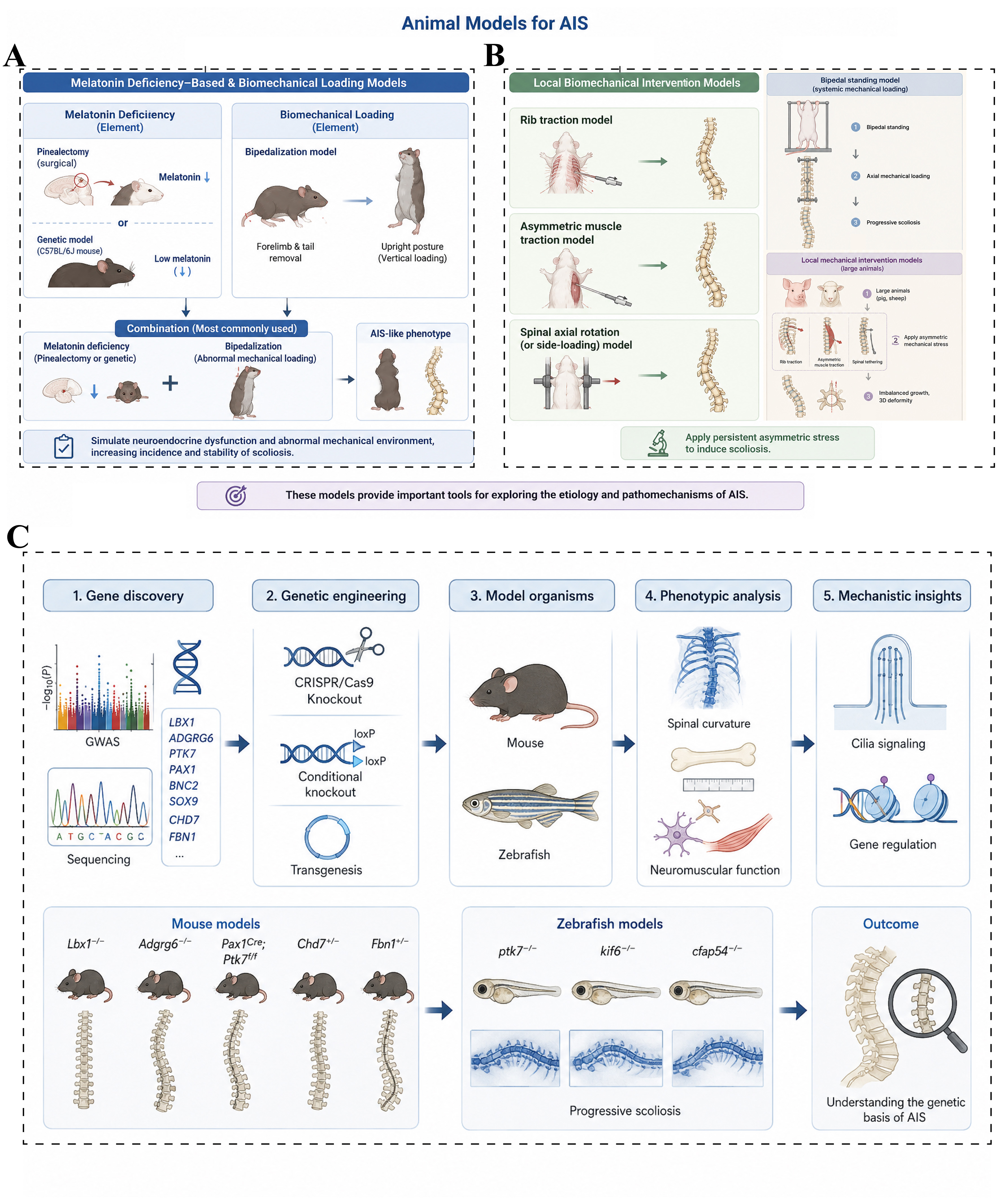
Figure 5. Animal models of scoliosis. (A) Melatonin deficiency-based models: Melatonin deficiency is established either by pinealectomy or by using animals with naturally low melatonin levels, followed by bipedalization to simulate abnormal vertical loading. The combined effects of melatonin deficiency and altered mechanical loading can induce AIS-like scoliosis; (B) Biomechanics-based models: Local mechanical intervention models, including rib traction, asymmetric muscle traction, and asymmetric spinal loading, induce scoliosis through sustained asymmetric mechanical stress and are used to investigate the mechanobiological mechanisms underlying AIS progression; (C) Genetic models: Based on AIS susceptibility genes identified through GWAS and sequencing analyses, mouse and zebrafish models are generated using gene-editing technologies. Phenotypic assessments of spinal curvature, growth and development, and neuromuscular function are then performed to elucidate the genetic factors and molecular mechanisms associated with AIS. AIS: Adolescent idiopathic scoliosis; GWAS: genome-wide association studies.
In addition to bipedal models, several approaches apply asymmetric mechanical forces directly on the growing spine, including rib tethering, asymmetric muscle traction, and spinal tethering[141,142]. These models are commonly used to investigate the effects of asymmetric loading on vertebral growth, disc morphology, and curve progression [Figure 5B]. Among them, porcine tethered scoliosis models are widely used because the size and growth characteristics of the porcine spine allow clinically relevant instrumentation and longitudinal assessment. By applying unilateral or asymmetric tethering during growth, these models can induce vertebral wedging, asymmetric disc growth, coronal curvature, and axial rotation[143-145]. Modified tethering techniques, including posterior or less invasive approaches, have further improved model consistency and reduced procedure-related morbidity.
Porcine tethering models are particularly valuable for studying growth modulation and for evaluating non-fusion corrective strategies, including vertebral body tethering and growth-preserving instrumentation[146,147]. However, the deformity is produced by externally imposed mechanical asymmetry and therefore does not reproduce the spontaneous onset, genetic susceptibility, neuromuscular regulation, or other biological features associated with AIS. Moreover, some tethering paradigms may more closely resemble mechanically induced or early-onset scoliosis than idiopathic adolescent scoliosis. These models should therefore be viewed primarily as platforms for studying mechanically driven curve progression and testing corrective interventions rather than as complete models of AIS pathogenesis.
Overall, melatonin-related and biomechanics-based models have contributed substantially to understanding how neuroendocrine signaling and asymmetric mechanical loading may influence scoliosis development. Their phenotypic characteristics, reproducibility, and clinical relevance vary considerably across species and modeling protocols. Accordingly, these models are best used to address specific mechanistic or translational questions rather than to reproduce the entire pathophysiological spectrum of human AIS.
Genetic models
Advances in genome-wide association studies (GWAS) and high-throughput sequencing have led to the identification of an increasing number of genetic loci and candidate genes associated with AIS[148] [Figure 5C]. Animal models based on these genetic findings provide valuable tools for investigating the molecular pathways that contribute to spinal development, growth regulation, and deformity formation. Frequently reported AIS-associated genes include LBX1, ADGRG6 (GPR126), PTK7, PAX1, BNC2, and SOX9. In parallel, genes implicated in syndromic forms of scoliosis, such as CHD7 and FBN1, have provided additional insight into biological pathways relevant to spinal deformity[149-152].
Using global knockout, conditional knockout, transgenic, and other genetic manipulation strategies, researchers have generated a range of animal models exhibiting spinal curvature, abnormal skeletal growth, vertebral developmental defects, or neuromuscular abnormalities. These models support a key role for genetic susceptibility in scoliosis pathogenesis while also highlighting the heterogeneity of the biological mechanisms involved. However, the phenotypes produced by individual gene perturbations often reflect specific developmental pathways and should not necessarily be regarded as direct reproductions of human AIS.
Zebrafish have become particularly useful for genetic studies of scoliosis because of their rapid development, high experimental throughput, and amenability to genetic manipulation[153]. Mutations or deficiencies involving PTK7, KIF6, and other genes related to ciliary function have been shown to produce progressive spinal curvature in zebrafish[154-156]. These models provide tractable systems for investigating how abnormalities in ciliary biology, cerebrospinal fluid dynamics, skeletal development, and related signaling pathways contribute to scoliosis-like phenotypes.
Animal models of vertebral fracture and spinal fusion
Vertebral fractures are common spinal injuries that can result from high-energy trauma, osteoporosis, or other conditions that compromise vertebral strength. Among these, osteoporotic vertebral compression fractures (OVCFs) are particularly prevalent in older adults and are associated with persistent back pain, progressive kyphotic deformity, functional impairment, and reduced quality of life. In parallel, spinal fusion is widely used to treat spinal instability, degenerative disorders, deformity, and other conditions requiring stabilization of affected motion segments. Understanding the biological processes underlying vertebral repair and successful spinal fusion therefore remains an important objective in spine research.
Animal models of vertebral fracture and spinal fusion provide essential platforms for investigating bone healing, graft incorporation, osteogenesis, and the biomechanical factors that influence fusion outcomes. They are also widely used to evaluate bone graft substitutes, biomaterials, growth factors, cellular therapies, and implantable devices before clinical translation. Developing reproducible models that reflect clinically relevant bone quality, defect characteristics, and surgical conditions is therefore critical to improving the translational value of preclinical studies.
Osteoporosis-associated vertebral compression fracture models
Osteoporosis-associated vertebral compression fracture models are widely used to investigate fracture development and repair under compromised bone-quality conditions. Osteoporotic phenotypes are commonly induced through ovariectomy (OVX), glucocorticoid administration, aging, or combined approaches, resulting in reduced bone mass, deterioration of trabecular architecture, and decreased vertebral mechanical strength[157-160]. Vertebral compression fractures are subsequently generated by applying controlled axial loading to the vertebral body until structural failure occurs [Figure 6A].
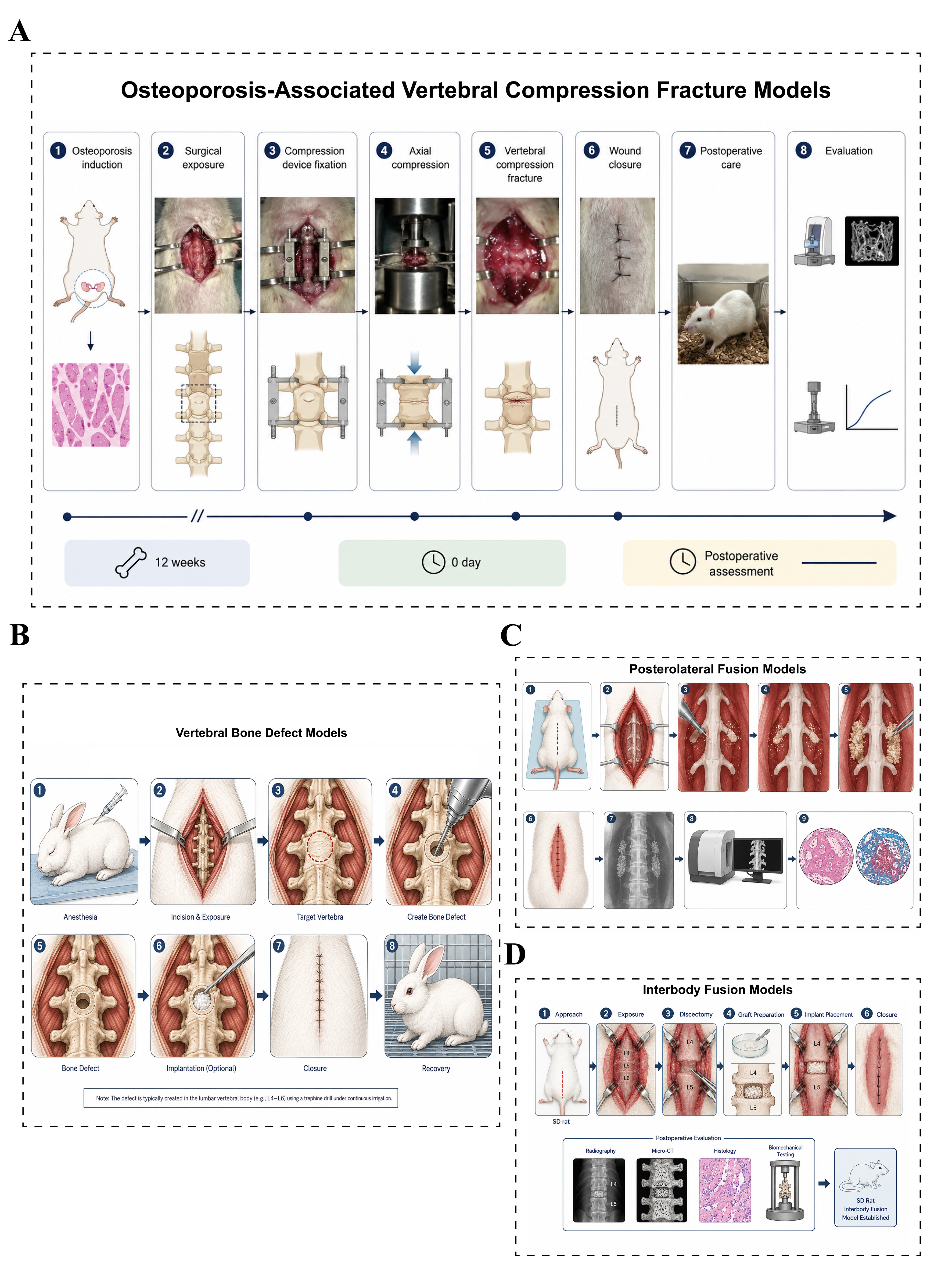
Figure 6. Animal models of vertebral fracture and spinal fusion. (A) Osteoporosis-associated vertebral compression fracture models: Bilateral OVX is performed to induce osteoporosis, followed by 12 weeks to establish stable bone loss. The target vertebral body is then exposed, and axial compression is applied using a customized device to induce a vertebral compression fracture. Fracture healing is evaluated by micro-CT, histology, and biomechanical testing; (B) Vertebral bone defect models: The target vertebral body is exposed through a posterior approach, and a standardized bone defect is created using a trephine or high-speed burr. Experimental materials are implanted into the defect, and bone regeneration is subsequently assessed by micro-CT and histological analysis; (C) Posterolateral fusion models: The bilateral L4-L5 transverse processes are exposed and decorticated, followed by implantation of test materials into the fusion bed. Fusion outcomes are evaluated using X-ray, micro-CT, and histological analysis; (D) Interbody fusion models: The target lumbar segment (L4-L5) is exposed, followed by discectomy and endplate preparation. Bone graft materials are implanted into the intervertebral space, and interbody fusion is assessed by X-ray, micro-CT, histological staining, and biomechanical testing. OVX: Ovariectomy; CT: computed tomography.
These models reproduce several clinically relevant features of OVCFs, including loss of vertebral height, trabecular disruption, deterioration of bone microarchitecture, and subsequent remodeling responses. Because the magnitude and rate of compressive loading can be experimentally controlled, fracture severity can be adjusted according to specific study objectives, providing relatively good reproducibility across standardized protocols. Such models are widely used to investigate fracture healing, bone remodeling, and the interaction between osteoporosis and vertebral repair, as well as to evaluate interventions such as bone cement, injectable biomaterials, tissue-engineered scaffolds, and anti-osteoporotic therapies[161].
Overall, osteoporosis-associated vertebral compression fracture models provide a useful preclinical platform for studying vertebral injury in osteoporotic bone and for assessing therapeutic strategies aimed at improving fracture stabilization, bone regeneration, and skeletal quality. However, the experimentally induced fracture pattern and healing environment remain simplified relative to the heterogeneous clinical presentations of human OVCF, and these differences should be considered when interpreting translational outcomes.
Vertebral bone defect models
Vertebral bone defect models are widely used to investigate bone regeneration and to evaluate tissue-engineering strategies. Standardized defects are typically created within the vertebral body using high-speed burrs, trephines, or bone drills, allowing the size and location of the defect to be controlled according to the experimental design[162,163] [Figure 6B].
Compared with vertebral compression fracture models, bone defect models offer several practical advantages, including reproducible defect geometry, controllable injury severity, and convenient implantation of biomaterials or cell-based constructs. These characteristics make them particularly suitable for evaluating the osteogenic and regenerative performance of bone graft substitutes, bioactive ceramics, tissue-engineered scaffolds, and stem cell-based therapies[164-166]. Rabbits and sheep are commonly used for vertebral bone defect studies because their vertebral dimensions provide sufficient surgical access and space for implantation, while also permitting radiological, histological, and biomechanical assessment of bone repair[167-171]. Large-animal models such as sheep are especially valuable when clinically relevant implant dimensions and load-bearing conditions are required.
Nevertheless, vertebral bone defect models primarily reproduce localized osseous loss rather than the complex fracture patterns and systemic skeletal abnormalities encountered in many clinical conditions. They are therefore best suited to studying bone regeneration and biomaterial performance rather than the full pathophysiology of vertebral fracture.
Posterolateral fusion models
Posterolateral fusion models are widely used to investigate the biological processes underlying spinal arthrodesis and to evaluate bone grafts and regenerative strategies. In these models, the bilateral posterolateral elements, typically including the transverse processes, are surgically exposed and decorticated, followed by implantation of autologous bone, allogeneic bone, or synthetic bone substitutes to promote new bone formation and bridging fusion between adjacent vertebral segments[172,173] [Figure 6C].
These models offer relatively standardized surgical procedures and well-established outcome measures, including radiographic assessment, histological analysis, manual palpation, and biomechanical testing. They are therefore frequently used to evaluate the osteogenic and fusion-promoting effects of biomaterials, bone graft substitutes, growth factors, and cell-based therapies[174,175]. However, posterolateral fusion occurs outside the intervertebral disc space and does not fully reproduce the axial loading environment or implant-endplate interactions encountered in interbody fusion. Accordingly, this model is particularly useful for studying bone formation and posterolateral arthrodesis, but its findings should be interpreted cautiously when extrapolated to other forms of clinical spinal fusion.
Interbody fusion models
Interbody fusion models more closely reproduce the surgical environment of clinical spinal fusion than posterolateral approaches. These models are typically established by removing the intervertebral disc and implanting a fusion cage, bone graft, or other interbody construct into the disc space [Figure 6D]. Because the implant is positioned within the load-bearing intervertebral space, these models allow evaluation of fusion progression, osseointegration, implant stability, and biomechanical performance under conditions that more closely resemble clinical interbody fusion. They are therefore widely used for the assessment of fusion cage design, bone graft substitutes, biomaterials, and tissue-engineering strategies[176-178].
With advances in three-dimensional printing and patient-specific implant design, interbody fusion models have become increasingly important for preclinical evaluation of novel spinal implants and regenerative constructs. Their principal advantage lies in the ability to assess interactions among the implant, vertebral endplates, bone graft material, and local mechanical environment. However, these models generally require more technically demanding surgical procedures and are often performed in medium- or large-animal species, resulting in greater experimental cost and complexity.
Emerging models for bone regeneration and biomaterial evaluation
Advances in regenerative medicine have increasingly expanded the application of vertebral fracture and spinal fusion models beyond conventional studies of bone healing and arthrodesis. Osteoporotic fracture, vertebral bone defect, and spinal fusion models are now frequently combined with tissue-engineering approaches to evaluate stem cell-based therapies, bioactive scaffolds, injectable hydrogels, gene delivery strategies, and other regenerative interventions.
At the same time, improvements in micro-CT, biomechanical testing, histomorphometric analysis, and multi-omics technologies have enabled more detailed assessment of bone regeneration across structural, functional, cellular, and molecular levels. These integrated approaches allow investigators to evaluate not only the extent of new bone formation and fusion, but also the underlying biological responses, material-tissue interactions, and remodeling processes.
Accordingly, contemporary vertebral fracture and spinal fusion models increasingly serve as translational platforms for both mechanistic investigation and preclinical evaluation of regenerative therapies. Their value lies in combining clinically relevant skeletal injury or fusion environments with multidimensional outcome assessment, thereby providing a more informative framework for comparing emerging biomaterials and bone-regenerative strategies.
Animal models of SCI
SCI is a devastating neurological condition resulting from traumatic or non-traumatic insults, including mechanical impact, compression, and ischemia, and can lead to persistent impairments in sensory, motor, and autonomic function. SCI substantially reduces quality of life and imposes a considerable socioeconomic burden worldwide. Despite advances in spinal surgery, acute care, and rehabilitation, the regenerative capacity of the adult spinal cord remains limited, and restoration of neurological function after severe injury remains a major clinical challenge. Reliable animal models are therefore essential for investigating SCI pathophysiology and for evaluating emerging therapeutic strategies.
The pathological evolution of SCI is generally described as primary and secondary injury. Primary injury results from the initial mechanical or ischemic insult and immediately disrupts neural and vascular structures. Secondary injury develops through a complex, dynamic cascade involving inflammation, oxidative stress, excitotoxicity, cell death, axonal degeneration, demyelination, glial scar formation, and remodeling of neural circuits. Because different clinical forms of SCI vary in injury mechanism, severity, and pathological progression, several complementary animal models have been developed to reproduce specific aspects of the human condition. The most commonly used approaches include contusion, compression, transection, and ischemia-based models.
Contusion injury models
Spinal cord contusion models are among the most widely used experimental approaches for traumatic SCI. They reproduce several key features of clinically relevant blunt spinal cord trauma, including an initial mechanical impact followed by progressive secondary tissue damage. In experimental settings, the spinal cord is typically exposed by laminectomy and subjected to a standardized impact using devices that allow controlled adjustment of injury severity through parameters such as impact force, displacement, or drop height[179] [Figure 7A].
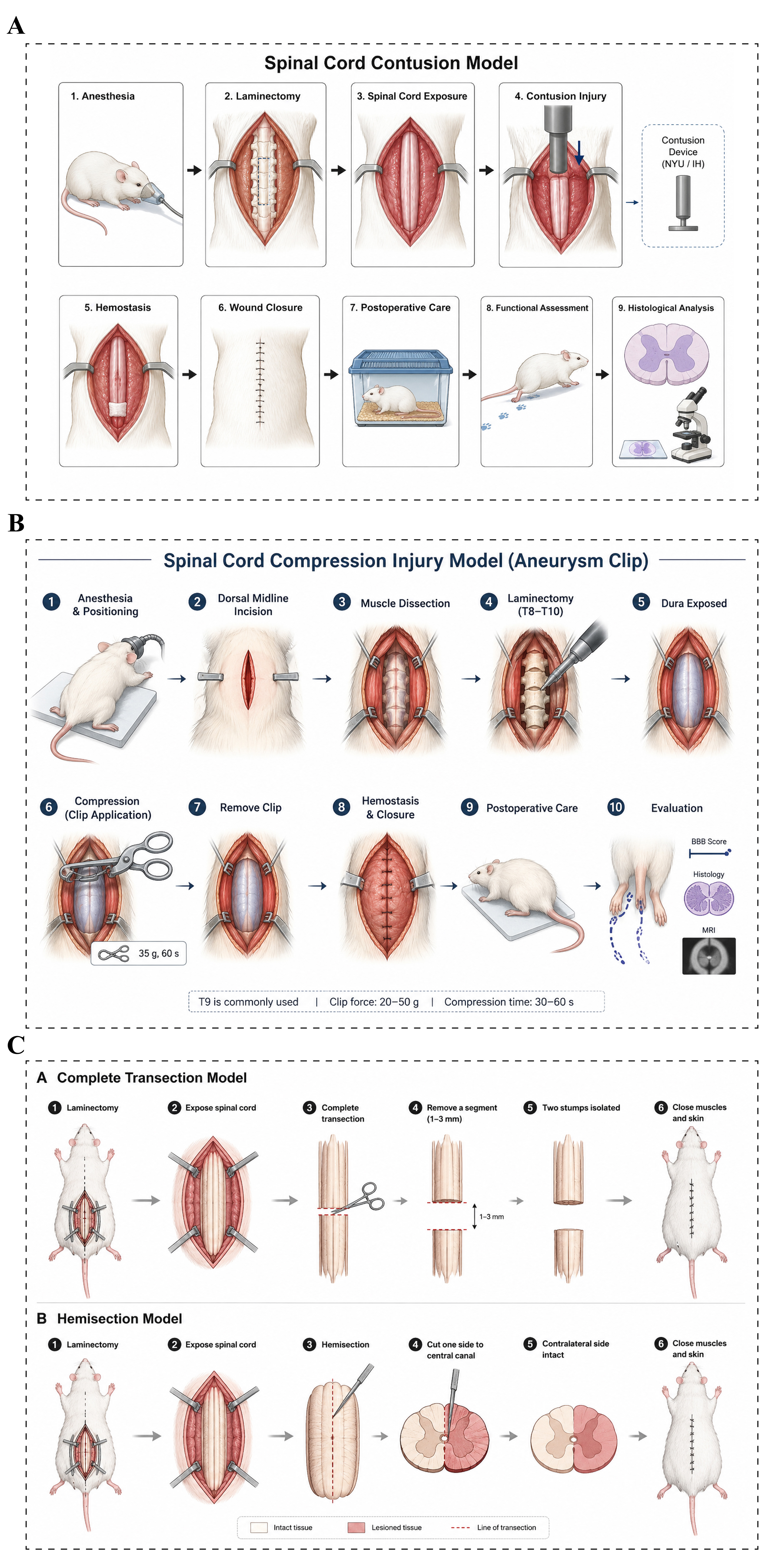
Figure 7. Animal models of SCI. (A) Contusion injury models: After anesthesia, the animal is placed in the prone position, and laminectomy is performed at the target thoracic level, typically T9-T10, to expose the spinal cord while preserving the dura. A standardized impactor, such as the NYU or Infinite Horizon Impactor, is then used to deliver a controlled contusion injury. After surgery, animals receive routine postoperative care, and injury severity and recovery are assessed using locomotor tests, histological analysis, and immunological evaluation; (B) Compression injury models: After laminectomy and spinal cord exposure under anesthesia, a calibrated aneurysm clip is applied to the target segment for 30-60 s to induce compression injury. Neurological impairment and recovery are subsequently evaluated using the BBB locomotor score, histological analysis, and imaging examinations; (C) Complete transection and hemisection models: In the complete transection model, the exposed spinal cord is completely severed at the target level, with a small segment of tissue removed when necessary to prevent reconnection. This model is widely used to study axonal regeneration, neural tissue engineering, and cell-based therapies. In the hemisection model, only one half of the spinal cord is transected while the contralateral side remains intact, making it particularly suitable for investigating neural plasticity, axonal regeneration, neural circuit reconstruction, and functional recovery. NYU: New York University impactor; IH: Infinite Horizon impactor; BBB: Basso, Beattie, and Bresnahan; MRI: magnetic resonance imaging; SCI: spinal cord injury.
Following contusion injury, animals typically develop pronounced motor deficits accompanied by acute pathological changes such as hemorrhage, edema, vascular disruption, and neuronal loss. Secondary injury subsequently evolves over time and may include inflammatory cell infiltration, axonal degeneration, demyelination, glial scar formation, and development of cystic cavities[180-182]. Because this model captures both the initial mechanical insult and the subsequent secondary injury cascade, it is widely used to investigate neuroprotective mechanisms, cell-based therapies, biomaterial-based interventions, and strategies to promote neural repair and functional recovery.
Recent advances in high-throughput and single-cell sequencing have further expanded the application of contusion models by revealing dynamic changes in the cellular composition and molecular state of the injured spinal cord. These studies have identified time-dependent responses involving microglia, infiltrating macrophages, reactive astrocytes, oligodendrocyte-lineage cells, and other neural and immune populations[183]. Such findings have improved understanding of the complex cellular interactions that shape secondary injury and repair after SCI.
Compression injury models
Spinal cord compression models reproduce neural injury associated with sustained or progressive mechanical compression, as may occur in vertebral fracture or dislocation, spinal canal stenosis, and space-occupying lesions. In contrast to contusion models, which primarily reproduce a brief mechanical impact, compression models allow the duration and magnitude of spinal cord deformation to be experimentally controlled. Common approaches include vascular clip compression, balloon compression, and screw- or device-based compression. Among these, vascular clip models are widely used because they provide relatively standardized and reproducible injury severity. In a typical procedure, a calibrated vascular clip is applied to the exposed spinal cord for a defined period, producing neuronal loss, axonal disruption, vascular compromise, and ischemic injury [Figure 7B][184,185].
Chronic compression models have also been developed to investigate slowly progressive spinal cord dysfunction, particularly in the context of degenerative cervical myelopathy. These models typically use expandable materials or gradually advancing compression devices to impose mild but persistent pressure on the spinal cord over an extended period[186,187]. The resulting pathological changes may include progressive axonal degeneration, demyelination, vascular dysfunction, and neurological decline, thereby reproducing selected features of chronic compressive myelopathy. Such models are particularly useful for studying the mechanisms of gradual neurological deterioration, the effects of prolonged compression, and functional recovery after surgical decompression.
Complete transection and hemisection models
Spinal cord transection models are widely used to investigate axonal regeneration, neural plasticity, and circuit reconstruction after SCI. Depending on the extent of tissue disruption, these models are generally classified as complete transection or hemisection injuries [Figure 7C].
In complete transection models, the spinal cord is fully severed, completely interrupting ascending and descending neural pathways. The clearly defined lesion boundary and absence of residual neural continuity make this model particularly useful for determining whether regenerating axons can traverse the injury site and for evaluating neural grafts, biomaterial scaffolds, and other strategies designed to promote structural reconnection[188]. However, complete transection is relatively uncommon in clinical SCI and therefore has limited value for reproducing the broader spectrum of traumatic spinal cord injuries encountered in patients.
Hemisection models, in contrast, disrupt one side of the spinal cord while preserving partial neural continuity on the contralateral side[189,190]. This configuration allows investigators to examine interactions between injured and spared pathways and to study compensatory plasticity during functional recovery. Because residual neural circuits can undergo structural and functional remodeling after hemisection, these models are particularly valuable for investigating neural plasticity, axonal sprouting, and circuit reorganization.
More recently, transection and hemisection models have been combined with neural tracing, optogenetic, and chemogenetic approaches to define the contribution of specific neuronal populations and pathways to post-injury remodeling and functional recovery. These techniques have expanded the utility of transection models for mechanistic studies of neural circuit repair after SCI.
Animal models of spinal infection
Spinal infection encompasses a heterogeneous group of disorders involving the vertebral bodies, intervertebral discs, paravertebral soft tissues, and epidural space. Major clinical entities include pyogenic spondylodiscitis, spinal tuberculosis, and implant-associated infection following spinal instrumentation[191-193]. Although advances in antimicrobial therapy, diagnostic imaging, and surgical management have improved clinical outcomes, spinal infection remains challenging because of delayed diagnosis, prolonged treatment, recurrence, and the risk of neurological complications. Its clinical burden may be further influenced by population aging, increasing use of immunosuppressive therapies, and the growing number of instrumented spinal procedures. Reliable animal models are therefore important for investigating disease pathogenesis, host-pathogen interactions, immune responses, and novel anti-infective strategies.
Unlike many noninfectious spinal disorders, spinal infection is characterized by dynamic interactions among invading pathogens, host immune responses, local tissue destruction, and subsequent repair. An informative animal model should therefore reproduce not only the establishment of a localized infectious lesion but also relevant pathological features such as inflammatory cell infiltration, bone and disc destruction, abscess formation, tissue remodeling, and, where appropriate, biofilm development. Current experimental models primarily include pyogenic spinal infection, spinal tuberculosis, and implant-associated infection models.
Pyogenic spondylodiscitis and spinal tuberculosis models
Pyogenic spondylodiscitis and spinal tuberculosis models are commonly established by direct inoculation of pathogenic microorganisms into the vertebral body or intervertebral disc [Figure 8A]. Depending on the experimental objective, inoculation can be performed within vertebral cancellous bone, the endplate region, or the intervertebral disc to induce different patterns of osseous and disc involvement. Following inoculation, animals may develop progressive inflammatory changes, bone destruction, disc space narrowing, and extension of infection into adjacent soft tissues. Disease severity and progression can be evaluated using radiological, histological, and microbiological methods. These models provide useful platforms for investigating host-pathogen interactions, tissue destruction, and therapeutic responses in spinal infection.
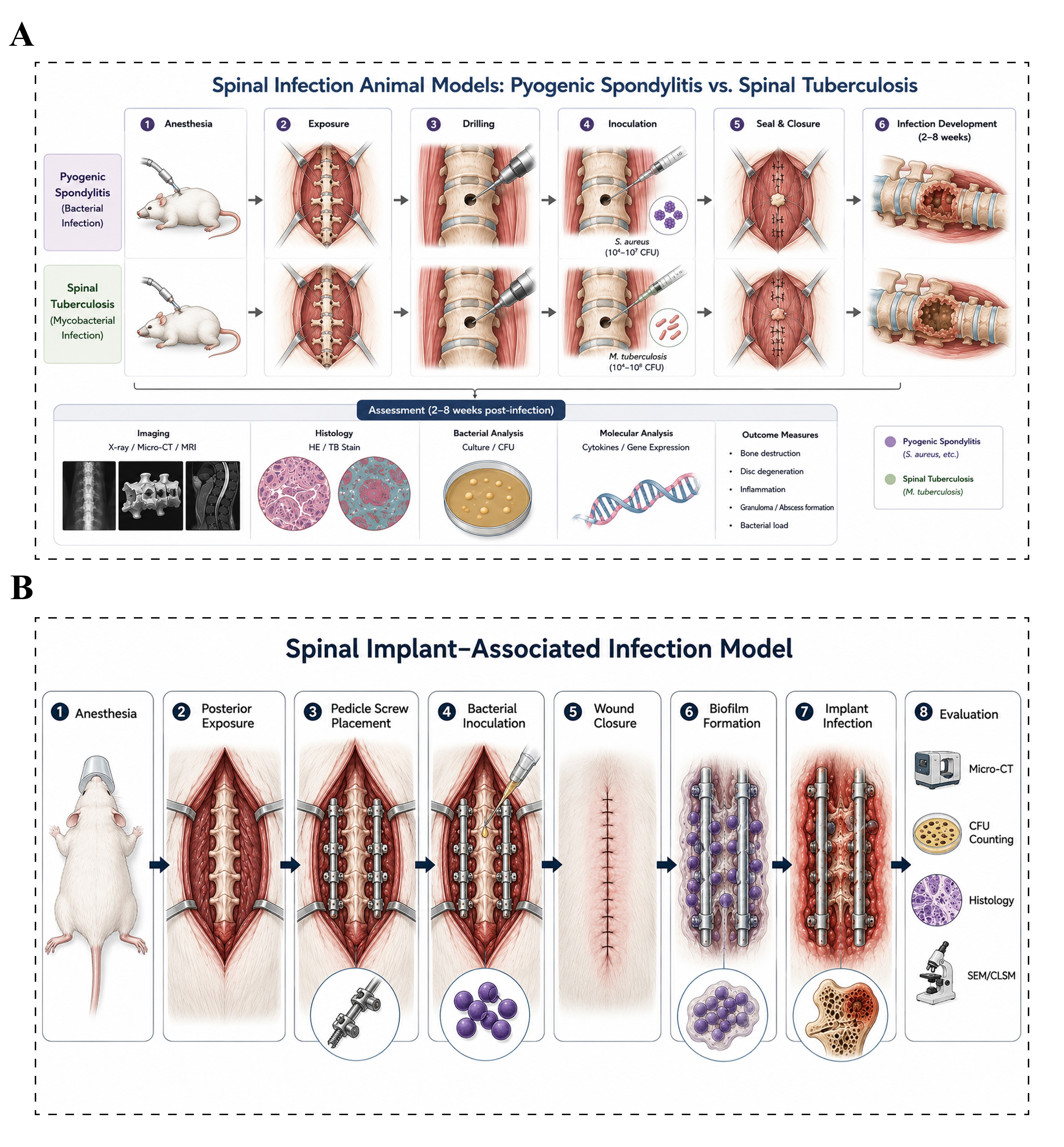
Figure 8. Animal models of spinal infection. (A) Pyogenic spondylodiscitis and spinal tuberculosis models: The target vertebral body is exposed under anesthesia, and a bone defect is created for inoculation with either pyogenic bacteria or Mycobacterium tuberculosis. Infection establishment, bone destruction, and inflammatory responses are subsequently evaluated using imaging, histological, and microbiological analyses. Apart from differences in the inoculated pathogen and disease course, the overall modeling procedures are largely similar; (B) Implant-associated infection models: Spinal implants, such as titanium screws or rods, are placed near the lamina or pedicle, followed by local inoculation of a bacterial suspension around the implant. Bacterial adhesion and biofilm formation on the implant surface gradually establish a stable implant-associated infection. Infection severity and tissue pathological changes are assessed by imaging, colony counting, histological analysis, and biofilm evaluation. Micro-CT: Micro-computed tomography; MRI: magnetic resonance imaging; HE: hematoxylin and eosin staining; TB: toluidine blue staining; CFU: colony-forming unit; SEM: scanning electron microscopy; CLSM: confocal laser scanning microscopy.
Staphylococcus aureus is the most commonly used pathogen for establishing pyogenic spondylodiscitis models, reflecting its clinical importance in vertebral and disc infections. Other microorganisms, including Escherichia coli, Pseudomonas aeruginosa, and methicillin-resistant Staphylococcus aureus (MRSA), have also been used to reproduce different infectious conditions. Depending on the pathogen, inoculum, and animal species, acute or subacute lesions can develop over days to weeks and may be characterized by inflammatory cell infiltration, bone destruction, disc space narrowing, and abscess formation[194,195]. Because of their relatively rapid and reproducible disease course, these models are well suited to studies of bacterial virulence, host inflammatory responses, infection-associated bone loss, and the efficacy of systemic antibiotics, local drug-delivery systems, and anti-infective biomaterials.
Spinal tuberculosis models are generally established by inoculating Mycobacterium tuberculosis (M. tuberculosis) or related mycobacterial strains into the vertebral body. Rabbits are frequently used because their vertebral size facilitates surgical inoculation and subsequent imaging and pathological assessment[196,197]. Compared with pyogenic infection models, however, spinal tuberculosis models require longer experimental periods and more stringent biosafety measures because of the slow growth and pathogenicity of M. tuberculosis. To improve experimental safety and feasibility, some studies have explored attenuated or alternative mycobacterial strains, although the pathological fidelity of these approaches should be interpreted in relation to the specific organism used[198].
Following infection, animals may gradually develop chronic granulomatous inflammation, caseous necrosis, progressive vertebral destruction, collapse, and, in advanced cases, spinal deformity or neurological compromise. Imaging modalities such as micro-CT and, in selected settings, positron emission tomography (PET)-CT can facilitate longitudinal assessment of lesion development and bone destruction. These models are used to evaluate antituberculous therapies and to investigate immune processes involving macrophages, T cells, granuloma formation, and host-directed therapeutic responses[199,200].
Fungal and parasitic spinal infection models have also been described using related inoculation strategies. However, these models remain relatively uncommon, and standardized experimental systems have not yet been established for most of these pathogens.
Implant-associated infection models
Implant-associated infection is an important complication of spinal instrumentation and is characterized by the ability of microorganisms to adhere to implant surfaces and form biofilms. Biofilm formation can substantially reduce bacterial susceptibility to antimicrobial agents and facilitate persistence by limiting effective host immune clearance. To reproduce these features experimentally, titanium screws, rods, plates, or other implant materials are placed in or adjacent to the spine and subsequently inoculated with selected bacterial strains[201,202] [Figure 8B]. Following inoculation, bacteria can adhere to the implant surface and develop organized biofilm communities, resulting in persistent local infection and surrounding tissue inflammation.
Compared with planktonic bacteria, biofilm-associated microorganisms exhibit altered metabolic states and increased tolerance to antimicrobial treatment, making implant-associated infections particularly difficult to eradicate. These models are therefore widely used to investigate biofilm formation, host responses to infected implants, and the interactions between microorganisms and biomaterial surfaces. They also provide useful preclinical platforms for evaluating antibacterial coatings, local drug-delivery systems, surface modifications, and novel implant materials designed to reduce bacterial adhesion and biofilm development[203].
However, outcomes can be strongly influenced by factors such as bacterial species and inoculum, implant material and surface properties, implantation site, and the timing of bacterial challenge. Standardization of these parameters is therefore important for improving reproducibility and for comparing anti-infective strategies across studies.
CONCLUSION
Animal models remain central to both basic and translational spine research. Across a broad range of conditions, including IVDD, LDH, AIS, vertebral fracture and spinal fusion, SCI, and spinal infection, animal models provide essential experimental systems for investigating disease mechanisms, validating therapeutic targets, and evaluating emerging interventions before clinical translation.
Over the past several decades, spinal animal models have evolved from systems designed primarily to reproduce gross morphological or histopathological changes toward platforms that increasingly incorporate cellular, molecular, biomechanical, and immunological mechanisms. Contemporary studies place greater emphasis on cell fate regulation, immune microenvironment remodeling, mechanotransduction, and tissue repair[204]. At the same time, advances in single-cell sequencing, spatial transcriptomics, and integrated multi-omics analysis have enabled more detailed characterization of disease-associated cell populations, molecular states, and intercellular interactions, thereby expanding the mechanistic information that can be obtained from animal models[205,206].
Despite these advances, no animal model can fully reproduce the anatomical, biomechanical, immunological, and temporal complexity of human spinal disorders. Model selection should therefore be driven by the specific biological question, disease stage, intervention, and outcome of interest rather than by the assumption that a single species or modeling approach is universally optimal. Where appropriate, complementary validation across different models may improve the robustness and translational relevance of experimental findings.
Sex should also be considered an important biological variable in the design and interpretation of spinal animal studies. Increasing evidence suggests that IVDD, pain-related phenotypes, inflammatory responses, tissue remodeling, and therapeutic outcomes may differ between males and females. These differences may reflect interactions among sex hormones, immune regulation, age-related endocrine changes, and biomechanical factors. Studies should therefore report sex and age explicitly, include both sexes when scientifically appropriate, and consider sex-stratified analyses when relevant to the experimental question.
Another important direction is developing multifactorial models that integrate two or more disease-relevant stimuli. Many spinal disorders arise from interactions among aging, abnormal mechanical loading, inflammation, metabolic dysfunction, and genetic susceptibility. Composite models may therefore reproduce selected aspects of this complexity more effectively than conventional single-factor approaches[207]. However, increased biological complexity must be balanced against reduced experimental controllability and reproducibility, and such models should be designed around clearly defined mechanistic or translational objectives.
Emerging technologies are also likely to broaden the scope of spinal disease modeling. Humanized animal models may improve the study of human-specific immune or molecular responses, whereas organoid and ex vivo systems can complement in vivo experiments by enabling more controlled investigation of human cell and tissue behavior. Integration of spatial multi-omics, advanced imaging, computational modeling, and artificial intelligence-assisted image analysis may further improve phenotyping, longitudinal assessment, and cross-scale interpretation of experimental data[208]. These approaches are likely to be most valuable when used as complementary components of integrated modeling frameworks rather than as replacements for conventional animal models.
Future development of spinal animal models is therefore likely to proceed along several converging directions: greater mechanistic precision, improved representation of clinically relevant disease heterogeneity, integration of complementary in vivo and human-based experimental systems, and more standardized outcome assessment. Advances in gene editing, regenerative medicine, biomaterials, biomanufacturing, and computational analysis may further strengthen the ability of preclinical models to connect mechanistic discovery with therapeutic evaluation.
Overall, the value of animal models lies not in their ability to reproduce every feature of human spinal disease, but in their capacity to answer defined scientific questions under controlled experimental conditions. Continued refinement of model design, greater attention to species- and sex-specific differences, and integration with emerging experimental and analytical technologies should improve the rigor, reproducibility, and translational relevance of future spine research.
DECLARATIONS
Acknowledgments
Chen F and Fan Y contributed equally to this work. The authors sincerely acknowledge the entire staff of the Department of Spine Surgery, Shandong Provincial Hospital Affiliated to Shandong First Medical University and Department of Spinal Surgery, who offered assistance throughout the course of this study.
Authors’ contributions
Contributed to the research design, data analysis, and manuscript writing: Chen F, Fan Y
Initiated and designed the study: Li T, Jing X
All authors were fully involved in the study and approved the final version of this manuscript.
Availability of data and materials
Not applicable.
AI and AI-assisted tools statement
During the preparation of this manuscript, the AI tool ChatGPT (OpenAI, GPT-5.6 Sol, released 2026-07-09) was used solely to polish the English language and improve linguistic clarity. The tool did not influence the study design, data collection, analysis, interpretation, or the scientific content of the work. All authors take full responsibility for the accuracy, integrity, and final content of the manuscript.
Financial support and sponsorship
This research was funded by Scientific Research Projects of Shandong Medical Association (YXH2025YS068).
Conflicts of interest
All authors declared that there are no conflicts of interest.
Ethical approval and consent to participate
Not applicable.
Consent for publication
Not applicable.
Copyright
© The Author(s) 2026.
Supplementary Materials
REFERENCES
1. Hartvigsen J, Hancock MJ, Kongsted A, et al. What low back pain is and why we need to pay attention. Lancet. 2018;391:2356-67.
2. GBD 2021 Low Back Pain Collaborators. Global, regional, and national burden of low back pain, 1990-2020, its attributable risk factors, and projections to 2050: a systematic analysis of the Global Burden of Disease Study 2021. Lancet Rheumatol. 2023;5:e316-29.
3. Hoffeld K, Lenz M, Egenolf P, et al. Patient-related risk factors and lifestyle factors for lumbar degenerative disc disease: a systematic review. Neurochirurgie. 2023;69:101482.
4. Dario AB, Ferreira ML, Refshauge KM, Lima TS, Ordoñana JR, Ferreira PH. The relationship between obesity, low back pain, and lumbar disc degeneration when genetics and the environment are considered: a systematic review of twin studies. Spine J. 2015;15:1106-17.
5. Dong Y, Peng R, Kang H, et al. Global incidence, prevalence, and disability of vertebral fractures: a systematic analysis of the global burden of disease study 2019. Spine J. 2022;22:857-68.
6. Safdarian M, Trinka E, Rahimi-Movaghar V, et al. Global, regional, and national burden of spinal cord injury, 1990-2019: a systematic analysis for the Global Burden of Disease Study 2019. Lancet Neurology. 2023;22:1026-47.
7. Po EYP, Yang M, Takemura N, et al. Global prevalence of adolescent idiopathic scoliosis between 1975 and 2025: a systematic review and meta-analysis. Spine. 2026;Epub ahead of print.
8. Wang S, Li M, Ren J, Tao J, Fang M, Kong L. Global prevalence and associated risk factors of scoliosis in children and adolescents: a systematic review and meta-analysis. BMC Public Health. 2025;25:3640.
9. Bae Y, Kim M, Jeong W, Jang S, Lee SW. Epidemiology and future burden of vertebral fractures: insights from the global burden of disease 1990-2021. Healthcare. 2025;13:1774.
10. Ding W, Hu S, Wang P, et al. Spinal cord injury: the global incidence, prevalence, and disability from the global burden of disease study 2019. Spine. 2022;47:1532-40.
11. Thavarajasingam SG, Subbiah Ponniah H, Philipps R, et al. Increasing incidence of spondylodiscitis in England: an analysis of the national health service (NHS) hospital episode statistics from 2012 to 2021. Brain Spine. 2023;3:101733.
12. Weckbach S, Lutz B, Wölfle-Roos JV, Reichel H. Infektionen der wirbelsäule: therapiestrategien. Chirurg. 2016;87:839-46.
13. Setton LA, Chen J. Mechanobiology of the intervertebral disc and relevance to disc degeneration. J Bone Joint Surg Am. 2006;88:52-7.
14. Hellenbrand DJ, Quinn CM, Piper ZJ, Morehouse CN, Fixel JA, Hanna AS. Inflammation after spinal cord injury: a review of the critical timeline of signaling cues and cellular infiltration. J Neuroinflammation. 2021;18:284.
15. Poletto DL, Crowley JD, Tanglay O, Walsh WR, Pelletier MH. Preclinical in vivo animal models of intervertebral disc degeneration. Part 1: a systematic review. JOR Spine. 2022;6:e1234.
16. Liang T, Gao B, Zhou J, et al. Constructing intervertebral disc degeneration animal model: a review of current models. Front Surg. 2023;9:1089244.
17. Zhang JJ, Song W, Luo WY, Wei M, Sun LB, Zou XN, et al. Autologous nucleus pulposus transplantation to lumbar 5 dorsal root ganglion after epineurium dissection in rats: a modified model of non-compressive lumbar herniated intervertebral disc. Chin Med J. 2011;124:2009-14.
18. Kawakami M, Tamaki T, Hayashi N, et al. Mechanical compression of the lumbar nerve root alters pain‐related behaviors induced by the nucleus pulposus in the rat. J Orthop Res. 2000;18:257-64.
19. Liu Y, Li J, Li H, et al. AMP-activated protein kinase activation in dorsal root ganglion suppresses mTOR/p70S6K signaling and alleviates painful radiculopathies in lumbar disc herniation rat model. Spine. 2019;44:E865-72.
20. Man GCW, Wang WWJ, Yim APY, et al. A review of pinealectomy-induced melatonin-deficient animal models for the study of etiopathogenesis of adolescent idiopathic scoliosis. Int J Mol Sci. 2014;15:16484-99.
21. Sharif-Alhoseini M, Khormali M, Rezaei M, et al. Animal models of spinal cord injury: a systematic review. Spinal Cord. 2017;55:714-21.
22. Alini M, Eisenstein SM, Ito K, et al. Are animal models useful for studying human disc disorders/degeneration? Eur Spine J. 2007;17:2-19.
23. Yao GL, Xie XS, Wang SJ, et al. NDP52-mediated autophagic degradation of CAPZA1 ameliorates intervertebral disc degeneration by suppressing cellular senescence. Free Radic Biol Med. 2026;251:480-95.
24. Qin T, Jiang Y, Xie Y, et al. Targeting Dnmt3a/m5C/RelA axis attenuates microglia inflammatory response and improves postoperative recovery in chronic compressive cervical spinal cord injury. Adv Sci. 2026;13:e75995.
25. Chen F, Peng F, Chen S, et al. PIEZO1‐GPX4 axis mediates mechanical stress‐induced vertebral growth plate dysplasia via ferroptosis activation. Adv Sci. 2025;12:e02052.
26. McLellan MA, Rosenthal NA, Pinto AR. Cre‐loxP‐mediated recombination: general principles and experimental considerations. Curr Protoc Mouse Biol. 2017;7:1-12.
27. Cong L, Ran FA, Cox D, et al. Multiplex genome engineering using CRISPR/Cas systems. Science. 2013;339:819-23.
29. Bowling S, Sritharan D, Osorio FG, et al. An engineered CRISPR-Cas9 mouse line for simultaneous readout of lineage histories and gene expression profiles in single cells. Cell. 2020;181:1410-22.e27.
30. Hunter C, Matyas J, Duncan NA. The notochordal cell in the nucleus pulposus: a review in the context of tissue engineering. Tissue Eng. 2003;9:667-77.
31. Hunter CJ, Matyas JR, Duncan NA. The three‐dimensional architecture of the notochordal nucleus pulposus: novel observations on cell structures in the canine intervertebral disc. J Anat. 2003;202:279-91.
32. Fan C, Wang W, Yu Z, et al. M1 macrophage-derived exosomes promote intervertebral disc degeneration by enhancing nucleus pulposus cell senescence through LCN2/NF-κB signaling axis. J Nanobiotechnol. 2024;22:301.
33. Ujigo S, Jonsson D, Bogestål Y, et al. Structural analysis of experimentally induced disc herniation-like changes in the rat. Spine Surg Relat Res. 2020;4:117-23.
34. Kjell J, Olson L. Rat models of spinal cord injury: from pathology to potential therapies. Dis Model Mech. 2016;9:1125-37.
35. Fan H, Tang H, Chen Z, et al. Inhibiting HMGB1-RAGE axis prevents pro-inflammatory macrophages/microglia polarization and affords neuroprotection after spinal cord injury. J Neuroinflammation. 2020;17:295.
36. Bennett GJ, Xie YK. A peripheral mononeuropathy in rat that produces disorders of pain sensation like those seen in man. Pain. 1988;33:87-107.
37. Marek GJ, Salek AA. Extending the specificity of DRL 72-s behavior for screening antidepressant-like effects of glutamatergic clinically validated anxiolytic or antidepressant drugs in rats. J Pharmacol Exp Ther. 2020;374:200-10.
38. Yan X, Ding JY, Zhang RJ, et al. FSTL1 accelerates nucleus pulposus cell senescence and intervertebral disc degeneration through TLR4/NF-κB pathway. Inflammation. 2024;47:1229-47.
39. Bu P, Peng R, Zhang J, et al. A one‐stone‐two-birds strategy for intervertebral disc repair: constructing a reductive chelation hydrogel to mitigate oxidative stress and promote disc matrix reconstruction. Adv Mater. 2024;37:2411290.
40. Virk SS, Coble D, Bertone AL, Hussein HH, Khan SN. Experimental design and surgical approach to create a spinal fusion model in a new zealand white rabbit (Oryctolagus cuniculus). J Invest Surg. 2017;30:226-34.
41. Geng G, Wang Q, Shi J, Yan J, Niu N, Wang Z. Establishment of a New Zealand rabbit model of spinal tuberculosis. J Spinal Disord Tech. 2015;28:E140-5.
42. Ma L, Hu Y, Wang Y, et al. From plant to prosthesis: an engineered biomimetic disc repairs senescence-associated intervertebral disc degeneration in goats. Bioact Mater. 2026;63:924-41.
43. Li Q, Hu B, Wang L, et al. Biomineralized PEEK cages containing osteoinductive CaP bioceramics promote spinal fusion in goats. Bioact Mater. 2025;45:128-47.
44. Zhou X, Zhang F, Wang D, et al. Micro fragmented adipose tissue promotes the matrix synthesis function of nucleus pulposus cells and regenerates degenerated intervertebral disc in a pig model. Cell Transplant. 2020;29:096368972090579.
45. Pekmezci M, Yilmaz G, Daglioglu K, et al. The effect of anterior spinal fusion on spinal canal development in an immature porcine model. Spine. 2009;34:E501-6.
46. Yang F, Xiao D, Zhao Q, et al. Fabrication of a novel whole tissue-engineered intervertebral disc for intervertebral disc regeneration in the porcine lumbar spine. RSC Adv. 2018;8:39013-21.
47. Nuckley DJ, Kramer PA, Del Rosario A, Fabro N, Baran S, Ching RP. Intervertebral disc degeneration in a naturally occurring primate model: radiographic and biomechanical evidence. J Orthop Res. 2008;26:1283-8.
48. Bailey JF, Fields AJ, Liebenberg E, Mattison JA, Lotz JC, Kramer PA. Comparison of vertebral and intervertebral disc lesions in aging humans and rhesus monkeys. Osteoarthritis Cartilage. 2014;22:980-5.
49. Wei F, Zhong R, Zhou Z, et al. In vivo experimental intervertebral disc degeneration induced by bleomycin in the rhesus monkey. BMC Musculoskelet Disord. 2014;15:340.
50. Capogrosso M, Milekovic T, Borton D, et al. A brain-spine interface alleviating gait deficits after spinal cord injury in primates. Nature. 2016;539:284-8.
51. Zhang ZG, Kang L, Zhou LP, et al. ASIC1a promotes nucleus pulposus derived stem cells apoptosis through modulation of SIRT3-dependent mitochondrial redox homeostasis in intervertebral disc degeneration. Redox Rep. 2025;30:2504120.
52. Jin Y, Wu O, Chen Q, et al. Hypoxia‐preconditioned BMSC‐derived exosomes induce mitophagy via the BNIP3-ANAX2 axis to alleviate intervertebral disc degeneration. Adv Sci. 2024;11:2404275.
53. Peng F, Sun M, Jing X, et al. Piezo1 promotes intervertebral disc degeneration through the Ca2+/F-actin/Yap signaling axis. Mol Med. 2025;31:90.
54. Ma Y, Yu X, Li W, et al. Animal models of internal endplate injury-induced intervertebral disc degeneration: a systematic review. J Invest Surg. 2024;37:2400478.
55. Elmounedi N, Keskes H. Establishment of intervertebral disc degeneration models; A review of the currently used models. J Orthop. 2024;56:50-6.
56. Hong JY, Kim H, Jeon W, et al. Animal models of intervertebral disc diseases: advantages, limitations, and future directions. Neurol Int. 2024;16:1788-818.
57. Masuda K, Aota Y, Muehleman C, et al. A novel rabbit model of mild, reproducible disc degeneration by an anulus needle puncture: correlation between the degree of disc injury and radiological and histological appearances of disc degeneration. Spine. 2005;30:5-14.
58. Elliott DM, Yerramalli CS, Beckstein JC, Boxberger JI, Johannessen W, Vresilovic EJ. The effect of relative needle diameter in puncture and sham injection animal models of degeneration. Spine. 2008;33:588-96.
59. Keorochana G, Johnson JS, Taghavi CE, et al. The effect of needle size inducing degeneration in the rat caudal disc: evaluation using radiograph, magnetic resonance imaging, histology, and immunohistochemistry. Spine J. 2010;10:1014-23.
60. Michalek AJ, Funabashi KL, Iatridis JC. Needle puncture injury of the rat intervertebral disc affects torsional and compressive biomechanics differently. Eur Spine J. 2010;19:2110-6.
61. Issy AC, Castania V, Castania M, et al. Experimental model of intervertebral disc degeneration by needle puncture in Wistar rats. Braz J Med Biol Res. 2013;46:235-44.
62. Mwale F, Masuda K, Pichika R, et al. The efficacy of Link N as a mediator of repair in a rabbit model of intervertebral disc degeneration. Arthritis Res Ther. 2011;13:R120.
63. Shi Y, Li F, Zhao T, et al. CRISPLD2 attenuates intervertebral disc degeneration by suppressing oxidative stress‐induced ferroptosis through the miR‐548I‐IL17A axis. Adv Sci. 2026;13:e16477.
64. Zhuang Y, Liu L, Liu M, et al. The sonic hedgehog pathway suppresses oxidative stress and senescence in nucleus pulposus cells to alleviate intervertebral disc degeneration via GPX4. Biochim Biophys Acta Mol Basis Dis. 2024;1870:166961.
65. Miao HY, Feng J, Liu W, et al. Mechanism of Duhuo Jisheng Decoction in regulating intervertebral disc degeneration via Wnt3a/YAP/ACSL4 pathway. Zhongguo Zhong Yao Za Zhi. 2025;50:5830-41.
66. Sakai D, Mochida J, Iwashina T, et al. Differentiation of mesenchymal stem cells transplanted to a rabbit degenerative disc model: potential and limitations for stem cell therapy in disc regeneration. Spine. 2005;30:2379-87.
67. Gullbrand SE, Ashinsky BG, Bonnevie ED, et al. Long-term mechanical function and integration of an implanted tissue-engineered intervertebral disc. Sci Transl Med. 2018;10:eaau0670.
68. Xia C, Zeng Z, Fang B, et al. Mesenchymal stem cell-derived exosomes ameliorate intervertebral disc degeneration via anti-oxidant and anti-inflammatory effects. Free Radic Biol Med. 2019;143:1-15.
69. Nishida K, Kang JD, Gilbertson LG, et al. Modulation of the biologic activity of the rabbit intervertebral disc by gene therapy: an in vivo study of adenovirus-mediated transfer of the human transforming growth factor beta 1 encoding gene. Spine. 1999;24:2419-25.
70. Navani A, Vardhan S, Aggarwal A, Navani R, Lana JF, Diwan SA. Functional spinal unit approach for orthobiologic injections for low back pain. pain physician. 2025;28:S145-56.
71. Liu S, Sun Y, Dong J, Bian Q. A mouse model of lumbar spine instability. J Vis Exp. 2021;170:e61722.
72. Oichi T, Taniguchi Y, Soma K, et al. A mouse intervertebral disc degeneration model by surgically induced instability. Spine. 2018;43:E557-64.
73. Li F, Chen M, Zhang M, et al. Targeting Piezo1 channel to alleviate intervertebral disc degeneration. J Orthop Translat. 2025;51:145-58.
74. Zhou C, Yao S, Fu F, et al. Morroniside attenuates nucleus pulposus cell senescence to alleviate intervertebral disc degeneration via inhibiting ROS-Hippo-p53 pathway. Front Pharmacol. 2022;13:942435.
75. Fu F, Bao R, Yao S, et al. Aberrant spinal mechanical loading stress triggers intervertebral disc degeneration by inducing pyroptosis and nerve ingrowth. Sci Rep. 2021;11:772.
76. Wang Y, Zhang W, Yang Y, et al. Osteopontin deficiency promotes cartilaginous endplate degeneration by enhancing the NF-κB signaling to recruit macrophages and activate the NLRP3 inflammasome. Bone Res. 2024;12:53.
77. Vergroesen PPA, Kingma I, Emanuel KS, et al. Mechanics and biology in intervertebral disc degeneration: a vicious circle. Osteoarthritis Cartilage. 2015;23:1057-70.
78. Neidlinger-Wilke C, Galbusera F, Pratsinis H, et al. Mechanical loading of the intervertebral disc: from the macroscopic to the cellular level. Eur Spine J. 2013;23:333-43.
79. Iatridis JC, Michalek AJ, Purmessur D, Korecki CL. Localized intervertebral disc injury leads to organ level changes in structure, cellularity, and biosynthesis. Cel Mol Bioeng. 2009;2:437-47.
80. Huang YC, Urban JPG, Luk KDK. Intervertebral disc regeneration: do nutrients lead the way? Nat Rev Rheumatol. 2014;10:561-6.
81. Neidlinger-Wilke C, Teixeira G, Greiner-Perth AK, Werner K, Wilke HJ, Liebsch C. Biology and mechanobiology of the intervertebral disc: challenges for regenerative therapies. Orthopädie. 2024;53:905-11.
82. Iatridis JC, Mente PL, Stokes IAF, Aronsson DD, Alini M. Compression-induced changes in intervertebral disc properties in a rat tail model. Spine. 1999;24:996-1002.
83. Maclean JJ, Lee CR, Alini M, Iatridis JC. The effects of short‐term load duration on anabolic and catabolic gene expression in the rat tail intervertebral disc. J Orthop Res. 2006;23:1120-7.
84. Stokes IAF, Iatridis JC. Mechanical conditions that accelerate intervertebral disc degeneration: overload versus immobilization. Spine. 2004;29:2724-32.
85. Fearing BV, Hernandez PA, Setton LA, Chahine NO. Mechanotransduction and cell biomechanics of the intervertebral disc. JOR Spine. 2018;1:e1026.
86. Ruffilli A, Viroli G, Neri S, et al. Mechanobiology of the human intervertebral disc: systematic review of the literature and future perspectives. Int J Mol Sci. 2023;24:2728.
87. Kaur A, Singh S, Vishwas S, Singh TG. Exploring the therapeutic potential of piezo1 in ageing-related neurodegenerative diseases. J Mol Neurosci. 2026;76:13.
88. Wu J, Chen Y, Liao Z, et al. Self-amplifying loop of NF-κB and periostin initiated by PIEZO1 accelerates mechano-induced senescence of nucleus pulposus cells and intervertebral disc degeneration. Mol Ther. 2022;30:3241-56.
89. Xiang Z, Zhang P, Jia C, et al. Piezo1 channel exaggerates ferroptosis of nucleus pulposus cells by mediating mechanical stress-induced iron influx. Bone Res. 2024;12:20.
90. Chen S, Li Z, Chen D, et al. Piezo1-mediated mechanotransduction promotes entheseal pathological new bone formation in ankylosing spondylitis. Ann Rheum Dis. 2023;82:533-45.
91. Dong ZR, Wang J, Huang YK, et al. Ciliary IFT88 inhibits intervertebral disc degeneration under excessive mechanical stress by regulating endplate cartilage calcification. J Adv Res. 2026;81:157-73.
92. Fan C, Chu G, Yu Z, et al. The role of ferroptosis in intervertebral disc degeneration. Front Cell Dev Biol. 2023;11:1219840.
93. Wang P, Xie Z, Deng L, et al. Macrophage-derived legumain ameliorates excessive mechanical stress-induced ferroptosis of nucleus pulposus cells and intervertebral disc degeneration via integrin αvβ3-Hippo signaling. Cell Mol Biol Lett. 2026;31:69.
94. Sakai D, Schol J. Cell therapy for intervertebral disc repair: clinical perspective. J Orthop Translat. 2017;9:8-18.
95. Gan Y, He J, Zhu J, et al. Spatially defined single-cell transcriptional profiling characterizes diverse chondrocyte subtypes and nucleus pulposus progenitors in human intervertebral discs. Bone Res. 2021;9:37.
96. Shi Z, Yuan T, Feng C, et al. Roles of hippo-YAP/TAZ signalling in intervertebral disc degeneration. Biomed Pharmacother. 2023;159:114099.
97. Hu T, Shi Z, Sun Y, et al. SEPHS1 attenuates intervertebral disc degeneration by delaying nucleus pulposus cell senescence through the Hippo-Yap/Taz pathway. Am J Physiol Cell Physiol. 2024;326:C386-99.
98. Zhang X, Shu S, Feng Z, Qiu Y, Bao H, Zhu Z. Microtubule stabilization promotes the synthesis of type 2 collagen in nucleus pulposus cell by activating hippo-yap pathway. Front Pharmacol. 2023;14:1102318.
99. Daly C, Ghosh P, Jenkin G, Oehme D, Goldschlager T. A review of animal models of intervertebral disc degeneration: pathophysiology, regeneration, and translation to the clinic. Biomed Res Int. 2016;2016:595216.
100. Jin L, Balian G, Li XJ. Animal models for disc degeneration-an update. Histol Histopathol. 2018;33:543-54.
101. Hoogendoorn RJ, Wuisman PI, Smit TH, Everts VE, Helder MN. Experimental intervertebral disc degeneration induced by chondroitinase ABC in the goat. Spine. 2007;32:1816-25.
102. Augustin JA, Burt KG, Barrett C, et al. Neuroimmune activation in a goat model of intervertebral disc degeneration. Cells. 2026;15:286.
103. Sakuma M, Fujii N, Takahashi T, Hoshino J, Miyauchi S, Iwata H. Effect of chondroitinase ABC on matrix metalloproteinases and inflammatory mediators produced by intervertebral disc of rabbit in vitro. Spine. 2002;27:576-80.
104. Imai Y, Okuma M, An HS, et al. Restoration of disc height loss by recombinant human osteogenic protein-1 injection into intervertebral discs undergoing degeneration induced by an intradiscal injection of chondroitinase ABC. Spine. 2007;32:1197-205.
105. Boxberger JI, Auerbach JD, Sen S, Elliott DM. An in vivo model of reduced nucleus pulposus glycosaminoglycan content in the rat lumbar intervertebral disc. Spine. 2008;33:146-54.
106. Risbud MV, Shapiro IM. Role of cytokines in intervertebral disc degeneration: pain and disc content. Nat Rev Rheumatol. 2014;10:44-56.
107. Le Maitre CL, Hoyland JA, Freemont AJ. Interleukin-1 receptor antagonist delivered directly and by gene therapy inhibits matrix degradation in the intact degenerate human intervertebral disc: an in situ zymographic and gene therapy study. Arthritis Res Ther. 2007;9:R83.
108. Phillips K, Cullen K, Chiverton N, et al. Potential roles of cytokines and chemokines in human intervertebral disc degeneration: interleukin-1 is a master regulator of catabolic processes. Osteoarthritis Cartilage. 2015;23:1165-77.
109. Kang R, Li H, Rickers K, Ringgaard S, Xie L, Bünger C. Intervertebral disc degenerative changes after intradiscal injection of TNF-α in a porcine model. Eur Spine J. 2015;24:2010-6.
110. Lai A, Moon A, Purmessur D, et al. Annular puncture with tumor necrosis factor-alpha injection enhances painful behavior with disc degeneration in vivo. Spine J. 2016;16:420-31.
111. Zheng J, Wang J, Liu H, et al. Alarmins S100A8/A9 promote intervertebral disc degeneration and inflammation-related pain in a rat model through toll-like receptor-4 and activation of the NF-κB signaling pathway. Osteoarthritis Cartilage. 2022;30:998-1011.
112. Dixon SJ, Lemberg KM, Lamprecht MR, et al. Ferroptosis: an iron-dependent form of nonapoptotic cell death. Cell. 2012;149:1060-72.
113. Ursini F, Maiorino M. Lipid peroxidation and ferroptosis: the role of GSH and GPx4. Free Radic Biol Med. 2020;152:175-85.
114. Swanson KV, Deng M, Ting JPY. The NLRP3 inflammasome: molecular activation and regulation to therapeutics. Nat Rev Immunol. 2019;19:477-89.
115. Broz P, Dixit VM. Inflammasomes: mechanism of assembly, regulation and signalling. Nat Rev Immunol. 2016;16:407-20.
116. Roughley PJ. Biology of intervertebral disc aging and degeneration: involvement of the extracellular matrix. Spine. 2004;29:2691-9.
117. Watanabe H, Kimata K, Line S, et al. Mouse cartilage matrix deficiency (cmd) caused by a 7 bp deletion in the aggrecan gene. Nat Genet. 1994;7:154-7.
118. Li SW, Prockop DJ, Helminen H, et al. Transgenic mice with targeted inactivation of the Col2 alpha 1 gene for collagen II develop a skeleton with membranous and periosteal bone but no endochondral bone. Genes Dev. 1995;9:2821-30.
119. Henry SP, Liang S, Akdemir KC, de Crombrugghe B. The postnatal role of Sox9 in cartilage. J Bone Miner Res. 2012;27:2511-25.
120. Bell DM, Leung KK, Wheatley SC, et al. SOX9 directly regulates the type-II collagen gene. Nat Genet. 1997;16:174-8.
121. Tsingas M, Ottone OK, Haseeb A, et al. Sox9 deletion causes severe intervertebral disc degeneration characterized by apoptosis, matrix remodeling, and compartment-specific transcriptomic changes. Matrix Biol. 2020;94:110-33.
122. Silberberg R, Aufdermaur M, Adler JH. Degeneration of the intervertebral disks and spondylosis in aging sand rats. Arch Pathol Lab Med. 1979;103:231-5.
123. Moskowitz RW, Ziv I, Denko CW, Boja B, Jones PK, Adler JH. Spondylosis in sand rats: a model of intervertebral disc degeneration and hyperostosis. J Orthop Res. 1990;8:401-11.
124. Tang SN, Walter BA, Heimann MK, et al. In vivo mouse intervertebral disc degeneration models and their utility as translational models of clinical discogenic back pain: a comparative review. Front Pain Res. 2022;3:894651.
125. Feng C, Liu H, Yang M, Zhang Y, Huang B, Zhou Y. Disc cell senescence in intervertebral disc degeneration: Causes and molecular pathways. Cell Cycle. 2016;15:1674-84.
126. Olmarker K, Rydevik B, Nordborg C. Autologous nucleus pulposus induces neurophysiologic and histologic changes in porcine cauda equina nerve roots. Spine. 1993;18:1425-32.
127. Obata K, Tsujino H, Yamanaka H, et al. Expression of neurotrophic factors in the dorsal root ganglion in a rat model of lumbar disc herniation. Pain. 2002;99:121-32.
128. Min Y, Wang C, Zhao P, Shi B. Spinal manipulative therapy promotes nucleus pulposus resorption and alleviates pain in a rat model of lumbar disc herniation via JNK-dependent regulation of inflammation and tissue remodeling. Am J Transl Res. 2026;18:1249-63.
129. Shamji MF, Setton LA, Jarvis W, et al. Proinflammatory cytokine expression profile in degenerated and herniated human intervertebral disc tissues. Arthritis Rheum. 2010;62:1974-82.
130. Olmarker K, Rydevik B, Holm S. Edema formation in spinal nerve roots induced by experimental, graded compression. An experimental study on the pig cauda equina with special reference to differences in effects between rapid and slow onset of compression. Spine. 1989;14:569-73.
131. Rydevik B, Brown MD, Lundborg G. Pathoanatomy and Pathophysiology of Nerve Root Compression. Spine. 1984;9:7-15.
132. Rydevik BL, Myers RR, Powell HC. Pressure increase in the dorsal root ganglion following mechanical compression: closed compartment syndrome in nerve roots. Spine. 1989;14:574-6.
133. Kobayashi S, Yoshizawa H, Yamada S. Pathology of lumbar nerve root compression Part 1: Intraradicular inflammatory changes induced by mechanical compression. J Orthop Res. 2004;22:170-9.
134. Boos N, Rieder R, Schade V, Spratt KF, Semmer N, Aebi M. 1995 Volvo Award in clinical sciences. The diagnostic accuracy of magnetic resonance imaging, work perception, and psychosocial factors in identifying symptomatic disc herniations. Spine. 1995;20:2613-25.
135. Machida M, Dubousset J, Imamura Y, Iwaya T, Yamada T, Kimura J. Role of melatonin deficiency in the development of scoliosis in pinealectomised chickens. J Bone Joint Surg Br. 1995;77:134-8.
136. Machida M, Dubousset J, Satoh T, et al. Pathologic mechanism of experimental scoliosis in pinealectomized chickens. Spine. 2001;26:E385-91.
137. Girardo M, Bettini N, Dema E, Cervellati S. The role of melatonin in the pathogenesis of adolescent idiopathic scoliosis (AIS). Eur Spine J. 2011;20:68-74.
138. Machida M, Murai I, Miyashita Y, Dubousset J, Yamada T, Kimura J. Pathogenesis of idiopathic scoliosis. Experimental study in rats. Spine. 1999;24:1985-9.
139. Chen F, Sun M, Peng F, et al. Compressive stress induces spinal vertebral growth plate chondrocytes apoptosis via Piezo1. J Orthop Res. 2023;41:1792-802.
140. Machida M, Dubousset J, Yamada T, et al. Experimental scoliosis in melatonin‐deficient C57BL/6J mice without pinealectomy. J Pineal Res. 2006;41:1-7.
141. Wijdicks SPJ, Lemans JVC, Overweg G, et al. Induction of a representative idiopathic-like scoliosis in a porcine model using a multidirectional dynamic spring-based system. Spine J. 2021;21:1376-86.
142. Braun JT, Ogilvie JW, Akyuz E, Brodke DS, Bachus KN. Creation of an experimental idiopathic-type scoliosis in an immature goat model using a flexible posterior asymmetric tether. Spine. 2006;31:1410-4.
143. Newton PO, Upasani VV, Farnsworth CL, et al. Spinal growth modulation with use of a tether in an immature porcine model. J Bone Joint Surg. 2008;90:2695-706.
144. Odent T, Cachon T, Peultier B, et al. Porcine model of early onset scoliosis based on animal growth created with posterior mini-invasive spinal offset tethering: a preliminary report. Eur Spine J. 2011;20:1869-76.
145. Zheng X, Sun X, Qiu Y, et al. A porcine early-onset scoliosis model created using a posterior mini-invasive method: a pilot study. J Spinal Disord Tech. 2014;27:E294-300.
146. Bogie R, Roth AK, Willems PC, Weegen W, Arts JJ, van Rhijn LW. The development of a representative porcine early-onset scoliosis model with a standalone posterior spinal tether. Spine Deform. 2017;5:2-10.
147. Zheng X, Qiu Y, Sun X, et al. A novel self-growing rod technique for early onset scoliosis: an experimental study based on a porcine scoliosis model. Zhonghua Yi Xue Za Zhi. 2016;96:1821-5.
148. Grauers A, Einarsdottir E, Gerdhem P. Genetics and pathogenesis of idiopathic scoliosis. Scoliosis Spinal Disord. 2016;11:45.
149. Takahashi Y, Kou I, Takahashi A, et al. A genome-wide association study identifies common variants near LBX1 associated with adolescent idiopathic scoliosis. Nat Genet. 2011;43:1237-40.
150. Qin X, Xu L, Xia C, et al. Genetic variant of GPR126 Gene is Functionally Associated with adolescent idiopathic scoliosis in Chinese population. Spine. 2017;42:E1098-103.
151. Xu L, Sheng F, Xia C, et al. Genetic variant of PAX1 gene is functionally associated with adolescent idiopathic scoliosis in the Chinese population. Spine. 2018;43:492-6.
152. De Salvatore S, Ruzzini L, Longo UG, et al. Exploring the association between specific genes and the onset of idiopathic scoliosis: a systematic review. BMC Med Genomics. 2022;15:115.
153. Grimes DT, Boswell CW, Morante NFC, Henkelman RM, Burdine RD, Ciruna B. Zebrafish models of idiopathic scoliosis link cerebrospinal fluid flow defects to spine curvature. Science. 2016;352:1341-4.
154. Hayes M, Gao X, Yu LX, et al. ptk7 mutant zebrafish models of congenital and idiopathic scoliosis implicate dysregulated Wnt signalling in disease. Nat Commun. 2014;5:4777.
155. Gray RS, Gonzalez R, Ackerman SD, et al. Postembryonic screen for mutations affecting spine development in zebrafish. Dev Biol. 2021;471:18-33.
156. Van Gennip JLM, Boswell CW, Ciruna B. Neuroinflammatory signals drive spinal curve formation in zebrafish models of idiopathic scoliosis. Sci Adv. 2018;4:eaav1781.
158. Turner AS. Animal models of osteoporosis - necessity and limitations. Eur Cell Mater. 2001;1:66-81.
159. Hofbauer LC, Compston JE, Saag KG, Rauner M, Tsourdi E. Glucocorticoid-induced osteoporosis: novel concepts and clinical implications. Lancet Diabetes Endocrinol. 2025;13:964-79.
160. Qi Z, Ye G, Liu Z, et al. A review of osteoporotic vertebral fracture animal models. Biomed Eng Online. 2025;24:40.
161. Cao W, Li D, Chen H, Luo Z, Zhang X, Li Y. Biomechanical study of porcine osteoporotic vertebral compression fracture model strengthened by trajectory-adjustable bone cement filling device. World Neurosurg. 2024;185:e357-66.
162. Yu Y, Lee S, Bock M, et al. Promotion of bone formation in a rat osteoporotic vertebral body defect model via suppression of osteoclastogenesis by ectopic embryonic calvaria derived mesenchymal stem cells. Int J Mol Sci. 2024;25:8174.
163. Malhotra A, Walle M, Paul GR, Kuhn GA, Müller R. Application of subject-specific adaptive mechanical loading for bone healing in a mouse tail vertebral defect. Sci Rep. 2021;11:1861.
164. Makino A, Hasegawa T, Yamamoto T, et al. Abaloparatide promotes bone repair of vertebral defects in ovariectomized rats by increasing bone formation. Bone. 2024;182:117056.
165. Sheyn D, Kallai I, Tawackoli W, et al. Gene-modified adult stem cells regenerate vertebral bone defect in a rat model. Mol Pharm. 2011;8:1592-601.
166. Liang H, Wang K, Shimer AL, Li X, Balian G, Shen FH. Use of a bioactive scaffold for the repair of bone defects in a novel reproducible vertebral body defect model. Bone. 2010;47:197-204.
167. Duan X, Liao HX, Zou HZ, Zhang ZJ, Ye JD, Liao WM. An injectable, biodegradable calcium phosphate cement containing poly lactic-co-glycolic acid as a bone substitute in ex vivo human vertebral compression fracture and rabbit bone defect models. Connect Tissue Res. 2018;59:55-65.
168. Zhu XS, Zhang ZM, Mao HQ, et al. A novel sheep vertebral bone defect model for injectable bioactive vertebral augmentation materials. J Mater Sci Mater Med. 2011;22:159-64.
169. Lamghari M, Huet H, Laurent A, Berland S, Lopez E. A model for evaluating injectable bone replacements in the vertebrae of sheep: radiological and histological study. Biomaterials. 1999;20:2107-14.
170. Wang H, Zhou Y, Li CQ, Chu TW, Wang J, Huang B. Tissue‑engineered bone used in a rabbit model of lumbar intertransverse process fusion: a comparison of osteogenic capacity between two different stem cells. Exp Ther Med. 2020;19:2570-8.
171. von Benecke J, Khan Z, Mclaren J, Shakesheff K, Birch N. A critical review of the methodology of ovine lumbar interbody fusion studies and recommendations for future study design. Bone Joint Res. 2026;15:58-72.
172. Broussolle T, Roux JP, Chapurlat R, Barrey C. Murine models of posterolateral spinal fusion: a systematic review. Neurochirurgie. 2023;69:101428.
173. Morse KW, Sun J, Hu L, et al. Development of murine anterior interbody and posterolateral spinal fusion techniques. J Bone Joint Surg Am. 2024;106:735-45.
174. Çetin E, Daldal İ, Eren A, et al. Epidermal growth factor enhances spinal fusion: posterolateral lumbar fusion model on rats. Acta Orthop Traumatol Turc. 2019;53:134-9.
175. Lam WMR, Zhuo WH, Yang L, et al. Mesenchymal stem cell exosomes enhance posterolateral spinal fusion in a rat model. Cells. 2024;13:761.
176. Yoshizato H, Morimoto T, Nonaka T, et al. Animal model for anterior lumbar interbody fusion: a literature review. Spine Surg Relat Res. 2024;8:373-82.
177. Duits A, Salvatori D, Schouten J, et al. Preclinical model for lumbar interbody fusion in small ruminants: rationale and guideline. J Orthop Translat. 2023;38:167-74.
178. Lobao A, Rice SW, Chapman J, Stauff MP, Song J. Rat spine fusion models for preclinical research: opportunities and challenges. ACS Biomater Sci Eng. 2025;11:7057-69.
179. Gayen CD, Bessen MA, Dorrian RM, et al. Survival model of thoracic contusion spinal cord injury in the domestic pig. J Neurotrauma. 2023;40:965-80.
180. Reinhardt DR, Stehlik KE, Satkunendrarajah K, Kroner A. Bilateral cervical contusion spinal cord injury: a mouse model to evaluate sensorimotor function. Exp Neurol. 2020;331:113381.
181. Gensel JC, Tovar CA, Hamers FPT, Deibert RJ, Beattie MS, Bresnahan JC. Behavioral and histological characterization of unilateral cervical spinal cord contusion injury in rats. J Neurotrauma. 2006;23:36-54.
182. Collazos-Castro JE, Soto VM, Gutiérrez-Dávila M, Nieto-Sampedro M. Motoneuron Loss associated with chronic locomotion impairments after spinal cord contusion in the rat. J Neurotrauma. 2005;22:544-58.
183. Yao XQ, Liu ZY, Chen JY, et al. Proteomics and bioinformatics reveal insights into neuroinflammation in the acute to subacute phases in rat models of spinal cord contusion injury. FASEB J. 2021;35:e21735.
184. Sobolev VE, Sysoev YI, Vyunova TV, Musienko PE. Animal models of spinal cord injury. Biomedicines. 2025;13:1427.
185. Ridlen R, McGrath K, Gorrie CA. Animal models of compression spinal cord injury. J Neurosci Res. 2022;100:2201-12.
186. Tanaka T, Murata H, Miyazaki R, et al. Human recombinant erythropoietin improves motor function in rats with spinal cord compression-induced cervical myelopathy. PLoS One. 2019;14:e0214351.
187. Yamamoto S, Kurokawa R, Kim P. Cilostazol, a selective Type III phosphodiesterase inhibitor: prevention of cervical myelopathy in a rat chronic compression model. J Neurosurg Spine. 2014;20:93-101.
188. Reshamwala R, Eindorf T, Shah M, et al. Induction of complete transection-type spinal cord injury in mice. J Vis Exp. 2020.
189. Wilson S, Fredericks DC, Safayi S, et al. Ovine hemisection model of spinal cord injury. J Invest Surg. 2021;34:380-92.
190. Ham TR, Farrag M, Soltisz AM, Lakes EH, Allen KD, Leipzig ND. Automated gait analysis detects improvements after intracellular σ peptide administration in a rat hemisection model of spinal cord injury. Ann Biomed Eng. 2019;47:744-53.
191. Placide R, Reznicek J. Evaluation and management of pyogenic spondylodiscitis: a review. J Clin Med. 2025;14:3477.
192. Principi N, Esposito S. Infectious discitis and spondylodiscitis in children. Int J Mol Sci. 2016;17:539.
193. Khanna K, Sabharwal S. Spinal tuberculosis: a comprehensive review for the modern spine surgeon. Spine J. 2019;19:1858-70.
194. Gamada H, Funayama T, Setojima Y, et al. Posterior fixation without debridement for pyogenic spondylodiscitis can promote infection control: initial evaluation of a pyogenic spondylodiscitis posterior fixation rat model. Eur Spine J. 2025;34:2165-75.
195. Chen WH, Jiang LS, Dai LY. A novel canine model of acute pyogenic spondylodiscitis. Neurosurg Rev. 2009;32:485-90.
196. Jia J, Zhang M, Cao Z, et al. The rabbit model for spinal tuberculosis: an overview. J Orthop Surg. 2024;32:10225536241266703.
197. Liu X, Jia W, Wang H, et al. Retraction: Establishment of a rabbit model of spinal tuberculosis using Mycobacterium tuberculosis strain H37Rv. Jpn J Infect Dis. 2015;68:89-97.
198. Qiao YJ, Song XY, Zhang LD, Li F, Zhang HQ, Zhou SH. Comparative study of a rabbit model of spinal tuberculosis using different concentrations of Mycobacterium tuberculosis. World J Orthop. 2025;16:101424.
199. Ma R, Zhang J, Chen Z, et al. Treatment of spinal tuberculosis in rabbits using bovine serum albumin nanoparticles loaded with isoniazid and rifampicin. Neurol Res. 2021;44:268-74.
200. Liu P, Jiang H, Li S, Lin Z, Jiang J. Determination of anti‐tuberculosis drug concentration and distribution from sustained release microspheres in the vertebrae of a spinal tuberculosis rabbit model. J Orthop Res. 2017;35:200-8.
201. Chen F, Schiffer NE, Song J. Animal models of orthopedic implant-associated infections and revisions. ACS Biomater Sci Eng. 2025;11:2052-68.
202. Gordon O, Miller RJ, Thompson JM, et al. Rabbit model of Staphylococcus aureus implant-associated spinal infection. Dis Model Mech. 2020;13:dmm045385.
203. Ishihama H, Ishii K, Nagai S, et al. An antibacterial coated polymer prevents biofilm formation and implant-associated infection. Sci Rep. 2021;11:3602.
204. González Martínez E, García-Cosamalón J, Cosamalón-Gan I, Esteban Blanco M, García-Suarez O, Vega JA. Biology and mechanobiology of the intervertebral disc. Neurocirugia. 2017;28:135-40.
205. Hu X, Wang Z, Zhang H, et al. Single-cell sequencing: new insights for intervertebral disc degeneration. Biomed Pharmacother. 2023;165:115224.
206. Longo SK, Guo MG, Ji AL, Khavari PA. Integrating single-cell and spatial transcriptomics to elucidate intercellular tissue dynamics. Nat Rev Genet. 2021;22:627-44.
207. Alini M, Diwan AD, Erwin WM, Little CB, Melrose J. An update on animal models of intervertebral disc degeneration and low back pain: Exploring the potential of artificial intelligence to improve research analysis and development of prospective therapeutics. JOR Spine. 2023;6:e1230.
Cite This Article
 Open Access
How to Cite
Chen F, Fan Y, Jing X, Li T. Animal models in spine research: applications, advantages, limitations, and future perspectives. Sci Orthop. 2026;1:5. https://dx.doi.org/10.20517/so.2026.12
Download Citation
If you have the appropriate software installed, you can download article citation data to the citation manager of your choice. Simply select your manager software from the list below and click on download.
Export Citation File
Type of Import
Tips on Downloading Citation
Citation Manager File Format
Type of Import
Direct Import: When the Direct Import option is selected (the default state), a dialogue box will give you the option to Save or Open the downloaded citation data. Choosing Open will either launch your citation manager or give you a choice of applications with which to use the metadata. The Save option saves the file locally for later use.
Indirect Import: When the Indirect Import option is selected, the metadata is displayed and may be copied and pasted as needed.
About This Article
Copyright

Data & Comments
Data
0








Comments
Comments must be written in English. Spam, offensive content, impersonation, and private information will not be permitted. If any comment is reported and identified as inappropriate content by OAE staff, the comment will be removed without notice. If you have any queries or need any help, please contact us at [email protected].