

fig8
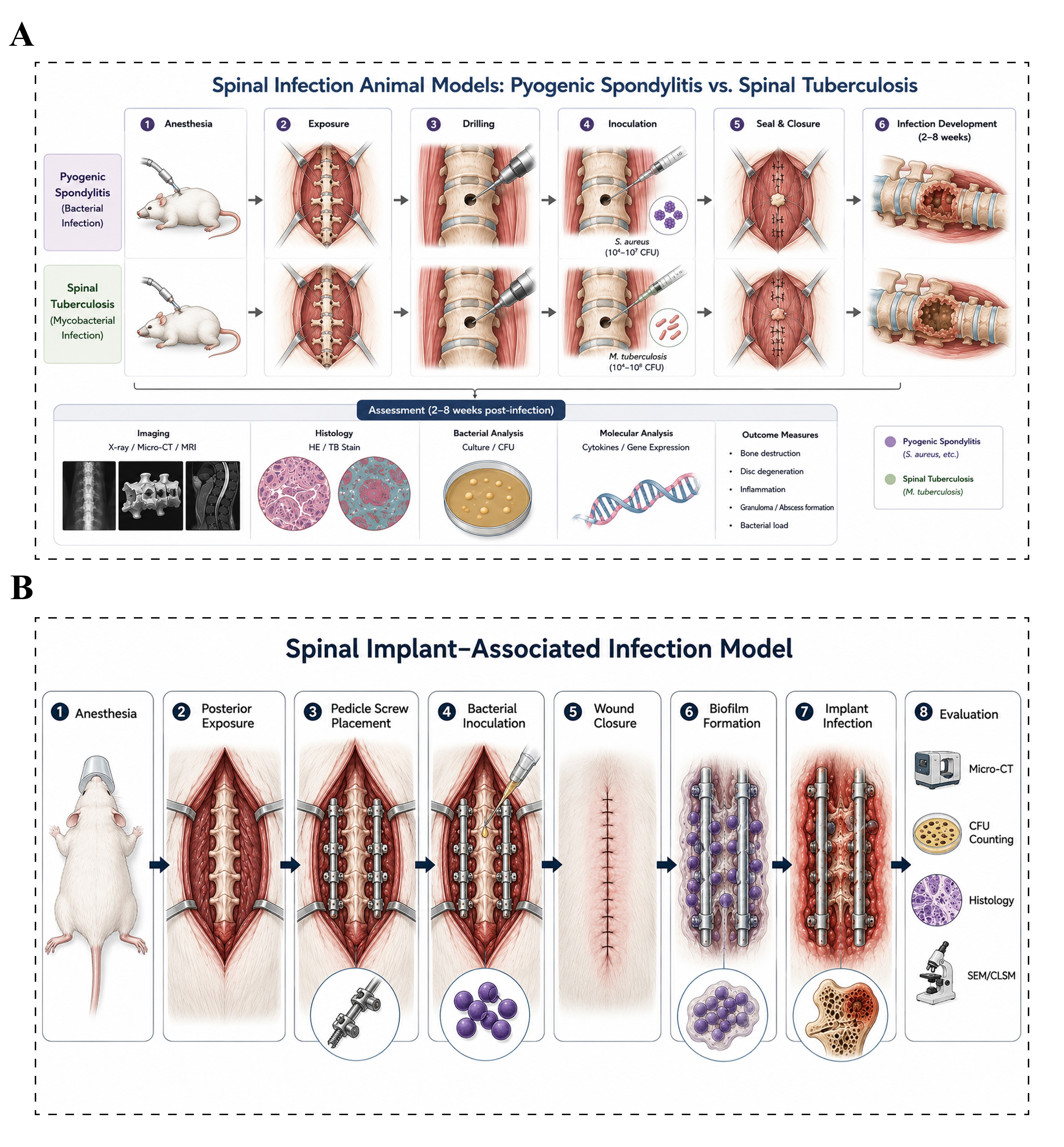
Figure 8. Animal models of spinal infection. (A) Pyogenic spondylodiscitis and spinal tuberculosis models: The target vertebral body is exposed under anesthesia, and a bone defect is created for inoculation with either pyogenic bacteria or Mycobacterium tuberculosis. Infection establishment, bone destruction, and inflammatory responses are subsequently evaluated using imaging, histological, and microbiological analyses. Apart from differences in the inoculated pathogen and disease course, the overall modeling procedures are largely similar; (B) Implant-associated infection models: Spinal implants, such as titanium screws or rods, are placed near the lamina or pedicle, followed by local inoculation of a bacterial suspension around the implant. Bacterial adhesion and biofilm formation on the implant surface gradually establish a stable implant-associated infection. Infection severity and tissue pathological changes are assessed by imaging, colony counting, histological analysis, and biofilm evaluation. Micro-CT: Micro-computed tomography; MRI: magnetic resonance imaging; HE: hematoxylin and eosin staining; TB: toluidine blue staining; CFU: colony-forming unit; SEM: scanning electron microscopy; CLSM: confocal laser scanning microscopy.

