

fig4
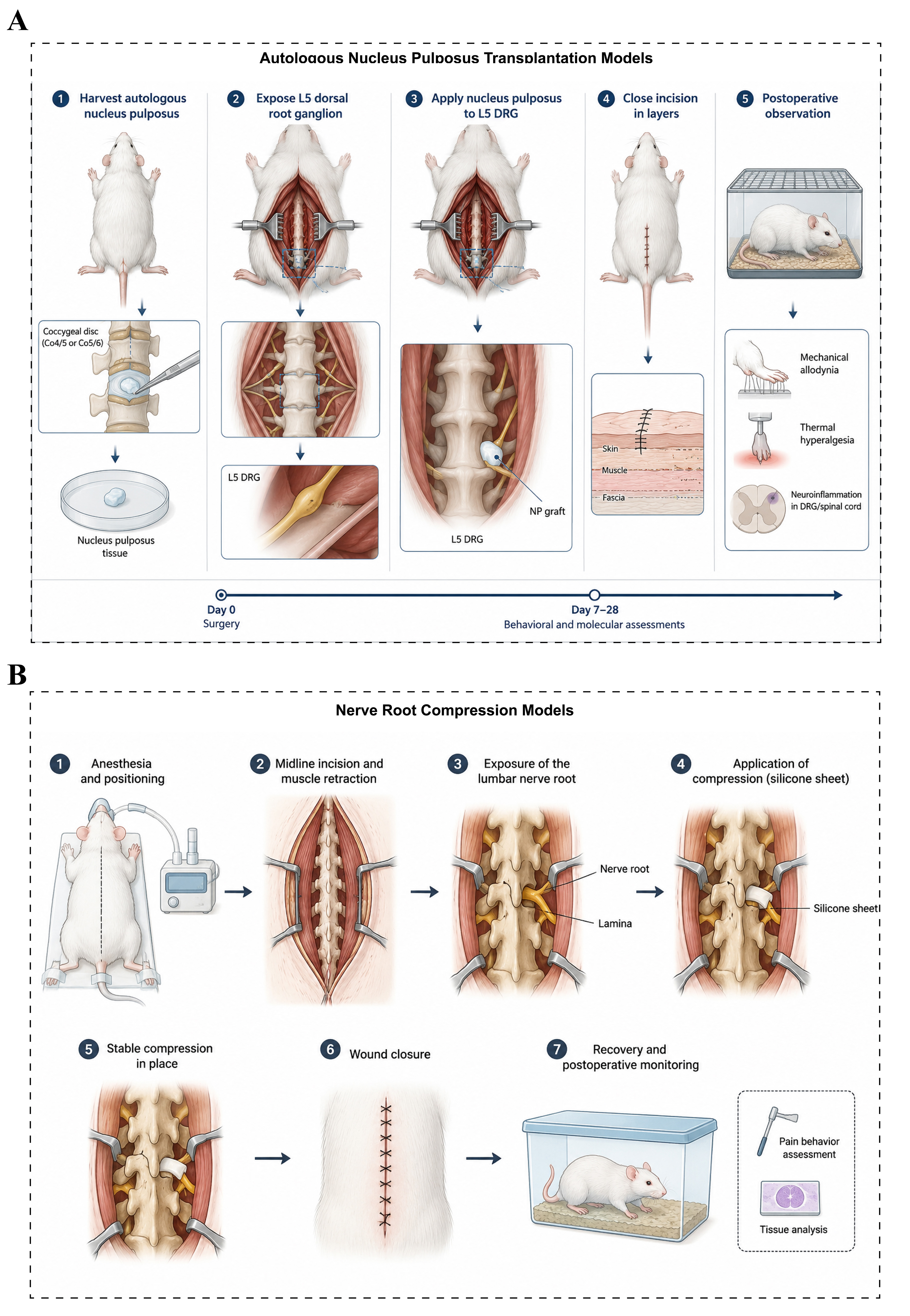
Figure 4. Animal models of lumbar disc herniation. (A) Autologous nucleus pulposus transplantation models: After anesthesia, the experimental animal is placed in the prone position, and autologous nucleus pulposus tissue is harvested from the caudal or lumbar intervertebral disc. The target lumbar nerve root (L4-L5 or L5-L6) is then exposed, and the harvested nucleus pulposus tissue is transplanted around the nerve root or dural sac. The incision is subsequently closed, followed by postoperative observation and relevant assessments; (B) Nerve root compression models: After anesthesia, the experimental animal is placed in the prone position, and the lamina and lumbar nerve root are exposed through a posterior midline lumbar incision. A silicone sheet, polyethylene tube, or other compression device is then placed around the nerve root to establish a sustained mechanical compression model. The incision is closed after surgery, followed by postoperative observation and evaluation of relevant outcome measures. NP: Nucleus pulposus; DRG: dorsal root ganglion.

