

fig6
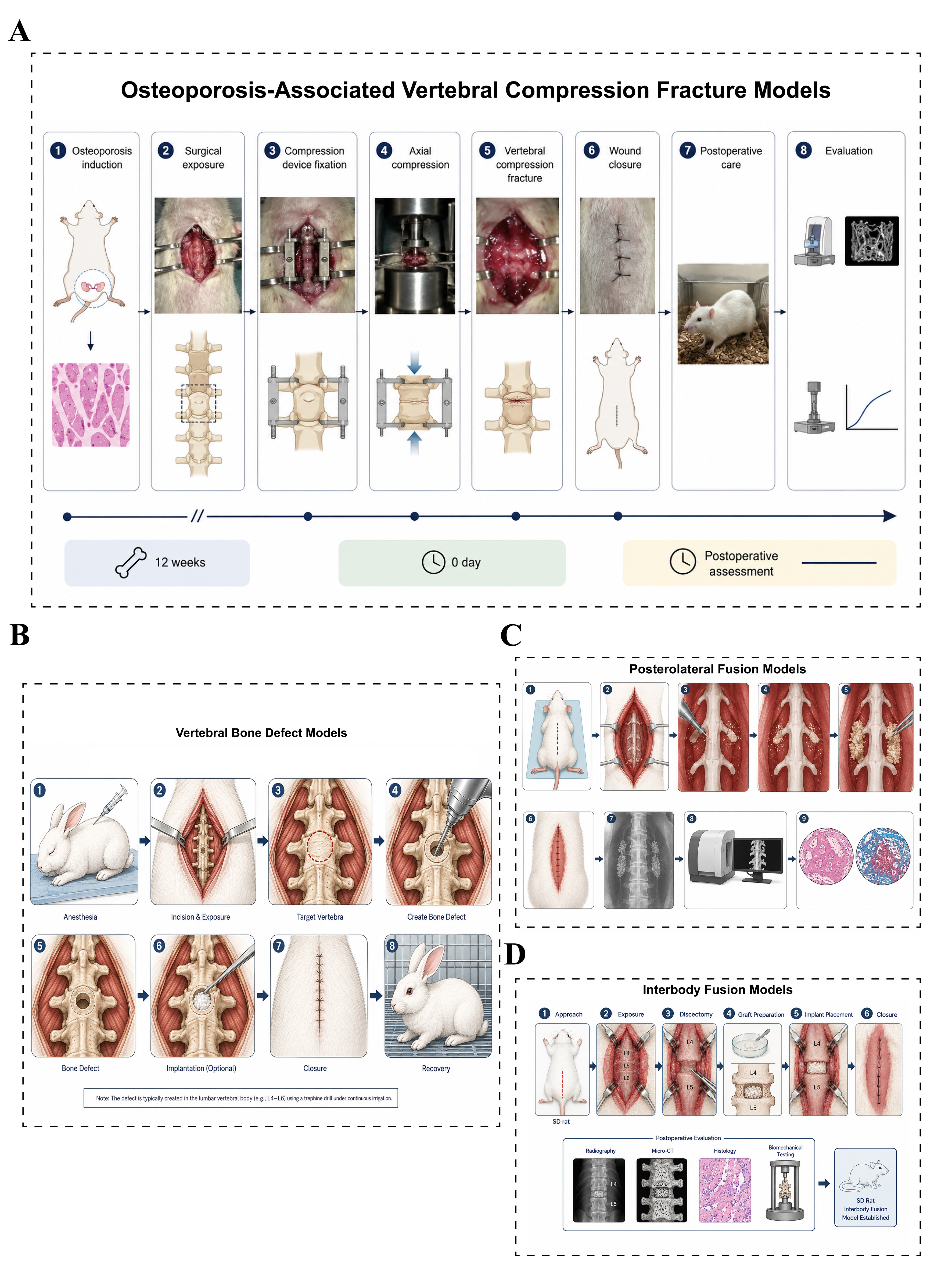
Figure 6. Animal models of vertebral fracture and spinal fusion. (A) Osteoporosis-associated vertebral compression fracture models: Bilateral OVX is performed to induce osteoporosis, followed by 12 weeks to establish stable bone loss. The target vertebral body is then exposed, and axial compression is applied using a customized device to induce a vertebral compression fracture. Fracture healing is evaluated by micro-CT, histology, and biomechanical testing; (B) Vertebral bone defect models: The target vertebral body is exposed through a posterior approach, and a standardized bone defect is created using a trephine or high-speed burr. Experimental materials are implanted into the defect, and bone regeneration is subsequently assessed by micro-CT and histological analysis; (C) Posterolateral fusion models: The bilateral L4-L5 transverse processes are exposed and decorticated, followed by implantation of test materials into the fusion bed. Fusion outcomes are evaluated using X-ray, micro-CT, and histological analysis; (D) Interbody fusion models: The target lumbar segment (L4-L5) is exposed, followed by discectomy and endplate preparation. Bone graft materials are implanted into the intervertebral space, and interbody fusion is assessed by X-ray, micro-CT, histological staining, and biomechanical testing. OVX: Ovariectomy; CT: computed tomography.

