

fig3
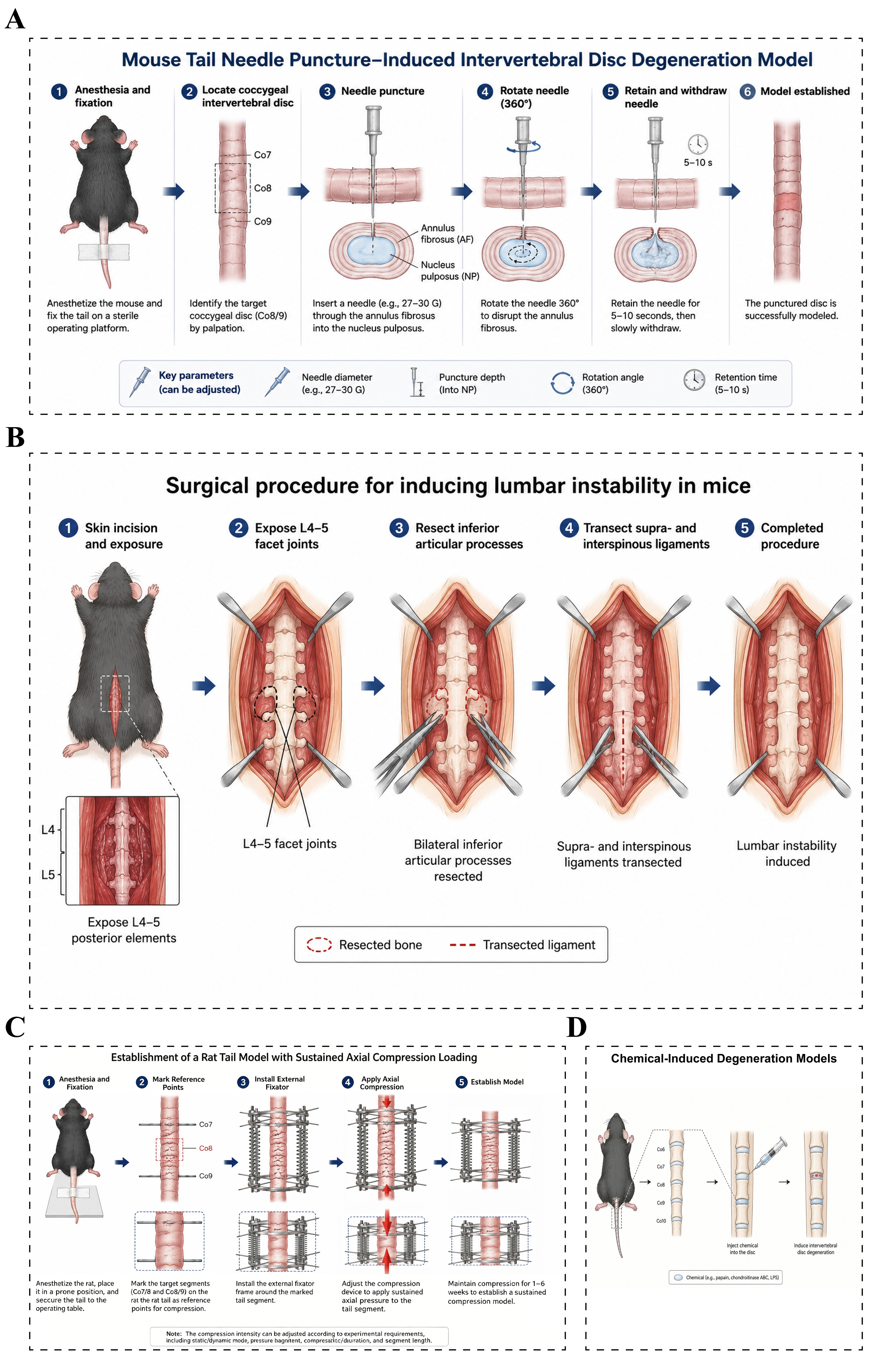
Figure 3. Animal models of intervertebral disc degeneration. (A) Needle puncture-induced intervertebral disc degeneration models: After anesthesia and fixation, the target caudal intervertebral disc in mice is identified, and a 27-30 G needle is inserted through the annulus fibrosus into the center of the nucleus pulposus. The needle is then rotated, retained briefly, and withdrawn, thereby causing annulus fibrosus injury and nucleus pulposus disruption to induce intervertebral disc degeneration; (B) Lumbar spinal instability models: After anesthesia, the posterior lumbar structures of the experimental mouse are exposed through a posterior midline approach. Under a microscope, the bilateral inferior articular processes are resected, and the supraspinous and interspinous ligaments are transected to disrupt posterior column stability and establish a lumbar motion segment instability model; (C) Mechanical loading models: Continuous axial loading is applied using spring-loading devices, Ilizarov external fixators, or customized compression systems to simulate a chronic mechanical overload environment; (D) Chemical-induced intervertebral disc degeneration models: Specific enzymes, inflammatory cytokines, or chemical reagents are injected into the intervertebral disc to disrupt the local microenvironment and rapidly induce intervertebral disc degeneration. NP: Nucleus pulposus.

