




Diagnostic and therapeutic applications of melanoma-derived exosomes in nanomedicine
 0
0 Abstract
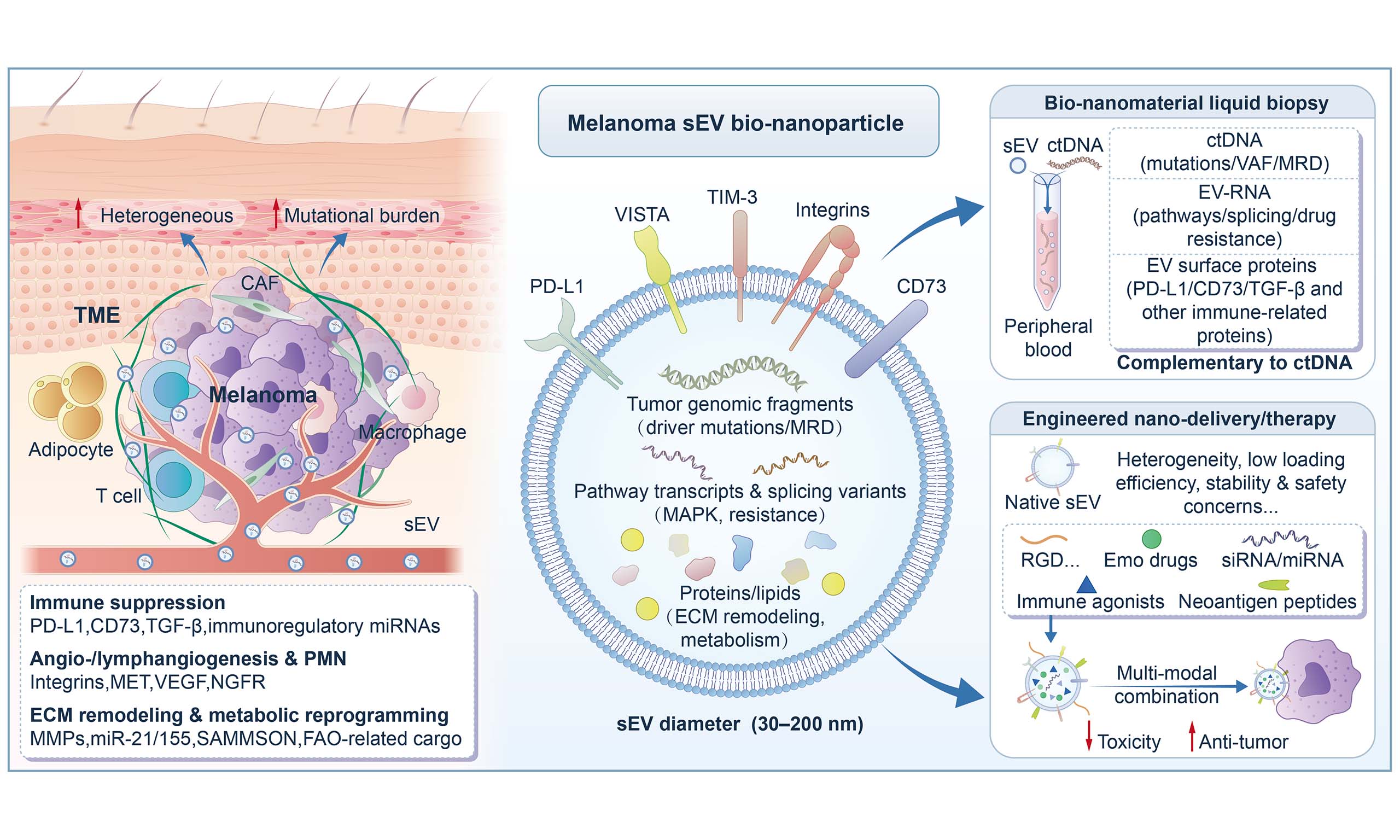
Melanoma remains challenging due to pronounced heterogeneity, early metastasis, and dynamic treatment resistance. Conventional tissue biopsy and imaging provide essential information but are limited by invasiveness, sampling bias, and an inability to capture longitudinal tumor-immune evolution. As endogenous bio-nanomaterials, small extracellular vesicles/exosomes circulate stably in body fluids and carry multi-omic cargo (DNA, RNA, proteins, lipids), enabling minimally invasive liquid biopsy and real-time disease monitoring. Beyond diagnostics, engineered exosomal nanomaterials can be functionalized and loaded with therapeutic payloads to improve targeted delivery and support combination regimens, including immunotherapy. This review highlights mechanistic roles of exosomes in melanoma progression, summarizes key liquid-biopsy biomarkers (e.g., exosomal PD-L1 and EV-miRNA/protein signatures), and discusses translational barriers such as standardization, scalable manufacturing, safety, and clinical validation.
Keywords
INTRODUCTION
Melanoma, as a malignant tumor, is characterized by pronounced heterogeneity and a strong propensity for metastasis; particularly in the context of continuous advances in immunotherapy and targeted therapy, how to achieve more refined stratification and monitoring across different stages of the disease spectrum remains a key challenge in clinical management[1-3]. The high incidence and poor prognosis of melanoma, together with the difficulty of early diagnosis and the development of therapeutic resistance in advanced stages, make it one of the most challenging cancers in clinical practice[4-7]. Traditional pathological biopsy and imaging examinations continue to serve as the cornerstone for staging and treatment response evaluation; however, their limited repeatability and inability to capture, in real time, the dynamic temporal evolution of clonal architecture and the immune microenvironment impose substantial constraints on clinical decision-making. Therefore, the development of novel, non-invasive diagnostic approaches is of particular importance.
Extracellular vesicles (EVs) are a heterogeneous group of membrane-enclosed particles released by cells into the extracellular space and are now recognized as important mediators of intercellular communication[8]. Among them, exosomes are generally defined as nanoscale vesicles of endosomal origin, most commonly ranging from 30 to 150 nm in diameter[9]. However, in many melanoma-related studies, these vesicles are still identified mainly on the basis of size range, isolation method, and conventional surface markers, rather than by direct verification of their endosomal biogenesis[10]. Accordingly, the terms “EVs”, “small extracellular vesicles (sEVs)”, and “exosomes” are sometimes used interchangeably in the published literature, which complicates cross-study comparison and interpretation[11]. For the sake of consistency and readability throughout this review, we primarily use the term “exosomes” to refer to nanoscale vesicular fractions that operationally correspond to exosome-enriched components in melanoma research[12]. At the same time, we acknowledge that, from a strict biogenetic perspective, part of these vesicles may be more accurately classified within the broader category of sEVs[13]. Unless the original studies explicitly distinguish different EV subpopulations, the term “exosomes” is used consistently in the following sections[14].
Melanoma-derived exosomes lie at the intersection of tumor biology and translational nanomedicine[15]. On the one hand, they actively participate in extracellular matrix (ECM) remodeling, immunosuppression, angiogenesis, metabolic reprogramming, and pre-metastatic niche formation, thus serving as important functional mediators of melanoma progression[16]. On the other hand, precisely because these exosomes carry multidimensional molecular information from tumor cells and their microenvironment in the form of nanoscale, lipid-bilayer-enclosed particles, they can also be developed as functionally informative analytes for liquid biopsy[17]. Furthermore, the very features that enable exosomes to mediate efficient intercellular communication—their lipid bilayer structure, complex molecular cargo, and surface components capable of interacting with recipient cells—also make them highly promising candidates for engineered drug delivery[18]. Therefore, the biological roles of melanoma exosomes in tumor progression and their translational applications in liquid biopsy and therapeutic delivery should not be viewed as two disconnected themes, but rather as two interrelated dimensions of the same bio-nanomaterial system[19]. Based on this logic, the present review first summarizes how exosomes shape melanoma progression and the immune microenvironment, and then further discusses how these properties support emerging applications in liquid biopsy and engineered therapeutic delivery[20].
TRANSLATIONAL POTENTIAL AND APPLICATIONS OF EXOSOMES IN MELANOMA RESEARCH
Nanomaterial-based exosomes
In this review, we focus on the most frequently cited EV subtype, exosomes, which are generally considered to originate from the endosomal-multivesicular body pathway and be released via exocytosis, with a typical size range of approximately 30-150 nm[21,22] [Figure 1].
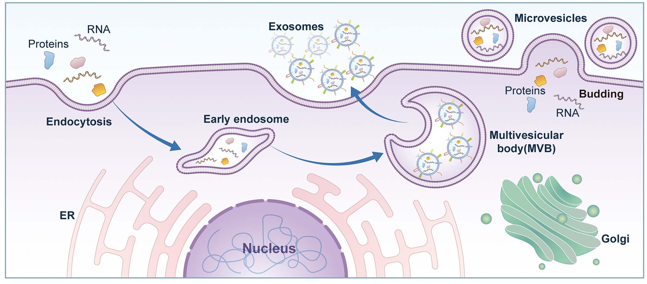
Figure 1. Biogenesis and release of exosomes. Exosome formation begins with plasma membrane invagination during endocytosis, leading to the formation of early endosomes. Early endosomes mature into late endosomes, whose limiting membranes invaginate to generate intraluminal vesicles (ILVs), ultimately forming multivesicular bodies (MVBs). Fusion of MVBs with the plasma membrane releases ILVs into the extracellular space as exosomes. Intracellular organelles, including the endoplasmic reticulum (ER) and Golgi apparatus, participate in the synthesis, sorting, and trafficking of exosome-associated components. ER: Endoplasmic reticulum; ILV: intraluminal vesicle; MVB: multivesicular body.
It is worth emphasizing that the significance of melanoma-associated exosomes lies not only in the rich biological information they carry, but also in the fact that they themselves constitute a class of bio-nanomaterials with a defined structural basis. Their lipid bilayer, surface membrane proteins and adhesion molecules, together with the nucleic acids, proteins, and lipids enclosed within them, collectively form the material basis through which exosomes exert their functions and determine their dual application potential in both liquid biopsy and therapeutic delivery[23]. Current engineering strategies applied to exosomes—whether donor-cell pretreatment, post-isolation surface modification, membrane fusion, or delivery-system construction—essentially involve nanoscale remodeling of their structure in order to further regulate biodistribution, cellular uptake, tissue penetration, and drug-release behavior in vivo[24]. Therefore, exosome design should not be understood merely as drug loading, but rather as a functional construction process centered on the coordinated relationships among membrane structure, surface molecules, and internal bioactive components[24].
It should be further noted that one of the major factors currently restricting progress in this field is the insufficient reproducibility and comparability across studies. First, differences in EV isolation methods directly affect the composition of the final vesicle population obtained. Ultracentrifugation, density-gradient centrifugation, size-exclusion chromatography (SEC), polymer precipitation, immunoaffinity capture, and microfluidic platforms differ in recovery rate, purity, processing time, and impurity removal capacity, and the EV subpopulations they enrich, as well as the background of non-vesicular particle contamination, are not equivalent[25]. This means that even when different studies all use the term “exosomes” or “sEVs”, their actual study objects may not be fully identical in structural composition and functional properties, thereby affecting cross-study comparisons of biomarker abundance, functional experimental outcomes, and delivery performance[26].
Second, source heterogeneity further increases the complexity of interpreting results. Melanoma-related EVs do not represent a single entity, but are influenced by multiple factors including donor-cell type, tumor stage, genetic background, hypoxic status, inflammatory stimulation, drug exposure, and culture conditions[26]. Differences in source may lead to significant variation in membrane molecular composition, internal bioactive content, and organ tropism, and consequently their biological effects and therapeutic performance are not directly comparable[23]. For liquid biopsy, this implies that EV signals detected in body fluids often represent composite readouts mixed from multiple origins; for drug-delivery studies, it underscores the need for more rigorous reporting of donor status, culture systems, and sample-processing workflows in order to improve interpretability and reproducibility[10].
In addition, differences among analytical platforms are another important source of limited comparability across studies. Common methods for EV characterization and detection currently include nanoparticle tracking analysis (NTA), transmission electron microscopy (TEM), Western blotting, flow cytometry, ExoView, ddPCR, sequencing, and mass spectrometry, and these platforms differ substantially in detection targets, sensitivity, quantification strategies, and normalization approaches[27]. Readouts for the same marker on different platforms are often difficult to directly align, meaning that even when studies show similar trends, they may still lack an integrated quantitative basis[28]. Therefore, future work should not only strengthen the description of EV structural features and design logic, but also reinforce the standardization of isolation, characterization, quantification, and reporting workflows in accordance with frameworks such as Minimal Information for Studies of Extracellular Vesicles (MISEV), so as to improve reproducibility, inter-study comparability, and the reliability of clinical translation[10].
Clinical applications of exosomes in liquid biopsy for melanoma
Liquid biopsy provides a noninvasive and repeatable assessment strategy for melanoma, making it particularly suitable for therapeutic response monitoring and recurrence surveillance[29,30]. Compared with circulating tumor DNA (ctDNA) and circulating tumor cells (CTCs), exosomes offer distinct potential advantages: their molecular signals are not restricted to tumor cells alone but may also originate from immune cells, stromal cells, and other components of the tumor microenvironment (TME), thereby more closely reflecting functional tumor-host interactions[17,31,32]. In addition, the lipid bilayer membrane confers a degree of protection to encapsulated nucleic acids and proteins, rendering longitudinal trend analysis during follow-up more feasible and robust[33]. Overall, exosome-based liquid biopsy is better positioned as a complementary dimension to existing biomarker systems, strengthening the evidence base for stratified diagnosis, treatment selection, and dynamic clinical decision-making, rather than serving as a direct replacement for any single established indicator[34].
Diagnosis and risk stratification: from single biomarkers to composite molecular signatures
At the level of diagnosis and risk stratification, exosomes are capable of simultaneously providing multi-level information[35]. At the nucleic acid level (e.g., mRNA/miRNA), signatures often correlate more closely with pathway activity and functional states; at the protein level, immune checkpoints and profiles related to invasion, migration, and angiogenesis can indicate disease activity and immune context; meanwhile, lipid composition and metabolism-related characteristics may reflect tumor metabolic reprogramming and structural differences in membranes[36]. Given the multi-omic nature of exosomes, single molecules are often insufficient to capture disease heterogeneity; therefore, a more rational approach involves constructing composite signatures and performing stratified validation across different stages or risk subgroups[37,38].
Prognostic assessment and monitoring of recurrence/metastasis
Given the high heterogeneity and significant risk of metastasis associated with melanoma, the clinical demand for continuous longitudinal monitoring is particularly acute[39,40]. The potential utility of exosomes in prognostic assessment is primarily manifested in their ability to: reflect fluctuations in tumor burden and systemic inflammatory/immune responses; indicate directional molecular changes prior to radiographically evident progression; and indirectly signal an increased risk of recurrence or metastasis through alterations in signaling pathways associated with matrix remodeling, immunosuppression, and angiogenesis[41-43].
Delivery potential of exosomes as nanocarriers
Material advantages of exosomes
Compared with synthetic nanomaterials, exosomes originate from the cellular membrane system, and their lipid bilayer structure and membrane protein composition generally confer superior biocompatibility and stability in vivo[44]. The receptors, adhesion molecules, and glycosylation patterns on the exosomal surface may influence target cell recognition, uptake, and intracellular trafficking pathways, thereby exhibiting delivery behaviors that more closely mimic physiological processes in certain contexts[45]. Furthermore, the ability of exosomes to encapsulate diverse cargos—including nucleic acids, proteins, and small molecules—provides a versatile platform for the development of multi-component intervention strategies[46-48]. It must be emphasized that exosomes are not entirely inert "empty shells"; their endogenous components may actively participate in signaling, which offers potential advantages while necessitating early evaluation of their safety profiles and application boundaries[49,50].
Synergistic points for combination therapy
In melanoma, insufficient response and acquired resistance remain the primary bottlenecks; consequently, the strategic positioning of exosomal delivery leans toward synergistic action[51]. When combined with immune checkpoint inhibitors, the delivery of immunomodulatory molecules to improve immune infiltration and effector T-cell function theoretically offers the potential to increase response rates or delay resistance. Within the context of BRAF/MEK targeted therapies, compensatory interventions can be designed to address resistance-related pathways and post-treatment microenvironmental rebound. Furthermore, when integrated with local modalities such as radiotherapy, ablation, or photothermal/photodynamic therapy, exosomes may facilitate the transition from "local control" to "systemic immune response" by enhancing immunogenic cell death and antigen presentation[52-54]. The commonality among these synergistic pathways is that exosomal delivery is not intended to replace existing therapies; instead, it seeks to amplify the systemic benefits of established regimens through more refined interventions at the local or cellular level, with efficacy validated by quantifiable incremental gains[55].
Therapeutic effects and applicability boundaries of exosome-mediated delivery
Whether exosome-mediated delivery is truly viable in vivo depends on the controllability of its biodistribution and clearance[56]. In practice, uptake by the reticuloendothelial system often limits the effective dose reaching the tumor; furthermore, variations in membrane composition and surface molecular profiles resulting from different sources and preparation protocols may lead to biased tissue distribution and undermine inter-study reproducibility. Simultaneously, the non-inert nature of exosomes implies that they may exert unintended regulatory effects in non-target tissues, necessitating vigilance regarding disturbances to immune homeostasis, particularly in the context of immunotherapy[57-59]. Moreover, the route of administration (e.g., intravenous, local injection, or subcutaneous/lymphatic pathways) has a decisive impact on exposure and safety, directly shaping its application scenarios and development strategies[56,57]. Consequently, effective exosomal delivery is not an inherent property; it requires a systematic trade-off between source selection, administration strategy, and downstream optimization to achieve relatively predictable in vivo performance[58].
In summary, as natural membranous nanocarriers, exosomes provide a versatile platform for melanoma delivery and combination therapy, owing to their biocompatibility, functionalized surfaces, and capacity to carry diverse types of cargo[55]. However, their clinical feasibility is collectively determined by in vivo drug distribution and clearance, immunological safety, and the consistency of large-scale manufacturing and quality control[54]. Consequently, a more rational translational positioning is to view exosomal delivery as a complementary component of combination therapy.
EXOSOME-MEDIATED REMODELING OF THE MELANOMA MICROENVIRONMENT
Melanoma, characterized by its high somatic mutational burden and pervasive epigenetic dysregulation, drives malignant progression primarily through mechanisms of immune evasion and metastatic dissemination[60-63]. Within this highly heterogeneous context, sEVs, as endogenous biological nanocarriers, are increasingly recognized as key mediators in regulating the TME in melanoma. Based on their nanoscale biological architecture, sEVs possess natural membranous encapsulation, stable circulation properties, and highly precise targeting capabilities, and are enriched with diverse bioactive molecules—including proteins, lipids, miRNAs, and long non-coding RNAs (lncRNAs)—thereby functioning both as platforms for signal transduction and as direct participants in structural and metabolic remodeling[64-67]. Tumor-derived sEVs from melanoma establish an immunosuppressive barrier, promote angiogenesis/lymphangiogenesis and pre-metastatic niche (PMN) formation, and drive ECM remodeling and metabolic reprogramming, thereby constructing a tumor microenvironment that sustains continuous tumor growth and metastasis[68]. Figure 2 illustrates the pleiotropic effects of tumor EVs in shaping the tumor microenvironment in melanoma, including the establishment of an immunosuppressive barrier [Figure 2A], the promotion of angiogenesis and lymphangiogenesis [Figure 2B], and ECM remodeling through stromal reprogramming [Figure 2C].
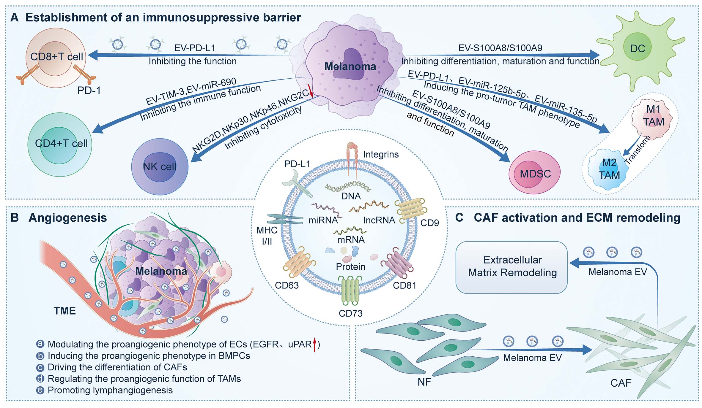
Figure 2. Pleiotropic roles of tumor-derived extracellular vesicles (EVs) in shaping the melanoma tumor microenvironment. (A) Tumor-derived EVs establish an immunosuppressive barrier through multiple mechanisms, including inhibition of CD8+ T-cell infiltration and function, impairment of CD4+ T-cell activity, suppression of natural killer (NK) cell cytotoxicity, induction of MDSCs, inhibition of DC maturation, and promotion of tumor-associated macrophage (TAM) polarization toward a pro-tumor M2-like phenotype. (B) Tumor-derived EVs promote angiogenesis and lymphangiogenesis by modulating endothelial cells (ECs), mobilizing bone marrow progenitor cells (BMPCs), driving CAF activation, and regulating pro-angiogenic TAM functions. (C) In melanoma, tumor-derived EVs contribute to CAF activation and ECM remodeling by promoting the transition of normal fibroblasts (NFs) into CAFs and by further enhancing matrix remodeling to support tumor progression. BMPC: Bone marrow progenitor cell; CAF: cancer-associated fibroblast; DC: dendritic cell; EC: endothelial cell; ECM: extracellular matrix; EV: extracellular vesicle; MDSC: myeloid-derived suppressor cell; NF: normal fibroblast; NK: natural killer; TAM: tumor-associated macrophage; TME: tumor microenvironment.
Establishment of an immunosuppressive barrier
sEVs-PD-L1-mediated T cell suppression and systemic immune evasion
The PD-1/PD-L1 axis is central to immune evasion in melanoma[69]. In melanoma, PD-L1 is not only expressed on the surface of tumor cells and multiple immune cell populations, but is also closely associated with responses to immune checkpoint inhibitors and clinical prognosis[70-73]. Recent studies have demonstrated that melanoma cells can deliver biologically active PD-L1 into the circulation via sEVs, thereby establishing a systemic “circulating immune checkpoint” and challenging the traditional notion that PD-L1 activity is confined to the local TME[74-77]. In metastatic melanoma models, PD-L1 displayed on the surface of tumor-derived exosomes can directly bind to PD-1 on CD8+ T cells, suppress TCR signaling, reduce cytokine secretion, and impair cytotoxic functions, thereby promoting tumor progression[74]. Blockade of sEVs-PD-L1 has been shown to restore T cell activity in vivo, induce systemic antitumor immune responses, and establish immunological memory, highlighting its independent and critical role in global immune regulation[30].
Myeloid immune cell reprogramming and indirect immunosuppression
At the bone marrow stage, melanoma-derived EVs can directly act on normal immature myeloid cells (IMCs)[78]. Studies based on murine models have shown that danger-associated molecular patterns (DAMPs) carried by tumor EVs activate TLR4 signaling in IMCs, driving their differentiation into immunosuppressive myeloid-derived suppressor cells (MDSCs) and markedly upregulating PD-L1 expression, thereby suppressing T cell activation and proliferation[78,79].
In addition, melanoma-derived EVs profoundly influence the differentiation and functional properties of antigen-presenting cells (APCs). Early studies have demonstrated that melanoma EVs inhibit the normal differentiation of monocytes into dendritic cells (DCs) and instead induce the formation of a CD14+ immature myeloid cell population[80,81]. This process is partially dependent on the enrichment of TGF-β signaling within EVs, ultimately leading to impaired DC maturation, reduced antigen-presenting capacity, and compromised T cell proliferation and effector functions[81-83]. Tumor-derived exosomes isolated from the plasma of melanoma patients similarly drive APCs toward a suppressive phenotype, with their immunomodulatory effects closely associated with the enrichment of immunosuppressive factors such as TGF-β within exosomes[82,84-86].
In summary, melanoma-derived sEVs amplify and transform myeloid cell functional reprogramming that is initially confined to the local tumor site into a systemic, body-wide immunosuppressive effect through multilevel, cascade-like signal transmission and cellular reprogramming, thereby establishing a structurally stable and self-sustaining immunosuppressive barrier within the tumor microenvironment[87-89], as summarized in Table 1.
Cargo molecules in extracellular vesicles and their immunomodulatory functions
| Cargo | Target cells | Mechanism of action | Citation |
| PD-L1 | CD8+ T cells | Inhibiting the function of CD8+ T cells | [90] |
| PD-L1 | CD8+ T cells | Induce exhaustion of tumor-specific CD8+ T cells | [91] |
| PD-L1 | Macrophages | Leading to M2 macrophage polarization | [92] |
| miR-214 | Macrophages | Inducing a proinflammatory activation of macrophages | [93] |
| miR-125b-5p | Macrophages | Inducing the pro-tumor TAMs phenotype | [94] |
| S100A8 and S100A9 | DCs | Interfering with the process of DCs maturation | [80] |
| Multiple miRNAs | DCs | Inhibiting the function of DCs | [95,96] |
| miR-690 | CD4+ T cells | Induce apoptosis in CD4+ T cells | [97] |
| TIM-3 | CD4+ T cells | Inhibiting the function of CD4+ T cells | [98] |
| MICA | NK cells | Inhibiting the function of NK cells | [85] |
| Hsp86 | MDSCs | Inducing the differentiation of MDSCs | [78,99] |
| Hsp70 | MDSCs | Enhance the immunosuppressive function of MDSCs | [84] |
Promotion of angiogenesis and pre-metastatic niche (ecological niche) formation
For melanoma to achieve sustained proliferative expansion and distant metastasis, it must reconstruct its vascular and lymphatic networks and pre-establish a PMN in distant organs. Recent studies indicate that melanoma-derived sEVs systemically promote angiogenesis and lymphangiogenesis and participate in PMN remodeling by delivering pro-angiogenic factors, integrins, and diverse non-coding RNAs, thereby creating favorable conditions for metastatic dissemination.
Induction of angiogenesis
To meet the increased demand for oxygen and nutrients associated with rapid proliferation, metastatic cancer cells can induce angiogenesis, remodel pre-existing vasculature, and recruit bone marrow-derived cells (BMDCs). Peinado et al.[100,101] demonstrated that melanoma-derived exosomes confer a pro-angiogenic phenotype to BMDCs through horizontal transfer of the tyrosine kinase receptor MET, thereby promoting the formation of highly vascularized PMNs in organs prone to metastasis such as the lung[100,101]. Subsequent validation studies further confirmed that sEVs derived from B16F10 melanoma mildly promote the formation of lung and bone metastatic lesions; although no significant reduction in metastatic burden was observed following treatment with MET-downregulated sEVs, a decreasing trend was still evident, indicating an important regulatory role of MET in this process[102].
Induction of lymphangiogenesis and pre-metastatic microenvironment formation
In addition to the vascular system, melanoma relies heavily on lymphatic routes to achieve early metastatic dissemination. Clinical studies have demonstrated that lymphangiogenesis is significantly associated with sentinel lymph node metastasis and reduced disease-free survival (DFS) in melanoma patients[103]. Hood et al. were among the first to propose that melanoma-derived sEVs can be transported through lymphatic vessels to lymph nodes, where they remodel lymph node architecture prior to the arrival of tumor cells and guide melanoma cell migration toward “hotspot” regions enriched in tumor-derived exosomes[104].
García-Silva et al. further systematically elucidated the molecular mechanisms underlying sEV-mediated lymphangiogenesis, demonstrating that metastatic melanoma sEVs enriched in NGFR (p75NTR) are internalized by lymphatic endothelial cells, activating MAPK/ERK and NF-κB pathways to promote lymphangiogenesis[105]. Genetic ablation of NGFR or pharmacological inhibition of its signaling effectively suppresses lymph node metastasis and improves survival in murine models[105].
Driving extracellular matrix remodeling and metabolic reprogramming
Melanoma invasion and distant dissemination are highly dependent on the dynamic adaptation of the ECM and the metabolic microenvironment. By delivering ECM-modifying factors and metabolic regulators, sEVs enable bidirectional reprogramming of both stromal cells and tumor cells themselves.
ECM remodeling and the CAF network
Within the tumor microenvironment, sEVs have been demonstrated to induce the transdifferentiation of normal fibroblasts into cancer-associated fibroblasts (CAFs) and to extensively remodel the ECM[106,107]. Studies show that sEVs derived from B16F0 murine melanoma promote the upregulation of CAF markers, including α-smooth muscle actin (α-SMA) and fibroblast activation protein (FAP), in NIH/3T3 fibroblasts while significantly enhancing their migratory capacity[94,106]. In co-culture systems, fibroblasts conditioned by these sEVs further facilitate melanoma cell proliferation and migration[96,106].
Melanoma sEVs are enriched in multiple oncogenic microRNAs, including miR-21 and miR-155-5p. miR-21 suppresses the expression of tissue inhibitor of metalloproteinases-3 (TIMP3) while upregulating multiple matrix metalloproteinases (MMPs), thereby markedly enhancing ECM degradation[108,109]. In addition, membrane-type 1 matrix metalloproteinase (MT1-MMP), a key enzyme involved in matrix breakdown, has been identified in melanoma EVs released via a melanosome-associated secretory pathway, suggesting a melanoma-specific vesicle release mechanism tightly linked to ECM destruction[107,109,110].
Metabolic reprogramming
Metabolic reprogramming represents another hallmark of melanoma progression. sEVs induce a "reverse Warburg effect" in stromal cells: exosomal miR-155 and miR-210 reprogram fibroblasts toward aerobic glycolysis, producing metabolites like lactate to fuel tumor cell oxidative phosphorylation[111]. In this context, melanoma-derived sEVs are increasingly recognized as key mediators that induce and coordinate this “reverse” metabolic coupling between stromal and tumor compartments.
In reciprocal tumor-stroma communication, adipocyte-derived sEVs likewise exert profound effects on melanoma metabolism. Lazar et al. reported that adipocyte-derived sEVs enhance the migratory and invasive capacities of melanoma cells and are enriched in proteins associated with fatty acid oxidation (FAO)[112]. In the presence of adipocyte sEVs, intracellular FAO levels in melanoma cells are markedly increased, offering a metabolic explanation for the association between obesity and poor melanoma prognosis[112]. Subsequent studies further demonstrated that FAO-related proteins and their fatty acid substrates can be directly transferred to melanoma cells via sEVs, establishing a form of intercellular metabolic support[112,113]. Collectively, these findings indicate that melanoma not only remodels the tumor microenvironment through its own secreted sEVs but can also acquire and exploit sEVs derived from distant metabolic organs, such as adipose tissue, to obtain exogenous metabolic resources that fuel tumor progression.
EXOSOME-BASED BIO-NANOMATERIAL LIQUID BIOPSY PLATFORM
In recent years, exosomes have evolved from mere subjects of research into practical tools for liquid biopsy. Particularly in melanoma, various cell types—including tumor cells, cancer-associated fibroblasts, and immune cells—release exosomes in significant quantities. These vesicles carry enriched miRNA, proteins, and DNA fragments, which contribute to upregulating invasion and metastasis pathways, stromal remodeling, and immune suppression. As EV research advances, growing evidence suggests that the application of exosomes in melanoma extends beyond tumor monitoring to include the evaluation of immunotherapy efficacy and the tracking of minimal residual disease[114,115]. Representative EV isolation strategies and cross-tumor EV liquid-biopsy examples are summarized in Figure 3[116-118], including commonly used EV isolation workflows from blood samples [Figure 3A], a representative cerebrospinal fluid-derived EV liquid-biopsy study in glioblastoma [Figure 3B], and a representative serum exosome-based biomarker analysis in lung cancer [Figure 3C].

Figure 3. Liquid-biopsy sampling and isolation of exosomes/small extracellular vesicles (sEVs), with representative applications in glioblastoma and lung cancer. (A) Overview of commonly used EV isolation strategies from clinical blood samples, including differential ultracentrifugation, polymer-based precipitation, ultrafiltration, and immunoaffinity capture. Reprinted with permission[116]. Copyright 2022, Frontiers. (B) Representative glioblastoma study using cerebrospinal fluid (CSF)-derived EV subfractions for liquid biopsy and immunomodulatory analysis. Reprinted with permission[117]. Copyright 2020, Springer Nature. (C) Representative lung cancer study using serum-derived exosomes for morphological validation and immune-related biomarker analysis, including exosomal PD-L1 evaluation and correlation with tumor immune features and clinical outcomes. Reprinted with permission[118]. Copyright 2021, Springer Nature. CSF: Cerebrospinal fluid; EVs: extracellular vesicles; PD-L1: programmed death-ligand 1.
From a nanomaterials perspective, melanoma-associated EVs represent a class of bio-nanoparticles actively released by tumor and immune cells. They typically range from 30 to 150 nm in diameter, are enveloped by a lipid bilayer, and express various surface molecules such as integrins, immune checkpoint molecules (e.g., PD-L1), and glycoproteins[108,119]. Their cargo includes encapsulated DNA fragments, mRNA, miRNA, lncRNA, as well as various functional proteins and lipids. Through intercellular communication, these EVs are involved in tumor invasion, metastasis, stromal remodeling, and the formation of an immunosuppressive microenvironment. They are recognized as key mediators in systemic tumor-host interactions[120,121]. An overview of the biological sources, sampling routes, downstream analytical workflow, proximal ocular EV sampling, nano-immune fingerprinting, ctDNA/ctEV complementarity, and clinical application continuum is shown in Figure 4A.
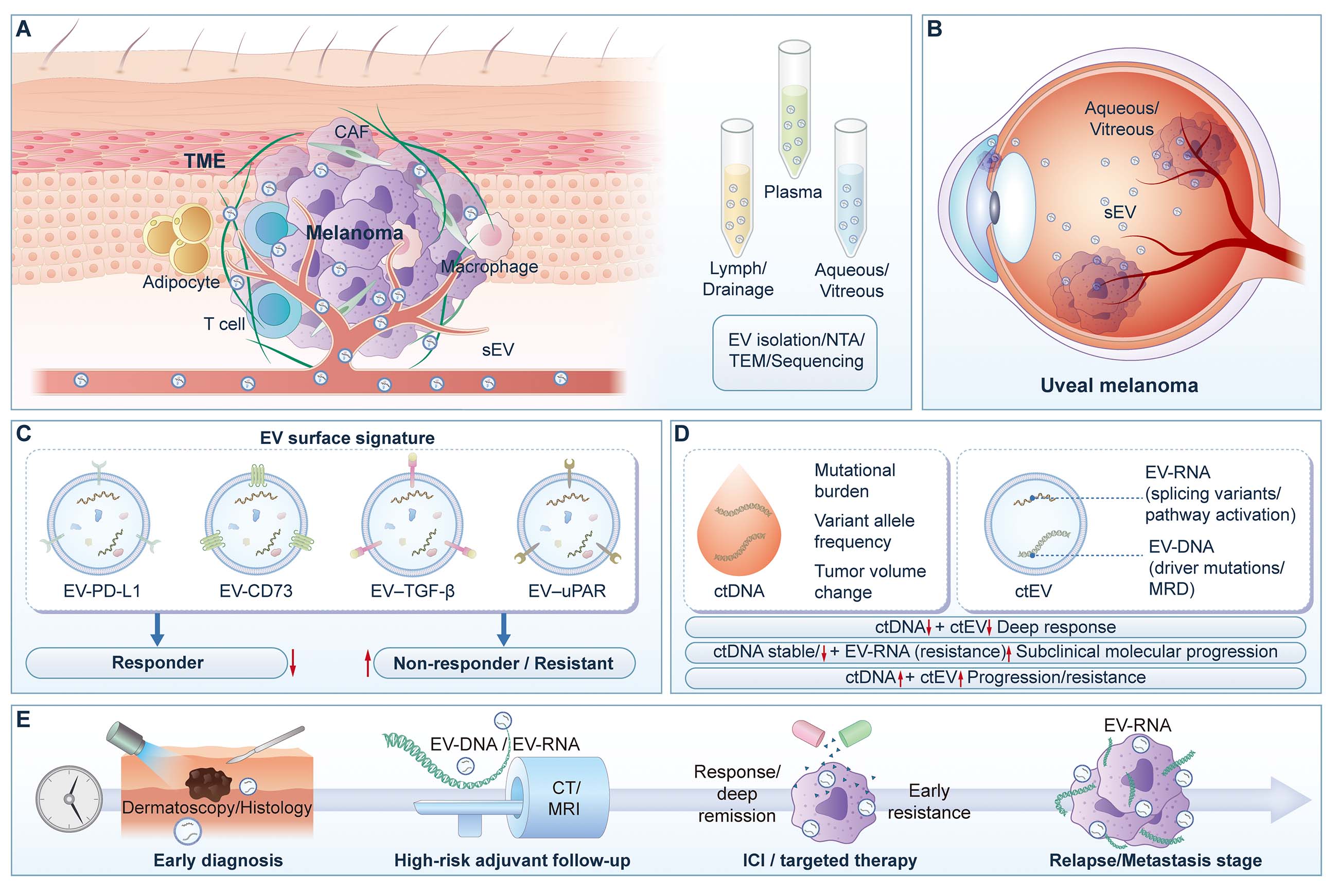
Figure 4. Structure and applications of melanoma EVs as bio-nanomaterial liquid-biopsy platforms. (A) Release of EVs from a heterogeneous melanoma TME into accessible biofluids, including plasma, lymphatic/drainage fluid, and ocular fluids, followed by downstream isolation and characterization workflows such as nanoparticle tracking analysis (NTA), transmission electron microscopy (TEM), and sequencing. (B) Schematic illustration of uveal melanoma and tumor-derived small extracellular vesicles (sEVs) in aqueous and vitreous humor, supporting ocular fluid-based EV sampling. (C) Representative EV surface “nano-immune fingerprint” composed of immunoregulatory markers, including EV-programmed death-ligand 1 (EV-PD-L1), EV-CD73, EV-transforming growth factor-β (EV-TGF-β), and EV-urokinase-type plasminogen activator receptor (EV-uPAR), for stratification of responders and non-responders/resistant cases. (D) Complementary information framework comparing circulating tumor DNA (ctDNA) with circulating tumor EVs (ctEVs), including EV-DNA and EV-RNA, for evaluation of mutational burden, molecular residual disease, functional transcripts, pathway activation, treatment response, and molecular progression/resistance. (E) Clinical continuum of EV-based applications across melanoma management, including early diagnosis, adjuvant surveillance, on-treatment monitoring during immune checkpoint inhibitor or targeted therapy, and relapse/metastasis detection. ctDNA: Circulating tumor DNA; ctEV: circulating tumor extracellular vesicle; EVs: extracellular vesicles; NTA: nanoparticle tracking analysis; TEM: transmission electron microscopy; TME: tumor microenvironment; uPAR: urokinase-type plasminogen activator receptor.
Nanoscale EV platforms and immunotherapy monitoring
Sample and methodological considerations for nanoscale liquid biopsy
As a class of bio-nanomaterials, exosomes are highly sensitive to sample processing conditions and detection platforms[23]. Regarding EV isolation techniques, while ultracentrifugation remains the standard method, approaches more commonly adopted in clinical settings include SEC, polymer-based precipitation, ultrafiltration, and immunoaffinity capture. Each method has its advantages and limitations, and the choice should be based on research objectives and clinical practicality[122].
In addition to peripheral blood-based sampling, ocular biofluids also provide clinically relevant proximal sources of tumor-derived vesicles in uveal melanoma, as illustrated in Figure 4B.
Dynamic readouts of exo-PD-L1
PD-L1, which is abundantly present in exosomes (exo-PD-L1), has been demonstrated to play a significant role in immunotherapy. PD-L1 is the target of immune checkpoint inhibitors, such as PD-1 inhibitors. Exo-PD-L1 can be regarded as an extension of the PD-1/PD-L1 axis, forming a systemic immunosuppressive layer in bodily fluids. This layer weakens the function of CD8+ T cells and promotes tumor immune evasion[74].
The functional role of exo-PD-L1 as a decoy implies that PD-L1 molecules on exosomes can competitively bind to the PD-1 receptor, thereby inhibiting the immune system's attack on the tumor. This mechanism may explain why some melanoma patients with high plasma PD-L1 levels still exhibit poor responses to immunotherapy. Although the efficacy of immune checkpoint inhibitors theoretically relies on PD-1/PD-L1 binding, exo-PD-L1 may interfere with this effective immune response through its decoy function.
With ongoing clinical research, the dynamic changes in exo-PD-L1 levels are increasingly recognized as a real-time monitoring indicator that more accurately reflects the systemic immune state. Multiple cohort studies have found that plasma exo-PD-L1 levels are closely associated with tumor burden, degree of immunosuppression, and clinical outcomes. High baseline levels are often observed in patients with greater tumor burden and poorer prognosis, while a significant decline during early treatment (approximately 2-6 weeks) is commonly seen in patients who achieve objective responses or disease control[123].
Multiparameter analysis of nano-immunosuppressive axis: CD73, TGF-β, and uPAR
Beyond exo-PD-L1, several other immunosuppressive biomarkers detectable in exosomes provide valuable insights into the immune microenvironment. Taking EV-CD73 as an example, CD73 is a key molecule in the adenosinergic immunosuppressive pathway[124]. By catalyzing the conversion of AMP to adenosine, CD73 inhibits effector T cell function while promoting the activity of regulatory T cells (Tregs) and MDSCs[125]. This mechanism plays a significant role in shaping the immunosuppressive tumor microenvironment[126]. Studies have shown that the expression of CD73+ EVs in the plasma of melanoma patients is significantly higher than in healthy controls, and their adenosine-generating capacity can suppress lymphocyte proliferation and IFN-γ secretion[127].
More importantly, clinical research has found that high or increasing levels of CD73+ EVs before or during early treatment in patients receiving PD-1 inhibitors are associated with treatment resistance and poorer prognosis[127]. Therefore, CD73+ EVs may serve as an early biomarker predictive of resistance and represent a potential therapeutic target for combination strategies, such as co-administration with A2A/A2B receptor antagonists, to enhance the efficacy of PD-1 inhibitors[128].
TGF-β-associated exosomes are another important marker in the immune microenvironment. TGF-β promotes fibrotic stromal remodeling, Treg recruitment, and exhaustion of effector T cells, mechanisms closely linked to immune escape and an immunosuppressive phenotype[129]. Multiple studies indicate that high levels of EV-TGF-β before or early in treatment suggest limited benefit from immunotherapy and poorer outcomes. Evidence supports including EV-TGF-β in risk stratification models for immunotherapy[87,130].
uPAR+ EVs are strongly associated with tumor invasiveness and metastatic potential[131]. Research has found that in patients with metastatic melanoma, early increases in uPAR+ EV levels during treatment are significantly correlated with drug resistance and shorter overall survival (OS). These exosomes, taken up by endothelial cells, can promote angiogenesis and increase vascular permeability, thereby facilitating conditions for tumor metastasis[132].
Therefore, combined assessment of biomarkers such as EV-CD73, EV-TGF-β, and EV-uPAR allows for a more precise characterization of the immunosuppressive microenvironment and identification of resistance mechanisms during immunotherapy[133]. Particularly in the course of treatment, when persistently high or rising exo-PD-L1 levels coincide with upregulation of EV-CD73 and/or EV-TGF-β, and ctDNA levels do not decrease or show signs of rebound, this pattern may indicate a high risk of immune escape and treatment resistance [Figure 4C][87,127,134].
EV-DNA and EV-RNA: driver mutation detection and pathway activity monitoring
EV-DNA
EV-DNA refers to tumor-derived DNA fragments encapsulated within nanoscale vesicles. Its primary advantages include high stability due to protective encapsulation and a relatively high tumor-origin fraction. Compared to cell-free DNA (cfDNA), EV-DNA can more effectively preserve tumor-specific genetic information[135,136]. This protection makes EV-DNA a more stable analyte for liquid biopsy applications, particularly advantageous for detecting low-frequency mutations[88].
Furthermore, EV-DNA can be used for confirming mutational profiles before and after targeted therapy and for tracking clonal evolution. Especially in patients where repeat tissue biopsies are challenging, EV-DNA serves as a complementary source to ctDNA, improving the success rate of mutation detection. During long-term targeted therapy, it can help identify newly emerging resistant clones. For example, in some patients undergoing targeted therapy, rapid changes in the mutational profile detected in EV-DNA may indicate the emergence of new resistance mechanisms, information which ctDNA alone might fail to capture in a timely manner[89].
EV-RNA
In contrast to EV-DNA, which focuses on the detection of structural mutations, EV-RNA primarily conveys functional information[137]. EV-RNA, including mRNA, miRNA, and lncRNA, reflects transcriptional activity within tumor cells and offers unique advantages in tracking non-mutational resistance mechanisms. Unlike ctDNA, which lacks functional readouts, EV-RNA can reveal tumor-associated changes such as transcription factor activation and gene splice variants, which are part of the adaptive evolution of cancer cells[138].
Through the analysis of EV-RNA, clinicians can identify key pathways involved in immune escape and detect gene splice variants at an early stage, enabling timely adjustment of treatment strategies to prevent the further development of resistance. Unlike ctDNA, EV-RNA captures more complex functional adaptations, illustrating how tumor cells may overcome therapeutic pressure through transcriptional regulation[138].
ctDNA and ctEV
ctDNA and circulating tumor-derived extracellular vesicles (ctEV, encompassing EV-DNA and EV-RNA) together form a multi-dimensional liquid biopsy framework with significant potential for tracking resistance mechanisms, monitoring minimal residual disease (MRD), and evaluating immunotherapy efficacy[139]. ctDNA primarily provides structural information such as tumor mutational burden, clonal expansion, and copy number variations. In contrast, ctEV, via its nanovesicular carriers, integrates DNA, RNA, and membrane protein-based functional biomarkers, thereby supplementing information on functional states such as immune evasion and metastatic potential [Figure 4D][108,140].
The major differences and complementary features of ctDNA and exosome-related biomarkers are summarized in Table 2.
Comparison of ctDNA and exosome-related biomarkers (EV-DNA, EV-RNA, and EV-proteins)
| Category | ctDNA | EV-DNA | EV-RNA | EV-proteins | Representative references |
| Main biological information | Mutation spectrum, clonal evolution, tumor burden, copy-number changes | Partial genomic alterations, MRD, and tumor-derived DNA information | Transcriptional activity, splice variants, functional states, and resistance-related pathways | Phenotypic states, immune microenvironment, membrane-molecule expression, and intercellular communication features | Medhin et al., 2023[114] |
| Information attribute | More structural information | Structural information combined with vesicle-protection features | More functional information | More phenotypic and microenvironmental information | Tsering et al., 2024[141] |
| Stability | Susceptible to fragmentation and degradation | Relatively more stable because of vesicular membrane protection | More stable than free RNA because of vesicular protection | Membrane proteins and intravesicular proteins retain a certain degree of protection | Tsering et al., 2024[141] |
| Detection methods | ddPCR, NGS, targeted sequencing | ddPCR, PCR, sequencing | qPCR, ddPCR, RNA-seq | ELISA, Western blot, flow cytometry, mass spectrometry, ExoView | Slusher et al., 2024[17] |
| Advantages | Relatively mature clinical foundation; suitable for monitoring mutations and tumor-burden changes | May complement blind spots of ctDNA detection and may be advantageous in some low-abundance settings | More sensitive to immune escape, resistance-associated transcriptional regulation, and functional changes | Can directly reflect surface markers and microenvironmental status | Medhin et al., 2023[114] |
| Limitations | Primarily reflects genomic-level information, with limited functional-state readouts | Insufficient standardization; isolation and quantification are strongly method-dependent | Large platform-related variation; preprocessing and normalization are complex | Quantitative consistency and cross-platform comparability remain limited | Slusher et al., 2024[17] |
| Potential applications in melanoma | Monitoring BRAF/NRAS mutations, tumor burden, and recurrence | MRD assessment, supplementation of structural-variation information, and combined monitoring | Early resistance detection, immune-state assessment, and functional pathway changes | Diagnosis, staging, immunotherapy-response evaluation, and prognostic stratification | Medhin et al., 2023[114] |
| Most suitable clinical scenarios | Treatment monitoring, recurrence warning, and tracking clonal evolution | Combined with ctDNA to improve capture of structural information | Monitoring functional resistance and immune escape | Assessing tumor phenotype and microenvironmental status | Slusher et al., 2024[17] |
| Clinical positioning | A relatively mature component of liquid biopsy | Still in development | High research and translational potential | Suitable as a complementary functional biomarker | Shen et al., 2023[142] |
| Overall assessment | Best suited to provide a structural map of the tumor | Complements the structural-information layer | Complements the functional-information layer | Complements the phenotypic/microenvironmental layer |
Diagnosis, staging, and differentiation of borderline lesions
EV-miRNA in early diagnosis and staging
EV-miRNAs are recognized as biomarkers encapsulated within nanoscale vesicles, holding significant promise for early diagnosis and staging of melanoma[143]. Utilizing high-throughput screening and machine learning, multiple studies have identified EV-miRNA panels capable of distinguishing melanoma from benign pigmented lesions, as well as early-stage from late-stage disease[144]. For instance, one study reported that a plasma-based EV-miRNA panel comprising miR-200c-3p, miR-144-3p, and miR-221-3p demonstrated favorable AUC values for melanoma diagnosis and staging. Notably, it showed high sensitivity and specificity (AUC > 0.85) for early-stage detection[144].
EV-proteins: elevating traditional serum biomarkers to the nanoscale
EV-proteins represent a nanoscale extension of traditional serum biomarkers, offering significant diagnostic and prognostic value. For example, classical melanoma markers such as S100B and melanoma inhibitory activity (MIA) have been detected in circulating EVs from patients with advanced melanoma, suggesting their potential utility as EV-based biomarkers[108,145]. Studies indicate that EV-S100B achieves an AUC value of approximately 0.85 in melanoma diagnosis, highlighting the advantage of this nanoscale protein biomarker[146].
Furthermore, research on communication proteins like EV-Connexin-43 demonstrates that their expression levels are significantly correlated with DFS and OS in melanoma[142]. ROC analysis shows that EV-Connexin-43 yields an AUC of about 0.77-0.78, supporting its potential as a supplementary marker for prognostic stratification and treatment intensity decisions[142]. Through small EV proteomics, researchers continue to identify candidate signature panels associated with metastasis risk and immune evasion—including adhesion molecules, cytoskeletal proteins, and metabolic enzymes—providing features for multi-marker panels and machine learning models[124].
Special lineages and proximal biofluids: the signal-to-noise advantage of nanoscale readouts
In special lineages of melanoma, such as uveal melanoma, EVs isolated from tumor-proximal biofluids (e.g., aqueous humor, vitreous) often carry a high abundance of tumor-associated information[147]. While the proteomic and miRNA signatures of these EVs overlap with those in peripheral blood, their enrichment is significantly higher. This confers a considerable advantage for "proximal nanoscale readouts" of localized lesions[148]. Studies show that EV concentrations in tumor-proximal fluids are typically greater than in peripheral blood, substantially improving the tumor signal-to-background noise ratio. This makes them particularly suitable for diagnosis, post-operative assessment of residual disease, and recurrence monitoring[149].
Multimodal integration: complementarity of nanoscale liquid biopsy with ctDNA and imaging
In summary, nanoscale readouts such as EV-miRNA and EV-proteins primarily reflect the phenotypic characteristics and microenvironmental state of tumors, whereas ctDNA conveys information on mutational burden and clonal expansion. By integrating these with imaging and traditional serum biomarkers, a multimodal framework encompassing "structure + function + behavior" can be constructed, enabling more precise whole-course patient management [Figure 4E][150]. In practical clinical pathways, EV-based liquid biopsy is likely to be initially adopted as a complementary tool alongside ctDNA and conventional markers within high-level medical centers. Its incremental value at specific clinical decision points can then be progressively validated through real-world cohort studies[151].
The combined analysis of EV-miRNA, EV-proteins, and ctDNA offers significant advantages in addressing clinical challenges. For instance, EV-miRNA can aid in distinguishing melanoma from benign lesions, EV-proteins enhance the diagnosis of advanced tumors, and ctDNA provides insights into mutational burden and clonal dynamics. Integrating these layers of information allows for more accurate early diagnosis, treatment stratification, and recurrence monitoring. Particularly in scenarios where imaging and traditional serum biomarkers yield ambiguous results, EVs liquid biopsy delivers additional clinical value, holding broad application prospects in early melanoma detection and the dynamic monitoring of immunotherapy response[152].
APPLICATIONS OF ENGINEERED EXOSOMES IN CUTANEOUS MELANOMA: CARGO LOADING, TARGETED DELIVERY, AND MULTIMODAL COMBINATION STRATEGIES
Properties and limitations of native exosomes
Melanoma is among the most aggressive cutaneous malignancies, and once it progresses to stage IV (metastatic disease), the 5-year survival rate drops to approximately 16%[153]. Native exosomes exhibit limited penetration into subcutaneous tumors, which is insufficient to meet therapeutic demands, and their practical application in melanoma therefore remains constrained by multiple challenges.
First, the purification and large-scale production of exosomes still face significant technical bottlenecks. Currently used isolation techniques—including ultracentrifugation, ultrafiltration, and immunoaffinity-based methods—are difficult to simultaneously satisfy pharmaceutical-grade requirements in terms of yield, purity, and preservation of biological activity[154]. Second, exosome stability severely limits their long-term storage and transportation. A study on exosomes derived from umbilical cord blood mesenchymal stem cells (UCB-MSCs) reported a 15%-20% loss of biological activity during production processes compliant with Good Manufacturing Practice (GMP) standards[155]. In addition, native exosomes generally exhibit low loading efficiency for gene therapeutics, RNA molecules, and conventional chemotherapeutic agents, highlighting the urgent need for more efficient and controllable cargo-loading strategies to improve delivery efficiency and therapeutic efficacy[156,157].
Moreover, surface chemical modification or the loading of exogenous bioactive agents may alter the native immunological properties of exosomes, potentially exposing immunosuppressive molecules or reshaping the local immune microenvironment, thereby eliciting unintended immunosuppressive or inflammatory responses[158-160].
Taken together, although native exosomes possess notable advantages as nanocarriers in terms of biocompatibility and functional versatility, their intrinsic limitations in purification efficiency, structural stability, cargo-loading capacity, and biosafety have driven increasing interest in engineering strategies aimed at overcoming key barriers to their application in tumor therapy and related fields.
Core objectives of engineering strategies
The central objective of engineered exosomes is to overcome the intrinsic limitations of native exosomes in practical applications by systematically enhancing targeting specificity, cargo-loading efficiency, and biosafety, thereby maximizing their value in disease diagnosis and therapy. As summarized in Figure 5, this includes representative cargo-loading strategies [Figure 5A], surface functionalization approaches for improving targeting specificity [Figure 5B], induction of tumor cell apoptosis through engineered exosomal delivery [Figure 5C], remodeling of the tumor immune microenvironment [Figure 5D], and theranostic co-delivery of therapeutic agents and imaging tracers [Figure 5E].
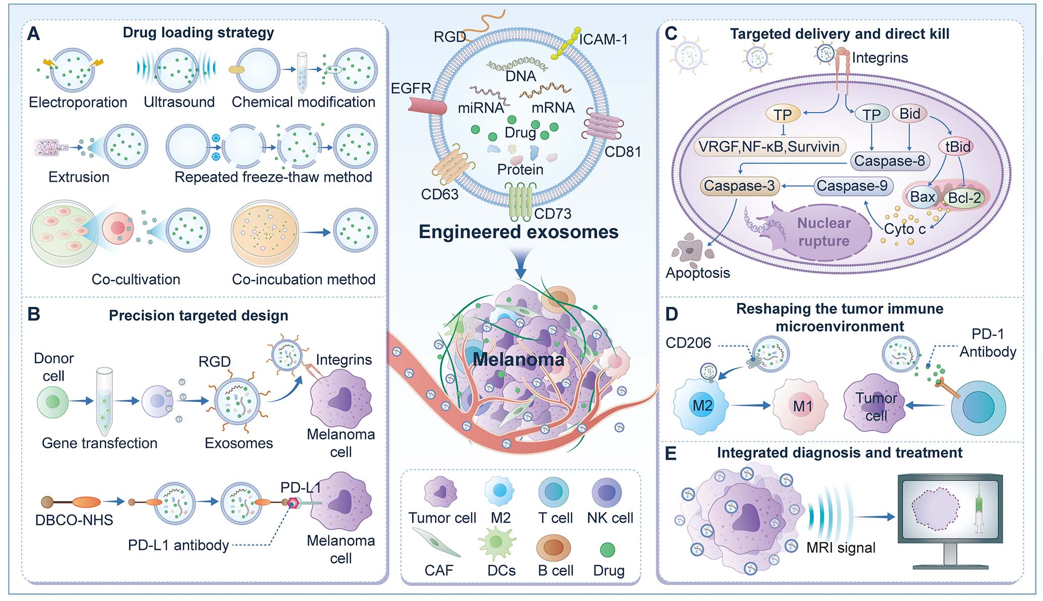
Figure 5. Engineering strategies for enhancing exosome drug loading, targeting, and therapeutic function. (A) Representative drug-loading strategies for engineered exosomes. (B) Surface functionalization approaches for improving targeting specificity. (C) One mechanism by which engineered exosomes induce tumor cell death, in which triptolide-loaded exosomes activate the intracellular caspase-3 pathway and trigger apoptosis. (D) Exosome-mediated remodeling of the tumor microenvironment through modulation of immune cells and immune-related molecules, including macrophages and T cells. (E) Co-delivery of therapeutic agents and imaging tracers by engineered exosomes for tumor-targeted theranostics. TP: Triptolide.
Classification and representative examples of exosomal drug-loading technologies
Exosomal drug-loading technologies can be broadly categorized into two main approaches: passive loading and active loading. These two strategies differ in terms of the types of therapeutics they are suitable for, operational complexity, and their impact on exosomal structural integrity and biological function[161].
Passive loading strategies
Passive loading strategies rely on the natural interactions between drug molecules and exosomes or endogenous cellular pathways, offering operational simplicity but generally lower loading efficiency, and mainly include co-incubation and co-culture approaches. Representative examples are shown in
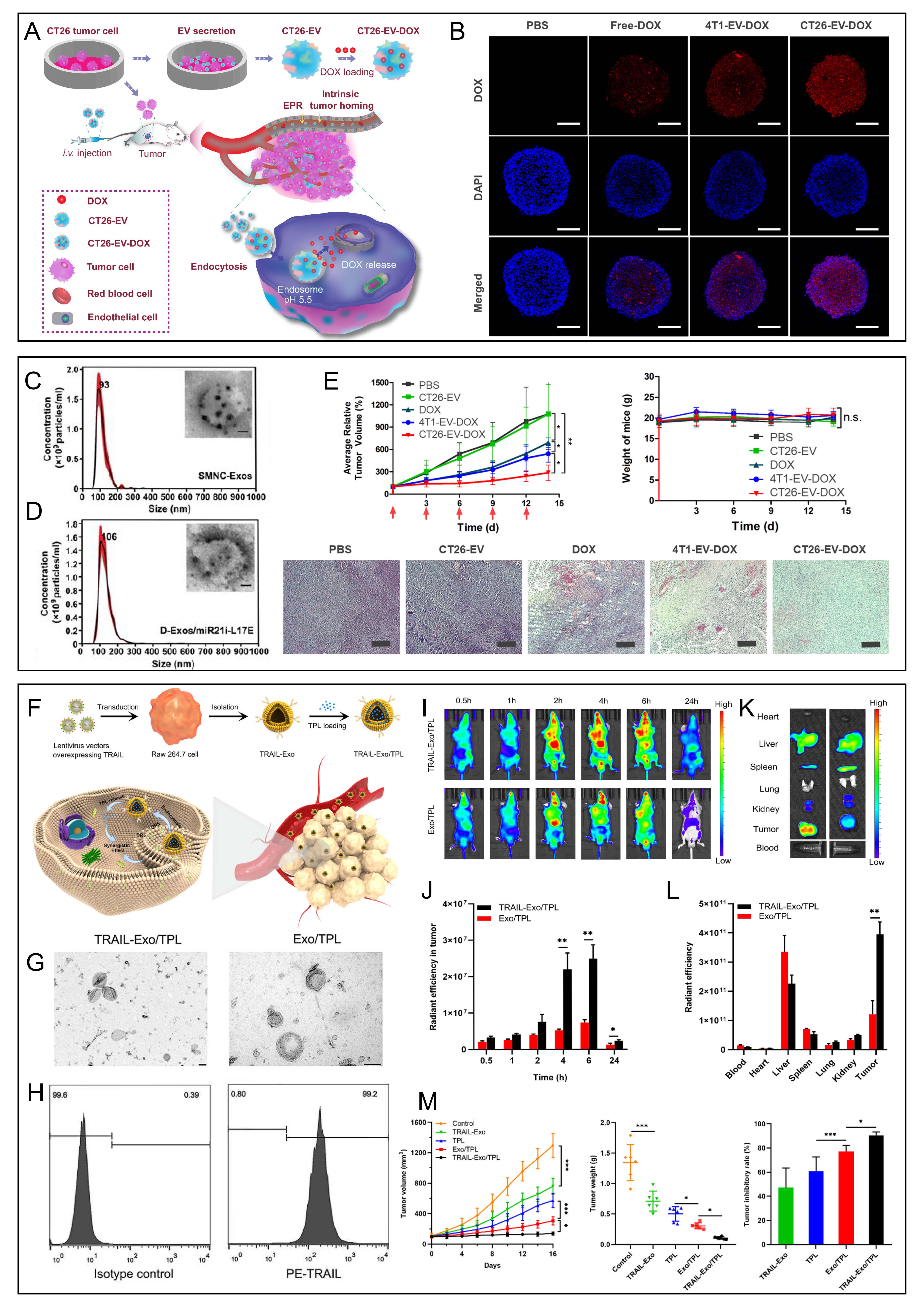
Figure 6. Representative studies of engineered exosome-based drug delivery, cellular uptake, biodistribution, and antitumor efficacy. (A, B, E) Tumor-derived extracellular vesicles loaded with doxorubicin for systemic delivery, tumor accumulation, three-dimensional tumor-spheroid penetration, and in vivo therapeutic evaluation. Scale bars: 200 µm in (B, E). Reprinted with permission[162]. Copyright 2022, Taylor & Francis. (C and D) Characterization of engineered blood exosomes and D-Exos/miR21i-L17E for tumor-targeting gene/chemo combination therapy. Reprinted with permission[163]. Copyright 2020, Ivyspring International Publisher. (F-M) Generation of TRAIL-expressing exosomes and triptolide-loaded engineered exosomes, together with ultrastructural characterization, surface-marker validation, in vivo imaging, biodistribution, and antitumor efficacy assessment. (G) scale bar: 100 nm. (J-M) *P < 0.05, **P < 0.01, and ***P < 0.001. Reprinted with permission[164]. Copyright 2021, American Chemical Society. DOX: Doxorubicin; TEM: transmission electron microscopy; TRAIL: tumor necrosis factor-related apoptosis-inducing ligand; TPL: triptolide.
Co-incubation is one of the simplest passive loading methods, in which exosomes are incubated with therapeutic agents under defined conditions to enable drug encapsulation via diffusion, lipid membrane fusion, or transmembrane transport mechanisms[165]. In melanoma therapy, doxorubicin-loaded exosomes generated by co-incubation exhibit favorable biocompatibility and low immunogenicity, thereby reducing systemic toxicity while enhancing drug accumulation at tumor sites[166]. Nevertheless, the drug-loading capacity of this method is limited and remains inefficient for macromolecular or hydrophilic therapeutics. For instance, when doxorubicin was loaded into HBc virus-like particle nanoparticles, incubation at 70 °C for 90 min achieved high loading efficiency, highlighting temperature as a critical determinant of encapsulation efficiency[167].
Technological advances in targeted delivery strategies
The innate tropism of exosomes primarily depends on the characteristics of the donor cells and generally lacks high efficiency and specificity toward diseased tissues. In the context of cutaneous melanoma therapy, this limitation significantly constrains both therapeutic efficacy and clinical applicability. Consequently, engineering strategies have focused on endowing exosomes with the ability to actively recognize and accumulate in melanoma cells or within the tumor microenvironment, which has become a major research focus in recent years. Current technological progress mainly concentrates on three directions: engineering of donor cells, direct functional modification of exosomes, and the design of intelligent targeting systems.
Donor cell engineering-based targeting strategies
This strategy involves genetically modifying donor cells (e.g., MSCs, HEK-293T cells) to stably express fusion proteins composed of targeting elements and exosomal membrane proteins such as Lamp2b or CD63, thereby enabling the continuous production of targeted exosomes at the source[168-170]. Its advantages include uniform targeting functionality and avoidance of potential carrier damage during subsequent purification.
To target integrin αvβ3, which is overexpressed on melanoma cell surfaces, researchers fused the high-affinity cyclic RGD (cRGD) peptide with the extracellular domain of lysosome-associated membrane protein 2B (Lamp2b), successfully generating targeted engineered exosomes [Figure 7A-F][171]. In B16-F10 melanoma-bearing mouse models, iRGD-modified exosomes loaded with doxorubicin exhibited over threefold higher fluorescence intensity at tumor sites compared to unmodified exosomes, accompanied by significant tumor growth inhibition, prolonged survival, and markedly reduced systemic toxicity[169,172,173].

Figure 7. Strategies for achieving exosome-targeted delivery in melanoma therapy. (A-F) Engineering of human umbilical cord mesenchymal stem cell-derived exosomes with cyclic arginine-glycine-aspartic acid (cRGD) peptide to encapsulate triptolide, thereby constructing a biomimetic targeted drug delivery system (cRGD-Exo/TP), with evaluation of survival, biodistribution, drug loading, encapsulation efficiency, drug release, and in vivo antitumor efficacy. *P < 0.05, **P < 0.01, and ***P < 0.001. Reprinted with permission[171]. Copyright 2022, Springer Nature. (G-J) Construction of pYEEIE peptide-functionalized Rhodiola rosea-derived exosome-like nanovesicles loaded with doxorubicin (pYEEIE-RELNs-DOX), including preparation workflow, ultrastructural characterization, in vivo imaging/tumor-targeting assessment, and antitumor efficacy evaluation in melanoma models. **P < 0.01, Reprinted with permission[192]. Copyright 2025, Frontiers. (K-O) Construction of an active-targeting exosome platform using a membrane-anchoring BODIPY group, polyethylene glycol (PEG) spacer, and cRGD targeting ligand, followed by assessment of integrin αvβ3 binding, exosome-marker expression, time-dependent tumor accumulation, and tumor suppression in vivo. ***P < 0.01, ***P < 0.0001, Reprinted with permission[172]. Copyright 2020, American Chemical Society. DL: Drug loading; DOX: doxorubicin; EE: encapsulation efficiency; RELN: Rhodiola rosea-derived exosome-like nanovesicle; TP: triptolide.
Direct functionalization of exosomes
Direct functionalization of exosomes refers to a class of engineering strategies in which targeting molecules or functional moieties are anchored onto the exosomal membrane after isolation and purification through chemical or physical approaches. This method does not require genetic modification of donor cells, offering high modularity, operational flexibility, and broad applicability, making it widely adopted for constructing precise exosome delivery systems.
Chemical conjugation is one of the most established direct modification methods. It primarily exploits reactive functional groups exposed on exosomal membrane proteins or lipids, such as amino, carboxyl, or thiol groups, to covalently link targeting ligands or functional molecules[174]. Bioorthogonal click chemistry (BCC), such as azide-alkyne cycloaddition, is considered an ideal tool for exosome surface modification due to its mild reaction conditions, high selectivity, and excellent biocompatibility[175]. For example, using copper-free BCC, researchers metabolically engineered donor cells to produce surface-modified EVs, which demonstrated effective tissue targeting and accumulation in rheumatoid arthritis and various tumor models, including breast, colon, and prostate cancers[176]. In melanoma, exosomes can potentially be functionalized via DBCO-NHS chemistry on their surface and subsequently conjugated with azide-modified PD-L1 antibodies, generating exosomes that target PD-L1-overexpressing melanoma cells. This strategy blocks the PD-1/PD-L1 immune checkpoint while enabling specific drug accumulation within tumor cells, enhancing the synergistic efficacy of immune checkpoint inhibitors and targeted therapeutics[177-180]. Importantly, this approach preserves the native targeting properties and bioactivity of exosomes while avoiding structural or functional damage to the exosomal membrane, making it suitable for in vivo imaging and precise therapy[122].
Additionally, Bolaños et al. conjugated gold nanorods to B16F-derived exosomes and chemically modified them with methotrexate (MTX). Leveraging MTX's high affinity for folate receptors overexpressed in melanoma cells, this engineered system achieved a gold accumulation in tumor tissue of 5.5% of the injected dose, compared with ~1% using conventional nanodelivery systems[181]. Consistent with the updated
Membrane fusion is another important direct functionalization technique. By co-processing exosomes with synthetic liposomes or engineered cell membrane vesicles under extrusion conditions, physical fusion and functional integration of distinct membrane components can be achieved[183-185]. For example, dendritic cell-derived exosomes fused with B16-F10 melanoma cell membranes via micro-extrusion produced hybrid vesicles with biomimetic properties. This hybrid system retained MHC molecules from dendritic cell exosomes and transmembrane proteins from melanoma cell membranes, enabling efficient homing to subcutaneous melanoma and lung metastases, and acting as an antigen presentation platform to enhance CD8+ T cell activation and proliferation, effectively inhibiting tumor growth and metastasis[186]. Furthermore, Gao et al. designed T cell-derived photoreactive hybrid γδ-T cell exosomes for targeted photodynamic therapy. Upon light irradiation, these exosomes induced melanoma cell apoptosis and generated reactive oxygen species (ROS) to achieve synergistic antitumor effects[187].
Design of multistage and smart targeting systems
To further enhance exosome penetration and targeting specificity in tumor tissues, researchers have developed multistage targeting systems and stimulus-responsive smart targeting systems, simulating programmed in vivo delivery and enabling precise recognition of the tumor microenvironment.
Multistage targeting systems activate different functional modules sequentially, mimicking hierarchical drug delivery mechanisms in vivo. For example, in the first targeting stage, the broad affinity of cRGD peptides for integrin αvβ3 allows exosomes to initially anchor on the endothelium of tumor neovasculature. In the second targeting stage, a matrix metalloproteinase-2 (MMP-2) sensitive peptide linker connects a cell-penetrating peptide to the exosome surface. Upon entering the tumor microenvironment, highly expressed MMP-2 specifically cleaves this linker, exposing and activating the cell-penetrating peptide, thereby facilitating exosome transvascular and dense ECM penetration and subsequent tumor parenchyma entry. Studies show that the tumor penetration depth of such multistage systems is over 2.5-fold greater than that of single-targeting systems[163,188]. Similarly, Kang et al. constructed an active targeting system (ASL) by combining a membrane-anchoring group (BODIPY)-spacer (PEG)-targeting ligand (cRGD peptide)[172]. In a B16-F10 melanoma mouse model, the system enabled precise tumor localization via specific cRGD-integrin interactions, integrating in vivo imaging, active targeting, and drug delivery functions [Figure 7K-O][172].
By precisely regulating delivery routes and the tumor immune microenvironment, multistage and stimulus-responsive smart exosome systems significantly enhance antitumor targeting and therapeutic efficacy, demonstrating broad clinical potential in immunotherapy and precision treatment of highly invasive tumors such as melanoma[189]. Representative examples of active loading and hybrid-engineering strategies include: electroporation-based exogenous loading of miR-195-5p into EVs for melanoma-targeted therapy sensitization [Figure 8A-F][190], ultrasound-assisted delivery in a murine melanoma model [Figure 8G-J][191], and fusion-based exosome-liposome hybrid nanoplatform co-delivering TP and miR497, included as a cross-tumor technical example of hybrid construction and evaluation [Figure 8K-N][183].
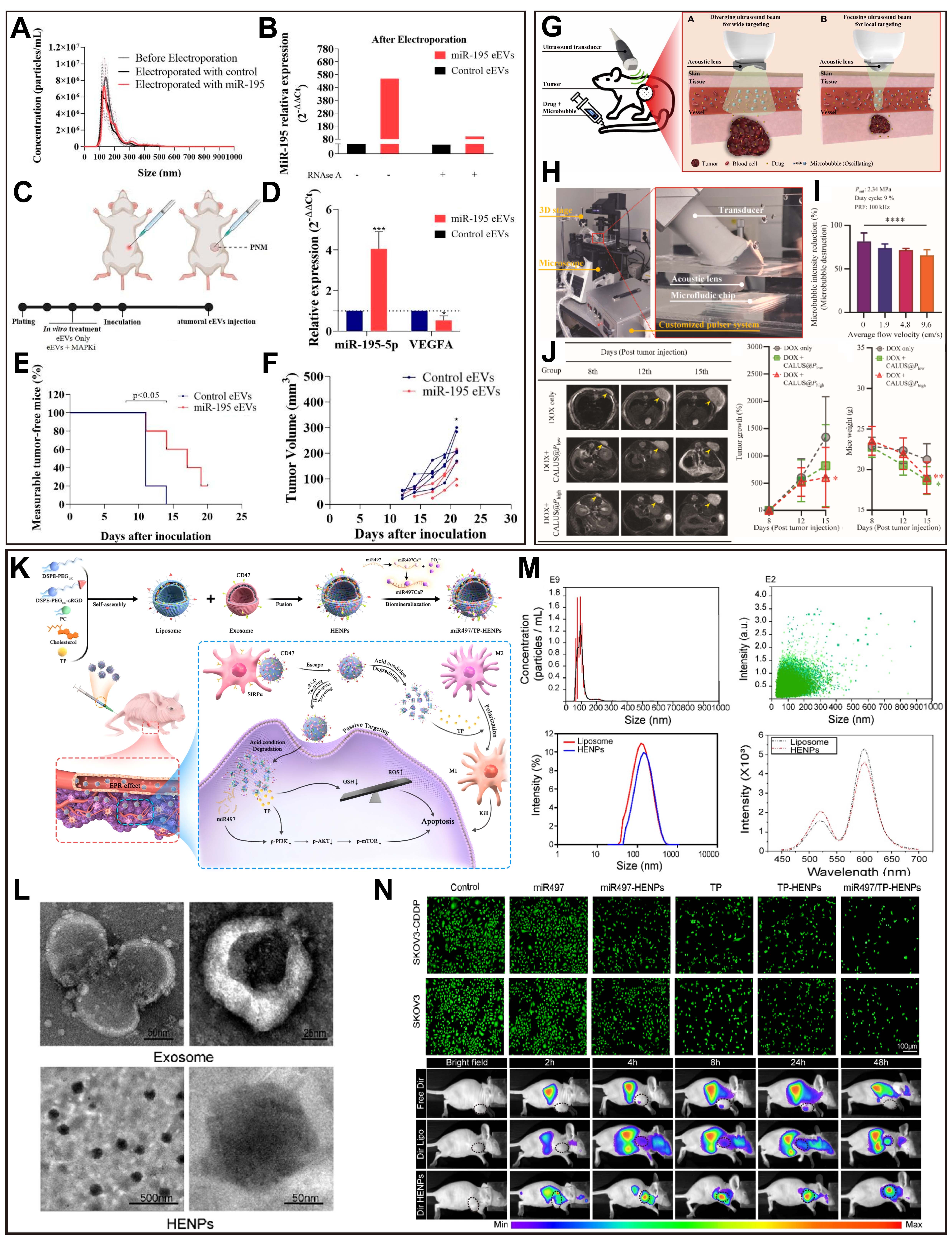
Figure 8. Application of active loading and hybrid-engineering strategies for exosome-mediated drug delivery. (A-F) Electroporation-based loading of miR-195-5p into extracellular vesicles (miR-195 eEVs) for melanoma treatment sensitization, including particle-size distribution, miRNA-loading validation, intratumoral administration, VEGFA modulation, measurable tumor-free rate, and tumor-volume evaluation. *P < 0.05, **P < 0.01, ***P < 0.001, Reprinted with permission[190]. Copyright 2023, MDPI. (G-J) Ultrasound-mediated drug delivery system using a customized convex acoustic lens-attached ultrasound (CALUS) platform for enhanced antitumor efficacy in a murine melanoma subcutaneous model, including in vitro system design, microbubble-intensity reduction, MRI assessment, tumor growth, and body-weight changes. ****P < 0.0001, Reprinted with permission[191]. Copyright 2023, Elsevier. (K-N) Cross-tumor example of exosome-liposome hybrid nanoparticles (HENPs) co-delivering triptolide (TP) and miR497, including hybrid-vesicle construction, ultrastructural characterization, particle-size/fluorescence characterization, and apoptosis assessment. Reprinted with permission[183]. Copyright 2022, Springer Nature. CALUS: Convex acoustic lens-attached ultrasound; eEVs: engineered extracellular vesicles; HENPs: hybrid exosome-liposome nanoparticles; MRI: magnetic resonance imaging; TP: triptolide; VEGFA: vascular endothelial growth factor A.
Multimodal exosome-based therapy and integrated theranostics
Rational design of combined chemotherapy and immunotherapy
To synergistically enhance cytotoxic tumor-cell killing and immune activation, engineered exosome-based systems may be designed either to improve targeted chemotherapy or to combine therapeutic delivery with immune microenvironment modulation. For example, pYEEIE-RELNs-DOX achieved targeted doxorubicin delivery and antitumor efficacy in melanoma-bearing mice[192], whereas exosome-mediated co-delivery of transforming growth factor-β receptor 1 kinase inhibitors and toll-like receptor 7/8 agonists provides a cross-tumor example of combining vesicle-based delivery with immunomodulatory therapy[193]. These examples support the rationale for exosome-assisted combination therapy, but melanoma-specific chemoimmunotherapy designs still require direct validation in appropriate preclinical models.
Integration of targeted therapy and gene-editing technologies
Exosomes are highly efficient carriers for the delivery of nucleic acid-based therapeutics, but the current cited evidence should be framed more cautiously. In melanoma, EVs loaded with miR-195-5p have been shown to sensitize melanoma cells to MAPK-targeted kinase inhibitors and to reduce tumor growth in vivo[190]. Beyond melanoma-specific EV data, engineered tumor-derived exosomes have also been explored in other tumor contexts to modulate chemoresistance and metastatic behavior[194], while non-EV RNAi nanoplatforms such as peptidomimetic lipid nanoparticles have demonstrated that STAT3 silencing can downregulate PD-L1 and inhibit melanoma growth[195]. Together, these findings support the conceptual feasibility of nucleic-acid-based nanodelivery, but they do not yet establish a mature exosome platform co-delivering BRAF V600E-specific siRNA and MEK inhibitors in melanoma.
Theranostics: the potential of exosomes in integrated imaging and therapy
By integrating imaging probes and therapeutic agents on the surface or within exosomes, real-time monitoring of treatment processes and quantitative evaluation of therapeutic efficacy can be achieved. In image-guided surgery, melanoma-targeting exosomes labeled with near-infrared (NIR) fluorescent dyes (such as Cy5.5 and DiR) can be directed to tumor tissues, enabling intraoperative, high-contrast visualization of micrometastatic lesions and metastatic lymph nodes. This approach assists surgeons in accurately delineating surgical margins and improves the completeness of tumor resection[196,197].
Challenges in precise targeted delivery of exosomes
Despite the substantial progress achieved in engineered exosome-based targeting strategies, their clinical translation still faces multiple challenges. As outlined in Figure 9, these include major translational goals, challenges, and potential solutions for melanoma liquid biopsy applications [Figure 9A-C], as well as corresponding issues in drug delivery and melanoma therapy [Figure 9D-F]. First, the heterogeneity of exosome sources leads to considerable variability in composition and biological function, compromising delivery stability and reproducibility, while standardized protocols for purification and quality control are still lacking[198]. Second, exosomes exhibit relatively short circulation half-lives in vivo and are prone to rapid clearance by the immune system, which markedly limits their delivery efficiency to target tissues[199]. Third, currently available loading techniques, such as electroporation and ultrasound-assisted loading, often suffer from limited drug encapsulation efficiency and may compromise exosomal membrane integrity. Fourth, targeting specificity remains insufficient, and achieving highly efficient and cell- or tissue-specific localization requires further systematic investigation. In addition, large-scale manufacturing and clinical translation are hindered by technical and regulatory barriers, including high production costs, batch-to-batch variability, and challenges in comprehensive safety evaluation. Finally, the lack of robust in vivo tracking and quantitative analytical technologies hampers accurate assessment of exosome biodistribution and therapeutic efficacy, thereby constraining the advancement of exosome-based strategies toward clinical application[200].
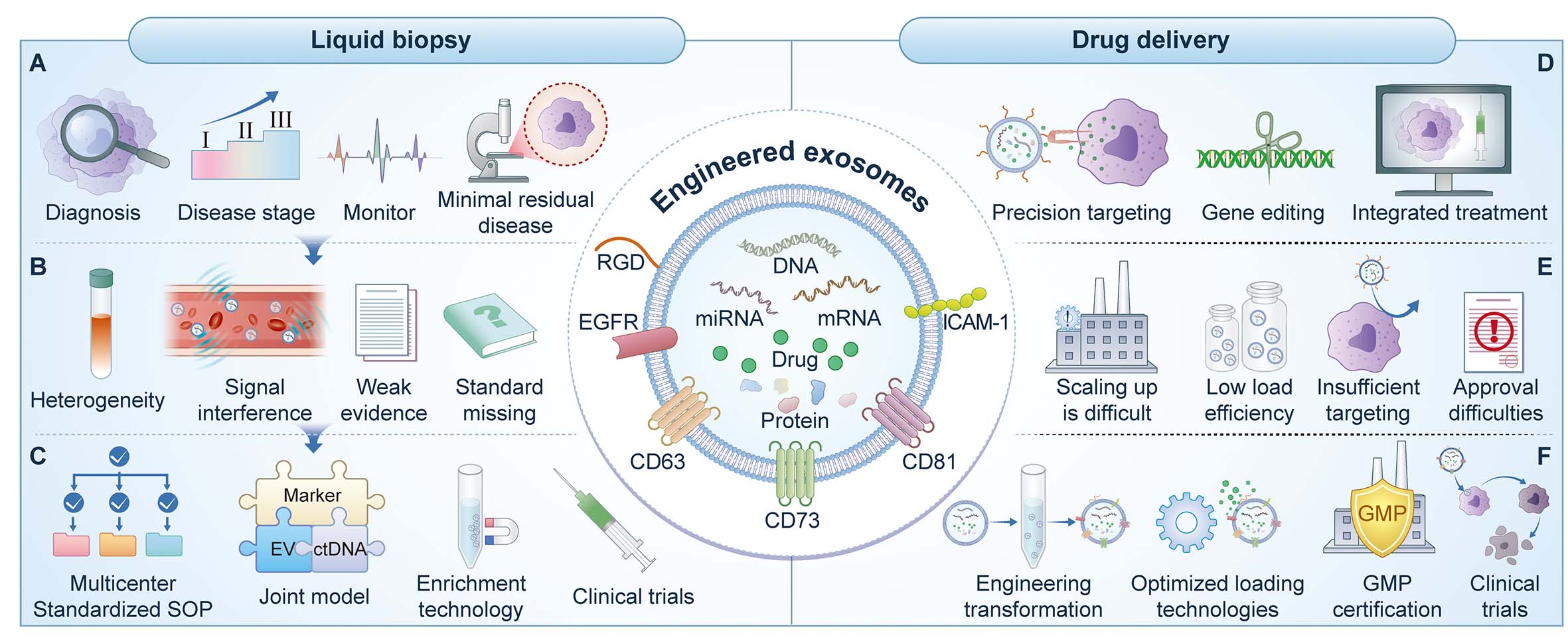
Figure 9. Goals and challenges in the translational application of engineered exosomes in melanoma. (A-C) Major goals, translational challenges, and potential solutions for the clinical application of engineered exosomes in melanoma liquid biopsy. (D-F) Major goals, translational challenges, and potential solutions for the clinical application of engineered exosomes in drug delivery and melanoma therapy.
It should be noted that although surface chemical modification of exosomes can markedly enhance target recognition, drug-loading capacity, and the programmability of the system, it may also alter their native immunological properties and in vivo behavior. On the one hand, operations such as covalent conjugation may interfere with the conformation of native membrane proteins, lipid organization, and glycosylation patterns on the exosomal surface, thereby affecting their interactions with the mononuclear phagocyte system, the complement system, and peripheral immune cells. On the other hand, different modification strategies may exert divergent effects on immunological characteristics: certain targeting ligands may strengthen recognition of target cells, but may also increase the risk of nonspecific uptake by immune cells; whereas polymer coating or charge regulation may help prolong circulation time and reduce hepatic and splenic clearance, yet may simultaneously weaken membrane fusion and endocytic efficiency. Therefore, evaluation of engineered exosomes should not remain limited to targeting efficiency or loading capacity, but should also comprehensively assess immunogenicity, inflammatory responses, complement activation tendency, clearance patterns in vivo, and tolerability upon repeated administration, so as to achieve a balance between functional enhancement and maintenance of biocompatibility[45,201,202].
Despite the substantial progress achieved in engineered exosome-targeting strategies, their clinical translation still faces multiple challenges. First, the native targeting capability of exosomes is not determined by a single molecule, but rather by the combined influence of donor-cell origin, membrane adhesion molecules and integrin profiles, lipid composition, glycosylation patterns, and the recipient tissue microenvironment. Current studies suggest that surface integrins, ICAM-1- and VCAM-1-related interaction axes, and other membrane proteins may all participate, to some extent, in the recognition, adhesion, and uptake of exosomes by tumor cells, endothelial cells, or immune cells; in melanoma, these interactions are also closely associated with pre-metastatic niche formation in lymph nodes, local immunosuppression, and selective uptake by tumor-associated cells[203-205].
However, based on the currently available in vivo evidence, this native targeting behavior is more accurately described as relative enrichment rather than an absolute advantage sufficient to achieve highly specific tumor localization. After systemic administration, exosomes still commonly undergo preferential uptake by the liver, spleen, and mononuclear phagocyte system, display short circulation half-lives, and show substantial variability in biodistribution. Marked differences also exist among exosomes from different sources with respect to organ tropism, cellular uptake profiles, and in vivo stability. Therefore, native targeting alone is usually insufficient to ensure highly selective accumulation in melanoma tissue or to significantly reduce off-target effects, and further enhancement still requires surface ligand modification, multistage targeting design, immune-evasion optimization, and more precise in vivo tracing strategies[201,202,206]. A comparative overview of engineered exosomes and clinically mature nanodelivery systems is provided in Table 3. The advantages, limitations, and representative applications of the major cargo-loading and surface-modification strategies are summarized in Table 4.
Comparison between engineered exosomes and clinically mature nanodelivery systems
| Category | Engineered exosomes | Lipid nanoparticles (LNPs) | Representative references |
| Origin | Naturally derived membranous nanovesicles secreted by cells | Artificially formulated nanodelivery systems assembled chemically | Bader et al., 2024[207] |
| Structural basis | Natural lipid bilayer, membrane proteins, glycosylation, and endogenous bioactive components | Composed of ionizable lipids, helper lipids, cholesterol, PEG-lipids, and related components | Kim et al., 2024[46] |
| Particle-size range | Usually about 30-150 nm, but strongly influenced by source and isolation method | Generally more controllable, with a more uniform size distribution | Bader et al., 2024[207] |
| Cargo types | Can carry RNA, DNA, proteins, small molecules, lipids, etc., while also containing endogenous cargo | Mainly suitable for nucleic-acid delivery and some small molecules, with more controllable cargo design | Kim et al., 2024[46] |
| Loading/encapsulation capacity | Affected by membrane integrity, donor-cell status, and loading method; large batch-to-batch variation | More mature process, with clearer optimization pathways for encapsulation efficiency and formulation | Bader et al., 2024[207] |
| Targeting mechanism | May possess some degree of natural homing/cellular preference and can be further surface-modified | Mainly depends on passive distribution and artificial targeting-ligand modification | Zhao et al., 2024[201] |
| Biocompatibility | Generally good natural biocompatibility and cell-communication properties | Formulations can be standardized, but immune responses and toxicity still require attention | Kim et al., 2024[46] |
| In vivo distribution | Strongly influenced by source, membrane composition, glycosylation, and surface modification; hepatic and splenic accumulation remains common | PK is easier to regulate, but RES uptake and liver distribution are also common | Gandek et al., 2023[208] |
| Immunological features | May retain natural immunomodulatory properties, but can also introduce complex immune effects | Immunogenicity is relatively more controllable, though PEG and ionizable lipids may still trigger reactions | Zhao et al., 2024[201] |
| Stability and storage | Long-term storage, freeze-thaw stability, and preservation of activity remain challenging | Formulation stability and industrial storage/transport experience are more mature | Nieuwland et al., 2024[209]; György et al., 2024[210] |
| Scale-up production | Difficulties remain in yield, purification, standardization, and GMP scale-up | Process and CMC pathway are more mature, with clearer industrial advantages | Alter et al., 2025[211] |
| Quality control | High subpopulation heterogeneity makes quality-attribute definition more complex | Critical quality attributes and release standards are easier to establish | Welsh et al., 2024[10]; Xu et al., 2025[55] |
| Regulatory maturity | Clinical translation and regulatory pathway are still evolving | More mature clinical and regulatory experience already exists | Li et al., 2025[212]; Mizenko et al., 2024[213] |
| Main advantages | Natural membrane structure, biocompatibility, complex cargo, and potential natural targeting | Mature process, scalable manufacturing, and relatively better batch consistency | Kim et al., 2024[46] |
| Main limitations | Heterogeneity, low yield, difficult characterization and standardization, and variable in vivo behavior | Limited biological-information content; specificity and tissue penetration still need optimization in some settings | Bader et al., 2024[207] |
Comparative summary of major cargo-loading and surface-modification strategies for engineered exosomes
| Strategy | Category | Basic principle | Main advantages | Main limitations | Typical efficiency/practical performance | Best suited cargo or application | Representative references |
| Passive incubation | Cargo loading | Exosomes are co-incubated with cargo, and loading mainly occurs through passive diffusion or membrane partitioning | Simple, mild, easy to perform, and relatively preserves vesicle integrity | Usually low and variable loading efficiency; not ideal for large hydrophilic cargos | Low to moderate; generally more suitable for hydrophobic small molecules than for nucleic acids or proteins | Hydrophobic drugs, lipophilic probes, exploratory loading studies | Sun et al., 2010[214] |
| Electroporation | Cargo loading | Electrical pulses transiently permeabilize the exosomal membrane, enabling charged cargos to enter | Widely used; suitable for siRNA, miRNA, and oligonucleotides; relatively accessible | May induce cargo aggregation and membrane damage; loading can be overestimated without rigorous purification | Moderate but highly variable; strongly affected by buffer composition and purification strategy | siRNA, miRNA, antisense oligonucleotides, some proteins | Wahlgren et al., 2012[215] |
| Sonication | Cargo loading | Ultrasound-induced mechanical shear temporarily disrupts membrane organization and facilitates cargo entry | Often improves loading compared with passive incubation | May affect membrane integrity, endogenous cargo composition, and biological activity | Moderate to relatively high, but often with greater structural perturbation | Small molecules, hydrophobic cargos, applications prioritizing loading over strict preservation of native vesicle properties | Kim et al., 2016[216] |
| Extrusion | Cargo loading/hybrid engineering | Exosomes and cargo are forced through membranes with defined pore sizes, promoting membrane disruption and reassembly | Can improve incorporation efficiency and generate relatively homogeneous engineered vesicles | Mechanically harsh; may disrupt membrane proteins and alter native EV identity | Moderate to relatively high, with increased risk of structural remodeling | Small molecules, membrane-associated cargos, EV-liposome hybrid preparation | Guo et al., 2021[217] |
| Donor-cell engineering | Endogenous cargo loading/surface engineering | Parent cells are genetically or biologically engineered so that secreted exosomes carry desired cargos or display targeting motifs | Preserves membrane integrity; enables biologically integrated loading and stable ligand display | Labor-intensive; dependent on cell status and cargo sorting; lower process control and variable yield | Variable, often moderate; especially useful for biologically active nucleic acids and proteins | miRNA, mRNA, proteins, membrane-displayed peptides, receptors, targeting ligands | Ohno et al., 2013[218] |
| Chemical conjugation | Surface modification | Covalent or affinity-based chemistry is used to attach ligands, dyes, polymers, or drugs to the exosomal surface | Stable and versatile surface functionalization; suitable for post-isolation engineering | May alter native membrane proteins, glycans, uptake behavior, and immunological properties | Generally efficient for surface decoration, but biological compatibility must be carefully evaluated | Targeting ligands, imaging probes, PEGylation, affinity tags | Pham et al., 2021[219] |
| Click chemistry | Surface modification | Bioorthogonal reactions are used to install functional moieties on the exosomal membrane in a controlled manner | High specificity, modularity, and controllability | Requires reactive group installation and additional synthetic steps; over-modification may affect vesicle behavior | High controllability for surface engineering rather than bulk cargo loading | Targeting ligands, theranostic probes, multifunctional exosome design | Smyth et al., 2014[220] |
| Membrane fusion | Surface modification/hybrid engineering | Exosomes are fused with liposomes or synthetic membranes using physical or chemical strategies | Combines native EV features with the tunability of synthetic nanocarriers | Generates hybrid vesicles rather than fully native exosomes; requires more complex characterization | Moderate to high engineering flexibility; useful for reformulation and surface remodeling | EV-liposome hybrids, combination delivery, membrane remodeling, synthetic lipid incorporation | Sato et al., 2016[154] |
CLINICAL TRANSLATION AND REGULATORY CONSIDERATIONS
Following over a decade of fundamental research and early-stage validation, work on melanoma-derived exosomes has generally shifted from descriptive studies to applied exploration[9]. Whether employed as liquid biopsy readouts or as delivery vehicles, their successful translation into clinical practice hinges on addressing a consistent set of critical questions: Is the clinical need clearly defined? Can the detection and manufacturing processes be standardized? Can the evidence be reproduced in real-world settings? And can the final product fit into an actionable regulatory framework[212]. Accordingly, this section outlines the core challenges that must be overcome for exosome-based applications to achieve clinical adoption, focusing on four key dimensions: the level of evidence, standardization of the detection pipeline, manufacturing and safety considerations for therapeutic carriers, and regulatory pathways and trial design[213].
Level of evidence and clinical problem definition
Regarding liquid biopsy, a substantial body of research currently remains at the stage of retrospective or exploratory cohort studies. Significant variability in sample sizes, follow-up durations, and endpoint definitions across studies makes it difficult to directly compare conclusions and limits clinical applicability[9]. The key to advancing the evidence base lies not in identifying additional candidate molecules, but in first defining actionable clinical problems and aligning them with testable endpoints and clear decision-making scenarios[221]. Examples include: (1) Recurrence early warning and adjustment of follow-up frequency in high-risk post-operative patient cohorts. (2) Dynamic monitoring of early response/resistance trends during immunotherapy, informing decisions such as switching regimens early or adding combination therapies. (3) Aid in interpreting cases with "pseudoprogression/inflammatory response". (4) Evaluation of incremental value on top of existing risk models, i.e., whether exosome-based readouts can improve predictive performance or patient stratification[213].
Standardization and quality control of the detection pipeline
Uncertainty in exosome detection often originates first from pre-analytical variables and variations in isolation and enrichment strategies[209]. Blood collection and processing procedures (type of anticoagulant, hemolysis, centrifugation conditions, storage temperature, and freeze-thaw cycles, etc.) can significantly impact particle counts and nucleic acid/protein readings; different enrichment methods (ultracentrifugation, SEC, immunoaffinity capture, polymer-based precipitation, and their combinations) define the measurand, thereby affecting the comparability of studies[210,222]. Even on the same platform, quantification and normalization strategies (by volume, particle number, protein amount, or internal references) can alter the robustness of conclusions and the boundaries for extrapolation[10].
Regulatory pathways and trial design
For the clinical application of exosomes in liquid biopsy, the framework more closely aligns with that of In Vitro Diagnostics (IVD) or Laboratory-Developed Tests (LDTs). The core focus is on analytical performance (accuracy, precision, linear range, limit of detection, interference resistance, inter-batch consistency) and clinical performance (demonstrating application value within a defined population and with clear
CONCLUSION
Based on the above discussion, a more feasible path forward can be summarized along three tracks: First, Problem-Driven Implementation of Liquid Biopsy: Prioritize clinical scenarios with well-defined decision-making pathways. Pre-specify sampling time points, thresholds, and model rules, with incremental value serving as the core evaluation metric[221]. Second, Delivery-Based Enhancement within a Combination Therapy Framework: Pair mechanistic pharmacodynamic readouts with clinical endpoints. Early focus should be placed on defining risk boundaries and manufacturability, moving beyond proof-of-concept demonstrations solely in vitro or in single animal models[59]. Third, Platform-Level Standardization: Establish actionable quality control systems and cross-center consistency evaluations for both detection and manufacturing. This is essential to advance exosomes from research entities to clinical products[10].
In summary, exosomes represent a nanoscale platform for melanoma, offering both biological information and material properties. The success of their clinical translation will depend less on technical feasibility alone and more on the establishment of standardized, reproducible, and regulatory-compliant systemic frameworks, as well as the demonstration of incremental benefit in well-defined clinical contexts[114]. In the future, the clinical value of exosomes in melanoma is expected to materialize along a "Standardization-Validation-Integration-Decision-making" pathway.
DECLARATIONS
Authors’ contributions
Conceptualization and writing - original draft: Li, L.; Zheng, X.; Shi, J.; Zhao, F.
Literature investigation and Figure arrangement: Chen, Y.; Jia, H.; Chen, J.
Writing-review & editing: Wang, X.; Luo, P.; Zhang, L.
Supervision and funding acquisition: Zhao, Z.; Lin, L.; Sun, Q.; Luo, Z.
Availability of data and materials
Not applicable.
AI and AI-assisted tools statement
Not applicable.
Financial support and sponsorship
This study was funded by National Natural Science Foundation of China (82301158). Key Laboratory of Dermatology (Anhui Medical University), Ministry of Education, Hefei, 230032 Anhui China (Number AYPYS2025-01).
Conflicts of interest
Luo, Z. serves as Guest Editor of the Special Issue “Biological Nanomaterials for Targeted Drug Delivery and Gene Therapy” in Microstructures. He is not involved in any stage of the editorial process, including reviewer selection, manuscript handling, or decision-making, while the other authors have declared that they have no conflicts of interest.
Ethical approval and consent to participate
Not applicable.
Consent for publication
Not applicable.
Copyright
© The Author(s) 2026.
REFERENCES
1. Keilholz, U.; Ascierto, P.; Dummer, R.; et al. ESMO consensus conference recommendations on the management of metastatic melanoma: under the auspices of the ESMO Guidelines Committee. Ann. Oncol. 2020, 31, 1435-48.
2. Atkins, M. B.; Curiel-Lewandrowski, C.; Fisher, D. E.; et al. The state of melanoma: emergent challenges and opportunities. Clin. Cancer. Res. 2021, 27, 2678-97.
3. Li, H.; Bian, Y.; Xiahou, Z.; Zhao, Z.; Zhao, F.; Zhang, Q. The cellular signaling crosstalk between memory B cells and tumor cells in nasopharyngeal carcinoma cannot be overlooked: Their involvement in tumor progression and treatment strategy is significant. J. Cancer. 2025, 16, 288-314.
4. Huang, S. K.; Hoon, D. S. Liquid biopsy utility for the surveillance of cutaneous malignant melanoma patients. Mol. Oncol. 2015, 10, 450-63.
5. Sivapalan, L.; Murray, J. C.; Canzoniero, J. V.; et al. Liquid biopsy approaches to capture tumor evolution and clinical outcomes during cancer immunotherapy. J. Immunother. Cancer. 2023, 11, e005924.
6. Patel, R. P.; Somasundram, P. M.; Smith, L. K.; Sheppard, K. E.; Mcarthur, G. A. The therapeutic potential of targeting minimal residual disease in melanoma. Clin. Transl. Med. 2023, 13, e1197.
7. Tirosh, I.; Izar, B.; Prakadan, S. M.; et al. Dissecting the multicellular ecosystem of metastatic melanoma by single-cell RNA-seq. Science 2016, 352, 189-96.
8. Whiteside, T. L. Biology of extracellular vesicles and the potential of tumor-derived vesicles for subverting immunotherapy of cancer. J. Immunother. Cancer. 2025, 13, e010376.
9. Zou, Y.; Zhou, Y.; Li, G.; Dong, Y.; Hu, S. Clinical applications of extracellular vesicles: recent advances and emerging trends. Front. Bioeng. Biotechnol. 2025, 13, 1671963.
10. Welsh, J. A.; Goberdhan, D. C. I.; O'driscoll, L.; et al. Minimal information for studies of extracellular vesicles (MISEV2023): from basic to advanced approaches. J. Extracell. Vesicle. 2024, 13, e12404.
11. Ragni, E. Extracellular vesicles: recent advances and perspectives. Front. Biosci. 2025, 30, 36405.
12. Tang, H.; Yu, D.; Zhang, J.; et al. The new advance of exosome-based liquid biopsy for cancer diagnosis. J. Nanobiotechnol. 2024, 22, 610.
13. Ghanam, J.; Lichá, K.; Chetty, V. K.; et al. Unravelling the significance of extracellular vesicle-associated DNA in cancer biology and its potential clinical applications. J. Extracell. Vesicle. 2025, 14, e70047.
14. Andre, M.; Caobi, A.; Miles, J. S.; Vashist, A.; Ruiz, M. A.; Raymond, A. D. Diagnostic potential of exosomal extracellular vesicles in oncology. BMC. Cancer. 2024, 24, 322.
15. Li, Z.; Gao, Y.; Cao, Y.; et al. Extracellular RNA in melanoma: advances, challenges, and opportunities. Front. Cell. Dev. Biol. 2023, 11, 1141543.
16. Zhan, Y.; Yang, Z.; Zeng, H.; Yu, J.; Chen, X.; Wu, Y. Extracellular vesicle-derived non-coding RNAs in remodeling melanoma. Biomed. Pharmacother. 2024, 172, 116213.
17. Slusher, N.; Jones, N.; Nonaka, T. Liquid biopsy for diagnostic and prognostic evaluation of melanoma. Front. Cell. Dev. Biol. 2024, 12, 1420360.
18. Liu, J. J. J.; Liu, D.; To, S. K. Y.; Wong, A. S. T. Exosomes in cancer nanomedicine: biotechnological advancements and innovations. Mol. Cancer. 2025, 24, 166.
19. Ovchinnikova, L. A.; Lomakin, Y. A. Extracellular vesicles as a source of biomarkers for cancer diagnosis. Acta. Nat. 2025, 17, 28-40.
20. Babaei, S.; Fadaee, M.; Abbasi-Kenarsari, H.; Shanehbandi, D.; Kazemi, T. Exosome-based immunotherapy as an innovative therapeutic approach in melanoma. Cell. Commun. Signal. 2024, 22, 527.
21. Di Bella, M. A. Overview and update on extracellular vesicles: considerations on exosomes and their application in modern medicine. Biology 2022, 11, 804.
22. Zhang, Y.; Lan, M.; Chen, Y. Minimal information for studies of extracellular vesicles (MISEV): ten-year evolution (2014-2023). Pharmaceutics 2024, 16, 1394.
23. Di Bella, M. A.; Taverna, S. Extracellular vesicles: diagnostic and therapeutic applications in cancer. Biology 2024, 13, 716.
24. Yang, C.; Xue, Y.; Duan, Y.; Mao, C.; Wan, M. Extracellular vesicles and their engineering strategies, delivery systems, and biomedical applications. J. Controlled. Release. 2024, 365, 1089-123.
25. Zhang, Q.; Jeppesen, D. K.; Higginbotham, J. N.; Franklin, J. L.; Coffey, R. J. Comprehensive isolation of extracellular vesicles and nanoparticles. Nat. Protoc. 2023, 18, 1462-87.
26. Carney, R. P.; Mizenko, R. R.; Bozkurt, B. T.; et al. Harnessing extracellular vesicle heterogeneity for diagnostic and therapeutic applications. Nat. Nanotechnol. 2024, 20, 14-25.
27. Li, B.; Wu, W.; Xu, W.; Qian, H.; Ji, C. Advances of extracellular vesicles isolation and detection frontier technology: from heterogeneity analysis to clinical application. J. Nanobiotechnol. 2025, 23, 678.
28. Woo, H.; Nam, Y.; Park, H. G.; Lee, H. Bridging laboratory innovation to translational research and commercialization of extracellular vesicle isolation and detection. Biosens. Bioelectron. 2025, 282, 117475.
29. Poggio, M.; Hu, T.; Pai, C.; et al. Suppression of exosomal PD-L1 induces systemic anti-tumor immunity and memory. Cell 2019, 177, 414-27.e13.
30. Urzì, O.; Olofsson Bagge, R.; Crescitelli, R. Extracellular vesicles in uveal melanoma - Biological roles and diagnostic value. Cancer. Lett. 2025, 615, 217531.
31. Dong, X.; Lin, Y.; Li, K.; et al. Consensus statement on extracellular vesicles in liquid biopsy for advancing laboratory medicine. Clin. Chem. Lab. Med. 2025, 63, 465-82.
32. Asleh, K.; Dery, V.; Taylor, C.; Davey, M.; Djeungoue-Petga, M.; Ouellette, R. J. Extracellular vesicle-based liquid biopsy biomarkers and their application in precision immuno-oncology. Biomark. Res. 2023, 11, 99.
33. Liu, J.; Hu, X.; Xin, W.; Wang, X. Exosomal non-coding RNAs: a new approach to melanoma diagnosis and therapeutic strategy. Curr. Med. Chem. 2024, 31, 6084-109.
34. Ricciardi, E.; Giordani, E.; Ziccheddu, G.; et al. Metastatic melanoma: liquid biopsy as a new precision medicine approach. Int. J. Mol. Sci. 2023, 24, 4014.
35. Minami, S.; Chikazu, D.; Ochiya, T.; Yoshioka, Y. Extracellular vesicle-based liquid biopsies in cancer: Future biomarkers for oral cancer. Transl. Oncol. 2023, 38, 101786.
36. Mondal, S. K.; Whiteside, T. L. Proteomic profiles of melanoma cell-derived exosomes in plasma: discovery of potential biomarkers of melanoma progression. Melanoma. Res. 2021, 31, 472-5.
37. Lattmann, E.; Räss, L.; Tognetti, M.; et al. Size-exclusion chromatography combined with DIA-MS enables deep proteome profiling of extracellular vesicles from melanoma plasma and serum. Cell. Mol. Life. Sci. 2024, 81, 90.
38. Clancy, J. W.; D'souza-Schorey, C. Tumor-derived extracellular vesicles: multifunctional entities in the tumor microenvironment. Annu. Rev. Pathol. Mech. Dis. 2023, 18, 205-29.
39. Helvind, N. M.; Brinch-Møller Weitemeyer, M.; Chakera, A. H.; et al. Stage-specific risk of recurrence and death from melanoma in Denmark, 2008-2021: a national observational cohort study of 25 720 patients with stage IA to IV melanoma. JAMA. Dermatol. 2023, 159, 1213.
40. Spiliopoulou, P.; Holanda Lopes, C. D.; Spreafico, A. Promising and minimally invasive biomarkers: targeting melanoma. Cells 2023, 13, 19.
41. Serratì, S.; Guida, M.; Di Fonte, R.; et al. Circulating extracellular vesicles expressing PD1 and PD-L1 predict response and mediate resistance to checkpoint inhibitors immunotherapy in metastatic melanoma. Mol. Cancer. 2022, 21, 20.
42. Sun, L.; Wei, X.; Zhao, Q.; et al. Dynamic change of PD-L2 on circulating plasma extracellular vesicles as a predictor of treatment response in melanoma patients receiving anti-PD-1 therapy. J. Extracell. Vesicle. 2025, 14, e70054.
43. Hsu, C.; Chandramoorthy, H. C.; Mohammed, J. S.; et al. Exosomes as key mediators in immune and cancer cell interactions: insights in melanoma progression and therapy. Arch. Dermatol. Res. 2025, 317, 729.
44. Serrano, D. R.; Juste, F.; Anaya, B. J.; et al. Exosome-based drug delivery: a next-generation platform for cancer, infection, neurological and immunological diseases, gene therapy and regenerative medicine. Pharmaceutics 2025, 17, 1336.
45. Johnson, V.; Vasu, S.; Kumar, U. S.; Kumar, M. Surface-engineered extracellular vesicles in cancer immunotherapy. Cancers 2023, 15, 2838.
46. Kim, H. I.; Park, J.; Zhu, Y.; Wang, X.; Han, Y.; Zhang, D. Recent advances in extracellular vesicles for therapeutic cargo delivery. Exp. Mol. Med. 2024, 56, 836-49.
47. Chen, Y.; Luh, F.; Ho, Y.; Yen, Y. Exosomes: a review of biologic function, diagnostic and targeted therapy applications, and clinical trials. J. Biomed. Sci. 2024, 31, 67.
48. Brezgin, S.; Danilik, O.; Yudaeva, A.; et al. Basic guide for approaching drug delivery with extracellular vesicles. Int. J. Mol. Sci. 2024, 25, 10401.
49. Abbasi-malati, Z.; Azizi, S. G.; Milani, S. Z.; et al. Tumorigenic and tumoricidal properties of exosomes in cancers; a forward look. Cell. Commun. Signal. 2024, 22, 130.
50. Xia, Y.; Zhang, J.; Liu, G.; Wolfram, J. Immunogenicity of extracellular vesicles. Adv. Mater. 2024, 36, 2403199.
51. Ziogas, D. C.; Theocharopoulos, C.; Koutouratsas, T.; Haanen, J.; Gogas, H. Mechanisms of resistance to immune checkpoint inhibitors in melanoma: what we have to overcome? Cancer. Treat. Rev. 2023, 113, 102499.
52. Fernandez, M. F.; Choi, J.; Sosman, J. New approaches to targeted therapy in Melanoma. Cancers 2023, 15, 3224.
53. Yin, G.; Guo, W.; Huang, Z.; Chen, X. Efficacy of radiotherapy combined with immune checkpoint inhibitors in patients with melanoma: a systemic review and meta-analysis. Melanoma. Res. 2022, 32, 71-8.
54. Van Delen, M.; Derdelinckx, J.; Wouters, K.; Nelissen, I.; Cools, N. A systematic review and meta-analysis of clinical trials assessing safety and efficacy of human extracellular vesicle-based therapy. J. Extracell. Vesicle. 2024, 13, e12458.
55. Xu, G.; Jin, J.; Fu, Z.; et al. Extracellular vesicle-based drug overview: research landscape, quality control and nonclinical evaluation strategies. Sig. Transduct. Target. Ther. 2025, 10, 255.
56. Su, X.; Wang, H.; Li, Q.; Chen, Z. Extracellular vesicles: a review of their therapeutic potentials, sources, biodistribution, and administration routes. Int. J. Nanomed. 2025, 20, 3175-99.
57. Kang, M.; Jordan, V.; Blenkiron, C.; Chamley, L. W. Biodistribution of extracellular vesicles following administration into animals: a systematic review. J. Extracell. Vesicle. 2021, 10, e12085.
58. Chen, T.; Chen, D.; Su, W.; Liang, J.; Liu, X.; Cai, M. Extracellular vesicles as vital players in drug delivery: a focus on clinical disease treatment. Front. Bioeng. Biotechnol. 2025, 13, 1600227.
59. Humbert, C.; Cordier, C.; Drut, I.; et al. GMP-compliant process for the manufacturing of an extracellular vesicles-enriched secretome product derived from cardiovascular progenitor cells suitable for a phase I clinical trial. J. Extracell. Vesicle. 2025, 14, e70145.
60. Akbani, R.; Akdemir, K. C.; Aksoy, B. A.; et al. Genomic classification of cutaneous melanoma. Cell 2015, 161, 1681-96.
61. Moran, B.; Silva, R.; Perry, A. S.; Gallagher, W. M. Epigenetics of malignant melanoma. Semin. Cancer. Biol. 2018, 51, 80-8.
62. Sarkar, D.; Leung, E. Y.; Baguley, B. C.; Finlay, G. J.; Askarian-Amiri, M. E. Epigenetic regulation in human melanoma: past and future. Epigenetics 2015, 10, 103-21.
63. Mannavola, F.; D’oronzo, S.; Cives, M.; et al. Extracellular vesicles and epigenetic modifications are hallmarks of melanoma progression. Int. J. Mol. Sci. 2019, 21, 52.
64. Kalluri, R.; Lebleu, V. S. The biology,function, and biomedical applications of exosomes. Science 2020, 367, eaau6977.
65. Willms, E.; Cabañas, C.; Mäger, I.; Wood, M. J. A.; Vader, P. Extracellular vesicle heterogeneity: subpopulations, isolation techniques, and diverse functions in cancer progression. Front. Immunol. 2018, 9, 738.
66. Zaborowski, M. P.; Balaj, L.; Breakefield, X. O.; Lai, C. P. Extracellular vesicles: composition, biological relevance, and methods of study. Bioscience 2015, 65, 783-97.
67. Yáñez-mó, M.; Siljander, P. R. M.; Andreu, Z.; et al. Biological properties of extracellular vesicles and their physiological functions. J. Extracell. Vesicle. 2015, 4, 27066.
68. Lespagnol, A.; Duflaut, D.; Beekman, C.; et al. Exosome secretion, including the DNA damage-induced p53-dependent secretory pathway, is severely compromised in TSAP6/Steap3-null mice. Cell. Death. Differ. 2008, 15, 1723-33.
69. Payandeh, Z.; Khalili, S.; Somi, M. H.; et al. PD-1/PD-L1-dependent immune response in colorectal cancer. J. Cell. Physiol. 2020, 235, 5461-75.
70. Jiang, Y.; Zhao, X.; Fu, J.; Wang, H. Progress and challenges in precise treatment of tumors with PD-1/PD-L1 blockade. Front. Immunol. 2020, 11, 339.
71. Wang, B.; Bai, W.; Ma, H.; Li, F. Regulatory effect of PD1/PD-Ligand 1 (PD-L1) on treg cells in patients with idiopathic pulmonary fibrosis. Med. Sci. Monit. 2020, 26, 927577.
72. Shergold, A. L.; Millar, R.; Nibbs, R. J. Understanding and overcoming the resistance of cancer to PD-1/PD-L1 blockade. Pharmacol. Res. 2019, 145, 104258.
73. Cloughesy, T. F.; Mochizuki, A. Y.; Orpilla, J. R.; et al. Neoadjuvant anti-PD-1 immunotherapy promotes a survival benefit with intratumoral and systemic immune responses in recurrent glioblastoma. Nat. Med. 2019, 25, 477-86.
74. Chen, G.; Huang, A. C.; Zhang, W.; et al. Exosomal PD-L1 contributes to immunosuppression and is associated with anti-PD-1 response. Nature 2018, 560, 382-6.
75. Ricklefs, F. L.; Alayo, Q.; Krenzlin, H.; et al. Immune evasion mediated by PD-L1 on glioblastoma-derived extracellular vesicles. Sci. Adv. 2018, 4, eaar2766.
76. Theodoraki, M.; Yerneni, S. S.; Hoffmann, T. K.; Gooding, W. E.; Whiteside, T. L. Clinical significance of PD-L1+ exosomes in plasma of head and neck cancer patients. Clin. Cancer. Res. 2018, 24, 896-905.
77. Yang, Y.; Li, C.; Chan, L.; et al. Exosomal PD-L1 harbors active defense function to suppress T cell killing of breast cancer cells and promote tumor growth. Cell. Res. 2018, 28, 862-4.
78. Fleming, V.; Hu, X.; Weller, C.; et al. Melanoma extracellular vesicles generate immunosuppressive myeloid cells by upregulating PD-L1 via TLR4 signaling. Cancer. Res. 2019, 79, 4715-28.
79. Du, Z.; Feng, Y.; Zhang, H.; Liu, J.; Wang, J. Melanoma-derived small extracellular vesicles remodel the systemic onco-immunity via disrupting hematopoietic stem cell proliferation and differentiation. Cancer. Lett. 2022, 545, 215841.
80. Maus, R. L. G.; Jakub, J. W.; Nevala, W. K.; et al. Human melanoma-derived extracellular vesicles regulate dendritic cell maturation. Front. Immunol. 2017, 8, 358.
81. Valenti, R.; Huber, V.; Filipazzi, P.; et al. Human tumor-released microvesicles promote the differentiation of myeloid cells with transforming growth factor-β-mediated suppressive activity on t lymphocytes. Cancer. Res. 2006, 66, 9290-8.
82. Hosseini, R.; Asef-kabiri, L.; Yousefi, H.; et al. The roles of tumor-derived exosomes in altered differentiation, maturation and function of dendritic cells. Mol. Cancer. 2021, 20, 83.
83. Marton, A.; Vizler, C.; Kusz, E.; et al. Melanoma cell-derived exosomes alter macrophage and dendritic cell functions in vitro. Immunol. Lett. 2012, 148, 34-8.
84. Düchler, M.; Czernek, L.; Peczek, L.; Cypryk, W.; Sztiller-Sikorska, M.; Czyz, M. Melanoma-derived extracellular vesicles bear the potential for the induction of antigen-specific tolerance. Cells 2019, 8, 665.
85. Sharma, P.; Diergaarde, B.; Ferrone, S.; Kirkwood, J. M.; Whiteside, T. L. Melanoma cell-derived exosomes in plasma of melanoma patients suppress functions of immune effector cells. Sci. Rep. 2020, 10, 92.
86. Whiteside, T. L. Immunosuppressive functions of melanoma cell-derived exosomes in plasma of melanoma patients. Front. Cell. Dev. Biol. 2023, 10, 1080925.
87. Crucitta, S.; Cucchiara, F.; Marconcini, R.; et al. TGF-β mRNA levels in circulating extracellular vesicles are associated with response to anti-PD1 treatment in metastatic melanoma. Front. Mol. Biosci. 2024, 11, 1288677.
88. Tatischeff, I. Extracellular vesicle-DNA: the next liquid biopsy biomarker for early cancer diagnosis? Cancers 2023, 15, 1456.
89. Belviso, I.; Pastore, R.; Mileo, A.; et al. The role of fibroblasts in melanoma development: from tumor microenvironment remodeling to pre-metastatic niche formation. Int. J. Mol. Sci. 2025, 26, 6132.
90. Lu, M.; Yang, Y. Exosomal PD-L1 in cancer and other fields: recent advances and perspectives. Front. Immunol. 2024, 15, 1395332.
91. Chen, J.; Song, Y.; Miao, F.; et al. PDL1-positive exosomes suppress antitumor immunity by inducing tumor-specific CD8+ T cell exhaustion during metastasis. Cancer. Sci. 2021, 112, 3437-54.
92. Liu, N.; Zhang, J.; Yin, M.; et al. Inhibition of xCT suppresses the efficacy of anti-PD-1/L1 melanoma treatment through exosomal PD-L1-induced macrophage M2 polarization. Mol. Ther. 2021, 29, 2321-34.
93. Christofides, A.; Strauss, L.; Yeo, A.; Cao, C.; Charest, A.; Boussiotis, V. A. The complex role of tumor-infiltrating macrophages. Nat. Immunol. 2022, 23, 1148-56.
94. Gerloff, D.; Lützkendorf, J.; Moritz, R. K.; et al. Melanoma-derived exosomal miR-125b-5p educates tumor associated macrophages (TAMs) by targeting lysosomal acid lipase A (LIPA). Cancers 2020, 12, 464.
95. Wang, R.; Zhu, T.; Hou, B.; Huang, X. An iPSC-derived exosome-pulsed dendritic cell vaccine boosts antitumor immunity in melanoma. Mol. Ther. 2023, 31, 2376-90.
96. Tucci, M.; Mannavola, F.; Passarelli, A.; Stucci, L. S.; Cives, M.; Silvestris, F. Exosomes in melanoma: a role in tumor progression, metastasis and impaired immune system activity. Oncotarget 2018, 9, 20826-37.
97. Zhou, J.; Yang, Y.; Wang, W.; et al. Melanoma-released exosomes directly activate the mitochondrial apoptotic pathway of CD4+ T cells through their microRNA cargo. Exp. Cell. Res. 2018, 371, 364-71.
98. Li, X.; Liu, Y.; Yang, L.; Jiang, Y.; Qian, Q. TIM-3 shuttled by MV3 cells-secreted exosomes inhibits CD4+ T cell immune function and induces macrophage M2 polarization to promote the growth and metastasis of melanoma cells. Transl. Oncol. 2022, 18, 101334.
99. De Cicco, P.; Ercolano, G.; Ianaro, A. The new era of cancer immunotherapy: targeting myeloid-derived suppressor cells to overcome immune evasion. Front. Immunol. 2020, 11, 1680.
100. Peinado, H.; Alečković, M.; Lavotshkin, S.; et al. Melanoma exosomes educate bone marrow progenitor cells toward a pro-metastatic phenotype through MET. Nat. Med. 2012, 18, 883-91.
101. Peinado, H.; Zhang, H.; Matei, I. R.; et al. Pre-metastatic niches: organ-specific homes for metastases. Nat. Rev. Cancer. 2017, 17, 302-17.
102. Kim, J.; Afshari, A.; Sengupta, R.; et al. Replication study: melanoma exosomes educate bone marrow progenitor cells toward a pro-metastatic phenotype through MET. eLife 2018, 7, e39944.
103. Rinderknecht, M.; Detmar, M. Tumor lymphangiogenesis and melanoma metastasis. J. Cell. Physiol. 2008, 216, 347-54.
104. Hood, J. L.; San, R. S.; Wickline, S. A. Exosomes released by melanoma cells prepare sentinel lymph nodes for tumor metastasis. Cancer. Res. 2011, 71, 3792-801.
105. García-Silva, S.; Benito-Martín, A.; Nogués, L.; et al. Melanoma-derived small extracellular vesicles induce lymphangiogenesis and metastasis through an NGFR-dependent mechanism. Nat. Cancer. 2021, 2, 1387-405.
106. Hu, T.; Hu, J. Melanoma-derived exosomes induce reprogramming fibroblasts into cancer-associated fibroblasts via Gm26809 delivery. Cell. Cycle. 2019, 18, 3085-94.
107. Shelton, M.; Anene, C.; Nsengimana, J.; Roberts, W.; Newton-Bishop, J.; Boyne, J. The role of CAF derived exosomal microRNAs in the tumour microenvironment of melanoma. Biochim. Biophys. Acta. 2021, 1875, 188456.
108. Benito-Martín, A.; Jasiulionis, M. G.; García-Silva, S. Extracellular vesicles and melanoma: new perspectives on tumor microenvironment and metastasis. Front. Cell. Dev. Biol. 2023, 10, 1061982.
109. Wang, C.; Wang, Y.; Chang, X.; et al. Melanoma-derived exosomes endow fibroblasts with an invasive potential via miR-21 target signaling pathway. Cancer. Manag. Res. 2020, 12, 12965-74.
110. Tagliatela, A. C.; Hempstead, S. C.; Hibshman, P. S.; et al. Coronin 1C inhibits melanoma metastasis through regulation of MT1-MMP-containing extracellular vesicle secretion. Sci. Rep. 2020, 10, 11958.
111. Pavlides, S.; Whitaker-Menezes, D.; Castello-Cros, R.; et al. The reverse Warburg effect: aerobic glycolysis in cancer associated fibroblasts and the tumor stroma. Cell. Cycle. 2014, 8, 3984-4001.
112. Lazar, I.; Clement, E.; Dauvillier, S.; et al. Adipocyte exosomes promote melanoma aggressiveness through fatty acid oxidation: a novel mechanism linking obesity and cancer. Cancer. Res. 2016, 76, 4051-7.
113. Clement, E.; Lazar, I.; Attané, C.; et al. Adipocyte extracellular vesicles carry enzymes and fatty acids that stimulate mitochondrial metabolism and remodeling in tumor cells. EMBO. J. 2020, 39, EMBJ2019102525.
114. Medhin, L. B.; Beasley, A. B.; Warburton, L.; Amanuel, B.; Gray, E. S. Extracellular vesicles as a liquid biopsy for melanoma: are we there yet? Semin. Cancer. Biol. 2023, 89, 92-8.
115. Wu, N.; Sun, H.; Sun, Q.; et al. Circulating microRNAs as diagnostic biomarkers for melanoma: a systematic review and meta-analysis. BMC. Cancer. 2023, 23, 414.
116. Hu, C.; Jiang, W.; Lv, M.; et al. Potentiality of exosomal proteins as novel cancer biomarkers for liquid biopsy. Front. Immunol. 2022, 13, 792046.
117. Wang, M.; Cai, Y.; Peng, Y.; Xu, B.; Hui, W.; Jiang, Y. Exosomal LGALS9 in the cerebrospinal fluid of glioblastoma patients suppressed dendritic cell antigen presentation and cytotoxic T-cell immunity. Cell. Death. Dis. 2020, 11, 896.
118. Shimada, Y.; Matsubayashi, J.; Kudo, Y.; et al. Serum-derived exosomal PD-L1 expression to predict anti-PD-1 response and in patients with non-small cell lung cancer. Sci. Rep. 2021, 11, 7830.
119. Kluszczynska, K.; Czyz, M. Extracellular vesicles-based cell-cell communication in melanoma: new perspectives in diagnostics and therapy. Int. J. Mol. Sci. 2023, 24, 965.
120. Van Niel, G.; D'angelo, G.; Raposo, G. Shedding light on the cell biology of extracellular vesicles. Nat. Rev. Mol. Cell. Biol. 2018, 19, 213-28.
121. Théry, C.; Witwer, K. W.; Aikawa, E.; et al. Minimal information for studies of extracellular vesicles 2018 (MISEV2018): a position statement of the International Society for Extracellular Vesicles and update of the MISEV2014 guidelines. J. Extracell. Vesicle. 2018, 7, 1535750.
122. Ullah, M. The future of bioorthogonal-chemistry for targeting of exosomes in precision medicine. Oncotarget 2022, 13, 1303-4.
123. Rayamajhi, S.; Sipes, J.; Tetlow, A. L.; Saha, S.; Bansal, A.; Godwin, A. K. Extracellular vesicles as liquid biopsy biomarkers across the cancer journey: from early detection to recurrence. Clin. Chem. 2024, 70, 206-19.
124. Xia, C.; Yin, S.; To, K. K. W.; Fu, L. CD39/CD73/A2AR pathway and cancer immunotherapy. Mol. Cancer. 2023, 22, 44.
125. Kurago, Z.; Guo, G.; Shi, H.; et al. Inhibitors of the CD73-adenosinergic checkpoint as promising combinatory agents for conventional and advanced cancer immunotherapy. Front. Immunol. 2023, 14, 1212209.
126. Saigí, M.; Mesía-Carbonell, O.; Barbie, D. A.; Guillamat-Prats, R. Unraveling the intricacies of CD73/adenosine signaling: the pulmonary immune and stromal microenvironment in lung cancer. Cancers 2023, 15, 5706.
127. Turiello, R.; Capone, M.; Morretta, E.; et al. Exosomal CD73 from serum of patients with melanoma suppresses lymphocyte functions and is associated with therapy resistance to anti-PD-1 agents. J. Immunother. Cancer. 2022, 10, e004043.
128. Yang, Y.; Zhu, L.; Xu, Y.; et al. The progress and prospects of targeting the adenosine pathway in cancer immunotherapy. Biomark. Res. 2025, 13, 75.
129. Rodrigues-junior, D. M.; Tsirigoti, C.; Lim, S. K.; Heldin, C.; Moustakas, A. Extracellular vesicles and transforming growth factor β signaling in cancer. Front. Cell. Dev. Biol. 2022, 10, 849938.
130. De Miguel-Perez, D.; Russo, A.; Gunasekaran, M.; et al. Baseline extracellular vesicle TGF-β is a predictive biomarker for response to immune checkpoint inhibitors and survival in non-small cell lung cancer. Cancer 2022, 129, 521-30.
131. Biagioni, A.; Laurenzana, A.; Menicacci, B.; et al. uPAR-expressing melanoma exosomes promote angiogenesis by VE-Cadherin, EGFR and uPAR overexpression and rise of ERK1,2 signaling in endothelial cells. Cell. Mol. Life. Sci. 2020, 78, 3057-72.
132. Porcelli, L.; Guida, M.; De, Summa. S.; et al. uPAR+ extracellular vesicles: a robust biomarker of resistance to checkpoint inhibitor immunotherapy in metastatic melanoma patients. J. Immunother. Cancer. 2021, 9, e002372.
133. Serratì, S.; Di Fonte, R.; Porcelli, L.; et al. Circulating extracellular vesicles are monitoring biomarkers of anti-PD1 response and enhancer of tumor progression and immunosuppression in metastatic melanoma. J. Exp. Clin. Cancer. Res. 2023, 42, 251.
134. Cordonnier, M.; Nardin, C.; Chanteloup, G.; et al. Tracking the evolution of circulating exosomal-PD-L1 to monitor melanoma patients. J. Extracell. Vesicle. 2020, 9, 1710899.
135. Hur, J. Y.; Lee, K. Y. Characteristics and clinical application of extracellular vesicle-derived DNA. Cancers 2021, 13, 3827.
136. Waldenmaier, M.; Schulte, L.; Schönfelder, J.; et al. Comparative panel sequencing of DNA variants in cf-, ev- and tumorDNA for pancreatic ductal adenocarcinoma patients. Cancers 2022, 14, 1074.
137. Miceli, R. T.; Chen, T. Y.; Nose, Y.; et al. Extracellular vesicles, RNA sequencing, and bioinformatic analyses: challenges, solutions, and recommendations. J. Extracell. Vesicle. 2024, 13, e70005.
138. Li, M.; Yu, Y.; Liu, G.; et al. A promising frontier of circulating messenger RNA in liquid biopsy: from mechanisms to clinical applications. Int. J. Cancer. 2025, 157, 1519-37.
139. Bamodu, O. A.; Chung, C.; Pisanic, T. R. Harnessing liquid biopsies: exosomes and ctDNA as minimally invasive biomarkers for precision cancer medicine. J. Liquid. Biopsy. 2023, 2, 100126.
140. Sánchez-Herrero, E.; Serna-Blasco, R.; Robado De Lope, L.; González-Rumayor, V.; Romero, A.; Provencio, M. Circulating tumor DNA as a cancer biomarker: an overview of biological features and factors that may impact on ctDNA analysis. Front. Oncol. 2022, 12, 943253.
141. Tsering, T.; Nadeau, A.; Wu, T.; Dickinson, K.; Burnier, J. V. Extracellular vesicle-associated DNA: ten years since its discovery in human blood. Cell. Death. Dis. 2024, 15, 668.
142. Shen, Y.; Li, M.; Liao, L.; Gao, S.; Wang, Y. Plasma exosome-derived connexin43 as a promising biomarker for melanoma patients. BMC. Cancer. 2023, 23, 242.
143. Lim, S. Y.; Boyd, S. C.; Diefenbach, R. J.; Rizos, H. Circulating MicroRNAs: functional biomarkers for melanoma prognosis and treatment. Mol. Cancer. 2025, 24, 99.
144. De Martino, E.; Gandin, I.; Azzalini, E.; et al. A group of three miRNAs can act as candidate circulating biomarkers in liquid biopsies from melanoma patients. Front. Med. 2023, 10, 1180799.
145. Trilla-Fuertes, L.; Gámez-Pozo, A.; Laso-García, F.; et al. Protein content of extracellular vesicles from patients with advanced melanoma changes upon progression to anti-PD1 therapy. Sci. Rep. 2026, 16, 5891.
146. Bollard, S.; Howard, J.; Casalou, C.; et al. Proteomic and metabolomic profiles of plasma-derived Extracellular Vesicles differentiate melanoma patients from healthy controls. Transl. Oncol. 2024, 50, 102152.
147. Sirivolu, S.; Peng, C.; Neviani, P.; Xu, B. Y.; Berry, J. L.; Xu, L. Comparative single vesicle analysis of aqueous humor extracellular vesicles before and after radiation in uveal melanoma eyes. Int. J. Mol. Sci. 2024, 25, 6035.
148. Luz, Pessuti. C.; Fialho, Costa. D.; Ribeiro, K. S.; et al. Characterization of Extracellular vesicles isolated from different liquid biopsies of uveal melanoma patients. J. Circ. Biomark. 2022, 11, 36-47.
149. Beasley, A. B.; Chen, F. K.; Isaacs, T. W.; Gray, E. S. Future perspectives of uveal melanoma blood based biomarkers. Br. J. Cancer. 2022, 126, 1511-28.
150. Marsavela, G.; Mcevoy, A. C.; Pereira, M. R.; et al. Detection of clinical progression through plasma ctDNA in metastatic melanoma patients: a comparison to radiological progression. Br. J. Cancer. 2021, 126, 401-8.
151. De, Simoni. E.; Spagnolo, F.; Gandini, S.; et al. Circulating tumor DNA-based assessment of molecular residual disease in non-metastatic melanoma. Cancer. Treat. Rev. 2024, 129, 102788.
152. Fathi, M.; Zarei, A.; Moghimi, A.; et al. Combined cancer immunotherapy based on targeting adenosine pathway and PD-1/PDL-1 axis. Expert. Opin. Ther. Targets. 2024, 28, 757-77.
153. Shan, Z.; Liu, F. Targeting ferroptosis, pyroptosis and necroptosis for cancer immunotherapy in melanoma: mechanistic insights and clinical perspectives. Front. Immunol. 2025, 16, 1629620.
154. Sato, Y. T.; Umezaki, K.; Sawada, S.; et al. Engineering hybrid exosomes by membrane fusion with liposomes. Sci. Rep. 2016, 6, 21933.
155. Lee, J.; Kim, J.; Na, K.; Ahn, H. GMP manufacturing of umbilical cord blood mesenchymal stem cell-released exosomes and verification of wound healing efficacy. Nanotheranostics 2025, 9, 95-109.
156. Yang, F.; Wang, M.; Guan, X. Exosomes and mimics as novel delivery platform for cancer therapy. Front. Pharmacol. 2022, 13, 1001417.
157. Thakur, A.; Sidu, R. K.; Zou, H.; Alam, M. K.; Yang, M.; Lee, Y. Inhibition of glioma cells’ proliferation by doxorubicin-loaded exosomes via microfluidics. Int. J. Nanomed. 2020, 15, 8331-43.
158. Zhao, Y.; Hang, R.; Li, H.; et al. Biomaterial surface-mediated macrophages exert immunomodulatory roles by exosomal CCL2-induced membrane integrin β1 trafficking in recipient cells. Adv. Sci. 2025, 12, 2409809.
159. Chen, J.; Zhang, G.; Wan, Y.; et al. Immune cell-derived exosomes as promising tools for cancer therapy. J. Controlled. Release. 2023, 364, 508-28.
160. Wang, W.; Jiang, Y.; Zhang, D.; et al. USP5 inhibition via bone marrow-targeted engineered exosomes for myeloproliferative neoplasms therapy. J. Nanobiotechnol. 2025, 23, 501.
161. Yin, Y.; Han, X.; Li, C.; et al. The status of industrialization and development of exosomes as a drug delivery system: a review. Front. Pharmacol. 2022, 13, 961127.
162. Nguyen, V. D.; Kim, H. Y.; Choi, Y. H.; Park, J.; Choi, E. Tumor-derived extracellular vesicles for the active targeting and effective treatment of colorectal tumors in vivo. Drug. Deliv. 2022, 29, 2621-31.
163. Zhan, Q.; Yi, K.; Qi, H.; et al. Engineering blood exosomes for tumor-targeting efficient gene/chemo combination therapy. Theranostics 2020, 10, 7889-905.
164. Jiang, L.; Gu, Y.; Du, Y.; Tang, X.; Wu, X.; Liu, J. Engineering exosomes endowed with targeted delivery of triptolide for malignant melanoma therapy. ACS. Appl. Mater. Interfaces. 2021, 13, 42411-28.
165. Liu, J.; Srivastava, S.; Li, T.; et al. On the feasibility of SERS-based monitoring of drug loading efficiency in exosomes for targeted delivery. Biosensors 2025, 15, 141.
166. Wu, J.; Jin, Z.; Fu, T.; et al. Extracellular vesicle-based drug delivery systems in cancer therapy. Int. J. Mol. Sci. 2025, 26, 4835.
167. Li, Z.; Ma, Y.; Ren, Y.; Lin, X.; Su, Z.; Zhang, S. Thermal-triggered loading and GSH-responsive releasing property of HBc particles for drug delivery. J. Controlled. Release. 2023, 362, 784-96.
168. Alvarez-erviti, L.; Seow, Y.; Yin, H.; Betts, C.; Lakhal, S.; Wood, M. J. A. Delivery of siRNA to the mouse brain by systemic injection of targeted exosomes. Nat. Biotechnol. 2011, 29, 341-5.
169. Wang, C.; Li, N.; Li, Y.; et al. Engineering a HEK-293T exosome-based delivery platform for efficient tumor-targeting chemotherapy/internal irradiation combination therapy. J. Nanobiotechnol. 2022, 20, 247.
170. Wu, K.; Chang, Y.; Chiang, R. Y.; Ding, D. CAP-LAMP2b-modified stem cells’ extracellular vesicles hybrid with CRISPR-Cas9 targeting ADAMTS4 to reverse IL-1β-induced aggrecan loss in chondrocytes. Int. J. Mol. Sci. 2025, 26, 9812.
171. Gu, Y.; Du, Y.; Jiang, L.; et al. αvβ3 integrin-specific exosomes engineered with cyclopeptide for targeted delivery of triptolide against malignant melanoma. J. Nanobiotechnol. 2022, 20, 384.
172. Kang, C.; Han, P.; Lee, J. S.; Lee, D.; Kim, D. Anchor, spacer, and ligand-modified engineered exosomes for trackable targeted therapy. Bioconjugate. Chem. 2020, 31, 2541-52.
173. Tian, Y.; Li, S.; Song, J.; et al. A doxorubicin delivery platform using engineered natural membrane vesicle exosomes for targeted tumor therapy. Biomaterials 2014, 35, 2383-90.
174. Tian, J.; Han, Z.; Song, D.; et al. Engineered exosome for drug delivery: recent development and clinical applications. Int. J. Nanomed. 2023, 18, 7923-40.
175. Macdougall, L. J.; Wiley, K. L.; Kloxin, A. M.; Dove, A. P. Design of synthetic extracellular matrices for probing breast cancer cell growth using robust cyctocompatible nucleophilic thiol-yne addition chemistry. Biomaterials 2018, 178, 435-47.
176. Lim, G. T.; You, D. G.; Han, H. S.; et al. Bioorthogonally surface-edited extracellular vesicles based on metabolic glycoengineering for CD44-mediated targeting of inflammatory diseases. J. . Extracell. Vesicle. 2021, 10, e12077.
177. Aare, M.; Lazarte, J. M. S.; Muthu, M.; Rishi, A. K.; Singh, M. Genetically bio-engineered PD-L1 targeted exosomes for immunotherapy of resistant triple negative breast cancer. Drug. Deliv. Transl. Res. 2025, 16, 693-710.
178. Wang, J. S.; Yassin, S.; Lin, A. Y. Utilization of exosome-based therapies to augment anti-PD-1/PD-L1 therapies. J. Immunother. Cancer. 2025, 13, e010603.
179. Yin, Z.; Yu, M.; Ma, T.; et al. Mechanisms underlying low-clinical responses to PD-1/PD-L1 blocking antibodies in immunotherapy of cancer: a key role of exosomal PD-L1. J. Immunother. Cancer. 2021, 9, e001698.
180. Salunkhe, S.; Dheeraj; Basak, M.; Chitkara, D.; Mittal, A. Surface functionalization of exosomes for target-specific delivery and in vivo imaging & tracking: strategies and significance. J. Controlled. Release. 2020, 326, 599-614.
181. Bolaños, K.; León, D.; Jara-Guajardo, P.; et al. In vivo targeted delivery of extracellular vesicle-gold nanorod hybrids to metastatic melanoma lung tumors. Int. J. Nanomed. 2025, 20, 6983-98.
182. Chen, H.; Jiang, Y.; Li, X. Adriamycin-loaded exosome with anti-CD20 aptamers selectively suppresses human CD20+ melanoma stem cells. Skin. Res. Technol. 2022, 29, e13259.
183. Li, L.; He, D.; Guo, Q.; et al. Exosome-liposome hybrid nanoparticle codelivery of TP and miR497 conspicuously overcomes chemoresistant ovarian cancer. J. Nanobiotechnol. 2022, 20, 50.
184. Xie, Q.; Gao, Y.; Chen, W.; et al. Membrane fusogenic liposomes facilitate the production of immunostimulatory extracellular vesicles for enhanced cancer therapy. J. Mater. Chem. B. 2025, 13, 7490-501.
185. Lee, Y.; Huang, C. Engineered perfluorochemical cancer-derived exosomes loaded with indocyanine green and camptothecin provide targeted photochemotherapy for effective cancer treatment. Int. J. Nanomed. 2025, 20, 327-42.
186. Ho, M. Y.; Liu, S.; Xing, B. Bacteria extracellular vesicle as nanopharmaceuticals for versatile biomedical potential. Nano. Converg. 2024, 11, 28.
187. Gao, Y.; Liu, J.; Wu, M.; et al. Photosensitive hybrid γδ-T exosomes for targeted cancer photoimmunotherapy. ACS. Nano. 2025, 19, 4251-68.
188. Zhang, M.; Hu, S.; Liu, L.; et al. Engineered exosomes from different sources for cancer-targeted therapy. Sig. Transduct. Target. Ther. 2023, 8, 124.
189. Hazekawa, M.; Nishinakagawa, T.; Hosokawa, M.; Ishibashi, D. Development of an organ-directed exosome-based siRNA-carrier derived from autologous serum for lung metastases and testing in the B16/BL6 spontaneous lung metastasis model. Pharmaceutics 2022, 14, 815.
190. Santos, N. L.; Bustos, S. O.; Reis, P. P.; Chammas, R.; Andrade, L. N. S. Extracellular vesicle-packaged miR-195-5p sensitizes melanoma to targeted therapy with kinase inhibitors. Cells 2023, 12, 1317.
191. Park, J. H.; Lee, S.; Jeon, H.; et al. A novel convex acoustic lens-attached ultrasound drug delivery system and its testing in a murine melanoma subcutaneous model. Int. J. Pharm. 2023, 642, 123118.
192. Duan, M.; Chen, B.; Yi, X.; et al. Doxorubicin delivery by pYEEIE peptide-functionalized rhodiola rosea-derived exosome-like nanovesicles for targeted melanoma therapy. Front. Pharmacol. 2025, 16, 1619998.
193. Lee, J. H.; Song, J.; Kim, I. G.; et al. Exosome-mediated delivery of transforming growth factor-β receptor 1 kinase inhibitors and toll-like receptor 7/8 agonists for combination therapy of tumors. Acta. Biomater. 2022, 141, 354-63.
194. Huang, C.; Zhou, Y.; Feng, X.; Wang, J.; Li, Y.; Yao, X. Delivery of engineered primary tumor-derived exosomes effectively suppressed the colorectal cancer chemoresistance and liver metastasis. ACS. Nano. 2023, 17, 10313-26.
195. Ehexige, E.; Bao, M.; Bazarjav, P.; et al. Silencing of STAT3 via peptidomimetic LNP-mediated systemic delivery of RNAi downregulates PD-L1 and inhibits melanoma growth. Biomolecules 2020, 10, 285.
196. Faruqu, F. N.; Wang, J. T.; Xu, L.; et al. Membrane radiolabelling of exosomes for comparative biodistribution analysis in immunocompetent and immunodeficient mice - a novel and universal approach. Theranostics 2019, 9, 1666-82.
197. Villa, A.; Crescenti, D.; De, Mitri. Z.; et al. Preclinical pharmacology of patient-derived extracellular vesicles for the intraoperative imaging of tumors. Theranostics 2024, 14, 6301-18.
198. Sadeghi, S.; Tehrani, F. R.; Tahmasebi, S.; Shafiee, A.; Hashemi, S. M. Exosome engineering in cell therapy and drug delivery. Inflammopharmacology 2023, 31, 145-69.
199. Antimisiaris, S. G.; Mourtas, S.; Marazioti, A. Exosomes and exosome-inspired vesicles for targeted drug delivery. Pharmaceutics 2018, 10, 218.
200. Kostyusheva, A.; Romano, E.; Yan, N.; Lopus, M.; Zamyatnin, A. A.; Parodi, A. Breaking barriers in targeted therapy: advancing exosome Isolation, Engineering, and imaging. Adv. Drug. Delivery. Rev. 2025, 218, 115522.
201. Zhao, S.; Di, Y.; Fan, H.; et al. Targeted delivery of extracellular vesicles: the mechanisms, techniques and therapeutic applications. Mol. Biomed. 2024, 5, 60.
202. Hu, M.; Han, Y.; Zhang, X.; et al. Extracellular vesicles for targeted drug delivery: advances in surface modification strategies and therapeutic applications. J. Transl. Med. 2025, 23, 1028.
203. Leary, N.; Walser, S.; He, Y.; et al. Melanoma-derived extracellular vesicles mediate lymphatic remodelling and impair tumour immunity in draining lymph nodes. J. Extracell. Vesicle. 2022, 11, e12197.
204. Zhang, W.; Zhong, W.; Wang, B.; et al. ICAM-1-mediated adhesion is a prerequisite for exosome-induced T cell suppression. Dev. Cell. 2022, 57, 329-43.e7.
205. Jackson Cullison, S. R.; Flemming, J. P.; Karagoz, K.; Wermuth, P. J.; Mahoney, M. G. Mechanisms of extracellular vesicle uptake and implications for the design of cancer therapeutics. J. Extracell. Bio. 2024, 3, e70017.
206. Song, H.; Chen, X.; Hao, Y.; Wang, J.; Xie, Q.; Wang, X. Nanoengineering facilitating the target mission: targeted extracellular vesicles delivery systems design. J. Nanobiotechnol. 2022, 20, 431.
207. Bader, J.; Brigger, F.; Leroux, J. Extracellular vesicles versus lipid nanoparticles for the delivery of nucleic acids. Adv. Drug. Delivery. Rev. 2024, 215, 115461.
208. Gandek, T. B.; Van Der Koog, L.; Nagelkerke, A. A comparison of cellular uptake mechanisms, delivery efficacy, and intracellular fate between liposomes and extracellular vesicles. Adv. Healthc. Mater. 2023, 12, 2300319.
209. Nieuwland, R.; Siljander, P. R. M. A beginner's guide to study extracellular vesicles in human blood plasma and serum. J. Extracell. Vesicle. 2024, 13, e12400.
210. György, B.; Pálóczi, K.; Balbisi, M.; et al. Effect of the 35 nm and 70 nm size exclusion chromatography (SEC) column and plasma storage time on separated extracellular vesicles. Curr. Issues. Mol. Biol. 2024, 46, 4337-57.
211. Alter, C. L.; Lotter, C.; Puligilla, R. D.; et al. Nano plasma membrane vesicle-lipid nanoparticle hybrids for enhanced gene delivery and expression. Adv. Healthc. Mater. 2024, 14, 2401888.
212. Li, Q.; Li, Y.; Shao, J.; et al. Exploring regulatory frameworks for exosome therapy: insights and perspectives. Health. Care. Sci. 2025, 4, 299-309.
213. Mizenko, R. R.; Feaver, M.; Bozkurt, B. T.; et al. A critical systematic review of extracellular vesicle clinical trials. J. Extracell. Vesicle. 2024, 13, e12510.
214. Sun, D.; Zhuang, X.; Xiang, X.; et al. A novel nanoparticle drug delivery system: the anti-inflammatory activity of curcumin is enhanced when encapsulated in exosomes. Mol. Ther. 2010, 18, 1606-14.
215. Wahlgren, J.; Karlson, T. D. L.; Brisslert, M.; et al. Plasma exosomes can deliver exogenous short interfering RNA to monocytes and lymphocytes. Nucleic. Acids. Res. 2012, 40, e130.
216. Kim, M. S.; Haney, M. J.; Zhao, Y.; et al. Development of exosome-encapsulated paclitaxel to overcome MDR in cancer cells. Nanomed. Nanotechnol. Biol. Med. 2016, 12, 655-64.
217. Guo, P.; Busatto, S.; Huang, J.; Morad, G.; Moses, M. A. A facile magnetic extrusion method for preparing endosome-derived vesicles for cancer drug delivery. Adv. Funct. Mater. 2021, 31, 2008326.
218. Ohno, S.; Takanashi, M.; Sudo, K.; et al. Systemically injected exosomes targeted to EGFR deliver antitumor MicroRNA to breast cancer cells. Mol. Ther. 2013, 21, 185-91.
219. Pham, T. C.; Jayasinghe, M. K.; Pham, T. T.; et al. Covalent conjugation of extracellular vesicles with peptides and nanobodies for targeted therapeutic delivery. J. Extracell. Vesicle. 2021, 10, e12057.
220. Smyth, T.; Petrova, K.; Payton, N. M.; et al. Surface functionalization of exosomes using click chemistry. Bioconjugate. Chem. 2014, 25, 1777-84.
221. Ferraz, L. S.; Souza, A. S. O.; Rodrigues, T.; Cabral-Castro, M. J. Liquid biopsy as a potential tool for diagnosis and clinical monitoring of cutaneous and uveal melanoma. J. Liquid. Biopsy. 2025, 9, 100306.
222. Uman, S.; Worthington, K.; Dominic, J.; Atluri, P.; Burdick, J. A. Comparison of technologies for manufacturing extracellular vesicles for therapeutic applications. Trends. Biotechnol. 2026, 44, 1079-99.
Cite This Article
 Open Access
How to Cite
Download Citation
Export Citation File:
Type of Import
Tips on Downloading Citation
Citation Manager File Format
Type of Import
Direct Import: When the Direct Import option is selected (the default state), a dialogue box will give you the option to Save or Open the downloaded citation data. Choosing Open will either launch your citation manager or give you a choice of applications with which to use the metadata. The Save option saves the file locally for later use.
Indirect Import: When the Indirect Import option is selected, the metadata is displayed and may be copied and pasted as needed.
About This Article
Special Topic
Copyright

Data & Comments
Data
0










Comments
Comments must be written in English. Spam, offensive content, impersonation, and private information will not be permitted. If any comment is reported and identified as inappropriate content by OAE staff, the comment will be removed without notice. If you have any queries or need any help, please contact us at [email protected].