

fig4
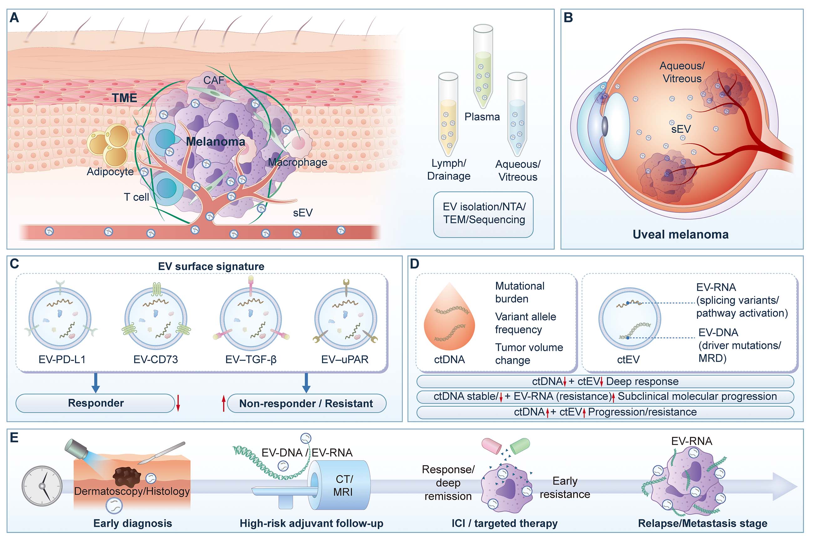
Figure 4. Structure and applications of melanoma EVs as bio-nanomaterial liquid-biopsy platforms. (A) Release of EVs from a heterogeneous melanoma TME into accessible biofluids, including plasma, lymphatic/drainage fluid, and ocular fluids, followed by downstream isolation and characterization workflows such as nanoparticle tracking analysis (NTA), transmission electron microscopy (TEM), and sequencing. (B) Schematic illustration of uveal melanoma and tumor-derived small extracellular vesicles (sEVs) in aqueous and vitreous humor, supporting ocular fluid-based EV sampling. (C) Representative EV surface “nano-immune fingerprint” composed of immunoregulatory markers, including EV-programmed death-ligand 1 (EV-PD-L1), EV-CD73, EV-transforming growth factor-β (EV-TGF-β), and EV-urokinase-type plasminogen activator receptor (EV-uPAR), for stratification of responders and non-responders/resistant cases. (D) Complementary information framework comparing circulating tumor DNA (ctDNA) with circulating tumor EVs (ctEVs), including EV-DNA and EV-RNA, for evaluation of mutational burden, molecular residual disease, functional transcripts, pathway activation, treatment response, and molecular progression/resistance. (E) Clinical continuum of EV-based applications across melanoma management, including early diagnosis, adjuvant surveillance, on-treatment monitoring during immune checkpoint inhibitor or targeted therapy, and relapse/metastasis detection. ctDNA: Circulating tumor DNA; ctEV: circulating tumor extracellular vesicle; EVs: extracellular vesicles; NTA: nanoparticle tracking analysis; TEM: transmission electron microscopy; TME: tumor microenvironment; uPAR: urokinase-type plasminogen activator receptor.





