


Insulin physiology and metabolic control: current concepts and perspectives
 0
0 
Abstract
Type 2 diabetes (T2D) results from progressive failure of multiple interconnected metabolic nodes. This review synthesizes evidence that β-cell dedifferentiation, mediated by loss of PDX-1, MAFA, and NKX6.1 and by epigenetic silencing of their promoters, may represent a leading mechanism of β-cell dysfunction, potentially preceding apoptotic death. Reduced hepatic insulin clearance through CEACAM1-dependent clearance pathways may contribute to a feedback loop amplifying systemic hyperinsulinemia and worsening peripheral insulin resistance. Adipose tissue dysfunction has been proposed to initiate cascading hepatic and muscular resistance in a subset of T2D phenotypes. Emerging mechanisms include gut microbiota-derived metabolites regulating incretin secretion; cellular senescence and mitochondrial dysfunction as drivers of metabolic aging; and altered bile acid metabolism affecting gluconeogenic regulation. Subcutaneous insulin, reversing the physiological portal-to-peripheral gradient, generates hepatic hypoinsulinemia and peripheral hyperinsulinemia despite adequate total doses. These advances will potentially identify stage-matched therapeutic windows: early resistance for lifestyle/sodium-glucose cotransporter 2 inhibitors (SGLT2i); compensatory phase for GLP-1Ra; advanced dedifferentiation for cell replacement. Precision medicine subtyping and multi-node targeting promise improved glycemic control and disease modification.
Keywords
INTRODUCTION
Insulin is a fundamental peptide hormone that regulates glucose and nutrient metabolism[1,2]. Its physiological effects depend on a coordinated lifecycle that extends from biosynthesis in pancreatic β-cells[3] to secretion, portal transport, peripheral action[4] and hepatic clearance[5]. Disruption at any of these stages can alter insulin availability, tissue-specific signaling, and glucose homeostasis. In type 2 diabetes (T2D), these processes become progressively dysregulated, yet their interactions are often considered separately. This review therefore integrates classical and emerging perspectives on insulin biosynthesis, secretion, portal transport, hepatic clearance, and peripheral signaling to examine how their coordinated disruption contributes to disease progression.
A central unresolved issue in T2D is how alterations across the insulin lifecycle interact to produce progressive metabolic dysfunction. Although pharmacological interventions can improve glycemic control, many patients experience continued disease progression[6,7], suggesting that models focused on individual components of insulin physiology may not fully capture the systemic nature of T2D. A more comprehensive framework is therefore needed to explain how disturbances in β-cell function, insulin quality, hepatic insulin handling, and peripheral insulin action propagate across the insulin system.
Several fundamental questions remain unresolved within this framework. First, it is still actively debated whether the progressive loss of functional β-cell mass in human T2D is driven predominantly by dedifferentiation or by apoptosis, with direct implications for whether β-cell failure may be reversible. Second, it remains unclear whether reduced hepatic insulin clearance[5] is a cause or a consequence of hepatic insulin resistance[8] and these competing explanations have rarely been reconciled. Third, the physiological consequences of disrupting the normal portal-to-peripheral insulin gradient - whether through disease progression or subcutaneous insulin replacement therapy - remain incompletely understood, particularly regarding their differential effects on hepatic and peripheral tissues. Finally, how emerging modulators of the insulin lifecycle interact with these classical processes - including gut microbiota-derived metabolites, cellular senescence, and altered bile acid metabolism - to influence disease trajectory is only beginning to be defined.
These questions reflect a common limitation in the current literature. The insulin lifecycle has traditionally been studied as a series of discrete compartments - biosynthesis[1,2], secretion[3], portal transit, hepatic clearance[5], and peripheral action[4] - each investigated within its own experimental tradition and largely in isolation from the others. Consequently, several partially overlapping, and at times apparently discordant, models of T2D pathogenesis have emerged without being examined jointly. Reconciling these perspectives requires an explicit account of how disruption at one stage of the insulin lifecycle propagates to, and is reinforced by, changes at the remaining stages. This need for an integrative account, rather than an additional node-specific description, that motivates the present review.
Accordingly, this review pursues five specific aims: (i) to critically evaluate the molecular determinants of β-cell identity maintenance and the evidence supporting dedifferentiation and apoptosis as mechanisms of β-cell dysfunction in T2D; (ii) to examine the mechanisms and clinical significance of hepatic insulin clearance[5], with particular attention to CEACAM1- and insulin-degrading enzyme (IDE)-dependent pathways and their proposed bidirectional relationship with peripheral insulin resistance[8]; (iii) to characterize how disruption of the physiological portal-to-peripheral insulin gradient, whether disease-driven or iatrogenic, differentially affects hepatic and peripheral insulin action[4]; (iv) to integrate emerging modulators of the insulin lifecycle, including gut microbiota, cellular senescence, and geroscience-related pathways, into this systemic framework; and (v) to translate these mechanistic perspectives into a staged model of disease progression capable of informing the sequencing and combination of precision-based therapeutic strategies.
METHODS
Review design and search strategy
This study was conducted as a narrative review with a structured literature search and integrative evidence synthesis. This design was selected because the review addresses multiple interconnected physiological and pathophysiological domains - from insulin biosynthesis and secretion to its clearance and peripheral resistance - rather than a single, predefined intervention, exposure, or clinical outcome. Methodological rigor was strengthened by considering the quality domains proposed by the Scale for the Assessment of Narrative Review Articles (SANRA), particularly explicit description of the search, appropriate referencing, and critical appraisal of the level and relevance of the evidence.
Electronic searches were conducted in PubMed/MEDLINE, Scopus, and Web of Science as primary bibliographic databases, supplemented by ScienceDirect, SAGE Journals, and Taylor & Francis Online to broaden retrieval of full-text articles. The principal search period was 1 January 2016 to 31 July 2026, with no language restriction. Seminal studies published before 2016 were retained when necessary to support established physiological or mechanistic concepts. Reference lists of key reviews and eligible primary studies were manually screened to identify additional relevant publications.
Search terms combined controlled vocabulary (including MeSH terms where available) with free-text keywords, covering insulin biosynthesis and processing, β-cell identity and dedifferentiation, glucose-stimulated secretion and KATP-channel signaling, incretins, hepatic, renal, and peripheral insulin clearance, CEACAM1 and IDE, insulin signaling and resistance, adipokines and ectopic lipid deposition, gut microbiota and bile acid-incretin signaling, hyperinsulinemia, cellular senescence, mitochondrial dysfunction, metabolic aging, and precision-medicine therapeutic strategies. Because the review spans mechanistically distinct domains, targeted supplementary searches were also performed for section-specific questions, avoiding reliance on a single master query that could reduce retrieval sensitivity across heterogeneous areas of insulin physiology [Supplementary Table 1].
Eligibility, selection, and evidence synthesis
Peer-reviewed publications were considered eligible if they provided direct evidence on normal insulin physiology, dysregulation at any point of its life cycle, or clinically relevant consequences for T2D and related metabolic disorders, including studies in humans, animal models, isolated tissues, primary cells, or organoids, as well as observational, interventional, and translational studies. Systematic reviews, meta-analyses, consensus statements, and authoritative reviews were used for higher-order synthesis, contextualization, and identification of landmark primary studies.
Studies unrelated to insulin physiology or metabolic control were excluded, along with those providing insufficient methodological detail, conference abstracts, non-peer-reviewed grey literature, editorials without original data, and duplicate reports. Titles and abstracts were screened, duplicate records were removed before screening, and potentially eligible records were assessed at full text. When several publications addressed the same mechanism, preference was given to the most recent and methodologically informative evidence, while retaining older landmark studies when necessary for historical or conceptual context.
For each selected source, the study model or population, the pathway or tissue examined, the principal mechanistic finding, translational or clinical relevance, and pertinent limitations were extracted. Evidence was organized according to the sequential and interconnected stages of the insulin life cycle addressed in the manuscript, and was synthesized narratively - not quantitatively - given the heterogeneity of study designs, experimental models, and biological endpoints. Interpretation of each finding considered study design, reproducibility across independent studies, concordance between experimental and human data, and clinical relevance; findings derived predominantly from animal or in vitro models were presented as mechanistic or hypothesis-generating evidence, distinguished from observations directly supported by human studies.
This review was not conducted as an intervention-focused systematic review or meta-analysis, and no quantitative pooling of effect estimates was planned. The structured search strategy, explicit eligibility criteria, domain-based organization of evidence, and critical distinction between preclinical and human evidence support a rigorous narrative synthesis appropriate to the broad physiological and translational scope of the article.
INSULIN BIOSYNTHESIS AND PROCESSING: THE MOLECULAR BLUEPRINT
Preproinsulin synthesis and endoplasmic reticulum processing
Pancreatic β-cells are highly specialized cells that produce and secrete insulin in response to hyperglycemia. The insulin gene (INS), located on human chromosome 11p15.5, encodes preproinsulin, a 110-amino-acid precursor whose transcription depends on the synergistic binding of key transcription factors to conserved promoter elements (RI/RIPE and C1/A1). The master regulator of β-cell identity is the pancreatic and duodenal homeobox 1 (PDX-1), acting alongside V-maf musculoaponeurotic fibrosarcoma oncogene homolog A (MAFA), neurogenic differentiation 1 (NEUROD1), and NK6 homeobox 1 (NKX6.1), factors that respond dynamically to glucose and nutrient stimuli through chromatin remodeling, including forkhead box A2 (FOXA2)-mediated H3K4me1 marks[9-11]. PDX-1 physically engages NEUROD1, while MAFA transcription at mature stages depends on upstream NKX6.1 activation. Together, the PDX-1/NEUROD1/MAFA triad can reprogram non-β-cells, including fibroblasts, induced pluripotent stem cells (iPSCs), and stem cells, into glucose-responsive insulin producers, establishing a foundation for β-cell replacement therapies[12-16].
In T2D, oxidative stress generated by hyperglycemia and lipotoxicity induces post-translational modifications (phosphorylation and ubiquitination) in PDX-1 and NKX6.1, reducing their binding to the MAFA promoter and triggering the activation of progenitor state genes [Neurogenin-3 (NEUROG3) and SRY of box 9 (SOX9)][14]. The result is β-cell dedifferentiation with loss of glucose responsiveness but without significant apoptosis[16,17]. This process has been confirmed in two independent models. Nkx6.1−/− mice fail to mature properly and recapitulate the human T2D β-cell profile, characterized by reduced MAFA expression, elevated immature markers, and impaired insulin secretion. In T2D patient-derived iPSCs, stress exposure triggers epigenetic silencing of MAFA and PDX-1 through promoter methylation and H3K27me3 deposition, reactivating a juvenile islet epigenome[13,18-22]. Single-cell CRISPR screens further confirm the therapeutic potential of these factors in terms of regeneration[13,23-26].
Insulin biosynthesis begins with translation of INS mRNA into preproinsulin, which is cotranslationally inserted into the endoplasmic reticulum (ER) lumen, where signal peptide cleavage generates proinsulin (86 amino acids). Three disulfide bonds (A6-A11, A7-B7, A20-B19) are then formed between cysteine residues, catalyzed by protein disulfide isomerase (PDI) and ER oxidoreductase (Ero1β), establishing the native insulin conformation. Proinsulin then traffics through the Golgi apparatus into immature secretory granules, where proprotein convertases (PC)1/3 and PC2 cleave two dibasic sites, followed by carboxypeptidase E (CPE)-mediated dipeptide trimming to yield mature insulin and equimolar C-peptide[27]. Mature insulin is stored as zinc-stabilized hexameric insulin crystals in the acidic (pH ~5.5) granules. Secretion of equimolar C-peptide to insulin makes it a clinically invaluable surrogate marker of endogenous insulin secretion[28-31].
Whether C-peptide exerts biological activity beyond its role as a biosynthetic by-product remains an unresolved controversy. Wahren et al. have shown that C-peptide binds to cell-surface receptors and activates intracellular signaling cascades, including Gi-protein-coupled pathways, protein kinase C epsilon (PKCε), and mitogen-activated protein kinase (MAPK), producing effects on renal tubular function, peripheral nerve conduction velocity, and microvascular flow in preclinical models and phase II clinical trials in patients with type 1 diabetes (T1D)[32].
Although preclinical data and small clinical trials suggest that C-peptide may be bioactive, a viewpoint review concluded that despite 30 years of research, its specific receptor and mechanism of action have not been identified. Whereas C-peptide shows therapeutic potential for vascular and nervous damage in diabetes, there is insufficient evidence to support its therapeutic use[33].
Proinsulin misfolding has emerged as a primary trigger of ER stress in T2D progression[28,34]. In Akita mutants fed a high-fat diet, hyperactivation of the inositol-requiring enzyme 1 alpha/spliced X-Box binding protein 1 (IRE1α/XBP1s) pathway depletes PDI/Ero1β and drives C/EBP homologous protein (CHOP)-mediated apoptosis independently of glucotoxicity[34,35]. Single-cell RNA sequencing identified stressed β-cell clusters with elevated binding immunoglobulin protein/glucose-regulated protein 78 kDa (BiP/GRP78) and reduced PDI levels correlating with impaired insulin secretion[36]. The β-cell dedicates approximately 50%-60% of its total protein synthesis capacity to insulin, demanding substantial constitutive ER folding. Accordingly, the unfolded protein response (UPR), orchestrated by IRE1α, PKR-like endoplasmic reticulum kinase (PERK), and activating transcription factor 6 (ATF6), functions not merely as a stress pathway but as a constitutively active adaptive mechanism[30].
An unresolved debate concerns the inflection point at which the UPR in pancreatic β-cells shifts from a homeostatic program to a maladaptive state that drives β-cell dysfunction[37]. Under glucose stimulation, IRE1α-mediated XBP1 splicing enhances ER folding capacity and promotes insulin secretion, while phosphorylation of PERK-mediated eukaryotic translation initiation factor 2 alpha (eIF2α) transiently reduces protein synthesis to protect the folding environment. These responses are essential for β-cell function[38].
Under sustained demand, as in obesity-associated insulin resistance, chronic IRE1α hyperactivation has been observed to degrade insulin mRNA via regulated IRE1-dependent decay (RIDD), promote c-Jun N-terminal Kinase (JNK)-mediated insulin receptor substrates 1 (IRS1) serine phosphorylation, and initiate CHOP-mediated pro-apoptotic signaling[39].
In human β-cells, it remains unclear whether this transition reflects a quantitative threshold of IRE1α activation, a qualitative switch in the oligomerization state of IRE1α, or cell-subtype-specific differences in UPR buffering capacity[31]. Thus, selectively modulating XBP1s without amplifying RIDD or CHOP could constitute an attractive strategy. However, it has not yet been achieved pharmacologically in human islets[40].
A study by Yau et al. further showed that mitoribosome insufficiency in β-cells impairs oxidative phosphorylation and amplifies UPR-mediated failure reminiscent of T2D islet pathology. Together, this illustrates the co-regulation of mitochondrial and ER quality control[41].
Several molecular regulators fine-tune ER processing. ER membrane protein complex subunit 10 (EMC10) modulates PDI activity to prevent proinsulin aggregation[42]. Peroxisome proliferator-activated receptor beta/delta (PPARβ/δ) agonists (GW501516) counteract stress-induced PDI inhibition[43]. Additionally, chemical chaperones [such as 4-phenylbutyric acid (PBA) and α-lipoic acid (α-LA)] mitigate ER overload by enhancing PDI/Ero1β flux and silencing PERK/activating transcription factor 4 (ATF4)[31]. CPE is non-redundant in granule maturation. For instance, β-cell-specific CPE deletion (βCpeKO) causes near-total loss of mature granules and elevates proinsulin. It also causes mitochondrial dysfunction and MAFA downregulation in response to high-glucose challenge. Moreover, human CPE mutations are associated with obesity and diabetes. For instance, insulin regulates CPE translation via PDX-1/sterol regulatory element-binding protein 1 (SREBP-1)/eukaryotic translation initiation factor 4 gamma 1 (eIF4G1), identifying maturation defects as early, translationally rescuable stressors[44].
Of note, murine β-cells express two INS (Ins1 and Ins2), whereas humans possess a single INS gene with greater dependence on NKX6.1, PDX-1, and MAFA for glucose-responsive expression. This difference is critically relevant when extrapolating findings from rodent knockout models to human physiology.
Transcriptional regulation and epigenetic control
The transcription factors PDX-1, NKX6.1, and MAFA form the core regulatory triad governing β-cell identity and INS gene expression. PDX-1 acts as the master regulator of pancreatic development and mature β-cell identity, while MAFA, a late-stage differentiation factor sensitive to oxidative stress, glucotoxicity, and lipotoxicity, represents a key early target of β-cell failure[16,17]. The clinical relevance of this triad is underscored by Genome-Wide Association Studies (GWAS) data identifying numerous T2D risk loci in proximity to these transcription factors and their targets[41].
Single-cell RNA sequencing has revealed remarkable heterogeneity within the β-cell population. A landmark study by Dror et al. demonstrated that epigenetic dosage defines at least two major β-cell subtypes with distinct functional and stress-response characteristics[45]. Additionally, Rutter et al. described subpopulations with differential connectivity within islet electrical networks[46]. The functionally dominant hub β-cells may bear disproportionate vulnerability to metabolic stress, though whether analogous heterogeneity characterizes human islets to the same degree remains an active area of investigation.
Epigenetic regulation through DNA methylation and histone modification adds a further layer of control. Using isolated human islets exposed to high glucose ex vivo, Hall et al. demonstrated that acute exposure to 19 mM glucose for 48 h alters DNA methylation at CpG sites proximal to PDX1, linking hyperglycemia to early epigenetic reprogramming of the β-cell. Complementing this, comparative studies between islets from donors with type 2 diabetes and non-diabetic donors - Dayeh et al., and more recently Rönn et al. - have identified stable methylation alterations in T2D risk genes, including CDKN1A and SLC2A2, reinforcing the idea that chronic metabolic context remodels the human islet methylome[47-49]. This work warrants careful interpretation: the conditions represent acute, supraphysiological glucose exposure ex vivo, and whether these methylation changes are causally upstream of β-cell dysfunction or a secondary epigenetic response to an already-dysfunctional metabolic state remains unclear. Longitudinal studies tracking methylation dynamics before and after clinical T2D onset are needed to establish causality. Complementary support comes from genome-wide methylation profiling of T2D islets, which identifies hypermethylation of PDX-1 and MAFA promoters as a consistent T2D epigenomic feature[14,50].
An integrative framework: the biosynthesis-quality-identity axis
The mechanisms previously described, including INS transcription, ER folding, granule maturation, and epigenetic maintenance of β-cell identity, are conventionally presented as parallel or sequential processes. We propose that they are better understood as a single homeostatic circuit: the BQI axis [Figure 1].
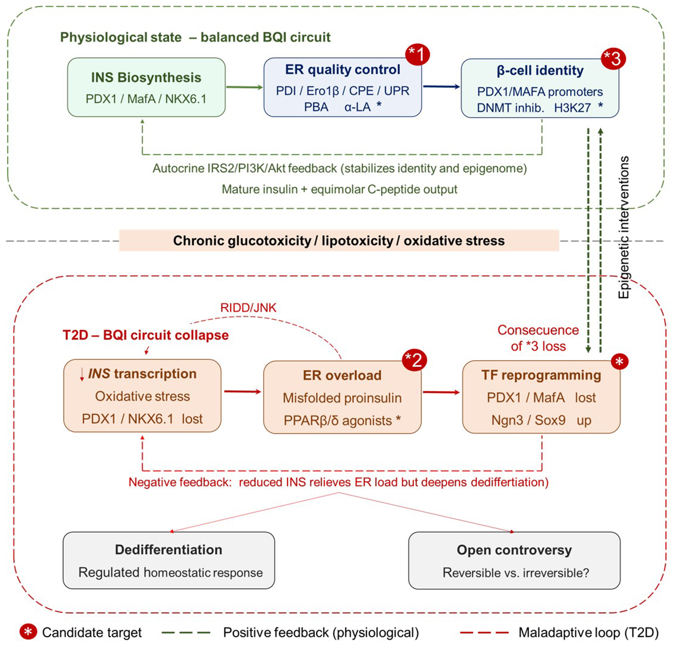
Figure 1. The BQI axis in pancreatic β-cells under physiological and T2D conditions. (Upper panel) Under physiological glucose stimulation, three interconnected nodes sustain β-cell homeostasis. (1) The PDX-1/MAFA/NKX6.1 TF triad drives INS gene expression; (2) ER quality-control machinery - PDI, Ero1β, PC1/3 and PC2, and CPE - ensures correct proinsulin processing, while a constitutively active but self-limiting UPR involving IRE1α/XBP1s, PERK/eIF2α, and ATF6 maintains folding homeostasis; (3) The β-cell identity node encompasses both the mature secretory phenotype and its epigenetic foundation - the PDX-1 and MAFA gene promoters maintained in an active, demethylated state decorated with H3K4me1 and H3K27ac marks. Autocrine insulin signaling through IRS2/PI3K/AKT (green dashed line) reinforces transcription factor binding and epigenetic maintenance, closing a positive homeostatic feedback loop that sustains all three nodes. (Lower panel) Chronic glucotoxicity and lipotoxicity disrupt the BQI circuit at three nodes. (1) Oxidative stress induces post-translational modifications of PDX-1 and NKX6.1, reducing INS transcription; (2) Excess proinsulin synthesis saturates PDI/Ero1β, generating misfolded aggregates and chronically activating IRE1α; IRE1β hyperactivation degrades INS mRNA via RIDD and activates JNK, further impairing IRS2 signaling (red dashed arrow, RIDD/JNK); (3) Loss of autocrine insulin input, combined with oxidative promoter modifications, drives progressive hypermethylation of PDX-1 and MAFA loci, silencing TF expression and reactivating progenitor-state genes (NEUROG3 and SOX9) - the TF reprogramming node (red box). This node carries no asterisk because it is the downstream pathological consequence of epigenome loss at node *3, not an independent therapeutic target. A negative feedback loop (red dashed arrow) illustrates that reduced INS output alleviates ER biosynthetic load but consolidates the dedifferentiated state. Candidate therapeutic targets are indicated by a red asterisk (*) badge in the upper-right corner of the relevant node. (*1) ER quality-control node, upper panel: chemical chaperones (PBA and α-LA) restore PDI/Ero1β folding flux and prevent proinsulin aggregation; PPARβ/δ agonists (GW501516) counteract stress-induced PDI inhibition and stabilize insulin receptor β-subunit conformation; (*2) ER overload node, lower panel: PPARβ/δ agonists act directly in the glucolipotoxic environment to restore PDI activity and reduce misfolded proinsulin burden; (*3) β-cell identity node, upper panel: epigenetic interventions - DNA methyltransferase (DNMT) inhibitors and H3K27me3 demethylases - reactivate PDX-1 and MAFA promoters hypermethylated during T2D progression. The green bidirectional dashed arrow in the right margin of the figure connects node *3 (upper panel) to the TF reprogramming node (lower panel), indicating that pharmacological restoration of the β-cell epigenome at *3 is the upstream intervention that reverse the downstream transcriptional collapse. Green dashed arrows, positive feedback (physiological); red dashed arrows, maladaptive loops (T2D). BQI: Biosynthesis-quality-identity; T2D: type 2 diabetes; INS: insulin; PDX1: pancreatic and duodenal homeobox 1; MAFA: MAF bZIP transcription factor A; NKX6.1: NK6 homeobox 1; TF: transcription factor; ER: endoplasmic reticulum; PDI: protein disulfide isomerase; Ero1β: endoplasmic reticulum oxidoreductase 1 beta; PC1/3: proprotein convertase 1/3; PC2: proprotein convertase 2; CPE: carboxypeptidase E; UPR: unfolded protein response; IRE1α: inositol-requiring enzyme 1 alpha; XBP1s: spliced X-box binding protein 1; PERK: protein kinase R-like endoplasmic reticulum kinase; eIF2α: eukaryotic translation initiation factor 2 alpha; ATF6: activating transcription factor 6; H3K4me1: histone H3 lysine 4 monomethylation; H3K27ac: histone H3 lysine 27 acetylation; IRS2: insulin receptor substrate 2; PI3K: phosphoinositide 3-kinase; AKT: protein kinase B; RIDD: regulated IRE1-dependent decay; JNK: c-Jun N-terminal kinase; NEUROG3: neurogenin 3; SOX9: SRY-box transcription factor 9; PBA: 4-phenylbutyric acid; α-LA: alpha-lipoic acid; PPARβ/δ: peroxisome proliferator-activated receptor beta/delta; DNMT: DNA methyltransferase; H3K27me3: histone H3 lysine 27 trimethylation.
Under physiological conditions, the PDX-1/MAFA/NKX6.1 triad drives INS transcription at a level matched to the β-cell’s ER folding capacity, maintained by PDI, Ero1β, and a tonically active but self-limiting UPR. Correct proinsulin processing by PC1/3, PC2, and CPE completes the production of mature insulin granules. Equimolar C-peptide release serves as a proxy for this entire upstream process. Mature secretory output reinforces transcription factor binding to the INS promoter through autocrine insulin signaling via IRS2/phosphoinositide 3-kinase (PI3K)/serine/threonine protein kinase AKT [also referred to as protein kinase B (PKB)], closing a positive feedback loop that stabilizes β-cell identity. The proinsulin/insulin ratio has been shown to be elevated in preclinical models of β-cell functional immaturity[51,52].
Chronic glucotoxicity and lipotoxicity disrupt this circuit at multiple nodes simultaneously: (i) oxidative stress modifies PDX-1 and NKX6.1 post-translationally, reducing INS transcription; (ii) excess proinsulin synthesis saturates PDI/Ero1β capacity, generating misfolded aggregates that constitutively activate IRE1α; (iii) chronic IRE1α activation degrades INS mRNA via RIDD and activates JNK, further impairing IRS2 signaling; (iv) loss of autocrine insulin input accelerates PDX-1 nuclear exclusion and MAFA promoter methylation, deepening the dedifferentiated state. The net result is a self-reinforcing collapse of the BQI axis in which dedifferentiation, far from representing a catastrophic failure, may constitute a regulated, homeostatic response that reduces ER biosynthetic load at the cost of secretory function.
This framework has three therapeutic implications not immediately apparent from a sequential model: (a) interventions targeting only glucose control may be insufficient if the epigenetic silencing of PDX-1/MAFA has already been established; (b) chemical chaperones (PBA, α-LA) and PDI activators (PPARβ/δ agonists) that restore ER quality control address a node upstream of transcription factor loss; and (c) complete restoration of β-cell function in advanced T2D may require simultaneous targeting of the epigenetic (demethylation of PDX-1/MAFA promoters), proteostatic (UPR rebalancing), and metabolic (reduction of glucolipotoxic load) arms of the BQI circuit - a multi-target strategy currently untested in human clinical trials. For clinical translation notes of this section, see Supplementary Materials.
INSULIN SECRETION: CLASSICAL MECHANISMS AND NOVEL INSIGHTS
The KATP channel paradigm: classical model of glucose-stimulated insulin secretion
Glucose-stimulated insulin secretion (GSIS) follows a well-defined metabolic and electrophysiological cascade. The process begins with the entry of glucose into the β-cell cytoplasm through specific transporters. In rodents, this function relies primarily on glucose transporter (GLUT) 2, a high-Km transporter. In human β-cells, a combination of GLUT2 and the low-Km transporter GLUT1 are responsible for glucose uptake. This difference reflects a more restrictive glycemic homeostatic control in humans compared to rodents. Notably, loss of GLUT2 from the β-cell plasma membrane has been reported as a marker of functional immaturity associated with constitutive insulin secretion[53-55] [Figure 2].
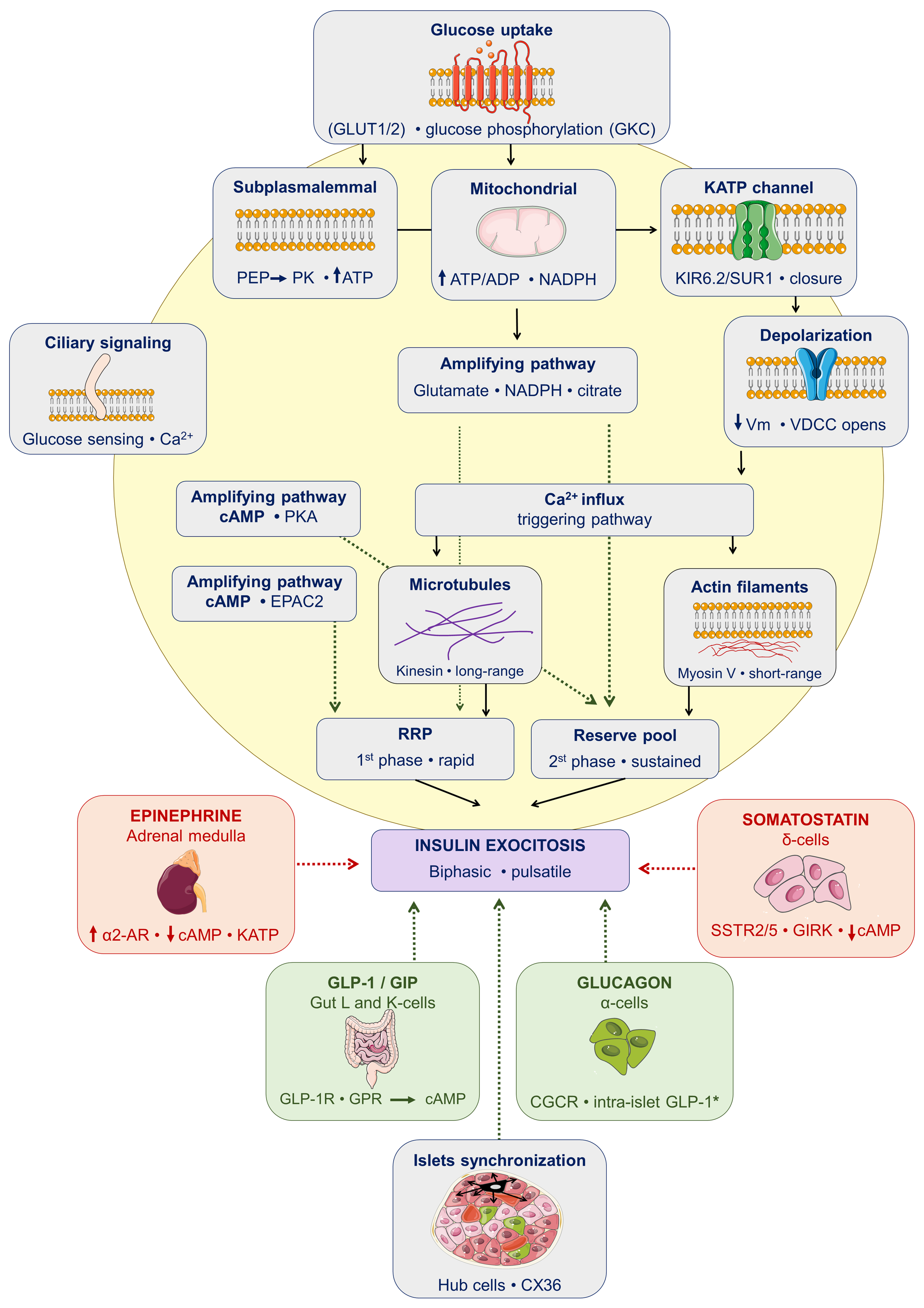
Figure 2. Paracrine, endocrine, and intracellular regulation of insulin secretion in pancreatic β-cells. The diagram distinguishes three tiers of regulatory input converging on the β-cell (yellow). Within the islet, α-cells provide paracrine stimulation via glucagon acting on GCGR to activate the cAMP-PKA-EPAC2 axis; under conditions of metabolic stress, α-cells also generate GLP-1* through alternative proglucagon processing by PC1/3, which acts on β-cell GLP-1 receptors. δ-cells exert tonic paracrine inhibition through somatostatin, which suppresses cAMP production and hyperpolarizes the β-cell membrane via SSTR2/5-coupled GIRK channels. Systemic endocrine signals include gut-derived incretins GLP-1 and GIP, which amplify GSIS through Gs-coupled receptor signaling; epinephrine, released from the adrenal medulla, inhibits secretion via α2-adrenergic receptors by suppressing cAMP and promoting KATP channel opening. Gap junction coupling through CX36 among hub and follower β-cells coordinates calcium oscillations across the islet syncytium, while primary cilia contribute to glucose sensing and intracellular Ca2+ coordination. Within the β-cell, glucose enters via GLUT1/2 and is phosphorylated by GCK, initiating glycolytic and mitochondrial flux that raises the cytoplasmic ATP/ADP ratio. Subplasmalemmal ATP is additionally supplied by PEP-dependent PK activity. Together these signals drive KATP channel closure (KIR6.2/SUR1), membrane depolarization, VDCC activation, and Ca2+ influx, the triggering pathway. An amplifying pathway, acting independently of KATP, enhances exocytosis through MCF (NADPH, glutamate, citrate) and cAMP-PKA-EPAC2 signaling. Insulin granule mobilization requires sequential cytoskeletal remodeling: kinesin-driven long-range transport along microtubules followed by myosin V-mediated short-range delivery along actin filaments. Granules from the RRP are released rapidly in the first secretory phase; RP granules sustain the prolonged second phase. The resulting biphasic, pulsatile insulin output is delivered to the portal circulation. Dashed green arrows, stimulatory; dashed red arrows, inhibitory. GLUT1/2: Glucose transporter 1/2; GCK: glucokinase; KATP: ATP-sensitive potassium channel; PEP: phosphoenolpyruvate; PK: pyruvate kinase; tATP: total ATP; ATP: adenosine triphosphate; ADP: adenosine diphosphate; NADPH: reduced nicotinamide adenine dinucleotide phosphate; KIR6.2: potassium inwardly rectifying channel subfamily J member 11; SUR1: sulfonylurea receptor 1; VDCC: voltage-dependent calcium channel; cAMP: cyclic adenosine monophosphate; PKA: protein kinase A; EPAC2: exchange protein directly activated by cAMP 2; RRP: readily releasable pool; RP: reserve pool; AR: adrenergic receptor; SSTR2/5: somatostatin receptor 2/5; GIRK: G protein-gated inwardly rectifying potassium channel; GLP-1: glucagon-like peptide-1; GIP: glucose-dependent insulinotropic polypeptide; GLP-1R: glucagon-like peptide-1 receptor; GPR: G protein-coupled receptor; GCGR: glucagon receptor; PC1/3: proprotein convertase 1/3; CX36: connexin 36; GSIS: glucose-stimulated insulin secretion; MCF: mitochondria-derived metabolic coupling factors.
Once inside the cell, glucose is phosphorylated by glucokinase (GCK, hexokinase IV), regarded as the true “glucose sensor” of the β-cell and the rate-limiting step of its metabolic flux. This phosphorylation represents the activating signal that triggers glycolysis and mitochondrial oxidative phosphorylation, thereby initiating the metabolic cascade leading to insulin granule exocytosis[56]. The net result of this metabolism is an increase in the cytoplasmic ATP/ADP ratio, which acts as the key effector signal on the ATP-sensitive potassium channel (KATP channel).
The KATP channel is composed of four inward rectifier potassium channel 6.2 (KIR6.2) pore-forming subunits and four sulfonylurea receptor 1 (SUR1) regulatory subunits. The rise in the ATP/ADP ratio triggers closure of this channel, producing plasma membrane depolarization, activation of voltage-dependent calcium channels (VDCCs), and a consequent massive influx of Ca2+ into the cell[30,56,57]. This calcium influx drives exocytosis of insulin-containing secretory granules through a biphasic process, determined by the existence of two distinct granule pools: the readily releasable pool (RRP) and the reserve pool (RP). RRP granules are rapidly released within the first 5-10 min following glucose elevation, giving rise to the first phase of secretion, followed by a more prolonged second phase that can extend for hours.
This so-called triggering pathway is complemented by an amplifying pathway that enhances insulin secretion beyond what calcium influx alone can achieve, through metabolic coupling factors including glutamate, reduced nicotinamide adenine dinucleotide phosphate (NADPH), long-chain acyl-CoAs, and malonyl-CoA [Figure 2]. A computational model by Deepa Maheshvare et al. systematically quantified the contributions of key enzymatic steps - particularly phosphofructokinase (PFK) activity and the ATP/ADP-dependent triggering step - to GSIS, confirming that the flux balance between glycolysis and mitochondrial oxidation is the primary determinant of secretory amplitude[56]. That model also highlighted that KATP-channel-independent mechanisms contribute substantially to secretory regulation.
Canonical insulin secretion mechanism under question
The discovery of the structure and function of the β-cell KATP channel unquestionably revolutionized our understanding of β-cell physiology. From a therapeutic standpoint, patients harboring mutations that lead to KATP inactivation and consequent loss of insulin secretory capacity have greatly benefited from sulfonylureas, which pharmacologically close the channel and restore insulin release. However, the central tenet of the canonical model, namely, that KATP closure depends on an increased mitochondrial ATP/ADP ratio, has come under scrutiny. Merrins et al. have recently proposed that this classical framework is incomplete and potentially incorrect. Their work provides genetic evidence that phosphoenolpyruvate (PEP) exerts metabolic control over pyruvate kinase (PK)-dependent ATP/ADP generation at the plasma membrane, which in turn governs KATP closure, calcium signaling, and insulin secretion[58] [Figure 2]. The authors identify three fundamental shortcomings of the dogmatic model: first, it fails to account for mitochondrial thermodynamics, which dictate how mitochondrial ATP production is accelerated in response to increased workload driven by rising ADP levels; second, it disregards metabolic compartmentation; and third, it neglects temporal compartmentation, a feature intrinsically linked to oscillatory metabolism[58].
In response to these limitations, Merrins et al. proposed a revised model built around four main integrated metabolic cycles: (1) spatial and temporal dimensions of metabolic signaling; (2) the functional relationships among cellular compartments, including the mitochondria, cytosol, and the subplasmalemmal space; (3) both “on-switch” signals that trigger secretion and “off-switch” signals that sustain metabolic and secretory oscillations; and (4) the redundancy among certain pathways, particularly redox components, which may help reconcile divergent findings reported in the literature[58]. Complementing this framework, Foster et al. provided direct genetic evidence through β-cell-specific deletion of the Pkm1 and Pkm2 isoforms of PK in mice, demonstrating that these isoforms are essential nutrient sensors linking PEP-driven ATP/ADP generation at the plasma membrane to KATP channel regulation and downstream calcium signaling[59] [Figure 2].
Efficient insulin secretion depends on intercellular islet communication. Hub β-cells (1%-10% of the population), which are characterized by high GCK and reduced PDX-1/NKX6.1 expression, act as pacemakers coordinating calcium dynamics and secretory activity of follower cells[60]. Loss of this connectivity, observed in T2D mouse models and following metabolic/inflammatory insults, contributes to the secretory dysfunction observed in diabetes [Figure 2]. Whether these events happen in the human pancreas requires further research.
Amplifying pathways and mitochondrial coupling factors
Recent studies have significantly deepened our understanding of the amplifying signals that drive GSIS. Mitochondria play a central role by generating metabolic coupling factors like glutamate, aspartate, citrate, and the NADPH redox state. These signals link mitochondrial activity directly to the exocytosis of secretory granules, working independently of the KATP channel pathway. Notably, pyruvate cycling pathways produce NADPH, which can directly modulate exocytosis by activating glutaredoxin and thioredoxin systems[57] [Figure 2].
One of the most fascinating aspects of β-cell biology is how these cells selectively prevent the expression of genes that would create metabolic “futile cycles” incompatible with proper glucose sensing. For instance, lactate dehydrogenase A (LDHA) and monocarboxylate transporter 1 (MCT1), which would allow lactate to act as a secretagogue and blur glucose specificity, are actively silenced in mature β-cells[57]. When this “disallowance” breaks down, it becomes an early marker of β-cell dedifferentiation and happens before any measurable secretory defects can be detected. This has been confirmed in human islets from donors with T2D, assigning it clinical relevance[30,57].
Incretin hormones add another layer to this regulatory network. Glucagon-like peptide-1 (GLP-1) and glucose-dependent insulinotropic polypeptide (GIP) are released from enteroendocrine L-cells and K-cells in the small intestine in response to nutrient intake. They bind to glucagon-like peptide-1 receptor (GLP-1R) and glucose-dependent insulinotropic polypeptide receptor (GIPR) receptors on β-cells, amplifying GSIS through the cyclic adenosine monophosphate (cAMP)-protein kinase A (PKA)-exchange protein directly activated by cAMP 2 (EPAC2) signaling pathway[30]. The physiological amplification provided by incretins accounts for a substantial 50%-70% of postprandial insulin secretion - the “incretin effect” [Figure 2]. One of the early features of T2D is the loss of this incretin effect, primarily due to impaired β-cell responsiveness to GLP-1 rather than reduced GLP-1 secretion. This mechanistic insight explains why GLP-1 receptor agonists (GLP-1RAs) have been clinically successful in treating T2D[61,62].
However, the native half-lives of GLP-1 and GIP are extremely short because dipeptidyl peptidase-4 (DPP-4) rapidly inactivates both hormones. For this reason, DPP-4 inhibitors are used therapeutically in T2D, as they prolong circulating GLP-1 and GIP levels, thereby lowering postprandial glycemia and improving overall glycemic control. Their effectiveness is also influenced by gastric emptying. In parallel, gut microbiota-derived metabolites such as short-chain fatty acids (SCFAs), secondary bile acids, and indole can directly stimulate incretin release from L-cells. This has led to growing interest in microbiota-targeted approaches, including probiotics, prebiotics, and dietary interventions, as potential adjuncts to improve glycemic control and support gut barrier function[63].
The limited translation from preclinical to clinical research reveals a significant gap in our understanding of insulin secretion dynamics. In rodent models, pulsatile secretion patterns and strong β-cell synchronization are well-documented. Clinical studies confirm pulsatility of secretion in patients, but the effects of β-cell heterogeneity on secretion patterns remain much less characterized[64-66]. The critical gap here is whether the impressive single-cell findings in the laboratory actually translate to the complex whole-organ secretion dynamics occurring in human patients. This uncertainty about how cellular heterogeneity impacts overall insulin output represents a major challenge in bridging preclinical mechanistic insights with clinical validation.
Hormonal regulation of insulin secretion
To avert hypoglycemia, β-cell secretion is subject to tonic inhibition from multiple sources. Pancreatic δ-cells produce somatostatin that activates somatostatin receptors (SSTR2/5) to suppress cAMP and hyperpolarize the cell membrane via G protein-gated inwardly rectifying potassium (GIRK) channels. Epinephrine produced by adrenal glands acts via α2-adrenergic receptors to inhibit cAMP while opening KATP channels and eventually antagonizing insulin secretion[67-69] [Figure 2].
K ATP-independent amplification pathways sustain insulin release. Glucagon from α-cells acts via GCGR-cAMP-PKA-EPAC2 signaling in granule priming and exocytosis, and enhances INS transcription via GCGR-PKA-cAMP response element-binding protein (CREB)[70]. Glucagon and GLP-1 both derive from preproglucagon, processed by PC2 in α-cells (glucagon) or PC1/3 in L-cells (GLP-1)[3]. GLP-1 and GIP amplify GSIS via Gs-coupled receptors. Under metabolic stress, α-cells also generate GLP-1 that augments β-cell GLP-1Ra signaling[71] [Figure 2].
Tirzepatide, a dual agonist of the GIPR and GLP-1R, is a highly effective therapy for T2D and obesity. In mouse models, tirzepatide-stimulated insulin secretion is mediated predominantly through GLP-1R, consistent with its lower potency at the GIPR. In contrast, studies in human islets show that GIPR antagonism consistently attenuates the insulinotropic response to tirzepatide, indicating a physiologically relevant role for GIPR in human tissue. Furthermore, tirzepatide increases both glucagon and somatostatin secretion in human islets, supporting the notion that it modulates islet hormone release through coordinated activation of both incretin receptors[72].
Other mechanisms underlying insulin secretion
Beyond the classical mechanisms, new molecular targets have been identified for the proper function of pancreatic β-cells. One layer of regulation relies on molecular motors, specialized proteins responsible for the intracellular transport of insulin granules. Following their formation on the trans-Golgi network, these granules need to be transported to the cell membrane for regulated exocytosis. The interlinked and dynamic cellular networks of tubulin and actin cytoskeletons are critical for insulin granule transport and secretion[73] [Figure 2].
Zhu et al. showed that microtubules are remodeled by glucose in β-cells from mice and humans. Furthermore, microtubules restrict GSIS in mice, acting as a “rheostat” for the β-cell. In fact, dysfunctional β-cells in diabetic mice display abnormally high microtubule density[74]. In the first phase of insulin secretion, actin filaments depolymerize into actin monomers, allowing the rapid release of insulin granules in RRP. In the second phase, actin filaments repolymerize into a new actin network that facilitates the transport of insulin granules to and release of insulin from the plasma membrane[75]. Thus, GSIS requires the coordination of both cytoskeleton networks: the transportation of insulin granules first involves long-range movement driven by kinesins on microtubules, with the subsequent handoff to myosin V for the short-range movement on actin filaments to the plasma membrane[75].
The primary cilium in β-cells regulates insulin secretion. These immotile organelles - confined between acinar cells and islet vasculature - function as sensory hubs coordinating glucose sensing, calcium influx for GSIS, and intercellular communication[76]. They also form axo-ciliary synapses with nerve fibers for neuroendocrine regulation[77]. Loss of primary cilia impairs β-cell differentiation, reduces insulin content, and disrupts glucose homeostasis, directly linking ciliary integrity to diabetes etiology[78,79] [Figure 2].
Pulsatile insulin secretion and paracrine islet regulation
Insulin is secreted in coordinated ultradian pulses (every 3-5 min) arising from synchronized β-cell calcium and metabolic oscillations across the islet. These pulses amplify hepatic insulin extraction and suppress glucagon secretion[80]. Disruption of pulsatility - elevated basal insulin with blunted peaks - is an early T2D defect exacerbated by hyperinsulinemia itself[8]. Islet paracrine regulation by glucagon (α-cells) and somatostatin (δ-cells) is a critical secretory control dimension disrupted in T2D[46].
Gap junction coupling through Connexin 36 (CX36) coordinates electrical activity across the islet syncytium, enabling the coherent calcium oscillations that underlie pulsatile secretion[46]. Diminished CX36 expression reduces oscillatory coherence and impairs GSIS in mouse models [Figure 2]. However, direct evidence in human islets for analogous changes in T2D remains limited, representing a gap between preclinical mechanistic insight and clinical validation.
Recent advances have transformed our understanding of insulin secretion, revealing that pulsatile release and oscillatory control depend on mechanisms far more complex than previously thought. As introduced previously, β-cell heterogeneity - including functionally specialized hub cells - directly governs the synchronization of calcium oscillations across subpopulations that is critical for coordinated pulsatile secretion[81,83].
The temporal component has emerged as another fundamental pillar: circadian rhythms critically regulate glucose metabolism and insulin secretion, adding a chronological dimension to secretion control that modulates the amplitude and frequency of ultradian pulses (every 3-5 min)[83]. This is complemented by the role of incretin hormones like GLP-1, which regulate glucose homeostasis and insulin secretion, with gut microbiota modulating their production and creating a gut-islet link that affects the response to insulin pulses[84].
In T2D, β-cell functional deterioration occurs in multiple progressive phases - reduction in cell number, functional exhaustion, and dedifferentiation. As noted above, loss of pulsatility is an early defect exacerbated by hyperinsulinemia itself. Islet paracrine regulation (glucagon from α-cells and somatostatin from δ-cells) is likewise disrupted in T2D, altering the secretory control that maintains coherent calcium oscillations[85-87].
It is crucial to recognize that abnormal insulin secretion is not exclusive to diabetes. In Cushing’s syndrome and paragangliomas, excess glucocorticoids or catecholamines generate insulin resistance and altered secretion with glucose intolerance without overt diabetes[88-90]. In obesity and metabolic syndrome, compensatory hypersecretion maintains normal glucose but signals existing resistance. For instance, in offspring of T2D parents, hyperinsulinemia precedes diabetes and predicts future risk[91-93]. These conditions demonstrate that abnormal secretion can occur independently of overt diabetes, serving as an early marker of metabolic dysregulation and identifying individuals at risk of progression to T2D. This underscores the loss of insulin pulsatility as a central event in the transition from metabolic compensation toward manifesting diabetes.
Abnormal insulin secretion in non-diabetic conditions: mechanisms and clinical diagnostic value
Dysregulated insulin secretion is a major but often overlooked feature of non-diabetic conditions. Although these disorders arise from diverse pathophysiological mechanisms, they share abnormal insulin release, ranging from insufficient secretion to hyperinsulinemic hypoglycemia (HH). HH occurs when insulin secretion is inappropriately elevated despite low blood glucose levels, reflecting a loss of normal glucose-dependent regulation of β-cell function[94].
Maturity-Onset Diabetes of the Young (MODY) due to hepatocyte nuclear factor 1-alpha (HNF1A) or HNF4A mutations (MODY3 and MODY1) causes impaired GSIS through transcriptional network instability, mitochondrial dysfunction, and exocytotic defects. Inheritance is autosomal dominant, and the phenotype includes early-onset hyperglycemia with preserved insulin sensitivity. Clinical recognition is essential, often characterized by a marked sulfonylurea sensitivity that allows targeted therapy and differentiation from T1D and T2D[95,96] [Table 1].
Spectrum of insulin secretion dysregulation beyond classical diabetes
| Condition | Phenotype | Mechanism | Key diagnostic clues | Reference |
| HNF1A/HNF4A-MODY | Early-onset hyperglycemia; preserved insulin sensitivity; family history | Impaired GSIS; transcriptional network instability; mitochondrial and exocytotic defects | Young onset; autosomal dominant inheritance; high sulfonylurea sensitivity | [95,96] |
| GCK-MODY | Mild, stable fasting hyperglycemia; minimal complications | Increased glucose threshold (set-point shift); altered GCK activity | Lifelong mild hyperglycemia; no treatment required; stable HbA1c | [97] |
| Neonatal diabetes (KATP mutations) | Neonatal-onset diabetes; insulin deficiency | K ATP channel activation; β-cell electrical silencing; reduced insulin secretion | Onset < 6 months; response to sulfonylureas; KCNJ11/ABCC8 mutations | [98,114] |
| CHI | Severe hypoglycemia in infancy; suppressed ketones | K ATP loss-of-function; inappropriate depolarization; metabolic dysregulation | Hypoglycemia with low ketones; diazoxide response variable; genetic subtypes | [100,115] |
| Insulinoma | Recurrent fasting hypoglycemia; neuroglycopenic symptoms | Autonomous insulin secretion; loss of glucose sensing | Whipple’s triad; high insulin, C-peptide, proinsulin; imaging localization | [102,103] |
| PBH | Postprandial hyperinsulinemic hypoglycemia, typically delayed-onset (months to years after surgery); neuroglycopenic symptoms triggered by meals | Exaggerated GLP-1-mediated incretin response driving β-cell insulin hypersecretion; β-cell hyperplasia/nesidioblastosis is an inconsistent and likely secondary finding | History of RYGB; hypoglycemia only postprandial (not fasting); improves with GLP-1 receptor blockade; inconsistent β-cell hyperplasia on histology; often recurs after partial pancreatectomy | [105-107] |
| NIPHS | Postprandial hyperinsulinemic hypoglycemia in non-operated patients; no fasting hypersecretion | Diffuse, dysregulated β-cell insulin release; nesidioblastosis inconsistent on histology | No prior bariatric surgery; postprandial only; negative imaging; partial response to diazoxide/octreotide | [108,109] |
| PCOS | Hyperandrogenism; oligomenorrhea; acanthosis nigricans | Hyperinsulinemia; insulin-androgen feedback amplification | Elevated insulin; clinical hyperandrogenism; metabolic syndrome features | [110] |
| Pancreatic cancer-associated dysglycemia | New-onset hyperglycemia; preclinical cancer state | Suppressed GSIS; exosomal signaling; ER stress | Recent-onset diabetes in adults; cancer risk indicators | [111] |
| Insulin autoimmune syndrome | Recurrent postprandial hypoglycemia; high total insulin | Insulin autoantibodies; delayed insulin release | Very high insulin; positive antibodies; no exogenous insulin | [103,112,113] |
GCK-MODY (MODY 2) results from a shift in the glucose-sensing threshold of the β-cell, leading to a higher glycemic set-point for insulin secretion without intrinsic secretory failure. This mechanism reflects altered enzymatic activity of GCK, the key glucose sensor in β-cells. The condition is caused by heterozygous inactivating mutations in the GCK gene. Phenotypically, it presents as mild, stable fasting hyperglycemia from early life, with little progression and minimal risk of complications. From a clinical perspective, correct diagnosis is essential, as most patients do not require pharmacological treatment, and unnecessary therapy can be avoided[97] [Table 1].
Neonatal diabetes from activating KATP channel mutations in potassium inwardly rectifying channel subfamily J member 11 (KCNJ11) or in ATP binding cassette subfamily C member 8 (ABCC8) causes persistent channel opening, electrically silencing the β-cell and preventing insulin release. The disease is manifested by the onset of diabetes by 6 months of age. Most patients respond to high-dose sulfonylureas, often enabling insulin therapy discontinuation[98,99] [Table 1].
Congenital hyperinsulinism (CHI) is caused by loss-of-function mutations in ABCC8/KCNJ11 (most common) or GCK, glutamate dehydrogenase 1 (GLUD1), short-chain 3-hydroxyacyl-CoA dehydrogenase (SCHAD), uncoupling protein 2 (UCP2), HNF1A, or HNF4A, defining distinct molecular subtypes. The phenotype is severe neonatal/infantile hypoglycemia with suppressed ketogenesis and neurological injury risk. Diffuse KATP-related forms are often diazoxide-unresponsive and may require near-total pancreatectomy; focal forms (somatic loss of heterozygosity at 11p15) can be cured by targeted resection[100,101] [Table 1].
While congenital forms highlight genetic causes of insulin hypersecretion early in life, acquired focal lesions represent an important cause of HH in adults.
Pancreatic insulinoma causes autonomous insulin hypersecretion independent of glucose levels, producing recurrent fasting hypoglycemia with neuroglycopenic symptoms. Most insulinomas are sporadic neuroendocrine tumors, with rare cases being associated with multiple endocrine neoplasia type 1 (MEN1). Diagnosis relies on Whipple’s triad plus inappropriately elevated insulin (≥ 3 mIU/mL), C-peptide (≥ 0.6 ng/mL), and proinsulin (≥ 5 pmol/L) during hypoglycemia with suppressed β-hydroxybutyrate[102,103].
From a clinical diagnostic standpoint, endogenous HH can be systematically evaluated using a supervised 72-h fast or mixed-meal testing, which allows documentation of inappropriate insulin secretion under controlled conditions[94]. Localization studies are then essential to identify focal causes, particularly insulinoma, and include imaging modalities such as endoscopic ultrasound, pancreatic protocol computed tomography (CT), and functional imaging with 68Ga-DOTA-exendin-4 PET/CT[104]. Establishing the underlying molecular or structural cause is critical for guiding treatment decisions - ranging from medical therapy with diazoxide (KATP channel activator) or octreotide (activates somatostatin receptor) to surgical intervention - and for enabling genetic counseling in inherited conditions such as CHI.
Post-bariatric hypoglycemia (PBH) following Roux-en-Y gastric bypass arises primarily from an exaggerated response to incretin, mainly GLP-1. GLP-1R blockade studies and protein-load secretion tests demonstrated that this is triggered by rapid gastric emptying/nutrient delivery after bypass anatomy, which stimulates excessive insulin secretion and produces postprandial hyperinsulinemia and hypoglycemia. In contrast, β-cell hyperplasia or nesidioblastosis - the histological correlate of islet mass expansion - shows inconsistent findings. While some studies find no increase in β-cell number, others document hypertrophy/hyperplasia in resected specimens. Even in patients with positive calcium-stimulation tests, histopathology confirms nesidioblastosis in only a subset of cases, with hypoglycemia frequently recurring after partial pancreatectomy. Consequently, incretin-mediated β-cell hyperfunction is accepted as the central and best-supported mechanism, while the causal role of β-cell hyperplasia/nesidioblastosis remains debated and is likely secondary or not universal[105-107] [Table 1].
Non-insulinoma pancreatogenous hypoglycemia syndrome (NIPHS) occurs in patients without prior surgery and has been histologically associated with nesidioblastosis in some cases. The hyperinsulinemia in NIPHS arises from an intrinsic, diffuse functional abnormality of pancreatic β-cells rather than from a discrete insulin-secreting tumor. For instance, β-cells show inappropriate, dysregulated insulin release that is not adequately suppressed as glucose falls, leading to postprandial hypoglycemia driven by nutrient-stimulated insulin secretion rather than continuous fasting hypersecretion. This diffuse, non-neoplastic β-cell dysfunction distinguishes NIPHS from insulinoma, where hyperinsulinemia stems from autonomous secretion by a focal clonal tumor largely independent of meal timing. Differentiation from insulinoma is critical to avoid unnecessary pancreatectomy. Its management includes dietary modification and, in severe cases, pharmacologic agents such as diazoxide, somatostatin analogs (octreotide), acarbose, or calcium channel blockers, with surgery reserved for refractory cases[108,109] [Table 1].
In polycystic ovary syndrome (PCOS), hyperinsulinemia acts as a central endocrine driver through amplification of insulin-androgen feedback loops, contributing to both metabolic and reproductive dysfunction. This mechanism may occur independently or in parallel with classical insulin resistance. Although PCOS is genetically complex and polygenic, multiple susceptibility loci influence insulin signaling and androgen synthesis pathways. The phenotype includes hyperandrogenism, oligomenorrhea, and clinical signs such as acanthosis nigricans, often accompanied by compensatory hyperinsulinemia. Clinically, understanding the primary role of hyperinsulinemia in some phenotypes supports targeted interventions aimed at reducing insulin levels, which can improve both metabolic and reproductive outcomes[110] [Table 1].
Pancreatic cancer-associated β-cell dysfunction arises from impaired GSIS due to tumor-induced endocrine disruption. Proposed mechanisms include the release of tumor-derived exosomes and induction of ER stress, which impair β-cell function. While not driven by classical germline mutations, this condition involves tumor-related molecular signaling pathways affecting glucose metabolism. Clinically, the disease is characterized by new-onset dysglycemia, often preceding the diagnosis of pancreatic cancer. This association is highly relevant, as recent-onset hyperglycemia in adults may serve as an early warning sign of pancreatic malignancy and warrants further evaluation in appropriate contexts[111] [Table 1].
Insulin autoimmune syndrome is caused by antibody-mediated disruption of insulin kinetics, leading to delayed insulin clearance and dissociation. Insulin-binding autoantibodies sequester circulating insulin and release it unpredictably, producing fluctuations in its bioavailability. This condition is not linked to a single genetic mutation but is associated with autoimmune predisposition and, in some populations, specific human leukocyte antigen (HLA) haplotypes. The phenotype consists of recurrent, often postprandial hypoglycemia with markedly elevated total insulin levels and positive anti-insulin antibodies in the absence of exogenous insulin use. Clinically, accurate diagnosis is essential to avoid misclassification as insulinoma or factitious hypoglycemia, and management focuses on dietary measures and immunomodulatory strategies when necessary[103,112,113] [Table 1]. For clinical translation notes of this section, see Supplementary Materials.
THE PORTAL TRANSIT AND HEPATIC INSULIN CLEARANCE
Portal delivery and first-pass extraction
Insulin exits β-cell islet microvasculature into the portal venous system, reaching the liver first via sinusoidal fenestrae. It has a short half-life of 4-6 min and is cleared at 700-1,000 mL/min[116]. Under physiological conditions, 50%-80% is cleared during the first hepatic passage, establishing a 2-3-fold portal-to-peripheral gradient. Kidneys handle 25%-40% of insulin disposal. This gradient suppresses hepatic glucose production while protecting against systemic hypoglycemia.
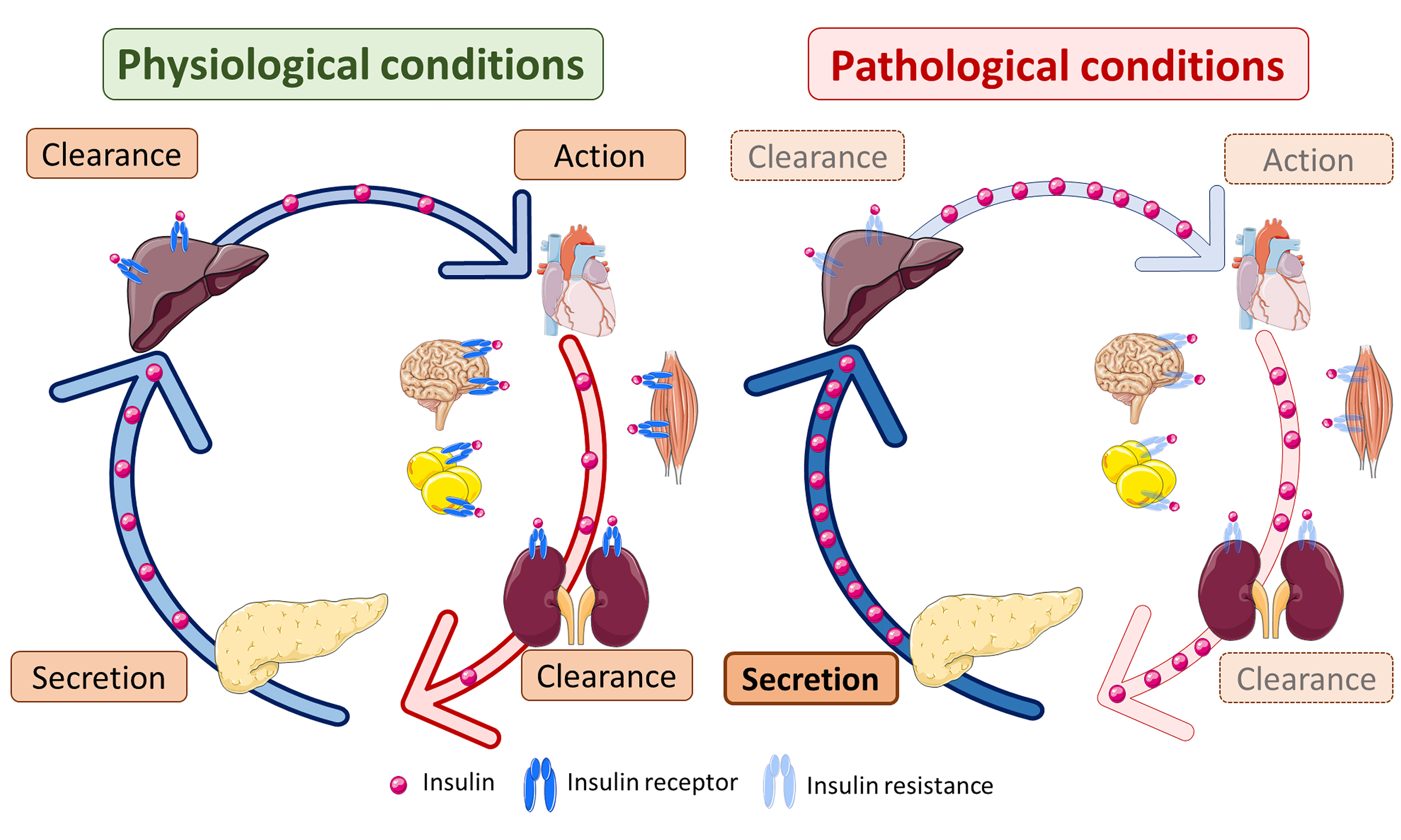
Insulin clearance, together with insulin secretion, regulates the physiologic level of insulin[5,117-119]. Hepatic insulin clearance occurs via receptor-mediated endocytosis: insulin receptor (INSR) activation triggers carcinoembryonic antigen-related cell adhesion molecule 1 (CEACAM1) phosphorylation, recruiting the insulin-INSR complex to clathrin-coated pits via adaptor protein 2 (AP2)[80,120]. Internalized insulin is degraded by IDE, PDI, and cathepsin D (CTSD), while the INSR is recycled. CEACAM1 coordinates receptor recycling and insulin degradation through fatty acid synthase (FASN) and src homology region 2-containing protein tyrosine phosphatase 2 (SHP2) interactions[5,116] [Figure 3A].
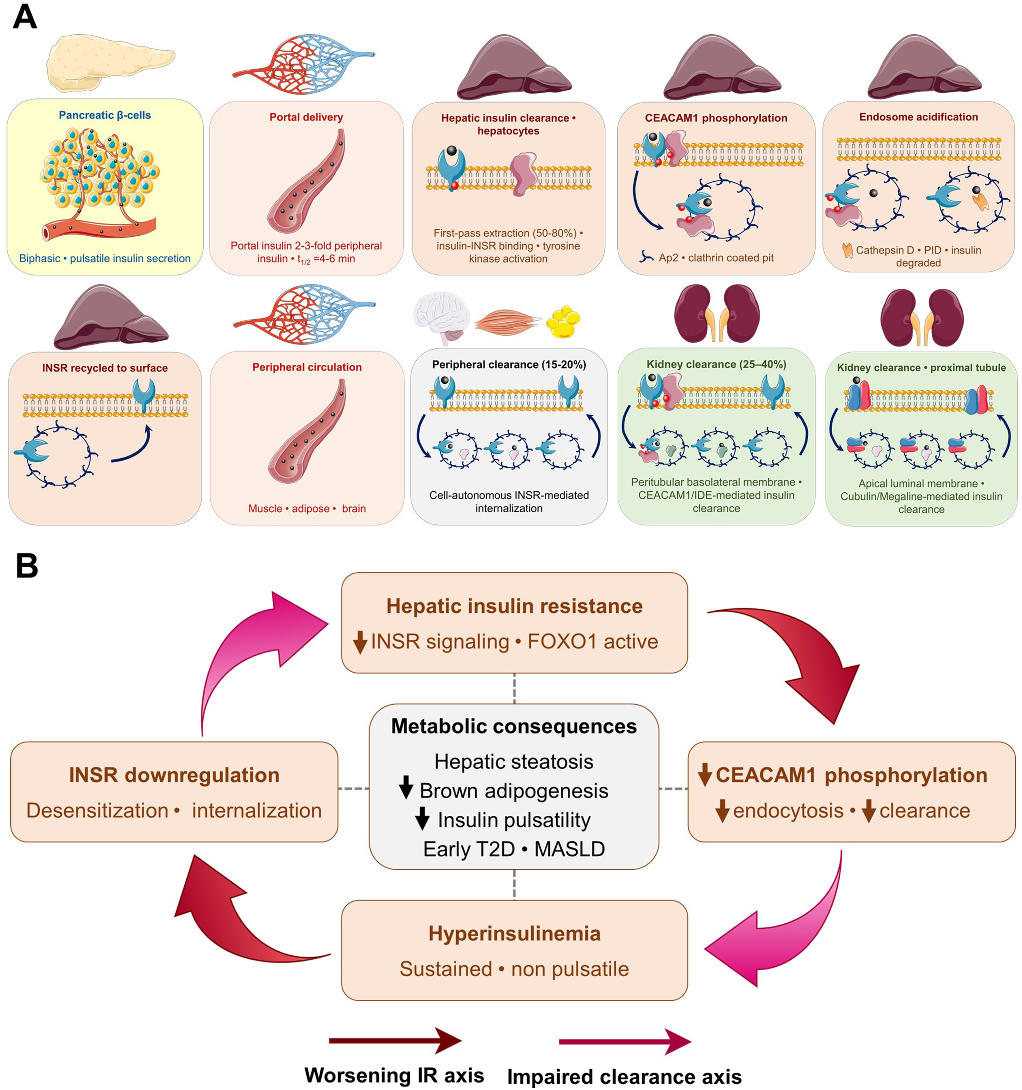
Figure 3. Hepatic insulin clearance and the clearance-resistance loop. (A) Hepatic insulin clearance. Portal delivery, receptor-mediated endocytosis, and consequences of impaired clearance. Under physiological conditions, insulin secreted in a biphasic, pulsatile pattern by pancreatic β-cells enters the portal venous system, establishing a portal-to-peripheral insulin gradient. Upon reaching the liver, 50%-80% of portal insulin is extracted during the first pass via receptor-mediated endocytosis in hepatocytes. Insulin binding to the INSR activates tyrosine kinase signaling, which phosphorylates CEACAM1; the phosphorylated CEACAM1-INSR-insulin complex is recruited to clathrin-coated pits via AP2 and internalized into acidified endosomes, where insulin is degraded by IDE, CTSD, and PDI, while the INSR is recycled to the plasma membrane. The residual ~50% of insulin reaches the peripheral circulation, where muscle, adipose tissue, brain, and erythrocytes contribute to disposal; the kidneys handle an additional 25%-40% through megalin-mediated reabsorption and IDE-dependent degradation in proximal tubule cells. Physiological hepatic clearance supports suppression of HGP, lipogenesis regulation, and glycogen synthesis. Conditions that impair hepatic clearance - including downregulation of CEACAM1, hepatic steatosis, obesity, and high-fat feeding - disrupt this axis and promote chronic hyperinsulinemia, peripheral insulin resistance, and MASLD. Subcutaneous insulin administration bypasses the portal route, abolishing the portal-to-peripheral gradient and resulting in relative hepatic under-insulinization alongside peripheral hyperinsulinemia; (B) The clearance-resistance loop. Pathological feedback between hepatic IR and impaired insulin clearance. Hepatic insulin resistance reduces INSR kinase activity and CEACAM1 phosphorylation, impairing receptor-mediated endocytosis and lowering hepatic insulin clearance. The resulting chronic hyperinsulinemia desensitizes and promotes INSR downregulation, further worsening hepatic insulin signaling and completing a self-reinforcing feedback cycle. Each node of this loop independently contributes to a common set of metabolic consequences (central panel). This clearance-resistance loop may represent an underappreciated and early driver of metabolic deterioration, operating independently of primary defects in β-cell secretory capacity. CEACAM1: Carcinoembryonic antigen-related cell adhesion molecule 1; INSR: insulin receptor; AP2: adaptor protein 2; IDE: insulin-degrading enzyme; CTSD: cathepsin D; PDI: protein disulfide isomerase; HGP: hepatic glucose production; MASLD: metabolic dysfunction-associated steatotic liver disease; IR: insulin resistance; T2D: type 2 diabetes.
Renal and peripheral clearance are integrated components of the insulin lifecycle. Proximal tubule cells remove insulin via megalin (LRP2)-mediated reabsorption and peritubular INSR-mediated uptake; urinary excretion accounts for less than 1% of the filtered load[116,119,121]. Chronic kidney disease prolongs insulin half-life, increasing hypoglycemia risk. Obesity may partially compensate by increasing renal plasma flow.
The renal contribution to endogenous insulin clearance is debated. Clamp studies show clearance correlates with lean mass and insulin sensitivity, not GFR[122]. Peripheral tissues (muscle, adipose, brain, erythrocytes) may account for 15%-20% of disposal via INSR-mediated internalization[123]. Clearance is lower in African American populations, potentially contributing to T2D susceptibility[124-127]. High-fat feeding suppresses hepatic clearance by up to 30%, underscoring it as a regulated adaptive process[128]. Dysregulated clearance promotes hyperinsulinemia, steatosis, and insulin resistance independently of β-cell output[5,8,80,125,129,130]. Recent studies on metabolic dysfunction-associated steatotic liver disease (MASLD)/metabolic dysfunction-associated fatty liver disease (MAFLD) and obesity further support that excess energy intake and hepatic steatosis impair hepatic insulin clearance and promote chronic hyperinsulinemia[117,131].
Contrary to the traditional liver-centric model, these data support an integrated system implicating kidneys and peripheral tissues in the active regulation of insulin clearance. Characterizing these distinct pathways offers a framework for patient phenotyping and targeted treatment allocation.
Subcutaneous insulin delivery and loss of the portal‑to‑peripheral gradient
As noted above, under physiological conditions, pancreatic insulin secretion creates a portal-to-peripheral insulin gradient, exposing the liver to insulin concentrations three to four times higher than those in peripheral tissues. This gradient is essential for the liver to effectively suppress hepatic glucose production while peripheral tissues maintain normal glucose uptake[132].
Subcutaneous insulin administration fundamentally abolishes this physiological distribution. When insulin is injected subcutaneously, it enters the peripheral circulation directly, resulting in relative hepatic insulin deficiency and peripheral hyperinsulinemia[133]. This unphysiological distribution has been consistently demonstrated across multiple studies.
Targeting insulin specifically to the liver corrected metabolic defects caused by peripheral delivery. When insulin was infused into the portal vein, glucose uptake was equally divided between liver and muscle. However, when insulin was delivered peripherally, muscle glucose uptake was four-fold greater than liver uptake, with liver glucose uptake being less than half of normal[134]. These defects could not be corrected by simply adjusting the peripheral insulin dose.
Comparative studies of portal vs. peripheral venous administration confirm that the portal route is superior for maintaining appropriate insulin concentrations and controlling hepatic glucose output. In peripherally infused animals, basal hepatic glucose output was significantly higher than in portally infused animals. Similarly, peripheral venous administration resulted in inadequate suppression of hepatic glucose production at basal insulin rates, while higher peripheral doses increased peripheral glucose uptake and hypoglycemia risk, a “double jeopardy” for insulin-treated patients[135].
Current subcutaneously injected insulin products result in hyperinsulin exposure in peripheral tissues (skeletal muscle and adipose) while almost completely avoiding the liver[136]. This increases the risk of hypoglycemic episodes and causes weight gain due to the unphysiological distribution.
The clinical implications are significant: under-insulinization of the liver at basal rates leads to increased hepatic glucose output and residual hyperglycemia, while higher peripheral doses increase the risk of hypoglycemia. This mechanism contributes to persistent hyperglycemia and diabetic complications despite insulin therapy. The physiological distribution is lost when insulin is injected subcutaneously, causing impaired regulation of hepatic glucose metabolism[133,134].
Development of hepato-preferential insulin analogs and intraperitoneal (IP) delivery systems aims to restore the portal-peripheral gradient and improve hepatic glucose control. Three therapeutic strategies aim to restore preferential hepatic exposure to insulin:
Hepato-Preferential Analogs (insulin-327): insulin-327 is a 22-carbon fatty diacid-acetylated insulin analog (Novo Nordisk) designed to be hepato-preferential. The molecule cannot cross the continuous capillary endothelium of muscle but freely accesses fenestrated hepatic sinusoids, physiologically replicating the portal gradient. In canine models, insulin-327 corrected deficits in hepatic glucose metabolism induced by peripheral insulin[134]. However, despite promising preclinical data, insulin-327 remains a preclinical model compound and lacks published human trials.
Oral insulin as an indirect hepato-preferential strategy: The oral route physiologically replicates the first hepatic pass: insulin is absorbed in the intestine and reaches the liver before systemic circulation[137]. ORMD-0801 (Oramed) is the first oral insulin analog in clinical trials. Oramed demonstrated reduction of hepatic steatosis and fibrosis in patients with T2D + metabolic dysfunction-associated steatohepatitis (MASH), a unique therapeutic convergence between hepato-preference and metabolic liver disease[138].
IP administration systems: IP administration restores a gradient similar to the portal route: insulin is absorbed in the peritoneal cavity and reaches the liver before distributing to systemic circulation[139]. This allows the liver to be exposed to higher insulin concentrations, similar to physiological pancreatic secretion. Pharmacokinetic modeling confirms that this space functions as a “virtual compartment” with rapid kinetics (min) compared to slow subcutaneous absorption (1-2 h)[140,141]. The advantage of this strategy is driven by three key advances: (1) mature continuous glucose monitoring (CGM) systems[139,142]; (2) thermostable insulin formulations[143,144] and (3) more robust control algorithms for closed-loop systems[140,141].
Therapeutic modulation of insulin clearance: opportunities and challenges from CEACAM1 to IDE
Studies in genetically modified mice have firmly established CEACAM1 as the central mediator of receptor-dependent hepatic insulin clearance. Liver-specific inactivation or global deletion of CEACAM1 leads to hyperinsulinemia, peripheral insulin resistance, and hepatic steatosis - phenocopying many features of metabolic syndrome and MASLD[79,117]. Conversely, forced hepatic overexpression of CEACAM1 curtails diet-induced insulin resistance[145]. These findings have been replicated across multiple mouse model systems, providing strong preclinical evidence. Direct human data on CEACAM1 polymorphisms and insulin clearance are more limited, though reduced hepatic CEACAM1 expression has been reported in patients with MASLD and MASH, especially as fibrosis progresses[146,147]. Moreover, lower circulating levels of CEACAM1 have been reported in Portuguese patients with dysregulated glucose homeostasis and with high fatty liver index and, reciprocally, reduced insulin clearance[148].
Mechanistically, the bidirectional relationship between insulin clearance and insulin resistance creates a pathological feedback loop: hepatic insulin resistance reduces CEACAM1 phosphorylation and insulin endocytosis, lowering clearance and raising systemic insulin concentrations. The resultant hyperinsulinemia desensitizes INSR and promotes receptor downregulation, further impairing signaling[8,117]. This “clearance-resistance loop” may be an underappreciated driver of metabolic deterioration in early T2D and MASLD[8]. Hyperinsulinemia also limits the pulsatility of insulin release and reduces brown adipogenesis, thereby decreasing energy expenditure and adding further metabolic burden[80] [Figure 3B].
The evidence supporting CEACAM1 as a therapeutic target is biologically compelling, but the translation into clinical tools remains incomplete. The available evidence falls into three different tiers:
Direct molecular targeting
Liver-specific reconstitution of CEACAM1 via adenoviral delivery fully reverses hyperinsulinemia, insulin resistance, and steatohepatitis in murine models, confirming that restoring this single protein is sufficient to normalize the entire metabolic phenotype[149]. Based on this, CEACAM1 enhancers and insulin receptor isoform B (INSR-B) agonists have been proposed as future therapeutic strategies targeting the hepatic clearance axis directly[116]. PPARδ agonists and retinoic acids (Vitamin A) transcriptionally upregulate Ceacam1 expression by binding to the highly conserved PPRE/RXR cis-regulatory element in the Ceacam1 promoter[150,151]. However, no CEACAM1-focused drug is currently approved.
Approved drugs with indirect effects on hepatic insulin clearance
Several existing drug classes improve insulin clearance as a secondary consequence of their metabolic actions:
Sodium-glucose cotransporter 2 inhibitors (SGLT2i) - Dapagliflozin and empagliflozin reduce hyperinsulinemia and improve hepatic insulin clearance in patients with T2D, partly by reducing glucotoxicity and hepatic steatosis. A clinical study with ipragliflozin demonstrated a direct improvement in the hepatic insulin clearance index (postprandial C-peptide/insulin molar ratio), alongside improved insulin resistance and liver function[116,152].
GLP-1a and tirzepatide - Weight loss and improved insulin sensitivity induced by these agents are associated with partial recovery of hepatic insulin clearance, though the mechanism does not appear to operate directly through CEACAM1 upregulation. The metabolic offloading of the liver appears to be the primary driver[5]. For instance, treating fat-fed mice with Exenatide, a GLP-1 receptor agonist, reversed metabolic abnormalities in wild-type but not in Ceacam1 null mice. This was mediated by inducing CEACAM1 expression in the liver and subsequently increasing hepatic insulin clearance to counter the increase in insulin secretion and maintain physiologic insulin levels[150].
Weight loss - dietary, pharmacological, or surgical - remains the intervention with the greatest documented impact on insulin clearance. A ~10% reduction in body weight increases insulin clearance and partially restores insulin sensitivity[5,152]. Sleeve gastrectomy in obese mice directly increased insulin clearance, confirming that weight loss-driven hepatic metabolic recovery is sufficient to improve this pathway[153]. Roux-en-Y gastric bypass similarly restored insulin-mediated regulation of hepatic lipoprotein production, further supporting the role of surgical weight loss in normalizing hepatic insulin handling[154].
No standardized biomarker of hepatic CEACAM1 activity exists. The C-peptide/insulin molar ratio [hepatic insulin clearance index (HICI)] has been proposed as a proxy; below 1 during glucose challenge, it may identify impaired clearance with greater sensitivity than homeostatic model assessment of insulin resistance (HOMA-IR)[116], but has not entered clinical guidelines. Hepatic CEACAM1 protein levels decline with advancing fibrosis in MASH[130] as well as with obesity[155]. Caloric restriction elevates CEACAM1 levels in parallel with decreasing body weight and MASH in selectively bred rats with low aerobic capacity[156,157]. CEACAM1 is biologically validated but clinically untargeted; current strategies act indirectly via weight loss, SGLT2i, or GLP-1Ra. Thus, direct CEACAM1-restoring pharmacotherapy remains an unexplored opportunity.
IDE is a zinc metalloprotease belonging to the M16 family that degrades insulin through a “clamshell”-like mechanism. It contains an inverted HXXEH zinc-binding catalytic motif and is found in multiple cellular compartments, including the cytosol, peroxisomes, mitochondria, endosomes, the plasma membrane, and the extracellular space. Although IDE is predominantly cytosolic, its subcellular distribution may vary according to cell type. Its proteolytic activity is regulated by metabolic signals and can be inhibited by ATP, oxidative modification, free long-chain fatty acids, and fatty acyl-CoAs. In addition to insulin, IDE degrades amyloid-β peptide, which is linked to Alzheimer’s disease, as well as glucagon[158,159].
IDE animal models yield contradictory clearance results: L-IDE-KO mice show no plasma clearance change[160]. Diet-induced obese mice with hepatic Ide ablation show no clearance alteration but develop hepatic insulin resistance[161]; however, Borges et al. found that postprandial IDE clearance is lost in dysmetabolic states[162]. Despite these contradictions, all models confirm that hepatic IDE regulates insulin signaling: its ablation activates glucagon signaling, dysregulates glucose metabolism, and exacerbates hyperinsulinemia in obese mice[160,161].
IDE has emerged as a promising therapeutic target for T2D, with both inhibition and activation strategies showing preclinical potential. IDE inhibitors represent the more extensively studied approach, with strong evidence demonstrating improved glucose tolerance in animal models. The landmark study by Maianti et al. identified the first IDE inhibitor (macrocycle 6bK) that improved glucose tolerance in vivo, reducing glucose and increasing insulin levels in both lean and obese mice with high selectivity[163]. Similarly, antibody-mediated IDE inhibition[164] improved insulin activity in streptozotocin (STZ)-induced diabetic mice following single IP administration. Conversely, Durham et al., using exosite inhibitors, found that targeting IDE improves glucose tolerance in obese mice, though it does not significantly increase insulin sensitivity[165]. These findings challenge the traditional view of IDE as the primary mediator of insulin clearance, indicating that alternative, IDE-independent mechanisms drive hormone degradation in vivo. However, significant challenges persist: traditional zinc-targeting inhibitors may cause toxicity to other metalloproteases, and pan-cellular ablation of Ide (IDE-KO mice) mimicking complete and constant IDE inhibition paradoxically causes diabetes through chronic insulin resistance[166,167], whereas partial/transient inhibition appears therapeutic[163,164].
Interestingly, IDE activators represent an emerging alternative strategy. Recent work[168] demonstrated that synthetic preimplantation factor (sPIF), an IDE activator, improved insulin secretion and glucose tolerance in diet-induced obese mice. This suggests that IDE activation could enhance β-cell function despite increasing insulin degradation[168]. Indole-based activators[169] have also been identified, though their application to diabetes remains preliminary compared to Alzheimer’s research.
Currently, neither IDE inhibitors nor activators have reached clinical application. Inhibitors show strong preclinical efficacy but face safety challenges and mechanistic paradoxes, while activators remain very preliminary despite promising recent evidence. Both approaches provide valuable insights into hepatic insulin resistance, but the complexity of IDE’s physiological role and context-dependent paradoxical effects (partial vs. complete inhibition, physiological vs. pathological conditions) continue to limit therapeutic development.
Molecular control of hepatic gluconeogenesis by insulin: the aquaporin-9 pathway
The liver occupies a central position in glucose metabolism through glycerol gluconeogenesis, accounting for approximately 5%-10% of hepatic glucose output under fasting conditions and rising substantially in insulin-resistant states. The essential step of glycerol entry into hepatocytes (both in rodents and humans) requires facilitated transport across the plasma membrane. This step is primarily mediated by aquaporin-9 (AQP9), the principal aquaglyceroporin expressed in hepatocytes. AQP9 is a rate-limiting step in glycerol gluconeogenesis, making it an important metabolic gatekeeper[170-172].
AQP9 expression is negatively regulated by insulin: insulin-mediated PI3K/AKT signaling phosphorylates forkhead box O1 (FoxO1), which is then sequestered from the Aqp9 gene promoter, suppressing AQP9 transcription[171,173]. This regulatory mechanism is physiologically coherent: postprandial insulin elevation suppresses glycerol entry and gluconeogenesis simultaneously, contributing to the postprandial switch from glucose production to glucose uptake. In insulin-resistant states, where FOXO1 is inadequately suppressed, AQP9 expression rises, amplifying glycerol-driven gluconeogenesis and contributing to fasting hyperglycemia. AQP9 downregulation in MASLD, as documented in human liver biopsies[174], appears to represent a late-stage compensatory mechanism to limit lipid accumulation and glycerol-driven glucose production.
The adipose tissue provides the circulating glycerol pool through lipolysis, mediated by adipocyte aquaglyceroporins AQP3 and AQP7, which facilitate glycerol efflux from adipocytes and are regulated by both insulin (which suppresses lipolysis) and leptin (which activates sympathetic innervation of white adipose tissue to promote lipolysis)[173]. This adipose-liver aquaglyceroporin axis integrates lipolytic regulation with hepatic glucose production, representing an important inter-organ communication pathway disrupted across multiple points in obesity and T2D[173,174]. The regulation of aquaglyceroporins by insulin and leptin through the PI3K/AKT/mammalian target of rapamycin (mTOR) pathway provides a mechanistic link between two major hormonal axes and hepatic gluconeogenic substrate supply[173]. For clinical translation notes of this section, see Supplementary Materials.
PERIPHERAL INSULIN SIGNALING: THE MOLECULAR ARCHITECTURE OF INSULIN ACTION
Molecular architecture of INSR proximal signaling
Insulin action in peripheral tissues is initiated by binding to the INSR, a heterotetrameric tyrosine kinase receptor composed of two extracellular α-subunits and two β-subunits. The β-subunits span the plasma membrane once and contain tyrosine phosphorylation sites and tyrosine kinase domains in their cytoplasmic tails. INSR is expressed as two isoforms, INSR-A and INSR-B, differing by the presence or absence of a 12-amino-acid sequence encoded by exon 11 segment in the C-terminus domain of the α-subunits. INSR-B predominates in metabolic tissues and mediates the classical metabolic actions of insulin. INSR-A, with higher affinity for insulin and insulin-like growth factor 2 (IGF-2), mediates mitogenic signaling. CEACAM1 preferentially promotes insulin endocytosis via INSR-A, which helps protect the expression of the high-affinity isoform on the hepatocyte surface[175]. The clinical relevance of INSR isoform imbalance in disease states is an active area of research[80,117,119].
Insulin binding to α-subunits initiates signaling by triggering conformational changes and trans-autophosphorylation of the β-subunit tyrosine residues (Tyr1158, Tyr1162 and Tyr1163). This activation of intracellular IRS1-4 generates critical docking sites for SH2-domain proteins, effectively bifurcating the signaling cascade into two canonical branches: the mitogenic Ras/MAPK arm and the metabolic PI3K/AKT pathway. Downstream of this metabolic branch, phosphorylated IRS recruits the p85 regulatory subunit of PI3K, activating the p110 catalytic subunit to generate phosphatidylinositol (3,4,5)-trisphosphate (PIP3) at the inner leaflet of the plasma membrane. PIP3 subsequently recruits phosphoinositide-dependent kinase 1 (PDK1) and mammalian target of rapamycin complex 2 (mTORC2), which phosphorylate and activate AKT[176-178].
Insulin action in the liver
In the liver, the metabolic PI3K/AKT pathway predominates, whereas the mitogenic Ras/MAPK branch plays a less significant role in hepatocytes under normal physiologic conditions[47]. There is general agreement that the acute suppression of hepatic glucose production by insulin is mediated by both a direct and an indirect effect on hepatocytes[117,179]. Portal insulin suppresses hepatic glucose production while promoting storage. AKT2 phosphorylates glycogen synthase kinase-3 (GSK3α/β), relieving the inhibition of glycogen synthase 2 (GYS2)[180-182]. Concurrent activation of protein phosphatase 1 (PP1) further dephosphorylates and activates glycogen synthase, accounting for ~30% of postprandial glucose disposal. AKT excludes FOXO1 from the nucleus, curtailing gluconeogenic transcription [phosphoenolpyruvate carboxykinase (PCK1) and glucose-6-phosphatase (G6PC1)] and reducing CREB/peroxisome proliferator-activated receptor gamma coactivator 1-alpha (PGC-1α) activity. Glycogenolysis decreases as AKT activates phosphodiesterase 3B (PDE3B) to degrade cAMP, counteracting the effects of glucagon[92,183,184]. Insulin also induces glycolysis by upregulating GCK via SREBP-1c and by elevating fructose-2,6-bisphosphate through PFK-2, which stimulates PFK-1 allosterically[2,185]. A complementary insulin-regulated gluconeogenic substrate pathway involves glycerol entry through AQP9. Insulin-mediated FOXO1 nuclear exclusion suppresses AQP9 transcription, thereby restricting glycerol-driven gluconeogenesis. Dysregulation of this axis in insulin-resistant states is discussed herein.
Insulin, particularly at high levels, drives de novo lipogenesis (DNL) as the primary anabolic cue. AKT activates mTORC1 to process and translocate SREBP-1c, a master transcriptional regulator of lipogenic genes. This induces the expression of FASN, acetyl-CoA carboxylase (ACC), and stearoyl-CoA desaturase (SCD1). Additionally, carbohydrate-enriched diets elevate glucose-6-phosphate levels, acting synergistically by causing nuclear translocation of carbohydrate response element-binding protein (ChREBP) to induce the transcription of lipogenic genes[2,46,186-188]. Insulin stabilizes apolipoprotein (Apo)B acutely to promote very low-density lipoprotein (VLDL) assembly. However, in obesity and T2D, “selective hepatic insulin resistance” preserves lipogenic signaling while impairing FOXO1 suppression, yielding hyperglycemia alongside steatosis. Impaired adipose lipolysis causes an excess of free fatty acids (FFAs) to flood the liver, overwhelming oxidation and fueling re-esterification and inflammation; ultimately causing progression of MASLD[8,189-191].
Despite the lipogenic action of insulin, the liver is physiologically protected against the high portal insulin concentrations. This protection operates in part through CEACAM1/FASN coupling: upon acute insulin pulses, CEACAM1 phosphorylation enhances receptor-mediated insulin internalization and targeting to endosomal compartments. Eventually, phosphorylated CEACAM1 recruits FASN from perinuclear regions, pulling it away from the internalized insulin-INSR complex in late endosomes to suppress FASN lipogenic activity. This results in the simultaneous dissociation of insulin from its receptor to undergo degradation as INSR-A recycles back to the membrane. This dual action of CEACAM1 - coordinating insulin clearance and lipogenesis - is intact even in early MASLD and challenges the concept of “selective hepatic insulin resistance” as an intrinsic signaling bifurcation, suggesting instead that it may be a consequence of impaired clearance (see controversy 1).
Insulin action in skeletal muscle
Skeletal muscle accounts for 60%-80% of postprandial glucose disposal, making it the dominant site of insulin-stimulated glucose uptake. In the basal state, GLUT4 glucose transporters are stored in intracellular vesicles (GSVs) and are retained by phosphorylated AS160 [TBC1 Domain Family Member 4 (TBC1D4)], which maintains Ras-related proteins Rab8A (RAB8A)/RAB10/RAB14 in their inactive GDP-bound form. Insulin activates the INSR-IRS1-PI3K-AKT2 cascade, which phosphorylates AS160 at sites such as Thr642, thereby relieving its inhibitory activity. This allows Rab to engage with the soluble N-ethylmaleimide-sensitive factor attachment protein receptor (SNARE) machinery [vesicle-associated membrane protein 2 (VAMP2), syntaxin4 (STX4), and synaptosomal-associated protein 23 (SNAP23)], driving GLUT4 to translocate to the plasma membrane and t-tubules. This mechanism amplifies glucose transport capacity by a factor of 10-40[2,192-194].
Upon entry, glucose is phosphorylated by hexokinase II (HKII), which traps it intracellularly to fuel glycogen synthesis and oxidation. AKT2 inactivates GSK3 and activates PP1, which dephosphorylates GYS1. This accounts for 30%-35% of postprandial glucose disposal[195-197]. Insulin further enhances glycolysis and oxidation by activating pyruvate dehydrogenase phosphatase 1 (PDP1) and channeling pyruvate into the Krebs cycle[198].
In addition to governing carbohydrate metabolism, insulin regulates protein turnover. AKT2 phosphorylates tuberous sclerosis complex (TSC)1/2, thereby activating mTORC1 and ras homolog enriched in brain (RHEB), which drive translation via the phosphorylation of eukaryotic translation initiation factor 4E-binding protein 1 (4EBP1) (releasing eIF4E) and ribosomal protein S6 kinase β-1 (S6K1)[199]. At the same time, nuclear exclusion of FOXO3 suppresses the expression of components of the ubiquitin-proteasome system, such as atrogin-1 (FBXO32) and muscle RING finger 1 (TRIM63/MuRF1), thereby reducing proteolysis[200].
Insulin action in adipose tissue
Adipose tissue is an active insulin target with multiple metabolic roles. Insulin powerfully suppresses lipolysis in adipocytes by activating PDE3B, reducing intracellular cAMP and inhibiting hormone-sensitive lipase (HSL) and adipose triglyceride lipase (ATGL). This anti-lipolytic action is among the most insulin-sensitive of all insulin responses, occurring at lower insulin concentrations than those required for glucose uptake. The consequence is that in insulin-resistant states, unsuppressed lipolysis maintains elevated plasma FFA flux, which in turn propagates insulin resistance in liver and muscle, a “lipotoxic feedforward” cycle central to metabolic disease progression[131,201].
Adipose tissue exhibits high insulin sensitivity, primarily channeling triglycerides into storage while curbing lipolysis. Insulin activates the IRS1/2-PI3K-AKT signaling pathway, which suppresses HSL activity. This is achieved through AKT-mediated activation of PDE3B, which hydrolyses cAMP and reduces PKA signaling. This reduces HSL phosphorylation at activating sites (Ser563/660) and perilipin-1 (PLIN1) at Ser517, thereby preventing comparative gene identification-58 (CGI-58) from being recruited to ATGL. This antilipolytic brake represents the body’s most sensitive insulin response (half-maximal at ~10-15 pM) and lowers circulating FFAs to indirectly enhance muscle glucose uptake and restrain hepatic gluconeogenesis[2,202-204].
Insulin also drives lipogenesis by triggering GLUT4 translocation to facilitate glucose uptake and re-esterification of glycerol-3-phosphate. Insulin also induces lipoprotein lipase (LPL) on capillary endothelial cells to hydrolyze triglyceride-rich lipoproteins, thereby increasing the influx of FFAs. Although less prominent than in the liver, insulin also upregulates enzymes involved in DNL via SREBP-1c. This consolidates postprandial lipids in adipocytes and prevents their deposition elsewhere in the body[202,205]. Insulin also suppresses the efflux of glycerol from adipocytes: the aquaglyceroporins AQP3 and AQP7, which facilitate glycerol exit from adipocytes, are regulated by insulin through the PI3K/AKT/mTOR pathway, thereby coupling adipose insulin action to the provision of gluconeogenic substrate for the liver[173,206,207].
The dynamic secretion and actions of adipokines, including adiponectin, leptin, resistin, tumor necrosis factor alpha (TNF-α), and interleukin 6 (IL-6), play key roles in regulating systemic insulin sensitivity. Failure of adipose tissue to suppress lipolysis appropriately constitutes a critical dysfunction that amplifies systemic metabolic disease. Unchecked FFA efflux floods the liver and muscle, driving ectopic lipid accumulation and lipotoxicity, while reduced secretion of protective adipokines, particularly adiponectin, exacerbates insulin resistance. Elucidating how adipose tissue dysfunction initiates and sustains insulin resistance, as well as the endocrine and paracrine pathways by which it regulates metabolic homeostasis, is therefore crucial.
Insulin action in the central nervous system
The brain is now recognized as a critical regulator of energy homeostasis through insulin signaling. In the hypothalamus, particularly in the arcuate and ventromedial nuclei, insulin activates the PI3K-AKT pathway in agouti-related neuropeptide (AgRP)/neuropeptide Y (NPY) neurons, thereby curbing food intake, stimulating proopiomelanocortin (POMC) neurons, and enhancing satiety. Administering insulin intracerebroventricularly in rodents confirms reduced feeding and improved peripheral sensitivity. Beyond appetite regulation, hypothalamic insulin signaling via vagal circuits suppresses hepatic gluconeogenesis independently of direct liver effects. Insulin also modulates dopaminergic reward circuits by influencing the activity of the dopamine transporter, with central resistance potentially fueling overconsumption in obesity[162,208-210].
INSR are expressed throughout the central nervous system, particularly in the hypothalamus, hippocampus, and striatum. Central insulin signaling suppresses hepatic glucose output through autonomic pathways, modulates appetite and energy expenditure, and influences peripheral insulin sensitivity, effects that have been demonstrated in human intranasal insulin studies[211]. Impaired central insulin signaling has been proposed as a mechanism linking insulin resistance to cognitive decline and the increased Alzheimer’s disease risk in T2D, though this connection requires further causal establishment in humans[212].
Mechanisms of insulin resistance: classical and novel perspectives
Insulin resistance, the impaired ability of target tissues to respond to normal insulin concentrations, is the central feature of T2D, obesity, and the metabolic syndrome. The classical model attributed insulin resistance primarily to serine phosphorylation of IRS1, reducing its affinity for the p85 PI3K subunit. This serine phosphorylation is driven by inflammatory kinases [JNK and the inhibitor of nuclear factor kappa-B kinase subunit β (IKKβ)] activated by saturated fatty acids, ceramides, diacylglycerols (DAGs), and pro-inflammatory cytokines including TNF-α and IL-6[178,202,213].
The lipid overflow hypothesis states that ectopic lipid accumulation in skeletal muscle and liver, particularly DAG and ceramide species arising from excess saturated fatty acid flux, disrupts insulin signaling through activation of PKCε (liver) and PKCθ (muscle), which phosphorylate IRS1/2 at inhibitory serine residues, reducing PI3K/AKT activation. This model is well supported by human metabolic studies, including isotope tracer studies and liver biopsy data correlating intrahepatic DAG content with hepatic insulin resistance independent of total liver fat[8,118,214].
A key paradox of hepatic insulin resistance deserves explicit attention: in MASLD, insulin fails to suppress gluconeogenesis (through the PI3K/AKT/FOXO1 axis) but continues to stimulate DNL (through mTORC1/SREBP-1c). This “selective” or “branch-specific” insulin resistance produces the clinical paradox of fasting hyperglycemia and concurrent hepatic lipid accumulation under the same hyperinsulinemic state. Recent evidence has attributed this selectivity to the differential sensitivity of IRS1 vs. IRS2 to inhibitory phosphorylation. This clinically important distinction has been documented in both rodent models and human liver biopsies from MASLD patients[8,118,214].
Muscle insulin resistance, an early indicator of the progression of T2D, entails impairment of IRS1 phosphorylation through serine modification by kinases such as IKKβ, JNK, and PKCθ. Intramyocellular lipid accumulation, particularly of DAGs and ceramides resulting from incomplete fatty acid beta-oxidation, activates PKCθ and protein phosphatase 2A (PP2A), thereby disrupting INSR signaling. Mitochondrial dysfunction exacerbates this process by reducing oxidative capacity, increasing reactive oxygen species (ROS), and hindering β-oxidation. This ultimately blunts the AS160-RAB-SNARE axis and reduces the availability of GLUT4 on the cell surface. Ectopic intramyocellular lipid accumulation, particularly ceramides derived from saturated fatty acids, impairs AKT activation through PP2A-mediated dephosphorylation. A growing body of evidence has shown that exercise restores insulin-stimulated GLUT4 translocation in insulin-resistant skeletal muscle in humans. This occurs through AMP-activated protein kinase (AMPK)-dependent and Ras-related C3 botulinum toxin substrate 1 (Rac1)-dependent mechanisms that bypass the defective PI3K/AKT pathway to provide a mechanistic rationale for exercise as a cornerstone of T2D management[192,215-219].
Across insulin-resistant tissues, three convergent pathological processes amplify and perpetuate the signaling defects described above. Chronic low-grade inflammation, initiated by macrophage infiltration of adipose tissue and propagated systemically via nuclear factor kappa-light-chain-enhancer of activated B cells (NF-κB) and inflammasome activation, generates interleukin 1-beta (IL-1β), TNF-α, and IL-6, which directly impair IRS1 signaling in liver, muscle, and β-cells[220-222]. Oxidative stress, arising from mitochondrial dysfunction in insulin-resistant tissues, activates JNK and p38 MAPK through ROS-mediated mechanisms and can directly damage INSR and IRS1 through oxidative modification[221-223]. ER stress, induced by lipid overloading and misfolded protein accumulation, activates the UPR through the PERK-eIF2α-ATF4 branch, suppressing IRS1 expression and promoting resistance through suppressor of cytokine signaling (SOCS)1/3 induction[221-223]. These three mechanisms are most granularly characterized in rodent models of diet-induced obesity. Human validation, from prospective inflammatory biomarker data, adipose biopsy crown-like structure quantification, and skeletal muscle ceramide and oxidative capacity measurements, confirms their operation but has not achieved comparable mechanistic resolution.
Integrated insulin-glucagon regulation of peripheral insulin action
Following a mixed meal, portal insulin levels increase 10-fold within 15-20 min to suppress hepatic glucose output, primarily by reducing gluconeogenesis and glycogenolysis. Peak peripheral disposal occurs at 60-
In normal non-obese individuals, insulin levels drop to 2-10 μIU/mL during fasting, shifting the insulin/glucagon molar ratio towards glucagon dominance. Hepatic glucose output of ~7 g/h sustains euglycemia through glucagon-driven gluconeogenesis and glycogenolysis. Adipose tissue lipolysis provides FFAs and glycerol for hepatic triglyceride esterification and substrate provision. Ketogenesis emerges during prolonged fasting or ketogenic conditions[226-231].
Below ~3.8 mmol/L of glucose, the counterregulatory hierarchy unfolds: insulin secretion halts while glucagon rises (at the ~2.5-3.5 mmol/L threshold), followed by epinephrine (at ~3.2 mmol/L), with growth hormone and cortisol lagging behind (at ~3.0 mmol/L). This culminates in neuroglycopenia below ~2.8 mmol/L[232,233]. In long-standing T1D and T2D, impaired glucagon responses due to lost islet paracrine cues or autonomic neuropathy heighten the risk of hypoglycemia[234-237].
Current controversies in peripheral insulin signaling
Is “selective hepatic insulin resistance” a coherent pathophysiological entity?
Selective hepatic insulin resistance, in which insulin fails to suppress gluconeogenesis via PI3K/AKT/FOXO1 yet still drives DNL via mTORC1/SREBP-1c, has been proposed to explain fasting hyperglycemia with hepatic steatosis in T2D. Petersen et al. attribute this to differential IRS1/IRS2 susceptibility to PKCε-mediated inhibitory phosphorylation, with preferential failure of the gluconeogenic arm[8,118,192,214]. However, the Najjar laboratory argues that acute insulin pulses suppress FASN activity and lipogenesis through the CEACAM1/FASN axis, a response preserved even in early MASLD[117,238]. Thus, apparent selectivity may arise from chronic hyperinsulinemia due to impaired hepatic insulin clearance, not primary signaling bifurcation, with distinct therapeutic implications for selecting the upstream intervention instead[117,152].
Does insulin resistance cause hyperinsulinemia, or does hyperinsulinemia drive insulin resistance?
The prevailing model positions insulin resistance as primary, with hyperinsulinemia as a compensatory β-cell response. An alternative hypothesis (Corkey et al.) proposes that primary hyperinsulinemia - from β-cell hypersecretion or impaired hepatic clearance - may itself drive insulin resistance via receptor downregulation and lipogenic substrate overload[117,239,240]. The clearance-resistance loop provides molecular evidence for this bidirectionality[238]. Epidemiological data show fasting hyperinsulinemia precedes measurable resistance by years[117], a temporal sequence inconsistent with a purely compensatory origin. The distinction is not merely semantic: if hyperinsulinemia is causal, therapies reducing insulin secretion or enhancing clearance (including CEACAM1 restoration[152] or hepato-preferential insulin delivery systems) acquire mechanistic primacy over peripheral sensitization strategies. Current evidence does not resolve this bidirectional relationship, and the two mechanisms are not mutually exclusive; however, the field has historically underweighted the causal role of hyperinsulinemia, particularly as it pertains to impaired insulin clearance, and this asymmetry warrants explicit recognition in future research designs.
DAG/ceramide lipotoxicity vs. mitochondrial dysfunction as the primary driver of muscle insulin resistance
Two mechanistic models dominate the skeletal muscle insulin resistance literature, and their relationship remains unresolved. The lipid metabolite hypothesis by Samuel et al. identifies specific DAG species, particularly sn-1,2-DAG, and ceramides from saturated fatty acid overload as proximal inhibitors of insulin signaling via PKCθ and PP2A, causing IRS1 serine phosphorylation and AKT inactivation[192,218,241]. The mitochondrial dysfunction hypothesis instead proposes that reduced oxidative capacity, with impaired β-oxidation, acylcarnitine accumulation, and elevated ROS, is the upstream lesion leading to lipid metabolite accumulation. Human studies have not consistently separated these mechanisms because both coexist in insulin-resistant muscle and may vary with disease stage, fiber type, and nutritional context. Acylcarnitine species may directly impair IRS1 signaling[242], potentially bridging the models, but this remains to be confirmed prospectively.
Adipose tissue dysfunction: initiator or amplifier of systemic insulin resistance?
Whether adipose or liver initiates systemic insulin resistance remains debated. The “adipocentric” model proposes adipose dysfunction as the primary lesion, with hepatic and muscular resistance secondary to FFA overload and inflammatory adipokine signaling[2,131,201,202]. This is supported by thiazolidinedione trials showing adipose sensitization reverses hepatic/muscular resistance[243]. However, lean individuals with lipodystrophy or genetic NAFLD variants develop profound systemic resistance without adipose dysfunction, suggesting that hepatic insulin resistance can play a primary role[244-247]. Longitudinal studies suggest intrahepatic lipid precedes adipose inflammation in early metabolic deterioration[248]. Current consensus favors a bidirectional relationship varying across T2D subtypes.
Physiological relevance of central insulin signaling in humans
Rodent studies established that hypothalamic insulin signaling suppresses hepatic glucose production, regulates appetite, and modulates insulin sensitivity[209,249,250]. Whether these mechanisms operate at physiologically relevant levels in humans remains contested. Intranasal insulin studies show effects on appetite and cognition consistent with central insulin action[211,251], but suppression of hepatic glucose output by hypothalamic insulin has not been convincingly demonstrated in humans. The brain’s contribution to postprandial glucose homeostasis appears smaller than in rodents, where the portal-hypothalamic-vagal axis accounts for more of insulin’s hepatic effect. The link between central insulin resistance and Alzheimer’s disease risk in T2D is plausible, but rests mainly on epidemiological association and mechanistic inference from animal models[212,252]. These limitations warrant caution when extrapolating circuit-level data to human metabolic physiology.
An integrated network model of peripheral insulin sensitivity
A tissue-by-tissue description of peripheral insulin sensitivity can give the misleading impression that each tissue operates as an independent, parallel module. The evidence and controversies discussed above support the view that insulin sensitivity across tissues constitutes a hierarchical, context-dependent system. Within this framework, dysfunction at any node can propagate through the organism via relative sensitivity thresholds, inter-organ crosstalk, and temporal dynamics[195,253,254]. This network can be described by four organizing principles:
Physiological insulin responses follow hierarchical tissue sensitivity thresholds
Insulin-responsive tissues exhibit varying hormone sensitivity. Adipose anti-lipolysis is half-maximally activated at approximately ~10-15 pM, which is reached under basal conditions[255,256]. Suppression of hepatic glucose output requires ~30-60 pM[257,258], while maximal skeletal muscle GLUT4 translocation necessitates around ~340-720 pM[259]. This hierarchy ensures that adipose anti-lipolysis acts as a substrate gatekeeper, suppressing FFA-mediated interference before muscle glucose disposal. Disruption of this process initiates cascades where excess FFA activates lipotoxic pathways in the liver and muscle, propagating upstream resistance. This suggests that adipose insulin resistance is temporally and mechanistically upstream of hepatic and muscular resistance in common forms of T2D, consistent with longitudinal data from high-risk cohorts[195] [Figure 4].
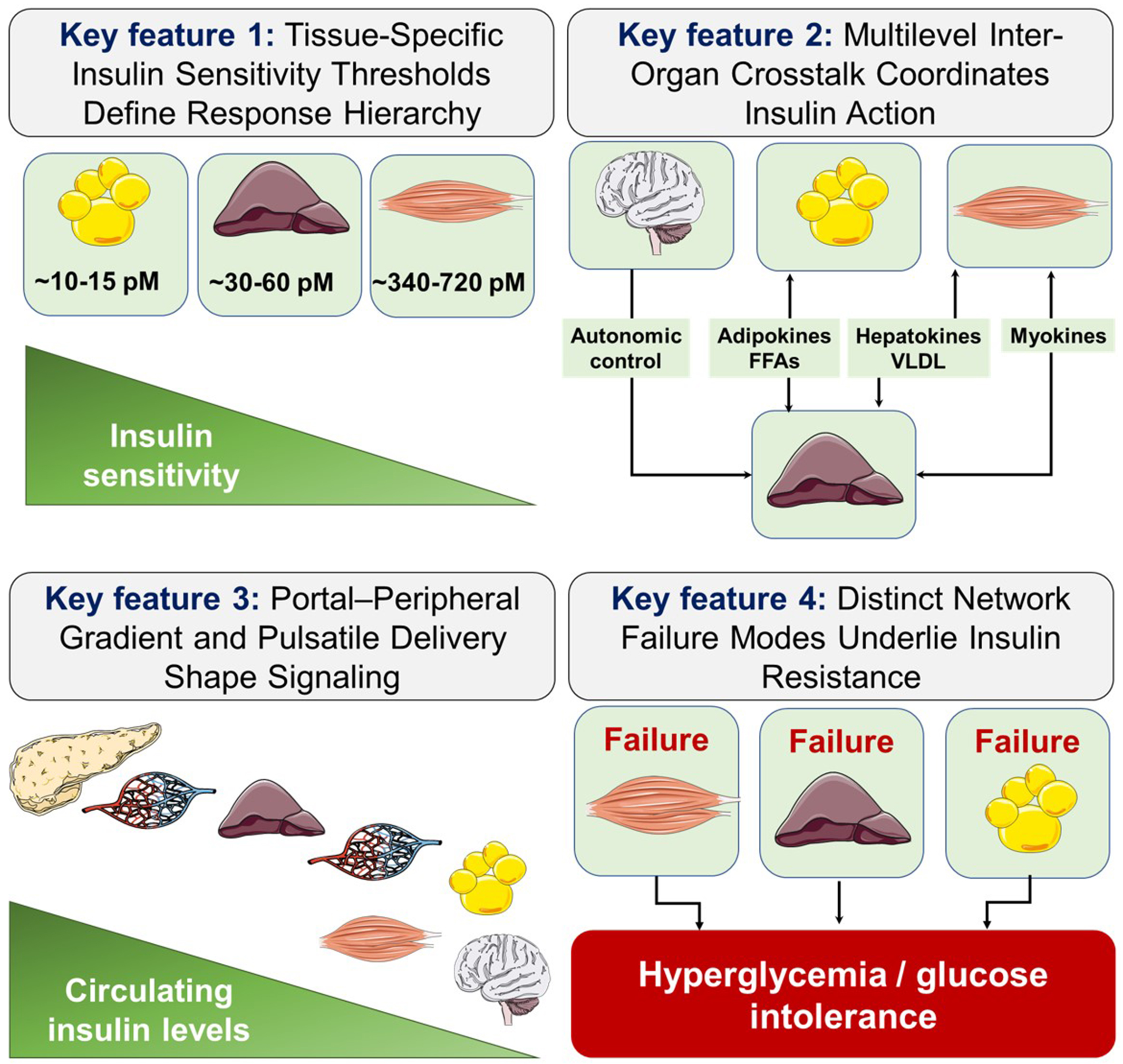
Figure 4. Hierarchical network model of peripheral insulin sensitivity and failure modes. The diagram summarizes four principles: a hierarchical threshold of tissue sensitivity to insulin, in which adipose tissue responds first, followed by liver and then muscle; inter-organ communication centered on the liver as a metabolic hub; a constitutive portal-pulsatile insulin architecture with higher portal than peripheral insulin concentrations; and three major failure modes of the network - adipose, hepatic, and muscular - that converge on hyperglycemia and glucose intolerance. This framework links physiological organization to the rationale for combination therapies that target multiple nodes of the network. FFAs: Free fatty acids; VLDL: very-low-density lipoprotein.
Inter-organ insulin communication depends on concurrent metabolic, hormonal, and neural signals
Inter-tissue coordination operates through multiple channels. Adipose-derived FFAs and adipokines (adiponectin, leptin, resistin) modulate hepatic and muscular signaling[260]. The liver communicates through VLDL-triglyceride secretion, hepatokines[261], and CEACAM1-mediated insulin clearance[117]. The hypothalamus modulates hepatic glucose output and fuel partitioning via autonomic pathways[262]. Skeletal muscle generates myokines during contraction, enhancing adipose lipolysis and hepatic fatty acid oxidation, constituting an exercise-dependent feedback loop[253]. Insulin resistance cannot be understood or effectively targeted by focusing on single tissues. The systemic phenotype emerges from the aggregate network state, and therapeutic perturbation of one node propagates with amplifying or compensatory effects depending on the states of other nodes [Figure 4].
Pulsatile portal insulin delivery functions as an integral component of insulin signaling
As described, insulin reaches the liver at portal concentrations 2-3 times higher than peripheral concentrations, with 50%-80% extracted during the first hepatic passage. This architecture is constitutive, not incidental: reversal of the gradient by subcutaneous insulin delivery produces consequences (weight gain, dyslipidemia, hyperglycemia) that arise from architectural disruption, not dosing problems. Integrative models must consider delivery architecture as a determinant of tissue-specific response. When the portal-pulsatile architecture is intact, physiological insulin responses follow hierarchical tissue sensitivity thresholds and unfold in their proper sequence [Figure 4]. Conversely, architectural disruption does not produce a uniform systemic failure but rather precipitates tissue-specific perturbations whose pattern depends on which node bears the greatest burden of compensation. This principle underlies the observation that insulin resistance is not a single disease state but a family of network failure profiles distinguishable by their initiating lesion and inter-organ propagation sequence.
Insulin resistance encompasses diverse network failure profiles
The controversies discussed above suggest that insulin resistance is initiated and sustained by distinct network failure profiles. Primary adipose expandability failure with ectopic lipid represents a different network state than primary hepatic CEACAM1 loss or primary muscle mitochondrial dysfunction, though all converge on fasting hyperinsulinemia and impaired glucose tolerance. Precision medicine stratification, such as Ahlqvist et al.’ data-driven clusters[263], can be reinterpreted as identifying which node failed first and which inter-organ channels are most disrupted. Therapeutic targeting that accounts for patient-specific network failure profiles represents the logical extension of precision endocrinology and explains treatment response heterogeneity[254]. This framework provides a conceptual basis for combination therapies addressing multiple nodes: GLP-1Ras (central appetite suppression, β-cell protection, hepatic glucose output), SGLT2i (renal glucose excretion, adipose lipolysis modulation, cardiac energetics), and thiazolidinediones (adipose expandability, ADIPOQ secretion), which synergize clinically to exceed single-model predictions [Figure 4].
In summary, peripheral insulin action is best understood as an integrated, hierarchically organized network where adipose tissue sets metabolic tone through exquisite low-insulin sensitivity, liver orchestrates fasting-to-fed transitions through insulin signaling and CEACAM1-dependent clearance, skeletal muscle executes postprandial glucose disposal, and central nervous system provides modulatory control. Network failure in T2D proceeds through tissue-specific routes, the sequence of which defines metabolic subtypes. Effective intervention requires identifying the primary failure node while accounting for network compensatory responses. Future research must move beyond single-tissue studies toward integrative models capturing inter-organ crosstalk dynamics, including pulsatile insulin delivery’s temporal dimension, as the physiological unit of analysis. For clinical translation notes of this section, see Supplementary Materials.
ADIPOBIOLOGY AND INSULIN RESISTANCE: THE ENDOCRINE DIMENSION
Adipose tissue as an active endocrine organ
Adipose tissue was long regarded as a passive lipid storage depot, but the discovery of leptin and adiponectin established it as a dynamic endocrine organ. Adipose tissue secretes biologically active molecules, termed adipokines, plus growth factors, inflammatory cytokines, lipid mediators, and extracellular vesicles that exert autocrine, paracrine, and endocrine effects on glucose metabolism, lipid homeostasis, vascular biology, inflammation, and angiogenesis[264].
With adipose expansion and dysfunction, secretion shifts from anti-inflammatory adipokines (adiponectin, omentin-1, adipolin, vaspin) that enhance insulin sensitivity to a pro-inflammatory profile. Expansion beyond lipid-storage capacity causes adipocyte hypertrophy, hypoxia, stress, and infiltration by pro-inflammatory M1-polarized macrophages, the hallmark of obese dysfunctional adipose tissue. This profile, dominated by leptin, resistin, visfatin, TNF-α, IL-6, monocyte chemoattractant protein-1 (MCP-1), and plasminogen activator inhibitor-1 (PAI-1), impairs insulin signaling and promotes atherogenesis[265,266].
Adipose tissue expandability varies between individuals, determining thresholds for overflow and ectopic fat deposition, with implications for T2D risk stratification beyond body mass index (BMI)-based classification. High expandability may preserve insulin sensitivity, whereas limited subcutaneous expandability favors ectopic fat accumulation at lower adiposity levels[267,268].
Key adipokines: mechanisms and clinical relevance
Leptin, produced mainly by subcutaneous white adipose tissue in proportion to fat mass, signals through leptin receptors (LEPR) (especially hypothalamic LEPRb) to suppress appetite, increase energy expenditure, and activate the sympathetic nervous system. In lipolysis, leptin has a direct effect: chronic signaling increases sympathetic tone in white adipose tissue, promoting lipolysis and reducing adipose mass without changes in food intake in rodents and humans. Leptin also regulates aquaglyceroporins: insulin and leptin coordinate AQP3 and AQP7 expression in adipocytes through the PI3K/AKT/mTOR pathway, linking these hormones to glycerol efflux. In obesity, despite high circulating leptin, central leptin resistance develops through reduced LEPRb signaling and SOCS3-mediated inhibition, blunting anti-obesity signals[269].
Adiponectin, the most abundant circulating adipokine, has insulin-sensitizing, anti-inflammatory, and cardioprotective effects. Through adiponectin receptors (AdipoR1 and AdipoR2), it activates AMPK and PPARα in muscle and liver, promoting fatty acid oxidation, suppressing hepatic gluconeogenesis, and enhancing glucose uptake. Circulating adiponectin is reduced in obesity and inversely correlates with visceral fat, insulin resistance, and T2D risk. The AdipoR agonist (AdipoRon) and derivatives are in preclinical and early clinical development[270].
Resistin (from adipose macrophages) promotes hepatic insulin resistance via NF-κB/SOCS3. TNF-α inhibits insulin signaling through IRS1 serine phosphorylation via JNK/IKKβ. IL-6 has concentration-dependent effects: high levels worsen hepatic insulin resistance; low levels may enhance muscle insulin sensitivity via AMPK[271]. Emerging adipokines [fibroblast growth factor 21 (FGF21), neuregulin-4 (Nrg4), lipocalin-2, omentin-1] extend this network linking adipose biology to β-cell function and hepatic metabolism. This adipose-islet axis - where adiponectin, FGF21, and omentin-1 support GSIS while resistin and pro-inflammatory cytokines impair it - is underappreciated in T2D progression and deserves further investigation[264].
Angiogenesis, growth factors, and adipose remodeling
Dysfunctional adipose tissue produces growth factors, including vascular endothelial growth factor (VEGF), hepatocyte growth factor (HGF), and platelet-derived growth factor (PDGF), that modulate vascularization, remodeling, and immune recruitment. Hypoxia in expanding adipose tissue stimulates hypoxia-inducible factor 1-α (HIF-1α)-driven VEGF expression; despite angiogenic signaling, vascularization in obesity is often insufficient, sustaining chronic hypoxia and adipokine dysregulation. Visceral adipose tissue, with more macrophages and proximity to the portal circulation, has greater metabolic consequences than subcutaneous fat, with direct clinical implications for cardiovascular risk stratification and therapeutic lifestyle intervention[272]. For clinical translation notes of this section, see Supplementary Materials.
GUT MICROBIOTA AND THE INSULIN LIFE CYCLE
Microbiota composition and insulin secretion
The gut microbiome, with 38 trillion cells and vast genetic diversity, profoundly affects host metabolism, immunity, and endocrine signaling. It influences the insulin life cycle through mechanisms affecting β-cell function, incretin secretion, hepatic metabolism, and insulin sensitivity[273].
SCFAs - butyrate, propionate, and acetate - from microbial fiber fermentation are primary metabolic regulators. SCFAs stimulate L-cell GLP-1 and peptide YY (PYY) secretion via G-protein-coupled receptor 41 (GPR41) and GPR43, enhancing the incretin effect and β-cell GSIS[273]. Butyrate improves β-cell function via AMPK phosphorylation and increases GLP-1. Propionate and butyrate also suppress adipocyte lipolysis and lipogenesis while increasing insulin-stimulated glucose uptake[274]. High-fiber diets support this by increasing GLP-1 and improving glycemia in T2D patients[275].
Specific taxa also influence insulin action. Akkermansia muciniphila, low in T2D and obesity, promotes barrier integrity and reduces metabolic endotoxemia; supplementation improves insulin sensitivity in metabolic syndrome[276]. Mendelian randomization by Sun et al. provided causal evidence linking genera like Flavonifractor and Clostridiaceae to T2D risk[277], overcoming limitations of cross-sectional studies.
Metabolic endotoxemia and insulin resistance
The gut microbiome promotes systemic insulin resistance via metabolic endotoxemia, where lipopolysaccharide (LPS) enters circulation due to impaired intestinal barrier function. LPS activates toll-like receptor 4 (TLR4) on macrophages, hepatocytes, and adipocytes. This triggers NF-κB-mediated inflammatory signaling that converges on IRS1 serine phosphorylation and JNK/IKKβ pathways[274]. Metabolic endotoxemia occurs in obese humans, with plasma LPS levels correlating with insulin resistance, HOMA-IR, and T2D risk[278].
Bile acid metabolism is another microbiome-metabolic axis. Bacterial hydrolases and dehydroxylases modify primary bile acids into secondary bile acids. These activate the G-protein-coupled receptor 5 (TGR5) on enteroendocrine L-cells, stimulating GLP-1 secretion, and the nuclear receptor farnesoid X receptor (FXR), modulating glucose and lipid metabolism[279]. T2D is linked to altered secondary bile acid profiles, potentially reducing incretin secretion[280]. RYGB-induced T2D improvement is partly attributed to altered bile acid profiles and increased GLP-1 secretion driven by microbiome changes[281].
Microbiome-based therapeutic strategies
Probiotic supplementation with Lactobacillus and Bifidobacterium species showed modest improvements in glycated hemoglobin and β-cell function in randomized T2D trials[282]. Patients receiving probiotics had lower glycated hemoglobin and improved β-cell function than metformin alone[283]. Fecal microbiota transplantation (FMT) from lean donors to individuals with T2D was tested in a randomized trial by
TEMPORAL DYNAMICS OF β-CELL FAILURE: DEDIFFERENTIATION, CLEARANCE ADAPTATION, AND DISEASE STAGING
A temporal framework for T2D progression
The mechanisms described above - β-cell biosynthesis, insulin secretion, portal delivery and hepatic clearance, and peripheral signaling - do not fail simultaneously in T2D. Prospective studies of high-risk individuals document a stereotyped temporal sequence, the recognition of which has fundamental implications for disease staging and therapeutic timing.
Peripheral insulin resistance develops first, driven by ectopic lipid accumulation, sedentary behavior, and genetic susceptibility. β-Cells compensate through insulin hypersecretion. Concurrently, hepatic steatosis reduces CEACAM1-mediated insulin endocytosis, lowering its clearance and raising systemic insulin - partially masking the emerging secretory deficit. Progressive glucolipotoxicity triggers β-cell dedifferentiation with loss of GSIS. When secretory compensation fails despite reduced clearance, fasting hyperglycemia and clinical T2D manifest[152].
This sequence reframes the natural history of T2D as a cascade of partially compensated failure rather than a linear decline, and identifies distinct intervention windows at each stage: insulin sensitization and lifestyle modification in the resistance-dominant phase; clearance restoration and β-cell identity preservation in the compensatory hypersecretion phase; and β-cell rescue or replacement strategies once dedifferentiation is advanced [Figure 5].
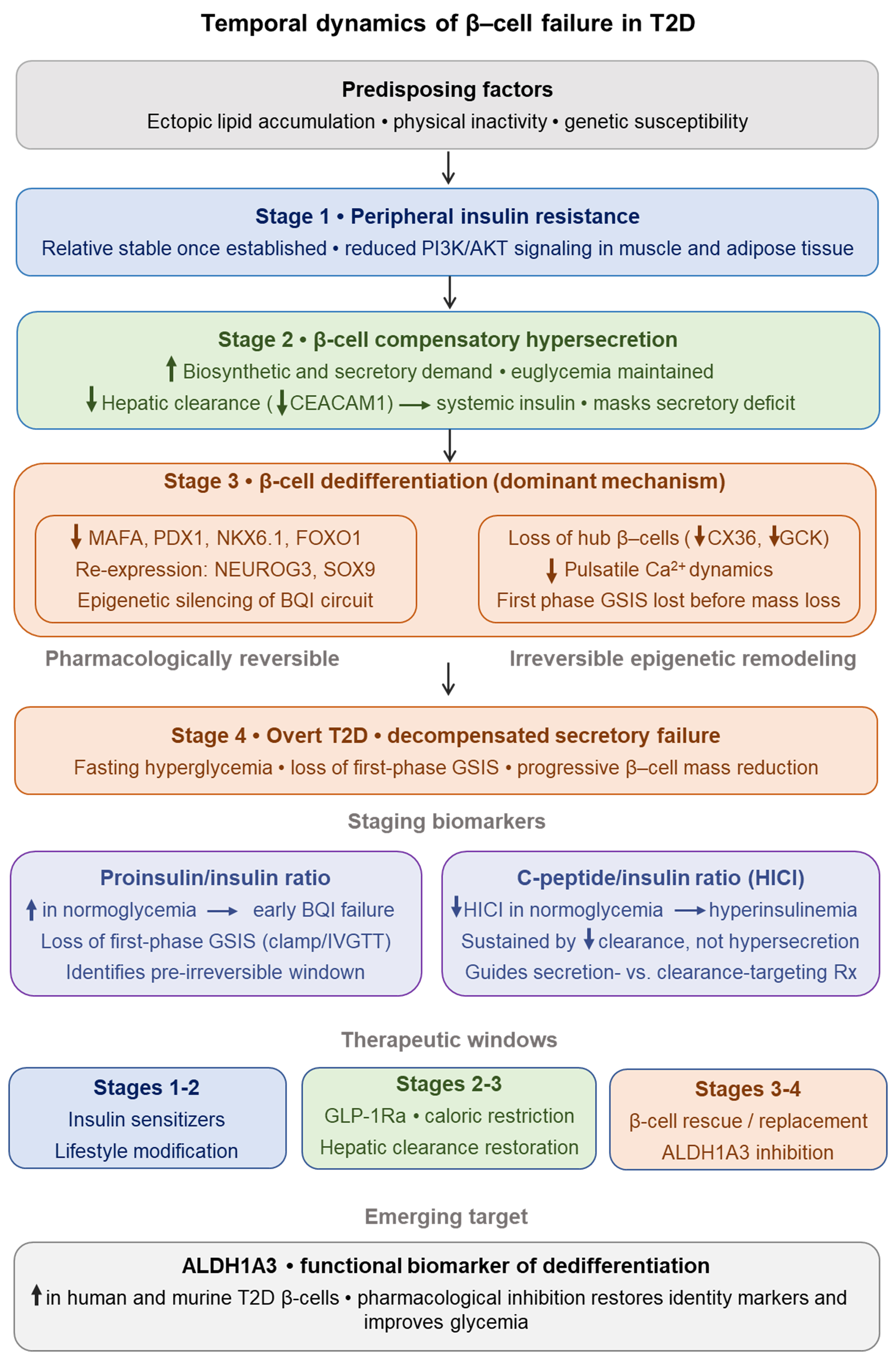
Figure 5. Temporal dynamics of β-cell failure in T2D. Predisposing factors (ectopic lipid, inactivity, genetic susceptibility) lead to peripheral insulin resistance (Stage 1) with reduced PI3K/AKT signaling in muscle and adipose tissue, driving compensatory β-cell hypersecretion (Stage 2) and reduced hepatic insulin clearance (CEACAM1), which raises systemic insulin and can mask secretory defects. Progressive β-cell stress causes loss of identity programs (MAFA, PDX-1, NKX6.1, FOXO1) and re-expression of progenitor/dedifferentiation markers (NEUROG3, SOX9) with disrupted hub β-cell signaling (CX36, GCK, first-phase GSIS lost), producing irreversible epigenetic remodeling and dedifferentiation (Stage 3) that precedes overt decompensated secretory failure and fasting hyperglycemia (Stage 4). Biomarkers (proinsulin/insulin and C-peptide/insulin ratios) and therapeutic windows (insulin sensitizers, GLP-1Ra, hepatic clearance restoration, β-cell rescue/replacement) are indicated. T2D: Type 2 diabetes; PI3K: phosphoinositide 3-kinase; AKT: protein kinase B; CEACAM1: carcinoembryonic antigen-related cell adhesion molecule 1; MAFA: MAF bZIP transcription factor A; PDX1: pancreatic and duodenal homeobox 1; NKX6.1: NK6 homeobox 1; FOXO1: forkhead box O1; NEUROG3: neurogenin 3; SOX9: SRY-box transcription factor 9; BQI: biosynthesis-quality-identity; CX36: connexin 36; GCK: glucokinase; Ca2+: calcium ion; GSIS: glucose-stimulated insulin secretion; HICI: hepatic insulin clearance index; IVGTT: intravenous glucose tolerance test; GLP-1Ra: glucagon-like peptide-1 receptor agonist; ALDH1A3: aldehyde dehydrogenase 1 family member A3; Rx: treatment.
Dedifferentiation as the dominant mechanism of β-cell failure
Classical models attributed progressive β-cell loss in T2D primarily to apoptosis driven by glucotoxicity, lipotoxicity, inflammatory cytokines, and IAPP deposition[285]. A paradigm shift emerged from work by
The molecular basis of this process, characterized by loss of MAFA, PDX-1, NKX6.1, and FOXO1 expression, reactivation of progenitor genes such as NEUROG3 and SOX9, and epigenetic silencing via hypermethylation of the PDX-1 and MAFA promoters, was previously described as failure of the BQI circuit. Here, that same process is contextualized within the temporal disease arc: dedifferentiation represents the downstream consequence of sustained glucolipotoxic load that the BQI circuit can no longer buffer.
Critically, the severity of metabolic stress determines reversibility. Under moderate glucolipotoxicity, dedifferentiation remains limited and pharmacologically reversible, a state amenable to GLP-1Ra, intensive glucose control, and caloric restriction[14]. Under severe or prolonged stress, irreversible epigenetic remodeling, mitochondrial dysfunction, and increased apoptotic loading narrow the therapeutic window[287]. Aldehyde dehydrogenase 1 isoform A3 (ALDH1A3) has emerged as a functional biomarker of the dedifferentiated β-cell state in both human and rodent T2D; its pharmacological inhibition restores β-cell identity markers and improves glycemia in diabetic mice, representing a potential staging and therapeutic target[288].
β-cell subpopulation vulnerability and hub cell loss
The β-cell population is not homogeneous. Single-cell RNA sequencing of human pancreatic islets has identified functionally distinct subpopulations with differential roles in coordinating islet secretory dynamics. A minority of “hub” or “leader” β-cells, characterized by high CX36 expression and specific metabolic gene signatures, orchestrates islet-wide oscillatory calcium dynamics and bears disproportionate responsibility for coordinated pulsatile secretion. These cells are identifiable by reduced PDX-1 and NKX6.1 expression alongside elevated GCK, a paradoxical combination that confers functional dominance but also heightened vulnerability to metabolic stress-induced dedifferentiation[60,289,290].
Loss of hub cells therefore has an outsized impact on secretory capacity relative to their fractional representation in the islet. This hierarchical architecture explains why early functional deficits in T2D, loss of first-phase insulin secretion and disrupted pulsatility, may precede significant reductions in total β-cell mass[290,291]. Additionally, single-cell transcriptomic analyses have revealed that endocrine cell alternative splicing is broadly dysregulated in T2D, adding a post-transcriptional dimension to the dedifferentiation landscape that is not captured by transcription factor immunostaining alone[292].
Disease staging and the therapeutic window
The temporal model outlined above offers a framework for staging T2D that goes beyond glycemic thresholds. The proinsulin/insulin ratio and the loss of first-phase GSIS, detectable by hyperglycemic clamp or intravenous glucose tolerance test, serve as clinical indicators of advancing dedifferentiation. An increasing proinsulin/insulin ratio, in the context of preserved fasting glucose, signals early BQI circuit failure and identifies individuals who may benefit most from interventions targeting β-cell identity before irreversible epigenetic changes occur.
The C-peptide/insulin molar ratio (HICI) provides a complementary staging dimension: a declining HICI in the context of normoglycemia indicates that hyperinsulinemia is sustained partly by reduced clearance rather than pure hypersecretion, a distinction with therapeutic implications for the choice between insulin secretion-reducing vs. clearance-restoring strategies[80,293,294].
Together, these biomarkers define a staging axis that ranges from compensated resistance with preserved β-cell identity, through dedifferentiation onset with clearance adaptation, to decompensated T2D with irreversible secretory failure. This axis may be more mechanistically informative than glycated hemoglobin A1c (HbA1c) alone and better aligned with the pathophysiological sequence described in this manuscript. For clinical translation notes of this section, see Supplementary Materials.
FROM PATHOLOGY TO TREATMENT: THE INSULIN LIFE CYCLE AS A THERAPEUTIC MAP
Therapeutic strategies targeting the insulin life cycle
The insulin life cycle framework provides a rational basis for organizing therapeutic targets. Different drug classes act at different phases: incretins enhance secretion; metformin reduces hepatic glucose production; SGLT2i lower glycemia by renal glucose excretion; insulin sensitizers (thiazolidinediones) improve peripheral signaling; GLP-1RAs additionally reduce appetite and body weight; and insulin replacement corrects absolute secretory deficiency[61,62].
GLP-1RAs: from insulin lifecycle modulation to precision therapeutics
GLP-1RAs have transformed T2D management by acting at multiple nodes of the insulin life cycle. By augmenting postprandial insulin secretion in a glucose-dependent manner, suppressing glucagon, delaying gastric emptying, and reducing appetite through central pathways, GLP-1RAs act on several nodes of metabolic regulation simultaneously[61,62,295,296]. Their clinical benefits also extend to cardiovascular and renal outcomes, while heterogeneity in glycemic response appears to depend in part on residual β-cell function, with C-peptide emerging as a potential predictive biomarker[61,297].
SGLT2i: a novel mechanism with pleiotropic benefits
SGLT2i reduce hyperglycemia by blocking renal glucose reabsorption, causing glucosuria independent of insulin. Lowered glucose and insulin, in turn, reduce β-cell demand, potentially slowing dedifferentiation. Cardiorenal benefits across multiple outcome trials involve mechanisms beyond glycemia: osmotic diuresis, natriuresis, reduced intraglomerular pressure, and direct cardiac energetic effects[298-302]. SGLT2i also reduce hepatic fat and improve transaminases in MASLD, consistent with reduced hepatic insulin resistance[118].
Precision medicine and disease subtyping
A major shift in T2D management philosophy is the recognition of T2D as a heterogeneous syndrome. The cluster-based classification proposed by Ahlqvist et al.[263] defined five T2D clusters based on six variables [glutamic acid decarboxylase antibodies (GADA), age at diagnosis, BMI, HbA1c, HOMA-B, HOMA-IR]: severe autoimmune diabetes (SAID), severe insulin-deficient diabetes (SIDD), severe insulin-resistant diabetes (SIRD), mild obesity-related diabetes (MOD), and mild age-related diabetes (MARD). These clusters showed differential risks of diabetic complications and potentially different optimal therapies.
A systematic review[61] found that reduced insulin secretion markers predicted a lesser glycemic response to GLP-1Ra, and reduced renal function predicted a lesser response to SGLT2i. A subsequent Bayesian causal forest analysis[62] validated an individualized SGLT2i vs. GLP-1Ra selection algorithm, showing predicted-response-based targeting improved short-term tolerability and long-term microvascular risk. These analyses represent early steps toward clinical precision medicine in T2D[61,62], though prospective validation in diverse populations is required.
Emerging targets: β-cell regeneration and clearance optimization
Several emerging therapeutic strategies address upstream life-cycle defects. ALDH1A3 inhibition to reverse β-cell dedifferentiation has shown promise in rodent T2D models[288] but awaits clinical translation. Stem cell-derived islet transplantation achieved insulin independence in a single T2D patient in a first-in-human pilot study, representing a promising but preliminary proof-of-concept for treating end-stage secretory deficiency; larger controlled trials are needed to confirm efficacy and safety.[303,304]. Therapeutic targeting of CEACAM1 or IDE to optimize hepatic insulin clearance remains at the preclinical stage[80,159]. Microbiome modulation through precision dietary interventions, probiotic formulations, and postbiotic therapies targeting the SCFA-GLP-1-insulin axis represents a low-risk adjunctive strategy with growing human evidence[284,305,306]. For clinical translation notes of this section, see Supplementary Materials.
AREAS OF CONTROVERSY AND OUTSTANDING QUESTIONS
Selective hepatic insulin resistance, whether it represents a branch-specific signaling defect, a cell-non-autonomous pathway, or a biologically adaptive response, remains mechanistically unresolved. The competing hypotheses, Shulman’s differential IRS1/IRS2 susceptibility model vs. the Najjar laboratory’s CEACAM1/clearance-driven account, are presented side by side, and their therapeutic implications are contrasted. Whether reduced hepatic insulin clearance is a cause or a consequence of hepatic insulin resistance remains unresolved[119,152]. The bidirectional “clearance-resistance loop” and its molecular basis have been described above.
The relative importance of β-cell dedifferentiation vs. apoptosis as the main mechanism of β-cell mass loss in human T2D remains debated. The molecular basis of dedifferentiation - loss of MAFA, PDX-1, and NKX6.1, reactivation of progenitor genes, and epigenetic silencing - is detailed above, as is its distinction from apoptosis. Still, human pancreatic studies are constrained by tissue availability and cross-sectional design, and by a lack of longitudinal data from serial pancreatic biopsies in living subjects. The surrogate biomarkers proposed for staging - proinsulin-to-insulin ratio, C-peptide decline trajectories, and HICI - were previously discussed, but they require prospective validation against autopsy data.
The causal role of gut microbiota in human T2D is increasingly supported by Mendelian randomization studies, although confirmation in large-scale interventional trials is still needed. The mechanisms linking microbial metabolites such as SCFAs, bile acids, and LPS to incretin secretion and insulin resistance, as well as the evidence for FMT, probiotics, and precision prebiotics, are discussed here in relation to their therapeutic potential as adjuncts. However, whether these interventions can produce clinically meaningful and durable improvements in patients with T2D remains to be established.
Whether central insulin resistance contributes to systemic glucose dysregulation in human T2D remains under investigation. Studies in rodents have provided evidence that hypothalamic insulin suppresses hepatic glucose output and regulates appetite. However, the translational limitations of these findings and the inconclusive human data from intranasal insulin studies should be critically considered. A better quantification of the magnitude and therapeutic relevance of central insulin action in whole-body glucose homeostasis is needed before clinical translation can be pursued.
Throughout this review, we explicitly flag the translational gap between rodent models and humans: the single human INS gene, species differences in GLUT expression, limited direct human evidence for CEACAM1 function, and uncertain magnitude of central insulin action in humans. Species differences in islet architecture, glucose transport, and incretin biology mean that many murine mechanistic findings require dedicated human validation. The precision medicine framework represents the most direct attempt to bridge this gap by grounding decisions in human-derived biomarker data rather than rodent mechanism alone. For clinical translation notes of this section, see Supplementary Materials.
METABOLIC AGING, CELLULAR SENESCENCE, AND GEROSCIENCE: A FRAMEWORK FOR FUTURE PRECISION STRATEGIES IN T2D
Reframing T2D as a disease of metabolic aging
A fundamental but underappreciated dimension of the insulin lifecycle is its temporal relationship with aging. β-Cell deterioration, hepatic steatosis, peripheral insulin resistance, and chronic inflammation share mechanistic overlap with hallmarks of biological aging. The Najjar laboratory has recently shown that reduction of hepatic CEACAM1 levels and impaired insulin clearance mediate age-related hepatic steatohepatitis and fibrosis and that protecting CEACAM1 prevents the metabolic dysfunction and liver injury associated with aging and bestows a survival advantage[307]. Reframing T2D as a disease of metabolic aging shifts therapeutic thinking toward upstream, disease-modifying intervention targeting aging processes that may simultaneously prevent the full spectrum of age-related metabolic dysfunction[101,308-310].
Senescent cells accumulate in metabolically stressed tissues (pancreatic islets, adipose tissue, liver, muscle) and are characterized by irreversible cell cycle arrest, apoptosis resistance, and secretion of a senescence-associated secretory phenotype (SASP), pro-inflammatory cytokines including IL-1β, IL-6, TNF-α, and MCP-1[310]. The SASP creates a pro-inflammatory milieu that impairs β-cell function, deepens insulin resistance, promotes hepatic steatosis, and disrupts peripheral insulin signaling.
Cellular senescence as an upstream therapeutic target
Pancreatic β-cells are particularly susceptible to stress-induced senescence from glucotoxicity, lipotoxicity, inflammatory cytokines, and oxidative stress, which induce DNA damage and senescence mediated by p16INK4a (protein of 16 kilodaltons/inhibitor of cyclin-dependent kinase 4a) and p21CIP1 (protein of 21 kilodaltons/CDK-interacting protein 1)[310,311]. Senescence may precede β-cell dedifferentiation or apoptosis, positioning it as a temporally earlier therapeutic target[310,312]. The convergence between cellular senescence, SASP, and inflammatory pathways (NF-κB, JNK, IKKβ) establishes senescence as a mechanistic hub linking aging to the entire insulin life cycle[313].
Senotherapeutics include senolytics (which eliminate senescent cells: dasatinib/quercetin, navitoclax, fisetin) and senomorphics [which suppress SASP: janus kinase (JAK)1/2 inhibitors, rapamycin, metformin][310,314]. While preclinical models demonstrate metabolic improvements, human evidence remains limited to small early-phase trials focused on safety and biomarker modulation rather than glycemic efficacy[310,315,316]. Senotherapeutics should be positioned as a conceptual framework to guide future precision-based clinical trials rather than advocating near-term clinical adoption.
Sirtuin activators and NAD+ biology: bridging geroscience and diabetology
The sirtuin family (SIRT1-7) interfaces energy sensing, aging, and metabolic regulation. SIRT1 activates PGC-1α (mitochondrial biogenesis master regulator) and suppresses NF-κB inflammatory signaling. SIRT3 maintains oxidative phosphorylation and suppresses mitochondrial ROS, directly countering two of the principal drivers of β-cell failure and peripheral insulin resistance[317-319]. Declining NAD+ with aging impairs sirtuin activity, contributing to senescence-inflammation and metabolic deterioration. NAD+ precursors (nicotinamide riboside, NMN) and sirtuin activators (SRT2104, resveratrol) have shown modest improvements in insulin sensitivity and inflammatory markers in early human trials, though larger trials with mechanistic endpoints are required to establish clinical significance[320-322].
Geroscience applications in precision risk stratification
The advent of high-resolution precision medicine tools has created an unprecedented opportunity to implement the shift from reactive to preventive diabetes medicine. Polygenic risk scores (PRS) for T2D achieve sufficient discriminatory power to stratify risk decades before hyperglycemia. Variants clustering around insulin secretion loci (TCF7L2, KCNJ11) identify different biological subtypes than those around insulin resistance loci (IRS1, PPARγ, FTO), enabling biologically informed stratification to direct prevention toward individuals likely to benefit from specific interventions[323,324].
Multi-cancer early detection (MCED) tests and epigenetic clocks illustrate the feasibility of population-level biological aging biomarkers. Epigenetic clocks provide quantitative surrogates of aging rate correlating with T2D incidence and mortality, potentially serving as actionable endpoints in senotherapy trials[310]. Together, PRS and biological aging metrics enable stratification by metabolic phenotype and aging rate, facilitating individualized intervention timing and target selection[310].
Integrating geroscience with diabetology: toward future precision-based and disease-modifying strategies
Current T2D management is reactive: treatment begins after hyperglycemia, targeting downstream manifestations of a years-long process. The geroscience-informed vision is substantively different: identify high-risk individuals through PRS and aging biomarkers, characterize the dominant life-cycle defect through molecular phenotyping, and deploy senescence-targeted or sirtuin-activating interventions to interrupt the causal chain between accelerated metabolic aging and clinical T2D before irreversible damage occurs[118,310,325].
Cellular senescence can be understood as a cross-cutting upstream mechanism degrading all insulin life cycle phases: SASP-mediated ER stress impairs β-cell biosynthesis; senescent β-cells lose secretory identity; hepatic senescence reduces insulin clearance; SASP cytokines drive inflammatory insulin resistance; adipose senescence amplifies dysfunctional adipokine secretion; and senescence-associated mitochondrial dysfunction accelerates insulin degradation. Cellular senescence is thus a unifying upstream regulator of insulin life cycle deterioration in metabolic aging[310,326].
Translating geroscience into practice faces challenges: identifying optimal targets, establishing therapeutic windows (senescence serves physiological roles), developing biomarkers tracking target engagement, and designing adequately powered trials[327]. For clinical translation notes of this section, see Supplementary Materials.
Limitations
This work has several limitations that should be acknowledged. First, as a narrative rather than a systematic review, this manuscript was not based on a pre-registered protocol or a formal quality/risk-of-bias appraisal of individual studies, and no quantitative synthesis was performed. Although a structured, multi-database search with explicit eligibility criteria was used to identify relevant evidence, the narrative synthesis and organization of findings around an integrative physiological framework carry an inherent risk of selective emphasis on findings that support the proposed model, and the interpretation should be considered accordingly.
Second, most of the mechanistic evidence synthesized here, particularly regarding β-cell dedifferentiation, the CEACAM1/IDE clearance pathway, and central and adipose insulin signaling, derives from rodent models or from human data that are cross-sectional rather than longitudinal, constraining direct causal inference in humans. Third, the BQI axis and the proposed staged model of disease progression are conceptual, hypothesis-generating frameworks intended to organize existing evidence and guide future study design; they have not themselves been prospectively tested and should not be regarded as validated diagnostic or therapeutic algorithms. Fourth, the candidate biomarkers discussed for patient staging, including the proinsulin-to-insulin ratio, the hepatic insulin clearance index, and senescence-associated markers, lack standardized assay methods and validated clinical cut-offs, and none has yet been incorporated into prospective trials or clinical guidelines. Fifth, the causal direction linking reduced hepatic insulin clearance to peripheral insulin resistance remains unresolved, as does the relative contribution of β-cell dedifferentiation vs. apoptosis to human β-cell mass loss; both are presented here as leading hypotheses rather than established mechanisms. Sixth, evidence for several emerging modulators, including gut microbiota-derived metabolites, bile acid signaling, and cellular senescence pathways, is largely correlative or derived from small interventional studies, and their incremental value over established therapies remains to be demonstrated in adequately powered trials. Finally, species differences, including the single human INS gene compared with the duplicated rodent Ins1/Ins2 genes, and differences in GLUT expression and islet architecture between rodents and humans, limit the direct extrapolation of several mechanistic findings discussed throughout this review. These limitations do not diminish the value of an integrative perspective but define the boundaries within which its conclusions should be interpreted, and they delineate priority areas for future prospective, mechanistic, and clinical investigation.
Notwithstanding these limitations, reframing the insulin life cycle as an interconnected network, rather than a set of independent nodes, offers a coherent explanation for previously discordant findings and a rational basis for staged, precision-based therapeutic strategies. Resolving the priority questions identified above will be essential to move this framework from a conceptual model toward validated, disease-modifying clinical practice.
CONCLUSIONS
The insulin lifecycle in health and T2D is best understood not as a linear cascade but as a hierarchical, multidimensional network where dysfunction at any node - biosynthesis, β-cell identity, secretion, hepatic clearance, or peripheral signaling - propagates systemic consequences. Within this framework, three interconnected processes emerge as central to disease progression. First, pancreatic β-cell dedifferentiation, driven by the loss of PDX-1, MAFA, and NKX6.1 and by epigenetic silencing of their promoters, may represent the dominant mechanism of β-cell functional loss, challenging the earlier apoptosis-centered paradigm and defining distinct, stage-specific therapeutic windows. Second, reduced hepatic insulin clearance, mediated by CEACAM1- and INSR-dependent endocytosis, appears to establish a bidirectional feedback loop with peripheral insulin resistance that amplifies compensatory hyperinsulinemia, although the direction of causality still requires validation in prospective human studies. Third, adipose tissue dysfunction - along with additional contributors such as gut microbiota-derived metabolites, cellular senescence, and altered insulin distribution kinetics from exogenous administration - further shapes the trajectory and heterogeneity of peripheral insulin resistance across T2D subtypes.
This integrated, network-based view carries direct clinical significance. It supports reconceptualizing T2D as a family of distinguishable altered molecular mechanisms rather than a single uniform disease. This framework identifies mechanistic biomarkers - proinsulin/insulin ratio, hepatic insulin clearance index, and biological aging measures - that can guide stage- and subtype-matched precision therapy. This includes early lifestyle/SGLT2i intervention, compensatory-phase GLP-1Ra, and cell-replacement strategies for advanced dedifferentiation.
DECLARATIONS
Acknowledgments
The image elements in the graphical abstract and figures were adapted from Servier Medical Art (https://smart.servier.com/), licensed under CC BY 4.0 (https://creativecommons.org/licenses/by/4.0/).
Authors’ contributions
Made substantial contributions to the concept and design of the study: Cózar-Castellano I, Perdomo G, Najjar SM
Investigation (searching databases, screening and extracting data): Urbano-Cano AL, Perdomo G
Formal analysis (synthesizing and comparing the selected studies): Perdomo G
Original draft preparation: Cózar-Castellano I, Urbano-Cano AL, Perdomo G, Najjar SM
Review and editing: Perdomo G, Najjar SM
Approved the final version of the manuscript: Cózar-Castellano I, Urbano-Cano AL, Perdomo G, Najjar SM
Availability of data and materials
Not applicable.
AI and AI-assisted tools statement
During the preparation of this manuscript, the AI tool Paperpal Prime (version 2.0.11, released 2026-06-01) was used solely for language editing. The tool did not influence the study design, data collection, analysis, interpretation, or the scientific content of the work. All authors take full responsibility for the accuracy, integrity, and final content of the manuscript.
Financial support and sponsorship
We acknowledge support from the Scientific Network Conexión Enfermedades Metabólicas (COMETA) funded by the Consejo Superior de Investigaciones Científicas (CSIC), Spain. This work was supported by grants (PID2019-110496RB-C21 and PID2022-136605OB-C21) to ICC and (PID2019-110496RB-C22 and PID2022-136605OB-C22) to Perdomo G, funded by MCIN/AEI/10.13039/501100011033 “ERDF A way of making Europe”. This work was also supported by NIH grants (R01-DK054254, R01-DK124126, and R01-DK129877) to Najjar SM. The work was also partially supported by the Osteopathic Heritage Foundation John J. Kopchick Eminent Research Chair at Najjar SM.
Conflicts of interest
Najjar SM is the Guest Editor of the Special Issue “Women Leading Metabolic Sciences” of Metabolism and Target Organ Damage and the Associate Editor of the journal Metabolism and Target Organ Damage. Najjar SM was not involved in any steps of editorial processing, notably including reviewers’ selection, manuscript handling and decision making. The other authors declare that there are no conflicts of interest.
Ethical approval and consent to participate
Not applicable.
Consent for publication
Not applicable.
Copyright
© The Author(s) 2026.
Supplementary Materials
REFERENCES
1. Tokarz VL, MacDonald PE, Klip A. The cell biology of systemic insulin function. J Cell Biol. 2018;217:2273-89.
2. Norton L, Shannon C, Gastaldelli A, DeFronzo RA. Insulin: the master regulator of glucose metabolism. Metabolism. 2022;129:155142.
3. Vogt ÉL, Kowaltowski AJ. GLP-1, Pancreatic β-cells, and insulin secretion: what we know and where we need to go. Diabetes. 2026;75:403-13.
4. Richter EA, Bilan PJ, Klip A. A comprehensive view of muscle glucose uptake: regulation by insulin, contractile activity, and exercise. Physiol Rev. 2025;105:1867-945.
5. Najjar SM, Caprio S, Gastaldelli A. Insulin clearance in health and disease. Annu Rev Physiol. 2023;85:363-81.
6. Sun H, Saeedi P, Karuranga S, et al. IDF diabetes atlas: global, regional and country-level diabetes prevalence estimates for 2021 and projections for 2045. Diabetes Res Clin Pract. 2022;183:109119.
7. American Diabetes Association Professional Practice Committee for Diabetes*. 9. Pharmacologic approaches to glycemic treatment: standards of care in diabetes-2026. Diabetes Care. 2026;49:S183-215.
8. Bo T, Gao L, Yao Z, et al. Hepatic selective insulin resistance at the intersection of insulin signaling and metabolic dysfunction-associated steatotic liver disease. Cell Metab. 2024;36:947-68.
9. Fu Z, Gilbert ER, Liu D. Regulation of insulin synthesis and secretion and pancreatic Beta-cell dysfunction in diabetes. Curr Diabetes Rev. 2013;9:25-53.
10. Lee K, Cho H, Rickert RW, et al. FOXA2 is required for enhancer priming during pancreatic differentiation. Cell Rep. 2019;28:382-93.e7.
11. Geusz RJ, Wang A, Lam DK, et al. Sequence logic at enhancers governs a dual mechanism of endodermal organ fate induction by FOXA pioneer factors. Nat Commun. 2021;12:6636.
12. Schaffer AE, Taylor BL, Benthuysen JR, et al. Nkx6.1 controls a gene regulatory network required for establishing and maintaining pancreatic Beta cell identity. PLoS Genet. 2013;9:e1003274.
13. Zhu Y, Liu Q, Zhou Z, Ikeda Y. PDX1, Neurogenin-3, and MAFA: critical transcription regulators for beta cell development and regeneration. Stem Cell Res Ther. 2017;8:240.
14. Son J, Accili D. Reversing pancreatic β-cell dedifferentiation in the treatment of type 2 diabetes. Exp Mol Med. 2023;55:1652-8.
15. Jeyagaran A, Urbanczyk M, Layland SL, Weise F, Schenke-Layland K. Forward programming of hiPSCs towards beta-like cells using Ngn3, Pdx1, and MafA. Sci Rep. 2024;14:13608.
16. Patel S, Remedi MS. Loss of β-cell identity and dedifferentiation, not an irreversible process? Front Endocrinol 2024;15:1414447.
17. Tang X, Kuo T, Wei Z. Editorial: pancreatic beta-cell dedifferentiation. Front Endocrinol. 2024;15:1524001.
18. Chen K, Zhang J, Huang Y, Tian X, Yang Y, Dong A. Single-cell RNA-seq transcriptomic landscape of human and mouse islets and pathological alterations of diabetes. iScience. 2022;25:105366.
19. Yang BT, Dayeh TA, Volkov PA, et al. Increased DNA methylation and decreased expression of PDX-1 in pancreatic islets from patients with type 2 diabetes. Mol Endocrinol. 2012;26:1203-12.
20. Kim H, Kulkarni RN. Epigenetics in β-cell adaptation and type 2 diabetes. Curr Opin Pharmacol. 2020;55:125-31.
21. Aigha II, Abdelalim EM. NKX6.1 transcription factor: a crucial regulator of pancreatic β cell development, identity, and proliferation. Stem Cell Res Ther. 2020;11:459.
22. Taylor BL, Liu FF, Sander M. Nkx6.1 is essential for maintaining the functional state of pancreatic beta cells. Cell Rep. 2013;4:1262-75.
23. Matsuoka TA, Artner I, Henderson E, Means A, Sander M, Stein R. The MafA transcription factor appears to be responsible for tissue-specific expression of insulin. Proc Natl Acad Sci U S A. 2004;101:2930-3.
24. Guo QS, Zhu MY, Wang L, et al. Combined transfection of the three transcriptional factors, PDX-1, NeuroD1, and MafA, causes differentiation of bone marrow mesenchymal stem cells into insulin-producing cells. Exp Diabetes Res. 2012;2012:672013.
25. Liu XD, Ruan JX, Xia JH, Yang SL, Fan JH, Li K. The study of regulatory effects of Pdx-1, MafA and NeuroD1 on the activity of porcine insulin promoter and the expression of human islet amyloid polypeptide. Mol Cell Biochem. 2014;394:59-66.
26. Liang J, Chirikjian M, Pajvani UB, Bartolomé A. MafA regulation in β-cells: from transcriptional to post-translational mechanisms. Biomolecules. 2022;12:535.
27. Hou N, Mogami H, Kubota-Murata C, Sun M, Takeuchi T, Torii S. Preferential release of newly synthesized insulin assessed by a multi-label reporter system using pancreatic β-cell line MIN6. PLoS One. 2012;7:e47921.
28. Sun J, Cui J, He Q, Chen Z, Arvan P, Liu M. Proinsulin misfolding and endoplasmic reticulum stress during the development and progression of diabetes. Mol Aspects Med. 2015;42:105-18.
29. Boyer CK, Bauchle CJ, Zhang J, Wang Y, Stephens SB. Synchronized proinsulin trafficking reveals delayed Golgi export accompanies β-cell secretory dysfunction in rodent models of hyperglycemia. Sci Rep. 2023;13:5218.
30. Cui D, Feng X, Lei S, et al. Pancreatic β-cell failure, clinical implications, and therapeutic strategies in type 2 diabetes. Chin Med J. 2024;137:791-805.
31. Zavarzadeh PG, Panchal K, Bishop D, et al. Exploring proinsulin proteostasis: insights into beta cell health and diabetes. Front Mol Biosci. 2025;12:1554717.
32. Wahren J, Kallas A, Sima AA. The clinical potential of C-peptide replacement in type 1 diabetes. Diabetes. 2012;61:761-72.
33. Chen J, Huang Y, Liu C, Chi J, Wang Y, Xu L. The role of C-peptide in diabetes and its complications: an updated review. Front Endocrinol. 2023;14:1256093.
34. Arunagiri A, Haataja L, Pottekat A, et al. Proinsulin misfolding is an early event in the progression to type 2 diabetes. Elife. 2019;8:e44532.
35. Izumi T, Yokota-Hashimoto H, Zhao S, Wang J, Halban PA, Takeuchi T. Dominant negative pathogenesis by mutant proinsulin in the Akita diabetic mouse. Diabetes. 2003;52:409-16.
36. Chen CW, Guan BJ, Alzahrani MR, et al. Adaptation to chronic ER stress enforces pancreatic β-cell plasticity. Nat Commun. 2022;13:4621.
37. Shrestha N, De Franco E, Arvan P, Cnop M. Pathological β-cell endoplasmic reticulum stress in type 2 diabetes: current evidence. Front Endocrinol. 2021;12:650158.
38. Diane A, Allouch A, Mu-U-Min RBA, Al-Siddiqi HH. Endoplasmic reticulum stress in pancreatic β-cell dysfunctionality and diabetes mellitus: a promising target for generation of functional hPSC-derived β-cells in vitro. Front Endocrinol. 2024;15:1386471.
39. An Y, Norris N, Li D, Gunton JE. Βeta-cells: stress, identity, failure and diabetes. Cells. 2026;15:475.
40. Zhang Y, Lin S, Yao J, et al. XBP1 splicing contributes to endoplasmic reticulum stress-induced human islet amyloid polypeptide up-regulation. Genes Dis. 2024;11:101148.
41. Yau B, Ghislain J, Kebede MA, Hughes J, Poitout V. The role of the beta cell in type 2 diabetes: new findings from the last 5 years. Diabetologia. 2025;68:2092-103.
42. Wat LW, Svensson KJ. Novel secreted regulators of glucose and lipid metabolism in the development of metabolic diseases. Diabetologia. 2024;67:2626-36.
43. Salvadó L, Barroso E, Gómez-Foix AM, et al. PPARβ/δ prevents endoplasmic reticulum stress-associated inflammation and insulin resistance in skeletal muscle cells through an AMPK-dependent mechanism. Diabetologia. 2014;57:2126-35.
44. Chen YC, Taylor AJ, Fulcher JM, et al. Deletion of carboxypeptidase E in β-cells disrupts proinsulin processing but does not lead to spontaneous development of diabetes in mice. Diabetes. 2023;72:1277-88.
45. Dror E, Fagnocchi L, Wegert V, et al. Epigenetic dosage identifies two major and functionally distinct β cell subtypes. Cell Metab. 2023;35:821-36.e7.
46. Rutter GA, Gresch A, Delgadillo Silva L, Benninger RKP. Exploring pancreatic beta-cell subgroups and their connectivity. Nat Metab. 2024;6:2039-53.
47. Hall E, Dekker Nitert M, Volkov P, et al. The effects of high glucose exposure on global gene expression and DNA methylation in human pancreatic islets. Mol Cell Endocrinol. 2018;472:57-67.
48. Dayeh T, Volkov P, Salö S, et al. Genome-Wide DNA methylation analysis of human pancreatic islets from type 2 diabetic and non-diabetic donors identifies candidate genes that influence insulin secretion. PLoS Genet. 2014;10:e1004160.
49. Rönn T, Ofori JK, Perfilyev A, et al. Genes with epigenetic alterations in human pancreatic islets impact mitochondrial function, insulin secretion, and type 2 diabetes. Nat Commun. 2023;14:8040.
50. Downing TL, Soto J, Morez C, et al. Biophysical regulation of epigenetic state and cell reprogramming. Nat Mater. 2013;12:1154-62.
52. Fernández-Díaz CM, Merino B, López-Acosta JF, et al. Pancreatic β-cell-specific deletion of insulin-degrading enzyme leads to dysregulated insulin secretion and β-cell functional immaturity. Am J Physiol Endocrinol Metab. 2019;317:E805-19.
53. De Vos A, Heimberg H, Quartier E, et al. Human and rat beta cells differ in glucose transporter but not in glucokinase gene expression. J Clin Invest. 1995;96:2489-95.
55. Rorsman P, Ashcroft FM. Pancreatic β-cell electrical activity and insulin secretion: of mice and men. Physiol Rev. 2018;98:117-214.
56. Deepa Maheshvare M, Raha S, König M, Pal D. A pathway model of glucose-stimulated insulin secretion in the pancreatic β-cell. Front Endocrinol. 2023;14:1185656.
57. Muñoz F, Fex M, Moritz T, Mulder H, Cataldo LR. Unique features of β-cell metabolism are lost in type 2 diabetes. Acta Physiol. 2024;240:e14148.
58. Merrins MJ, Corkey BE, Kibbey RG, Prentki M. Metabolic cycles and signals for insulin secretion. Cell Metab. 2022;34:947-68.
59. Foster HR, Ho T, Potapenko E, et al. β-cell deletion of the PKm1 and PKm2 isoforms of pyruvate kinase in mice reveals their essential role as nutrient sensors for the K(ATP) channel. Elife. 2022;11:e79422.
60. Rutter GA, Hodson DJ, Chabosseau P, Haythorne E, Pullen TJ, Leclerc I. Local and regional control of calcium dynamics in the pancreatic islet. Diabetes Obes Metab. 2017;19:30-41.
61. Young KG, McInnes EH, Massey RJ, et al. ; ADA/EASD PDMI. Treatment effect heterogeneity following type 2 diabetes treatment with GLP1-receptor agonists and SGLT2-inhibitors: a systematic review. Commun Med. 2023;3:131.
62. Cardoso P, Young KG, Nair ATN, et al. ; MASTERMIND consortium. Phenotype-based targeted treatment of SGLT2 inhibitors and GLP-1 receptor agonists in type 2 diabetes. Diabetologia. 2024;67:822-36.
63. Trapanese V, Dagostino A, Natale MR, et al. Bidirectional interactions between the gut microbiota and incretin-based therapies. Life. 2025;15:843.
64. Meier JJ, Veldhuis JD, Butler PC. Pulsatile insulin secretion dictates systemic insulin delivery by regulating hepatic insulin extraction in humans. Diabetes. 2005;54:1649-56.
66. Miranda MA, Macias-Velasco JF, Lawson HA. Pancreatic β-cell heterogeneity in health and diabetes: classes, sources, and subtypes. Am J Physiol Endocrinol Metab. 2021;320:E716-31.
68. Zabuliene L, Ilias I. Epinephrine also acts on beta cells and insulin secretion. World J Clin Cases. 2024;12:1712-3.
69. Dalle S, Abderrahmani A. Receptors and signaling pathways controlling beta-cell function and survival as targets for anti-diabetic therapeutic strategies. Cells. 2024;13:1244.
70. Zhang Y, Han C, Zhu W, et al. Glucagon potentiates insulin secretion via β-cell GCGR at physiological concentrations of glucose. Cells. 2021;10:2495.
71. Shuai H, Xu Y, Ahooghalandari P, Tengholm A. Glucose-induced cAMP elevation in β-cells involves amplification of constitutive and glucagon-activated GLP-1 receptor signalling. Acta Physiol. 2021;231:e13611.
72. El K, Douros JD, Willard FS, et al. The incretin co-agonist tirzepatide requires GIPR for hormone secretion from human islets. Nat Metab. 2023;5:945-54.
73. Barmaver SN, Gu G, Kaverina I. Roles of molecular motors in insulin-secreting beta cells. Curr Opin Cell Biol. 2025;97:102582.
74. Zhu X, Hu R, Brissova M, et al. Microtubules negatively regulate insulin secretion in pancreatic β cells. Dev Cell. 2015;34:656-68.
75. Li W, Li A, Yu B, et al. In situ structure of actin remodeling during glucose-stimulated insulin secretion using cryo-electron tomography. Nat Commun. 2024;15:1311.
76. Hughes JW, Cho JH, Conway HE, et al. Primary cilia control glucose homeostasis via islet paracrine interactions. Proc Natl Acad Sci U S A. 2020;117:8912-23.
77. Müller A, Klena N, Pang S, et al. Structure, interaction and nervous connectivity of beta cell primary cilia. Nat Commun. 2024;15:9168.
78. Gerdes JM, Christou-Savina S, Xiong Y, et al. Ciliary dysfunction impairs beta-cell insulin secretion and promotes development of type 2 diabetes in rodents. Nat Commun. 2014;5:5308.
79. Kluth O, Stadion M, Gottmann P, et al. Decreased expression of Cilia genes in pancreatic islets as a risk factor for type 2 diabetes in mice and humans. Cell Rep. 2019;26:3027-36.e3.
80. Najjar SM, Perdomo G. Hepatic insulin clearance: mechanism and physiology. Physiology. 2019;34:198-215.
81. Rubio-Navarro A, Gómez-Banoy N, Stoll L, et al. A beta cell subset with enhanced insulin secretion and glucose metabolism is reduced in type 2 diabetes. Nat Cell Biol. 2023;25:565-78.
82. Peng X, Ren H, Yang L, et al. Readily releasable β cells with tight Ca2+-exocytosis coupling dictate biphasic glucose-stimulated insulin secretion. Nat Metab. 2024;6:238-53.
83. Zhang Z, Wang S, Gao L. Circadian rhythm, glucose metabolism and diabetic complications: the role of glucokinase and the enlightenment on future treatment. Front Physiol. 2025;16:1537231.
84. Grasset E, Puel A, Charpentier J, et al. Gut microbiota dysbiosis of type 2 diabetic mice impairs the intestinal daily rhythms of GLP-1 sensitivity. Acta Diabetol. 2022;59:243-58.
85. Xing J, Chen C. Hyperinsulinemia: beneficial or harmful or both on glucose homeostasis. Am J Physiol Endocrinol Metab. 2022;323:E2-7.
86. Dahiya R, Singh AP, Rawat A. Immunometabolic reprogramming and β-cell dedifferentiation: integrated mechanisms driving type 2 diabetes progression. Diabetes Res Clin Pract. 2026;232:113111.
87. Ren H, Li Y, Xie B, et al. Pancreatic islet oscillation rhythmicity arises from δ and α cell interactions. Cell Syst. 2026;17:101587.
88. Abe I, Islam F, Lam AK. Glucose intolerance on phaeochromocytoma and paraganglioma-the current understanding and clinical perspectives. Front Endocrinol. 2020;11:593780.
89. Zhang W, Yu J, Chen Y, et al. Glucose disorders in patients with pheochromocytoma/paraganglioma: profile and influence effects in a large cohort with 705 patients. Endocr Pract. 2025;31:269-77.
90. DeFronzo RA, Auchus RJ. Cushing syndrome, hypercortisolism, and glucose homeostasis: a review. Diabetes. 2025;74:2168-78.
91. Janssen JAMJL. Hyperinsulinemia and its pivotal role in aging, obesity, type 2 diabetes, cardiovascular disease and cancer. Int J Mol Sci. 2021;22:7797.
92. Peng H, Wang M, Guo H, et al. Parental transmission of type 2 diabetes risk in offspring: a prospective family-based cohort study in northern China. Nutrients. 2025;17:1361.
93. Cao Y, Xu R, Zhang J. Insulin resistance in obese children and adolescents: from mechanisms to screening and exercise intervention. Front Pediatr. 2026;14:1679527.
94. Gϋemes M, Rahman SA, Kapoor RR, et al. Hyperinsulinemic hypoglycemia in children and adolescents: recent advances in understanding of pathophysiology and management. Rev Endocr Metab Disord. 2020;21:577-97.
95. Fajans SS, Bell GI. MODY: history, genetics, pathophysiology, and clinical decision making. Diabetes Care. 2011;34:1878-84.
96. Tosur M, Philipson LH. Precision diabetes: lessons learned from maturity-onset diabetes of the young (MODY). J Diabetes Investig. 2022;13:1465-71.
97. Stankute I, Dobrovolskiene R, Danyte E, Steponaviciute R, Schwitzgebel VM, Verkauskiene R. Pancreatic beta-cell function dynamics in youth with GCK, HNF1A, and KCNJ11 genes mutations during mixed meal tolerance test. Pediatr Diabetes. 2022;23:1009-16.
98. Bowman P, Sulen Å, Barbetti F, et al.; Neonatal Diabetes International Collaborative Group. Effectiveness and safety of long-term treatment with sulfonylureas in patients with neonatal diabetes due to KCNJ11 mutations: an international cohort study. Lancet Diabetes Endocrinol. 2018;6:637-46.
99. Lee MY, Gloyn AL, Maahs DM, Prahalad P. Management of neonatal diabetes due to a KCNJ11 mutation with automated insulin delivery system and remote patient monitoring. Case Rep Endocrinol. 2023;2023:8825724.
100. Zhang H, Yuan MX, Pan Q. Insulin autoimmune syndrome: a chinese expert consensus statement. Aging Med. 2025;8:e70007.
101. Zhang Z, He X, Sun Y, Li J, Sun J. Type 2 diabetes mellitus: a metabolic model of accelerated aging - multi-organ mechanisms and intervention approaches. Aging Dis. 2025;17:1399-422.
102. Brown LR. Whipple’s triad: the often-overshadowed legacy of Allen Oldfather Whipple. Br J Surg. 2021;108:e76.
103. Gao Y, Wang N, Huang Z, Bai N. Exogenous insulin autoimmune syndrome: a case report and literature review. Front Endocrinol. 2025;16:1700742.
104. Sidrak MMA, De Feo MS, Corica F, et al. Role of exendin-4 functional imaging in diagnosis of insulinoma: a systematic review. Life. 2023;13:989.
105. Salehi M, Peterson R, Tripathy D, Pezzica S, DeFronzo R, Gastaldelli A. Differential effect of gastric bypass versus sleeve gastrectomy on insulinotropic action of endogenous incretins. Obesity. 2023;31:2774-85.
106. Rayas M, Pezzica S, Honka H, et al. GLP-1 enhances β-cell response to protein ingestion and bariatric surgery amplifies it. Obesity. 2025;33:104-15.
107. Dawes D, Trivedi P, Lawler H. Nesidioblastosis in patients with severe postbariatric hypoglycemia: a monocentric case series. J Endocr Soc. 2025;9:bvaf125.
108. Dieterle MP, Husari A, Prozmann SN, et al. Diffuse, adult-onset nesidioblastosis/Non-Insulinoma Pancreatogenous Hypoglycemia Syndrome (NIPHS): review of the literature of a rare cause of hyperinsulinemic hypoglycemia. Biomedicines. 2023;11:1732.
109. Ramos-Cardona JC, Rafiq M, Quinn Martinez S. Noninsulinoma hyperinsulinemic hypoglycemia syndrome emerging post-nissen fundoplication. JCEM Case Rep. 2026;4:luaf326.
110. Houston EJ, Templeman NM. Reappraising the relationship between hyperinsulinemia and insulin resistance in PCOS. J Endocrinol. 2025;265:e240269.
111. Topsakal Ş, Yaylalı GF, Yarar Z, et al. Multicenter study on the clinical characteristics, diagnosis, and treatment outcomes of insulinoma: insights from 15 medical centres. Clin Endocrinol. 2025;103:57-65.
112. Church D, Cardoso L, Kay RG, et al. Assessment and management of anti-insulin autoantibodies in varying presentations of insulin autoimmune syndrome. J Clin Endocrinol Metab. 2018;103:3845-55.
113. Lin M, Chen Y, Ning J. Insulin autoimmune syndrome: a systematic review. Int J Endocrinol. 2023;2023:1225676.
114. Garcin L, Mericq V, Fauret-Amsellem AL, Cave H, Polak M, Beltrand J. Neonatal diabetes due to potassium channel mutation: response to sulfonylurea according to the genotype. Pediatr Diabetes. 2020;21:932-41.
115. Yavas Abali Z, Bas F, Houghton JAL, et al. Comprehensive clinical and molecular characterization with long-term outcomes in 40 patients with congenital hyperinsulinism. Endocrine. 2025;89:416-28.
116. Ghadieh HE, Gastaldelli A, Najjar SM. Role of insulin clearance in insulin action and metabolic diseases. Int J Mol Sci. 2023;24:7156.
117. Lee WH, Najjar SM, Kahn CR, Hinds TD Jr. Hepatic insulin receptor: new views on the mechanisms of liver disease. Metabolism. 2023;145:155607.
118. Truong XT, Lee DH. Hepatic insulin resistance and steatosis in metabolic dysfunction-associated steatotic liver disease: new insights into mechanisms and clinical implications. Diabetes Metab J. 2025;49:964-86.
119. Grazia Revello M, Percivalle E, Zannino M, Rossi V, Gerna G. Development and evaluation of a capture ELISA for IgM antibody to the human cytomegalovirus major DNA binding protein. J Virol Methods. 1991;35:315-29.
120. DeAngelis AM, Heinrich G, Dai T, et al. Carcinoembryonic antigen-related cell adhesion molecule 1: a link between insulin and lipid metabolism. Diabetes. 2008;57:2296-303.
121. Parvathareddy VP, Wu J, Thomas SS. Insulin resistance and insulin handling in chronic kidney disease. Compr Physiol. 2023;13:5069-76.
122. van Baar MJB, van Bommel EJM, Smits MM, et al. Whole-body insulin clearance in people with type 2 diabetes and normal kidney function: Relationship with glomerular filtration rate, renal plasma flow, and insulin sensitivity. J Diabetes Complications. 2022;36:108166.
123. Meijer RI, Barrett EJ. The insulin receptor mediates insulin’s early plasma clearance by liver, muscle, and kidney. Biomedicines. 2021;9:37.
124. Lee CC, Haffner SM, Wagenknecht LE, et al. Insulin clearance and the incidence of type 2 diabetes in Hispanics and African Americans: the IRAS family study. Diabetes Care. 2013;36:901-7.
125. Polidori DC, Bergman RN, Chung ST, Sumner AE. Hepatic and extrahepatic insulin clearance are differentially regulated: results from a novel model-based analysis of intravenous glucose tolerance data. Diabetes. 2016;65:1556-64.
126. Ladwa M, Bello O, Hakim O, et al. Exploring the determinants of ethnic differences in insulin clearance between men of Black African and White European ethnicity. Acta Diabetol. 2022;59:329-37.
127. Fosam A, Sikder S, Abel BS, et al. Reduced insulin clearance and insulin-degrading enzyme activity contribute to hyperinsulinemia in African Americans. J Clin Endocrinol Metab. 2020;105:e1835-46.
128. Bojsen-Møller KN, Lundsgaard AM, Madsbad S, Kiens B, Holst JJ. Hepatic insulin clearance in regulation of systemic insulin concentrations-role of carbohydrate and energy availability. Diabetes. 2018;67:2129-36.
129. Asare-Bediako I, Paszkiewicz RL, Kim SP, et al. Variability of directly measured first-pass hepatic insulin extraction and its association with insulin sensitivity and plasma insulin. Diabetes. 2018;67:1495-503.
130. Zaidi S, Asalla S, Muturi HT, et al. Loss of CEACAM1 in hepatocytes causes hepatic fibrosis. Eur J Clin Invest. 2024;54:e14177.
131. Lee WH, Kipp ZA, Bates EA, Pauss SN, Martinez GJ, Hinds TD Jr. The physiology of MASLD: molecular pathways between liver and adipose tissues. Clin Sci. 2025;139:1015-46.
132. Edgerton DS, Moore MC, Gregory JM, Kraft G, Cherrington AD. Importance of the route of insulin delivery to its control of glucose metabolism. Am J Physiol Endocrinol Metab. 2021;320:E891-7.
133. Kraft G, Coate KC, Smith M, et al. Profound sensitivity of the liver to the direct effect of insulin allows peripheral insulin delivery to normalize hepatic but not muscle glucose uptake in the healthy dog. Diabetes. 2023;72:196-209.
134. Edgerton DS, Scott M, Farmer B, et al. Targeting insulin to the liver corrects defects in glucose metabolism caused by peripheral insulin delivery. JCI Insight. 2019;5:126974.
135. Rizza RA, Westland RE, Hall LD, et al. Effect of peripheral versus portal venous administration of insulin on postprandial hyperglycemia and glucose turnover in alloxan-diabetic dogs. Mayo Clin Proc. 1981;56:434-8.
136. Shao J, Zaro JL, Shen WC. Tissue barriers and novel approaches to achieve hepatoselectivity of subcutaneously-injected insulin therapeutics. Tissue Barriers. 2016;4:e1156804.
137. Arbit E, Kidron M. Oral insulin delivery in a physiologic context: review. J Diabetes Sci Technol. 2017;11:825-32.
138. Zhang Y, Zhou W, Shen L, et al. Safety, pharmacokinetics, and pharmacodynamics of oral insulin administration in healthy subjects: a randomized, double-blind, phase 1 trial. Clin Pharmacol Drug Dev. 2022;11:606-14.
139. Dirnena-Fusini I, Åm MK, Fougner AL, Carlsen SM, Christiansen SC. Physiological effects of intraperitoneal versus subcutaneous insulin infusion in patients with diabetes mellitus type 1: a systematic review and meta-analysis. PLoS One. 2021;16:e0249611.
140. Schiavon M, Cobelli C, Dalla Man C. Modeling intraperitoneal insulin absorption in patients with type 1 diabetes. Metabolites. 2021;11:600.
141. Lo Presti J, Galderisi A, Doyle FJ 3rd, et al. Intraperitoneal insulin delivery: evidence of a physiological route for artificial pancreas from compartmental modeling. J Diabetes Sci Technol. 2023;17:751-6.
142. Dalla Libera A, Toffanin C, Drecogna M, Galderisi A, Pillonetto G, Cobelli C. In silico design and validation of a time-varying PID controller for an artificial pancreas with intraperitoneal insulin delivery and glucose sensing. APL Bioeng. 2023;7:026105.
143. Richter B, Bongaerts B, Metzendorf MI. Thermal stability and storage of human insulin. Cochrane Database Syst Rev. 2023;11:CD015385.
144. Sen S, Ali R, Onkar A, et al. Synthesis of a highly thermostable insulin by phenylalanine conjugation at B29 Lysine. Commun Chem. 2024;7:161.
145. Al-Share QY, DeAngelis AM, Lester SG, et al. Forced hepatic overexpression of CEACAM1 curtails diet-induced insulin resistance. Diabetes. 2015;64:2780-90.
146. Lee W. The CEACAM1 expression is decreased in the liver of severely obese patients with or without diabetes. Diagn Pathol. 2011;6:40.
147. Muturi HT, Ghadieh HE, Abdolahipour R, et al. Loss of CEACAM1 in endothelial cells causes hepatic fibrosis. Metabolism. 2023;144:155562.
148. Patarrão RS, Meneses MJ, Ghadieh HE, et al. Insights into circulating CEACAM1 in insulin clearance and disease progression: evidence from the portuguese PREVADIAB2 study. Eur J Clin Invest. 2024;54:e14344.
149. Russo L, Ghadieh HE, Ghanem SS, et al. Role for hepatic CEACAM1 in regulating fatty acid metabolism along the adipocyte-hepatocyte axis. J Lipid Res. 2016;57:2163-75.
150. Ghadieh HE, Muturi HT, Russo L, et al. Exenatide induces carcinoembryonic antigen-related cell adhesion molecule 1 expression to prevent hepatic steatosis. Hepatol Commun. 2018;2:35-47.
151. Muturi HT, Ghadieh HE, Asalla S, et al. Conditional deletion of CEACAM1 in hepatic stellate cells causes their activation. Mol Metab. 2024;88:102010.
152. Najjar SM, Abdolahipour R, Ghadieh HE, et al. Regulation of insulin clearance by non-esterified fatty acids. Biomedicines. 2022;10:1899.
153. Soares GM, Lopes LES, Balbo SL, et al. Sleeve gastrectomy-induced weight loss increases insulin clearance in obese mice. Int J Mol Sci. 2023;24:1729.
154. Yildirim V, Ter Horst KW, Gilijamse PW, et al. Bariatric surgery improves postprandial VLDL kinetics and restores insulin-mediated regulation of hepatic VLDL production. JCI Insight. 2023;8:e166905.
155. Heinrich G, Muturi HT, Rezaei K, et al. Reduced hepatic carcinoembryonic antigen-related cell adhesion molecule 1 level in obesity. Front Endocrinol. 2017;8:54.
156. Wisløff U, Najjar SM, Ellingsen O, et al. Cardiovascular risk factors emerge after artificial selection for low aerobic capacity. Science. 2005;307:418-20.
157. Bowman TA, Ramakrishnan SK, Kaw M, et al. Caloric restriction reverses hepatic insulin resistance and steatosis in rats with low aerobic capacity. Endocrinology. 2010;151:5157-64.
158. González-Casimiro CM, Merino B, Casanueva-Álvarez E, et al. Modulation of insulin sensitivity by insulin-degrading enzyme. Biomedicines. 2021;9:86.
159. Leissring MA, González-Casimiro CM, Merino B, Suire CN, Perdomo G. Targeting insulin-degrading enzyme in insulin clearance. Int J Mol Sci. 2021;22:2235.
160. Villa-Pérez P, Merino B, Fernández-Díaz CM, et al. Liver-specific ablation of insulin-degrading enzyme causes hepatic insulin resistance and glucose intolerance, without affecting insulin clearance in mice. Metabolism. 2018;88:1-11.
161. Merino B, Fernández-Díaz CM, Parrado-Fernández C, et al. Hepatic insulin-degrading enzyme regulates glucose and insulin homeostasis in diet-induced obese mice. Metabolism. 2020;113:154352.
162. Borges DO, Patarrão RS, Ribeiro RT, et al. Loss of postprandial insulin clearance control by Insulin-degrading enzyme drives dysmetabolism traits. Metabolism. 2021;118:154735.
163. Maianti JP, McFedries A, Foda ZH, et al. Anti-diabetic activity of insulin-degrading enzyme inhibitors mediated by multiple hormones. Nature. 2014;511:94-8.
164. Fursht O, Liran M, Nash Y, et al. Antibody-mediated inhibition of insulin-degrading enzyme improves insulin activity in a diabetic mouse model. Front Immunol. 2022;13:835774.
165. Durham TB, Toth JL, Klimkowski VJ, et al. Dual Exosite-binding Inhibitors of Insulin-degrading Enzyme Challenge Its Role as the Primary Mediator of Insulin Clearance in Vivo. J Biol Chem. 2015;290:20044-20059.
166. Farris W, Mansourian S, Chang Y, et al. Insulin-degrading enzyme regulates the levels of insulin, amyloid beta-protein, and the beta-amyloid precursor protein intracellular domain in vivo. Proc Natl Acad Sci U S A. 2003;100:4162-7.
167. Abdul-Hay SO, Kang D, McBride M, Li L, Zhao J, Leissring MA. Deletion of insulin-degrading enzyme elicits antipodal, age-dependent effects on glucose and insulin tolerance. PLoS One. 2011;6:e20818.
168. Sanz-González A, Cózar-Castellano I, Broca C, et al. Pharmacological activation of insulin-degrading enzyme improves insulin secretion and glucose tolerance in diet-induced obese mice. Diabetes Obes Metab. 2023;25:3268-78.
169. Kraupner N, Dinh CP, Wen X, et al. Identification of indole-based activators of insulin degrading enzyme. Eur J Med Chem. 2022;228:113982.
170. Ohgusu Y, Ohta KY, Ishii M, et al. Functional characterization of human aquaporin 9 as a facilitative glycerol carrier. Drug Metab Pharmacokinet. 2008;23:279-84.
171. Jelen S, Wacker S, Aponte-Santamaría C, et al. Aquaporin-9 protein is the primary route of hepatocyte glycerol uptake for glycerol gluconeogenesis in mice. J Biol Chem. 2011;286:44319-25.
172. Calamita G, Gena P, Ferri D, et al. Biophysical assessment of aquaporin-9 as principal facilitative pathway in mouse liver import of glucogenetic glycerol. Biol Cell. 2012;104:342-51.
173. Rodríguez A, Catalán V, Gómez-Ambrosi J, Frühbeck G. Aquaglyceroporins serve as metabolic gateways in adiposity and insulin resistance control. Cell Cycle. 2011;10:1548-56.
174. Rodríguez A, Gena P, Méndez-Giménez L, et al. Reduced hepatic aquaporin-9 and glycerol permeability are related to insulin resistance in non-alcoholic fatty liver disease. Int J Obes. 2014;38:1213-20.
175. Li Calzi S, Choice CV, Najjar SM. Differential effect of pp120 on insulin endocytosis by two variant insulin receptor isoforms. Am J Physiol. 1997;273:E801-8.
176. White MF. IRS proteins and the common path to diabetes. Am J Physiol Endocrinol Metab. 2002;283:E413-22.
178. Taheri R, Mokhtari Y, Yousefi AM, Bashash D. The PI3K/Akt signaling axis and type 2 diabetes mellitus (T2DM): from mechanistic insights into possible therapeutic targets. Cell Biol Int. 2024;48:1049-68.
179. Lewis GF, Carpentier AC, Pereira S, Hahn M, Giacca A. Direct and indirect control of hepatic glucose production by insulin. Cell Metab. 2021;33:709-20.
180. Cross DA, Alessi DR, Cohen P, Andjelkovich M, Hemmings BA. Inhibition of glycogen synthase kinase-3 by insulin mediated by protein kinase B. Nature. 1995;378:785-9.
181. Miao R, Fang X, Wei J, Wu H, Wang X, Tian J. Akt: a potential drug target for metabolic syndrome. Front Physiol. 2022;13:822333.
182. Jiang S, Zhai L, Shao Y, Sun F, Liu X. Targeting the PI3K/AKT signaling pathway: an important molecular mechanism of herbal medicine in the treatment of MASLD/MASH. Front Nutr. 2025;12:1743899.
183. DiPilato LM, Ahmad F, Harms M, Seale P, Manganiello V, Birnbaum MJ. The role of PDE3B phosphorylation in the inhibition of lipolysis by insulin. Mol Cell Biol. 2015;35:2752-60.
184. Creasy KT, Mehta MB, Schneider CV, et al. Ppp1r3b is a metabolic switch that shifts hepatic energy storage from lipid to glycogen. Sci Adv. 2025;11:eado3440.
185. Okuma H, Tsuchiya K. Tissue-specific activation of insulin signaling as a potential target for obesity-related metabolic disorders. Pharmacol Ther. 2024;262:108699.
186. Yokoyama A, Suzuki S, Okamoto K, Sugawara A. The physiological and pathophysiological roles of carbohydrate response element binding protein in the kidney. Endocr J. 2022;69:605-12.
187. Li Y, Wu S, Zhao X, et al. Key events in cancer: dysregulation of SREBPs. Front Pharmacol. 2023;14:1130747.
188. Li B, Piao S, Fu Y, et al. Lipid metabolism-MAFLD crosstalk: mechanisms and therapy. Front Endocrinol. 2026;17:1785178.
189. Kamagate A, Qu S, Perdomo G, et al. FoxO1 mediates insulin-dependent regulation of hepatic VLDL production in mice. J Clin Invest. 2008;118:2347-64.
190. Carli F, Della Pepa G, Sabatini S, Vidal Puig A, Gastaldelli A. Lipid metabolism in MASLD and MASH: from mechanism to the clinic. JHEP Rep. 2024;6:101185.
191. Palomurto S, Virtanen KA, Kärjä V, et al. Metabolic dysfunction-associated steatotic liver disease alters fatty acid profiles in the liver and adipose tissue. J Clin Endocrinol Metab. 2025;111:e23-31.
192. Petersen MC, Shulman GI. Mechanisms of insulin action and insulin resistance. Physiol Rev. 2018;98:2133-223.
193. Lee SH, Park SY, Choi CS. Insulin resistance: from mechanisms to therapeutic strategies. Diabetes Metab J. 2022;46:15-37.
194. Accili D, Deng Z, Liu Q. Insulin resistance in type 2 diabetes mellitus. Nat Rev Endocrinol. 2025;21:413-26.
195. da Silva Rosa SC, Nayak N, Caymo AM, Gordon JW. Mechanisms of muscle insulin resistance and the cross-talk with liver and adipose tissue. Physiol Rep. 2020;8:e14607.
196. Wasserman DH. Insulin, muscle glucose uptake, and hexokinase: revisiting the road not taken. Physiology. 2022;37:115-27.
197. Fontana F, Giannitti G, Marchesi S, Limonta P. The PI3K/Akt pathway and glucose metabolism: a dangerous liaison in cancer. Int J Biol Sci. 2024;20:3113-25.
198. Kumar V, Greenberg ML. Emerging roles of pyruvate dehydrogenase phosphatase 1: a key player in metabolic health. Front Physiol. 2025;16:1596636.
199. Szwed A, Kim E, Jacinto E. Regulation and metabolic functions of mTORC1 and mTORC2. Physiol Rev. 2021;101:1371-426.
200. Chen K, Gao P, Li Z, et al. Forkhead Box O signaling pathway in skeletal muscle atrophy. Am J Pathol. 2022;192:1648-57.
201. Clemente-Suárez VJ, Redondo-Flórez L, Beltrán-Velasco AI, et al. The role of adipokines in health and disease. Biomedicines. 2023;11:1290.
202. Santoro A, McGraw TE, Kahn BB. Insulin action in adipocytes, adipose remodeling, and systemic effects. Cell Metab. 2021;33:748-57.
203. Li Y, Li Z, Ngandiri DA, Llerins Perez M, Wolf A, Wang Y. The molecular brakes of adipose tissue lipolysis. Front Physiol. 2022;13:826314.
204. Savova MS, Mihaylova LV, Tews D, Wabitsch M, Georgiev MI. Targeting PI3K/AKT signaling pathway in obesity. Biomed Pharmacother. 2023;159:114244.
205. Shimano H, Sato R. SREBP-regulated lipid metabolism: convergent physiology - divergent pathophysiology. Nat Rev Endocrinol. 2017;13:710-30.
206. Galli M, Hameed A, Żbikowski A, Zabielski P. Aquaporins in insulin resistance and diabetes: more than channels! Redox Biol 2021;44:102027.
207. Ferré P, Phan F, Foufelle F. SREBP-1c and lipogenesis in the liver: an update1. Biochem J. 2021;478:3723-39.
208. Mitchell CS, Begg DP. The regulation of food intake by insulin in the central nervous system. J Neuroendocrinol. 2021;33:e12952.
209. Kullmann S, Blum D, Jaghutriz BA, et al. Central insulin modulates dopamine signaling in the human striatum. J Clin Endocrinol Metab. 2021;106:2949-61.
210. Chen W, Cai W, Hoover B, Kahn CR. Insulin action in the brain: cell types, circuits, and diseases. Trends Neurosci. 2022;45:384-400.
212. Pliszka M, Szablewski L. Insulin signaling in alzheimer's disease: association with brain insulin resistance. Int J Mol Sci. 2026;27:1222.
213. Martínez Báez A, Ayala G, Pedroza-Saavedra A, González-Sánchez HM, Chihu Amparan L. Phosphorylation codes in IRS-1 and IRS-2 are associated with the activation/inhibition of insulin canonical signaling pathways. Curr Issues Mol Biol. 2024;46:634-49.
214. Uehara K, Santoleri D, Whitlock AEG, Titchenell PM. Insulin regulation of hepatic lipid homeostasis. Compr Physiol. 2023;13:4785-809.
215. Draznin B. Molecular mechanisms of insulin resistance: serine phosphorylation of insulin receptor substrate-1 and increased expression of p85alpha: the two sides of a coin. Diabetes. 2006;55:2392-7.
216. Szendroedi J, Yoshimura T, Phielix E, et al. Role of diacylglycerol activation of PKCθ in lipid-induced muscle insulin resistance in humans. Proc Natl Acad Sci U S A. 2014;111:9597-602.
217. Stocks B, Zierath JR. Post-translational modifications: the signals at the intersection of exercise, glucose uptake, and insulin sensitivity. Endocr Rev. 2022;43:654-77.
218. McKenna CF, Stierwalt HD, Zemski Berry KA, et al. Intramuscular diacylglycerol accumulates with acute hyperinsulinemia in insulin-resistant phenotypes. Am J Physiol Endocrinol Metab. 2024;327:E183-93.
219. Ramos-Jiménez A, Rubio-Valles M, Guereca-Arvizuo J, et al. Canonical and alternative pathways (insulin and exercise) of GLUT4 synthesis, signaling, intracellular clustering, and recruitment to the plasma membrane. Int J Mol Sci. 2026;27:3475.
220. Li H, Meng Y, He S, et al. Macrophages, chronic inflammation, and insulin resistance. Cells. 2022;11:3001.
221. Zhang Y, Pei Z, Wen Z, et al. Nuclear receptor-driven immunometabolic crosstalk: immune-centric pharmacology targeting the inflamed nexus. Front Cell Dev Biol. 2025;13:1706384.
222. Yu L, Qian J, Li X, et al. Insulin resistance: mechanisms and therapeutic interventions. Mol Biomed. 2026;7:12.
223. Ayer A, Fazakerley DJ, James DE, Stocker R. The role of mitochondrial reactive oxygen species in insulin resistance. Free Radic Biol Med. 2022;179:339-62.
224. Dimitriadis GD, Maratou E, Kountouri A, Board M, Lambadiari V. Regulation of postabsorptive and postprandial glucose metabolism by insulin-dependent and insulin-independent mechanisms: an integrative approach. Nutrients. 2021;13:159.
225. Habegger KM. Cross talk between insulin and glucagon receptor signaling in the hepatocyte. Diabetes. 2022;71:1842-51.
226. Marliss EB, Aoki TT, Unger RH, Soeldner JS, Cahill GF Jr. Glucagon levels and metabolic effects in fasting man. J Clin Invest. 1970;49:2256-70.
227. Bock G, Chittilapilly E, Basu R, et al. Contribution of hepatic and extrahepatic insulin resistance to the pathogenesis of impaired fasting glucose: role of increased rates of gluconeogenesis. Diabetes. 2007;56:1703-11.
228. Rojas JM, Schwartz MW. Control of hepatic glucose metabolism by islet and brain. Diabetes Obes Metab. 2014;16:33-40.
229. Petersen MC, Vatner DF, Shulman GI. Regulation of hepatic glucose metabolism in health and disease. Nat Rev Endocrinol. 2017;13:572-87.
230. Capozzi ME, Coch RW, Koech J, et al. The limited role of glucagon for ketogenesis during fasting or in response to SGLT2 inhibition. Diabetes. 2020;69:882-92.
231. Smith K, Taylor GS, Peeters W, et al. Elevations in plasma glucagon are associated with reduced insulin clearance after ingestion of a mixed-macronutrient meal in people with and without type 2 diabetes. Diabetologia. 2024;67:2555-67.
232. De Feo P, Gallai V, Mazzotta G, et al. Modest decrements in plasma glucose concentration cause early impairment in cognitive function and later activation of glucose counterregulation in the absence of hypoglycemic symptoms in normal man. J Clin Invest. 1988;82:436-44.
233. Sprague JE, Arbelaez AM. Glucose counterregulatory responses to hypoglycemia. Pediatr Endocrinol Rev. 2011;9:463-73.
234. Cryer PE. Hierarchy of physiological responses to hypoglycemia: relevance to clinical hypoglycemia in type I (insulin dependent) diabetes mellitus. Horm Metab Res. 1997;29:92-6.
235. Beall C, Ashford ML, McCrimmon RJ. The physiology and pathophysiology of the neural control of the counterregulatory response. Am J Physiol Regul Integr Comp Physiol. 2012;302:R215-23.
236. Cryer PE. Mechanisms of hypoglycemia-associated autonomic failure in diabetes. N Engl J Med. 2013;369:362-72.
237. Stanley S, Moheet A, Seaquist ER. Central mechanisms of glucose sensing and counterregulation in defense of hypoglycemia. Endocr Rev. 2019;40:768-88.
238. Ter Horst KW, Vatner DF, Zhang D, et al. Hepatic insulin resistance is not pathway selective in humans with nonalcoholic fatty liver disease. Diabetes Care. 2021;44:489-98.
239. Corkey BE. Banting lecture 2011: hyperinsulinemia: cause or consequence? Diabetes 2012;61:4-13.
240. Bergman RN. Pancreatic β cell function versus insulin resistance: application of the hyperbolic law of glucose tolerance. J Clin Invest. 2024;134:e176738.
241. Samuel VT, Petersen KF, Shulman GI. Lipid-induced insulin resistance: unravelling the mechanism. Lancet. 2010;375:2267-77.
242. Aguer C, McCoin CS, Knotts TA, et al. Acylcarnitines: potential implications for skeletal muscle insulin resistance. FASEB J. 2015;29:336-45.
243. Zhang F, Pan X, Zhang X, Tong N. The effect of thiazolidinediones on body fat redistribution in adults: a systematic review and meta-analysis of randomized controlled trials. Obes Rev. 2024;25:e13675.
245. Gonzalez-Cantero J, Martin-Rodriguez JL, Gonzalez-Cantero A, Arrebola JP, Gonzalez-Calvin JL. Insulin resistance in lean and overweight non-diabetic Caucasian adults: Study of its relationship with liver triglyceride content, waist circumference and BMI. PLoS One. 2018;13:e0192663.
246. Chen VL, Wright AP, Halligan B, et al. Body composition and genetic lipodystrophy risk score associate with nonalcoholic fatty liver disease and liver fibrosis. Hepatol Commun. 2019;3:1073-84.
247. Klein RJ, Viana Rodriguez GM, Rotman Y, Brown RJ. Divergent pathways of liver fat accumulation, oxidation, and secretion in lipodystrophy versus obesity-associated NAFLD. Liver Int. 2023;43:2692-700.
248. Ahadi M, Molooghi K, Masoudifar N, Namdar AB, Vossoughinia H, Farzanehfar M. A review of non-alcoholic fatty liver disease in non-obese and lean individuals. J Gastroenterol Hepatol. 2021;36:1497-507.
249. Obici S, Zhang BB, Karkanias G, Rossetti L. Hypothalamic insulin signaling is required for inhibition of glucose production. Nat Med. 2002;8:1376-82.
251. Benedict C, Hallschmid M, Hatke A, et al. Intranasal insulin improves memory in humans. Psychoneuroendocrinology. 2004;29:1326-34.
252. Rhea EM, Leclerc M, Yassine HN, et al. State of the science on brain insulin resistance and cognitive decline due to Alzheimer’s disease. Aging Dis. 2024;15:1688-725.
253. Fan YH, Zhang S, Wang Y, Wang H, Li H, Bai L. Inter-organ metabolic interaction networks in non-alcoholic fatty liver disease. Front Endocrinol. 2024;15:1494560.
254. Fajkić A, Lam YW, Jahić R, Ćavar I, Markotić A, Belančić A. From adipose dysfunction to multi-organ steatosis: defining the metabolic steatotic axis. Curr Issues Mol Biol. 2026;48:178.
255. Jensen MD, Caruso M, Heiling V, Miles JM. Insulin regulation of lipolysis in nondiabetic and IDDM subjects. Diabetes. 1989;38:1595-601.
256. Stumvoll M, Jacob S, Wahl HG, et al. Suppression of systemic, intramuscular, and subcutaneous adipose tissue lipolysis by insulin in humans. J Clin Endocrinol Metab. 2000;85:3740-5.
257. Rizza RA, Mandarino LJ, Gerich JE. Dose-response characteristics for effects of insulin on production and utilization of glucose in man. Am J Physiol. 1981;240:E630-9.
258. Cherrington AD. Banting Lecture 1997. Control of glucose uptake and release by the liver in vivo. Diabetes. 1999;48:1198-214.
259. Kolb H, Stumvoll M, Kramer W, Kempf K, Martin S. Insulin translates unfavourable lifestyle into obesity. BMC Med. 2018;16:232.
260. Xourafa G, Korbmacher M, Roden M. Inter-organ crosstalk during development and progression of type 2 diabetes mellitus. Nat Rev Endocrinol. 2024;20:27-49.
261. Iglesias P. The endocrine role of hepatokines: implications for human health and disease. Front Endocrinol. 2025;16:1663353.
262. Pocai A, Lam TK, Gutierrez-Juarez R, et al. Hypothalamic K(ATP) channels control hepatic glucose production. Nature. 2005;434:1026-31.
263. Ahlqvist E, Storm P, Käräjämäki A, et al. Novel subgroups of adult-onset diabetes and their association with outcomes: a data-driven cluster analysis of six variables. Lancet Diabetes Endocrinol. 2018;6:361-9.
264. Hemat Jouy S, Mohan S, Scichilone G, Mostafa A, Mahmoud AM. Adipokines in the crosstalk between adipose tissues and other organs: implications in cardiometabolic diseases. Biomedicines. 2024;12:2129.
265. Guria S, Hoory A, Das S, Chattopadhyay D, Mukherjee S. Adipose tissue macrophages and their role in obesity-associated insulin resistance: an overview of the complex dynamics at play. Biosci Rep. 2023;43:BSR20220200.
266. Mirabelli M, Misiti R, Sicilia L, et al. Hypoxia in human obesity: new insights from inflammation towards insulin resistance-a narrative review. Int J Mol Sci. 2024;25:9802.
268. Janssen JAMJL. The causal role of ectopic fat deposition in the pathogenesis of metabolic syndrome. Int J Mol Sci. 2024;25:13238.
269. Picó C, Palou M, Pomar CA, Rodríguez AM, Palou A. Leptin as a key regulator of the adipose organ. Rev Endocr Metab Disord. 2022;23:13-30.
270. Han Y, Sun Q, Chen W, et al. New advances of adiponectin in regulating obesity and related metabolic syndromes. J Pharm Anal. 2024;14:100913.
271. Al-Mansoori L, Al-Jaber H, Prince MS, Elrayess MA. Role of inflammatory cytokines, growth factors and adipokines in adipogenesis and insulin resistance. Inflammation. 2022;45:31-44.
272. Sung HK, Doh KO, Son JE, et al. Adipose vascular endothelial growth factor regulates metabolic homeostasis through angiogenesis. Cell Metab. 2013;17:61-72.
273. Dulai AS, Min M, Sivamani RK. The gut microbiome’s influence on incretins and impact on blood glucose control. Biomedicines. 2024;12:2719.
274. Zhang S, Zhang Y, Li J, et al. Butyrate and propionate are negatively correlated with obesity and glucose levels in patients with type 2 diabetes and obesity. Diabetes Metab Syndr Obes. 2024;17:1533-41.
275. Wang Y, Liu J, Verbeke K, Retamal NG, Akkerman R, de Vos P. Dietary fiber and glucagon-like peptide-1 receptor agonists in obesity management: converging mechanisms, interactions, and strategies for durable weight control. Adv Nutr. 2026;17:100647.
276. Zeng Z, Chen M, Liu Y, et al. Role of Akkermansia muciniphila in insulin resistance. J Gastroenterol Hepatol. 2025;40:19-32.
277. Sun K, Gao Y, Wu H, Huang X. The causal relationship between gut microbiota and type 2 diabetes: a two-sample Mendelian randomized study. Front Public Health. 2023;11:1255059.
278. Del Cornò M, Aureli A, Varano B, Conti L. Endotoxins and metabolic endotoxemia in obesity and associated noncommunicable diseases: a focus on sex differences. Biomolecules. 2026;16:226.
279. He Y, Shaoyong W, Chen Y, et al. The functions of gut microbiota-mediated bile acid metabolism in intestinal immunity. J Adv Res. 2026;80:351-70.
280. Alqahtani MS. The gut microbiota-metabolic axis: emerging insights from human and experimental studies on type 2 diabetes mellitus-a narrative review. Medicina. 2025;61:2017.
281. Hernández-Montoliu L, Rodríguez-Peña MM, Puig R, et al. A specific gut microbiota signature is associated with an enhanced GLP-1 and GLP-2 secretion and improved metabolic control in patients with type 2 diabetes after metabolic Roux-en-Y gastric bypass. Front Endocrinol. 2023;14:1181744.
282. Baroni I, Fabrizi D, Luciani M, et al. Probiotics and synbiotics for glycemic control in diabetes: a systematic review and meta-analysis of randomized controlled trials. Clin Nutr. 2024;43:1041-61.
283. Memon H, Abdulla F, Reljic T, et al. Effects of combined treatment of probiotics and metformin in management of type 2 diabetes: a systematic review and meta-analysis. Diabetes Res Clin Pract. 2023;202:110806.
284. Wu Z, Zhang B, Chen F, et al. Fecal microbiota transplantation reverses insulin resistance in type 2 diabetes: a randomized, controlled, prospective study. Front Cell Infect Microbiol. 2022;12:1089991.
285. Costes S, Bertrand G, Ravier MA. Mechanisms of beta-cell apoptosis in type 2 diabetes-prone situations and potential protection by GLP-1-based therapies. Int J Mol Sci. 2021;22:5303.
286. Khin PP, Lee JH, Jun HS. A brief review of the mechanisms of β-cell dedifferentiation in type 2 diabetes. Nutrients. 2021;13:1593.
287. Manduchi E, Descamps HC, Liu J, et al. Epigenetic adaptation of beta cells across lifespan and disease. Nat Metab. 2026;8:941-56.
288. Son J, Du W, Esposito M, et al. Genetic and pharmacologic inhibition of ALDH1A3 as a treatment of β-cell failure. Nat Commun. 2023;14:558.
289. Dorrell C, Schug J, Canaday PS, et al. Human islets contain four distinct subtypes of β cells. Nat Commun. 2016;7:11756.
290. Salem V, Silva LD, Suba K, et al. Leader β-cells coordinate Ca2+ dynamics across pancreatic islets in vivo. Nat Metab. 2019;1:615-29.
291. Johnston NR, Mitchell RK, Haythorne E, et al. Beta cell hubs dictate pancreatic islet responses to glucose. Cell Metab. 2016;24:389-401.
292. Wang J, Wen S, Chen M, et al. Regulation of endocrine cell alternative splicing revealed by single-cell RNA sequencing in type 2 diabetes pathogenesis. Commun Biol. 2024;7:778.
293. Lebowitz MR, Blumenthal SA. The molar ratio of insulin to C-peptide. An aid to the diagnosis of hypoglycemia due to surreptitious (or inadvertent) insulin administration. Arch Intern Med. 1993;153:650-5.
294. Nadeau KJ, Arslanian SA, Bacha F, et al. ; TODAY Study Group. Insulin clearance at randomisation and in response to treatment in youth with type 2 diabetes: a secondary analysis of the TODAY randomised clinical trial. Diabetologia. 2025;68:676-87.
295. Badve SV, Bilal A, Lee MMY, et al. Effects of GLP-1 receptor agonists on kidney and cardiovascular disease outcomes: a meta-analysis of randomised controlled trials. Lancet Diabetes Endocrinol. 2025;13:15-28.
296. Moiz A, Filion KB, Tsoukas MA, Yu OH, Peters TM, Eisenberg MJ. Mechanisms of GLP-1 receptor agonist-induced weight loss: a review of central and peripheral pathways in appetite and energy regulation. Am J Med. 2025;138:934-40.
297. Gavigan C, Donner T. Predictors of responsiveness to GLP-1 receptor agonists in insulin-treated patients with type 2 diabetes. J Diabetes Res. 2023;2023:9972132.
298. Zinman B, Wanner C, Lachin JM, et al. ; EMPA-REG OUTCOME Investigators. Empagliflozin, cardiovascular outcomes, and mortality in type 2 diabetes. N Engl J Med. 2015;373:2117-28.
299. Neal B, Perkovic V, Mahaffey KW, et al. ; CANVAS Program Collaborative Group. Canagliflozin and cardiovascular and renal events in type 2 diabetes. N Engl J Med. 2017;377:644-57.
300. Zelniker TA, Braunwald E. Mechanisms of cardiorenal effects of sodium-glucose cotransporter 2 inhibitors: JACC state-of-the-art review. J Am Coll Cardiol. 2020;75:422-34.
301. Rangaswami J, Bhalla V, de Boer IH, et al. ; American Heart Association Council on the Kidney in Cardiovascular Disease; Council on Arteriosclerosis, Thrombosis and Vascular Biology; Council on Cardiovascular and Stroke Nursing; Council on Clinical Cardiology; and Council on Lifestyle and Cardiometabolic Health. Cardiorenal protection with the newer antidiabetic agents in patients with diabetes and chronic kidney disease: a scientific statement from the American Heart Association. Circulation. 2020;142:e265-86.
302. Herrington WG, Staplin N, Wanner C, et al. ; The EMPA-KIDNEY Collaborative Group. Empagliflozin in patients with chronic kidney disease. N Engl J Med. 2023;388:117-27.
303. Wu J, Li T, Guo M, et al. ; Clinical Group. Treating a type 2 diabetic patient with impaired pancreatic islet function by personalized endoderm stem cell-derived islet tissue. Cell Discov. 2024;10:45.
304. Scholz H, Sordi V, Piemonti L. Cautious optimism warranted for stem cell-derived islet transplantation in type 2 diabetes. Transpl Int. 2024;37:13358.
305. Chong S, Lin M, Chong D, Jensen S, Lau NS. A systematic review on gut microbiota in type 2 diabetes mellitus. Front Endocrinol. 2024;15:1486793.
306. Ji H, Su S, Chen M, Liu S, Liu S, Guo J. The role of gut microbiota in insulin resistance: recent progress. Front Microbiol. 2025;16:1633029.
307. Zaidi S, Asalla S, Abdolahipour R, et al. Diminished CEACAM1 level plays a critical role in age-related hepatic fibrosis. Mech Ageing Dev. 2025;228:112122.
308. Sunjaya AP, Sunjaya AF. Targeting ageing and preventing organ degeneration with metformin. Diabetes Metab. 2021;47:101203.
309. Cortez BN, Pan H, Hinthorn S, et al. Heterogeneity of increased biological age in type 2 diabetes correlates with differential tissue DNA methylation, biological variables, and pharmacological treatments. Geroscience. 2024;46:2441-61.
310. Syed AR, Aloti RA, Awad BJ, et al. Cellular senescence and metabolic aging in type 2 diabetes: mechanistic insights and translational implications. Front Endocrinol. 2026;17:1799261.
311. Yan Q, Zhang H, Ma Y, et al. AQP1 mediates pancreatic β cell senescence induced by metabolic stress through modulating intracellular H2O2 level. Free Radic Biol Med. 2025;226:171-84.
312. Murakami T, Inagaki N, Kondoh H. Cellular senescence in diabetes mellitus: distinct senotherapeutic strategies for adipose tissue and pancreatic β cells. Front Endocrinol. 2022;13:869414.
313. Cha J, Aguayo-Mazzucato C, Thompson PJ. Pancreatic β-cell senescence in diabetes: mechanisms, markers and therapies. Front Endocrinol. 2023;14:1212716.
314. Suda M, Paul KH, Tripathi U, Minamino T, Tchkonia T, Kirkland JL. Targeting cell senescence and senolytics: novel interventions for age-related endocrine dysfunction. Endocr Rev. 2024;45:655-75.
315. Saliev T, Singh PB. Targeting senescence: a review of senolytics and senomorphics in anti-aging interventions. Biomolecules. 2025;15:860.
316. Palmer AK, Spinelli R, Prata LGL, et al. Senotherapeutics for metabolic disease and diabetic complications. J Intern Med. 2026;299:2-19.
317. Covarrubias AJ, Perrone R, Grozio A, Verdin E. NAD+ metabolism and its roles in cellular processes during ageing. Nat Rev Mol Cell Biol. 2021;22:119-41.
319. Šešelja K, Šimunić E, Sobočanec S, et al. SIRT3-mediated mitochondrial regulation and driver tissues in systemic aging. Genes. 2025;16:1497.
320. Schultz MB, Sinclair DA. Why NAD+ declines during aging: it’s destroyed. Cell Metab. 2016;23:965-6.
321. Chini CCS, Cordeiro HS, Tran NLK, Chini EN. NAD metabolism: role in senescence regulation and aging. Aging Cell. 2024;23:e13920.
322. Christen S, Redeuil K, Goulet L, et al. The differential impact of three different NAD+ boosters on circulatory NAD and microbial metabolism in humans. Nat Metab. 2026;8:62-73.
323. Smith K, Deutsch AJ, McGrail C, et al. Multi-ancestry polygenic mechanisms of type 2 diabetes. Nat Med. 2024;30:1065-74.
324. Xia M, Li X. Updates of precision medicine in type 2 diabetes. Camb Prism Precis Med. 2023;1:e24.
325. Kraus WE, Bhapkar M, Huffman KM, et al. ; CALERIE Investigators. 2 years of calorie restriction and cardiometabolic risk (CALERIE): exploratory outcomes of a multicentre, phase 2, randomised controlled trial. Lancet Diabetes Endocrinol. 2019;7:673-83.
326. Iwasaki K, Abarca C, Aguayo-Mazzucato C. Regulation of cellular senescence in type 2 diabetes mellitus: from mechanisms to clinical applications. Diabetes Metab J. 2023;47:441-53.
Cite This Article
 Open Access
How to Cite
Download Citation
Export Citation File:
Type of Import
Tips on Downloading Citation
Citation Manager File Format
Type of Import
Direct Import: When the Direct Import option is selected (the default state), a dialogue box will give you the option to Save or Open the downloaded citation data. Choosing Open will either launch your citation manager or give you a choice of applications with which to use the metadata. The Save option saves the file locally for later use.
Indirect Import: When the Indirect Import option is selected, the metadata is displayed and may be copied and pasted as needed.
About This Article
Special Topic
Copyright

Data & Comments
Data
0










Comments
Comments must be written in English. Spam, offensive content, impersonation, and private information will not be permitted. If any comment is reported and identified as inappropriate content by OAE staff, the comment will be removed without notice. If you have any queries or need any help, please contact us at [email protected].