fig1
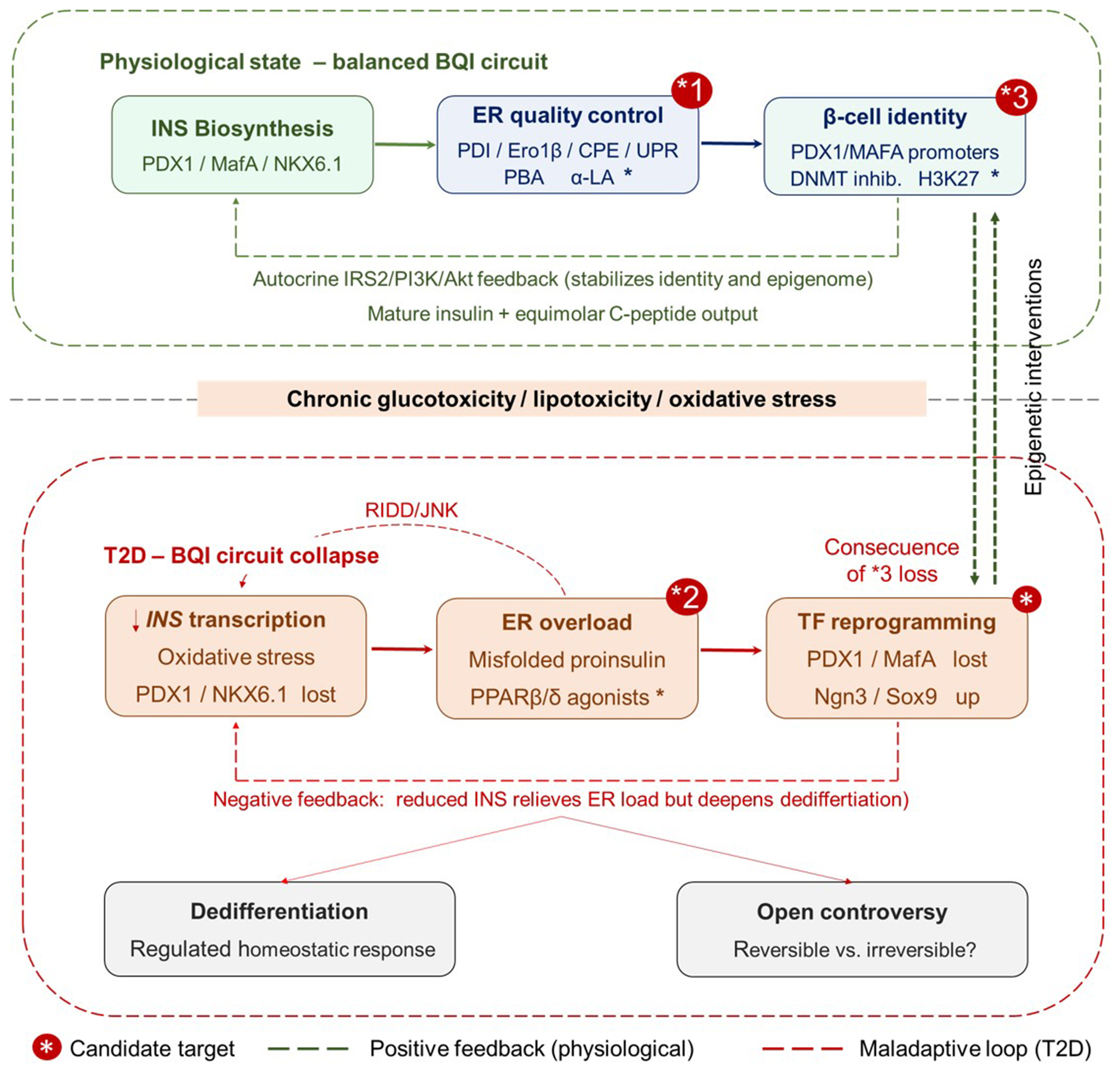
Figure 1. The BQI axis in pancreatic β-cells under physiological and T2D conditions. (Upper panel) Under physiological glucose stimulation, three interconnected nodes sustain β-cell homeostasis. (1) The PDX-1/MAFA/NKX6.1 TF triad drives INS gene expression; (2) ER quality-control machinery - PDI, Ero1β, PC1/3 and PC2, and CPE - ensures correct proinsulin processing, while a constitutively active but self-limiting UPR involving IRE1α/XBP1s, PERK/eIF2α, and ATF6 maintains folding homeostasis; (3) The β-cell identity node encompasses both the mature secretory phenotype and its epigenetic foundation - the PDX-1 and MAFA gene promoters maintained in an active, demethylated state decorated with H3K4me1 and H3K27ac marks. Autocrine insulin signaling through IRS2/PI3K/AKT (green dashed line) reinforces transcription factor binding and epigenetic maintenance, closing a positive homeostatic feedback loop that sustains all three nodes. (Lower panel) Chronic glucotoxicity and lipotoxicity disrupt the BQI circuit at three nodes. (1) Oxidative stress induces post-translational modifications of PDX-1 and NKX6.1, reducing INS transcription; (2) Excess proinsulin synthesis saturates PDI/Ero1β, generating misfolded aggregates and chronically activating IRE1α; IRE1α hyperactivation degrades INS mRNA via RIDD and activates JNK, further impairing IRS2 signaling (red dashed arrow, RIDD/JNK); (3) Loss of autocrine insulin input, combined with oxidative stress-associated epigenetic modifications, drives progressive hypermethylation of PDX-1 and MAFA loci, silencing TF expression and reactivating progenitor-state genes (NEUROG3 and SOX9) - the TF reprogramming node (red box). This node carries no asterisk because it is the downstream pathological consequence of epigenome loss at node *3, not an independent therapeutic target. A negative feedback loop (red dashed arrow) illustrates that reduced INS output alleviates ER biosynthetic load but consolidates the dedifferentiated state. Candidate therapeutic targets are indicated by a red asterisk (*) badge in the upper-right corner of the relevant node. (*1) ER quality-control node, upper panel: chemical chaperones (PBA and α-LA) restore PDI/Ero1β folding flux and prevent proinsulin aggregation; PPARβ/δ agonists (GW501516) counteract stress-induced PDI inhibition and stabilize insulin receptor β-subunit conformation; (*2) ER overload node, lower panel: PPARβ/δ agonists act directly in the glucolipotoxic environment to restore PDI activity and reduce misfolded proinsulin burden; (*3) β-cell identity node, upper panel: epigenetic interventions - DNA methyltransferase (DNMT) inhibitors and H3K27me3 demethylases - reactivate PDX-1 and MAFA promoters hypermethylated during T2D progression. The green bidirectional dashed arrow in the right margin of the figure connects node *3 (upper panel) to the TF reprogramming node (lower panel), indicating that pharmacological restoration of the β-cell epigenome at *3 is the upstream intervention that reverses the downstream transcriptional collapse. Green dashed arrows, positive feedback (physiological); red dashed arrows, maladaptive loops (T2D). BQI: Biosynthesis-quality-identity; T2D: type 2 diabetes; INS: insulin; PDX1: pancreatic and duodenal homeobox 1; MAFA: MAF bZIP transcription factor A; NKX6.1: NK6 homeobox 1; TF: transcription factor; ER: endoplasmic reticulum; PDI: protein disulfide isomerase; Ero1β: endoplasmic reticulum oxidoreductase 1 beta; PC1/3: proprotein convertase 1/3; PC2: proprotein convertase 2; CPE: carboxypeptidase E; UPR: unfolded protein response; IRE1α: inositol-requiring enzyme 1 alpha; XBP1s: spliced X-box binding protein 1; PERK: protein kinase R-like endoplasmic reticulum kinase; eIF2α: eukaryotic translation initiation factor 2 alpha; ATF6: activating transcription factor 6; H3K4me1: histone H3 lysine 4 monomethylation; H3K27ac: histone H3 lysine 27 acetylation; IRS2: insulin receptor substrate 2; PI3K: phosphoinositide 3-kinase; AKT: protein kinase B; RIDD: regulated IRE1-dependent decay; JNK: c-Jun N-terminal kinase; NEUROG3: neurogenin 3; SOX9: SRY-box transcription factor 9; PBA: 4-phenylbutyric acid; α-LA: alpha-lipoic acid; PPARβ/δ: peroxisome proliferator-activated receptor beta/delta; DNMT: DNA methyltransferase; H3K27me3: histone H3 lysine 27 trimethylation.