




Versatile and remarkable potential of transition metal dichalcogenides for advanced phototherapy and biosensing
 0
0 
Abstract
Transition metal dichalcogenides (TMDs) have emerged as a versatile class of two-dimensional materials whose strong light-matter interactions and tunable physicochemical properties enable a broad range of biomedical applications. This Review provides a mechanism-driven overview of TMDs as multifunctional platforms for phototherapeutic and optical biosensing technologies, with emphasis on how their atomic structure, electronic configuration, excitonic behavior, and surface chemistry govern optical absorption, energy relaxation, and biointerfacing processes. We first summarize the fundamental optoelectronic characteristics of TMDs that distinguish them from conventional nanomaterials, including strong exciton binding, efficient nonradiative energy dissipation, and layer-dependent symmetry and band structure. Building on these principles, recent advances in TMD-enabled phototherapeutic applications, such as photothermal therapy and photodynamic therapy, are discussed, highlighting how strong near-infrared absorption, high photothermal conversion efficiency, and structural robustness enable precise and durable light-triggered treatments. We then examine emerging optical biosensing modalities based on TMDs, including multiphoton and nonlinear optical imaging, surface-enhanced Raman scattering, and photoacoustic imaging. Across these platforms, exciton-mediated nonlinear responses, defect- and edge-induced charge-transfer pathways, and efficient optothermal transduction allow TMDs to overcome long-standing limitations in sensitivity, photostability, and imaging depth associated with traditional contrast agents. Finally, we discuss current challenges and future opportunities for translating TMD-enabled photonic and optothermal functionalities into scalable, biocompatible, and clinically relevant technologies, positioning TMDs as unifying materials for next-generation theranostic and biosensing platforms.
Keywords
INTRODUCTION
Two-dimensional (2D) materials have fundamentally reshaped the landscape of nanoscience by enabling the isolation of atomically thin solids whose physical properties differ profoundly from their bulk counterparts[1-3]. Since the advent of graphene, extensive efforts have focused on identifying alternative 2D material systems that combine structural stability with rich electronic, optical, and chemical functionality[4-6]. Among these, transition metal dichalcogenides (TMDs) have emerged as a particularly versatile class of layered materials, distinguished by their intrinsic band gaps, strong excitonic effects, and chemically accessible surfaces[7-9]. These attributes position TMDs at the intersection of condensed matter physics, materials chemistry, and biomedical engineering, enabling applications beyond those achievable with zero-bandgap or weakly interacting 2D systems[10-12].
TMDs encompass a broad family of compounds with the general formula MX2, where M is a transition metal, and X is a chalcogen. Their layered crystal structure, composed of covalently bonded X-M-X sheets held together by weak van der Waals interactions, enables reliable exfoliation down to the monolayer limit[5,11]. This dimensional reduction induces profound changes in electronic structure, including indirect-to-direct bandgap transitions, enhanced Coulomb interactions, and strong spin-orbit coupling[13,14]. As a result, monolayer and few-layer TMDs exhibit tightly bound excitons, pronounced optical absorption, and highly tunable electronic states that are exceptionally sensitive to thickness, composition, phase, strain, and environmental screening. These characteristics distinguish TMDs from conventional semiconductor nanomaterials and form the physical basis for their unique optoelectronic and photothermal behavior[7,11,14].
In parallel with their rapid development in electronics and photonics, TMDs have attracted increasing attention as functional nanomaterials for biomedical applications[15-17]. Their two-dimensional geometry affords a large specific surface area and abundant active sites for chemical modification, enabling efficient loading of therapeutic agents, photosensitizers, and targeting ligands[15]. At the same time, their optical absorption spans the visible and near-infrared (NIR) regions, overlapping with biological transparency windows that are critical for in vivo imaging and therapy[18]. Unlike many organic dyes or molecular probes, TMDs exhibit exceptional photostability and resistance to photobleaching, allowing sustained optical excitation without degradation. These features collectively render TMDs promising candidates for light-driven biomedical technologies, where robustness, efficiency, and tunability are essential[19].
A particularly compelling aspect of TMD-based systems is their ability to unify imaging and therapy within a single material platform. Phototherapeutic approaches such as photothermal therapy (PTT) and photodynamic therapy (PDT) rely on localized light-matter interactions to induce hyperthermia or generate reactive oxygen species (ROS), respectively[20,21]. TMDs naturally lend themselves to these modalities due to their strong NIR absorption, efficient nonradiative relaxation, and high photothermal conversion
Beyond therapy, TMDs have also emerged as powerful materials for advanced optical biosensing and imaging. Their exciton-dominated optical response and reduced dielectric screening dramatically amplify nonlinear optical processes, including two-photon absorption (TPA), second-harmonic generation (SHG), and photoacoustic (PA) signal generation[26-28]. These properties enable imaging modalities that combine deep tissue penetration, high spatial resolution, and intrinsic material contrast. In nonlinear optical imaging, TMDs act as active signal-generating media rather than passive labels, with nonlinear responses that can be spatially resolved and spectrally tuned[26,29]. In surface-enhanced Raman scattering (SERS), defect engineering, edge exposure, and electrochemical modulation in TMDs create efficient charge-transfer pathways that enable metal-free or hybrid Raman enhancement with high uniformity and single-molecule sensitivity[30-32]. In photoacoustic imaging (PAI), TMD nanosheets serve as robust optothermal transducers, converting absorbed optical energy into acoustic signals with high efficiency and stability, even in deep-tissue environments[33,34].
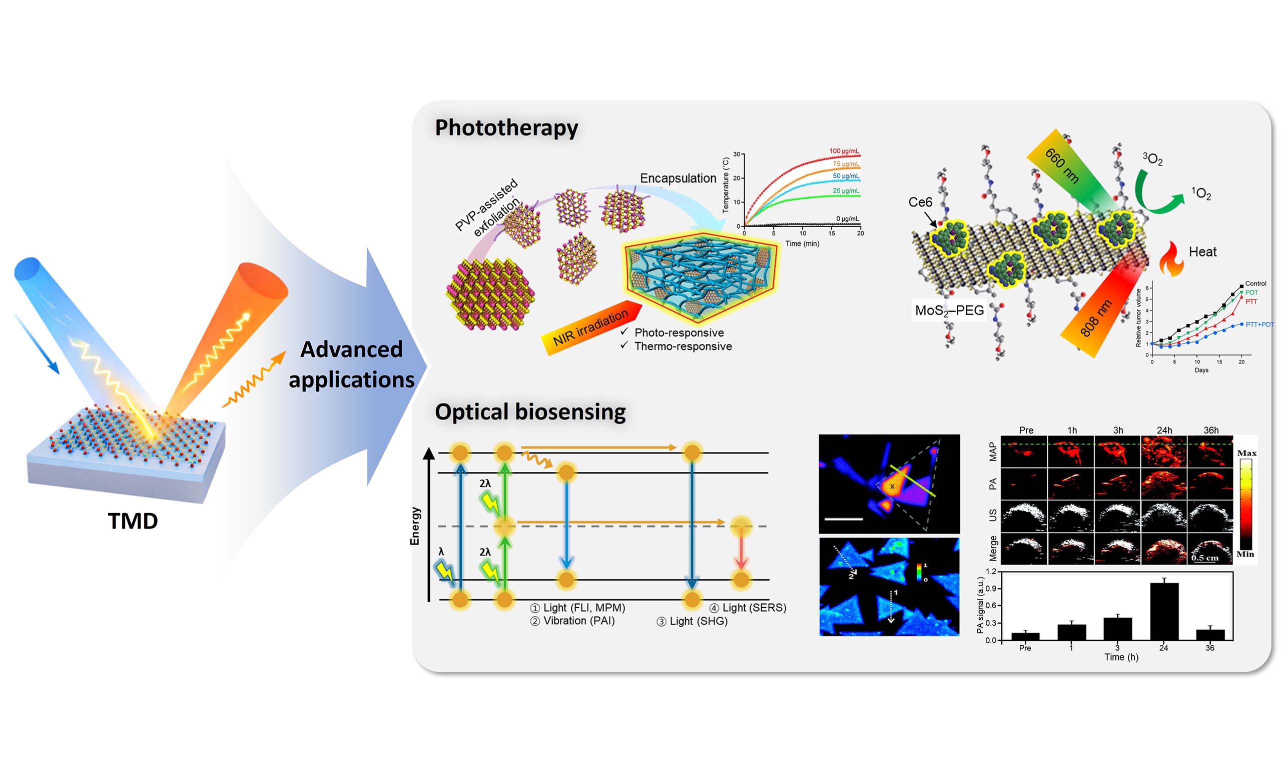
In this review, we provide a cohesive and materials-centric perspective on TMDs as multifunctional platforms for phototherapeutic and optical biosensing applications. Previous reviews have provided valuable insights into various aspects of TMD-based biomedical applications, often emphasizing specific material properties or individual application domains[5,7,35]. Building upon these efforts, this review adopts an integrated, mechanism-oriented perspective that highlights the common physical origins underlying these diverse functionalities. The fundamental properties of TMDs are comprehensively analyzed, including atomic structure, electronic band structure, thermal transport, optical response, mechanical behavior, and biocompatibility. These properties underpin their performance in biological environments. On this foundation, advanced TMD-enabled implementations are examined, with emphasis on photothermal and combined therapies, nonlinear optical imaging, SERS, and PAI. By integrating physical mechanisms with representative experimental demonstrations, this review aims to elucidate how the unique attributes of TMDs can be strategically harnessed to overcome the limitations of conventional imaging agents and therapeutic materials[15]. We highlight the emergence of TMDs not merely as alternative nanomaterials, but as adaptable, exciton-enabled platforms that bridge imaging and therapy at the nanoscale. We anticipate that the insights presented here will guide the rational design of next-generation TMD-based systems for precision diagnostics, image-guided therapy, and multifunctional biomedical technologies. To provide a concise overview of the advanced application fields, Table 1 schematically summarizes the major phototherapeutic, biosensing, and theranostic modalities enabled by TMDs, together with the underlying material attributes that govern their functionality.
Classification of advanced TMD applications
| Key feature | Optical response | Dominant energy transfer | Advanced applications | Ref. |
| Strong NIR absorption & high photothermal conversion efficiency | VIS-NIR | Photon → heat (nonradiative relaxation) | Photothermal-based applications (PTT; PAI) | [25,33,34,36] |
| Tunable band structure & catalytic activity | VIS-NIR | Photon → charge transfer/ROS generation | Multimodal phototherapy (PDT; photocatalytic, chemodynamic therapy) | [22,24,37,38] |
| Exciton-dominated nonlinear susceptibility | NIR excitation/VIS emission | TP-PL, SHG | Nonlinear optical imaging (MPM, SHG imaging) | [26-29,39] |
| Defect- and edge-induced electronic states | VIS | Charge-transfer-enhanced Raman scattering | SERS | [31,32,40,41] |
| Large surface area & hybrid interfaces | Material dependent | Coupled optical-thermal processes | Integrated theranostics (biosensing-guided phototherapy) | [42,43] |
PRINCIPLES AND APPLICATIONS
Fundamental features of TMD
Atomic structure
The synthesis of TMDs dates back to the 1960s, when layered compounds such as MoS2 and WS2 were prepared in bulk form via chemical vapor transport. Their scientific prominence expanded rapidly in the early 2010s after the isolation of graphene highlighted the broader potential of atomically thin solids[44,45]. Structurally, TMDs are typically described by repeating MX2 units, where M is a transition metal, and X is a chalcogen (S, Se, or Te). A substantial subset of TMD compounds forms layered crystals, while others adopt non-layered structures depending on the transition-metal series and the preferred coordination environment. Figure 1A shows the metals that enable both layered and non-layered structures in TMD materials. Approximately several dozen TMDs are known to exhibit layered polymorphs, and the range of accessible compositions and structures provides a broad materials space for tailoring physicochemical functions[5].
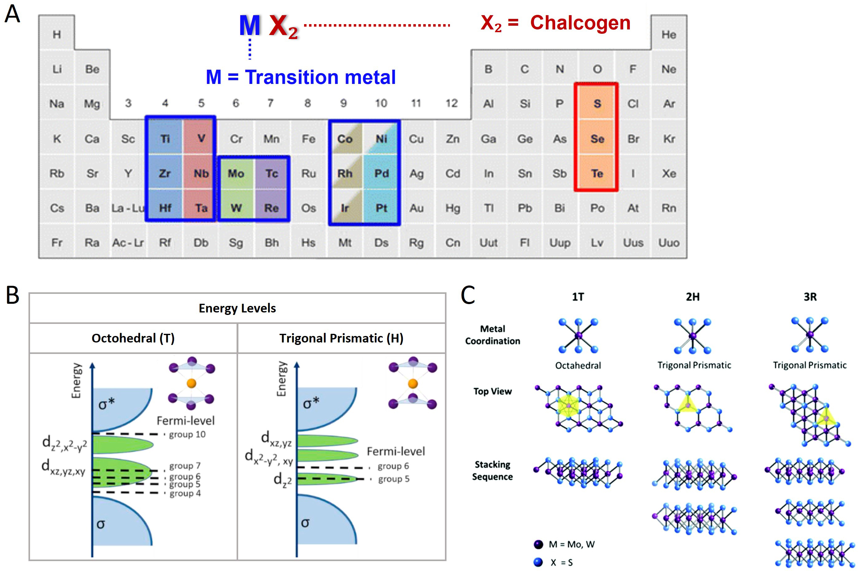
Figure 1. (A) The general MX2 formula of TMDs and the periodic table distribution of the transition metal (M) and chalcogen (X) elements forming the structure. Non-layered structures were identified for the M elements (Co, Rh, Ir, and Ni), which are indicated by half-colored boxes, whereas fully colored boxes represent layered structures, adapted with permission[46]. Copyright 2015, Springer Nature Link; (B) schematic representation of the basic electron energy diagram of octahedral and trigonal prismatic coordinate transition metals, adapted with permission[47]. Copyright 2021, Elsevier B.V.; (C) representative crystal structure of MoS2 and WS2: Octahedral (1T), Trigonal prismatic (2H) and Trigonal prismatic (3R) polytypes, adapted with permission[48]. Copyright 2017, Royal Society of Chemistry.
In layered TMDs, each structural unit consists of a transition-metal plane sandwiched between two chalcogen planes (X-M-X), forming a hexagonal in-plane lattice. Adjacent X-M-X layers are stacked through weak van der Waals interactions, enabling mechanical exfoliation or chemical delamination to monolayer and few-layer forms. The interlayer spacing is typically on the order of 5.9-6.5 Å, depending on the metal-chalcogen combination and stacking sequence[49,50]. At the atomic level, the metal center is most commonly coordinated either in a trigonal prismatic geometry (2H/1H family) or in an octahedral geometry (1T family), as schematically shown in Figure 1B[51]. These coordination motifs give rise to multiple polymorphs. The 2H phase exhibits trigonal prismatic coordination with hexagonal symmetry, while the 1T phase is characterized by octahedral coordination and may also appear in distorted variants (e.g., 1T′-like structures) depending on composition, strain, or electronic instabilities. Notably, certain TMDs such as ReS2 deviate from these conventional polymorphic frameworks by adopting a distorted 1T (1T′-like) structure, characterized by reduced crystal symmetry and the formation of Re-Re chains within the plane. This structural distortion leads to pronounced in-plane anisotropy and unusually weak interlayer coupling, effectively rendering its electronic and structural properties nearly layer-independent[52,53]. A third common stacking variant, the 3R phase, preserves a local coordination similar to 2H but differs in the relative layer registry along the c-axis [Figure 1C]. This polymorphism, combined with weak interlayer bonding, underpins the structural tunability of TMDs and supports systematic control of interlayer coupling, anisotropy, and surface-accessible active sites—features that are repeatedly exploited in TMD-enabled optical sensing and therapeutic platforms[54,55].
Electronic structure
The electronic properties of TMDs are governed primarily by the occupancy and crystal-field splitting of transition-metal d orbitals, which couple strongly to lattice symmetry and structural phase. Because chalcogen p orbitals generally lie well below the Fermi level, the d-electron configuration at the metal center plays a dominant role in determining whether a given TMD phase behaves as a semiconductor or a metal[5,56]. In MX2 compounds, transition metals often adopt an oxidation state close to +4, while chalcogens are close to -2, and the resulting d-electron count varies across the transition-metal series[46,57]. This electron count can be estimated by subtracting the oxidation state from the valence electron count of the neutral metal atom; thus, the accessible d-electron configurations span a broad range that correlates with phase preference and band topology. For example, group-4 (d0) compounds and many group-6 (d2) compounds frequently stabilize trigonal prismatic 2H-type coordination, whereas compounds with higher d-electron occupancy may favor octahedral coordination and metallic behavior. Group-5 (d1) systems can occupy an intermediate regime and may exhibit multiple coordination environments, depending on growth conditions and external perturbations, whereas distorted octahedral structures can become energetically favorable in certain compositions[47,58].
The layer number further modulates the band structure through quantum confinement and interlayer coupling. As an illustrative case, density functional theory calculations for monolayer 2H-MoS2 [Figure 2A] indicate that the conduction band minimum and valence band maximum coincide at the K point, yielding a direct band gap where optical transitions can occur without momentum exchange (Δk ≈ 0). In contrast, as the layer number increases, MoS2 evolves toward an indirect gap as the VBM shifts toward Γ and additional valleys become energetically competitive[59,60]. In contrast to conventional TMDs such as MoS2, certain materials like PtS2 exhibit a more pronounced thickness-dependent electronic transition, evolving from a semiconducting monolayer to a semimetallic bulk phase, thereby highlighting the strong influence of interlayer coupling on band structure modulation[61,62]. The most pronounced layer-dependent changes are often observed near valleys and band extrema that involve hybridized metal d orbitals and chalcogen p orbitals. Charge-density analysis [Figure 2B] suggests that pz-like chalcogen character contributes more strongly to out-of-plane interactions and is therefore more sensitive to stacking and dielectric environment, while in-plane d-orbital contributions remain comparatively localized within the X-M-X plane. Collectively, these features indicate that phase stability governed by d-orbital filling, symmetry-driven band splitting, and thickness-dependent interlayer coupling jointly define the electronic landscape of TMDs, providing multiple adjustment parameters for engineering light-matter interaction and carrier dynamics in optical biosensing and phototherapy.
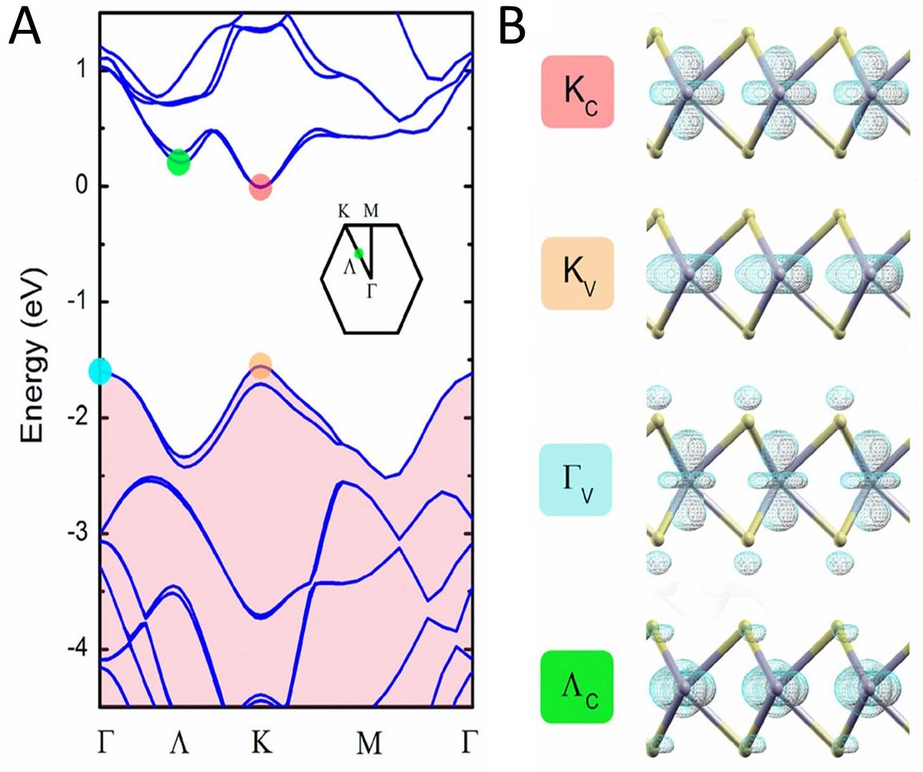
Figure 2. (A) theoretical electronic band structure of monolayer MoS2; (B) spatial electron distributions corresponding to the four-point states marked in (A). This figure is adapted with permission[59]. Copyright 2014, American Chemical Society.
Thermal conductivity
Thermal transport in TMDs is governed by lattice vibrations (phonons), which carry heat through collective atomic motion. Accordingly, the thermal conductivity and heat capacity depend strongly on the dispersion and scattering of phonon branches, including longitudinal acoustic (LA), transverse acoustic (TA), and flexural acoustic (ZA) modes[63].
Spectral analyses of thermal conductivity indicate that LA phonons often contribute substantially due to their relatively high group velocities. In many-layered TMDs, the acoustic-optical phonon gap can reduce certain anharmonic scattering channels, thereby extending the lifetime of heat-carrying acoustic phonons in specific frequency ranges, which can significantly influence κ [Figure 3A-D]. Within a simplified Boltzmann transport framework, the lattice thermal conductivity can be expressed as:
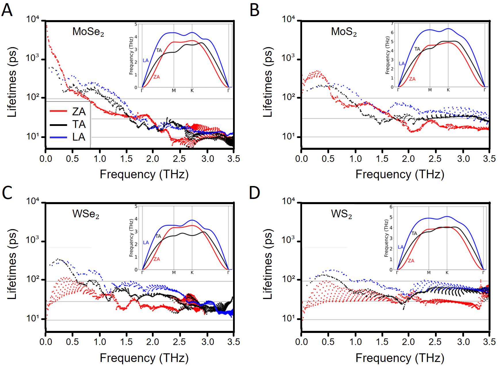
Figure 3. The acoustic mode-resolved lifetimes of monolayer TMDs. (A) MoSe2; (B) MoS2; (C) WSe2; (D) WS2. This figure is adapted with permission[64]. Copyright 2024, American Physical Society.
where Cv represents the specific heat capacity, vg the phonon group velocity, and 𝜏 the phonon lifetime. Longer phonon lifetimes increase the mean distance traveled before scattering and therefore enhance heat transport efficiency. In this context, differences in phonon lifetimes and group velocities offer a microscopic explanation for the observed ordering of thermal conductivity among representative monolayers (e.g., WS2 > MoS2 > WSe2 > MoSe2 under comparable assumptions), where heavier atoms tend to reduce vg and increase scattering, particularly at elevated temperatures[64].
Crystal phase and defects provide additional degrees of control. Structural disorder in octahedral or distorted phases can increase phonon scattering and reduce κ relative to more ordered trigonal prismatic lattices. For example, reversible phase transitions in certain TMDs have been associated with pronounced reductions in κ, consistent with bond-length nonuniformity and enhanced anharmonicity in the distorted lattice[65]. The role of defects can be described using a mean-free-path form:
where vs is an effective sound velocity and Λ is the average phonon mean free path. As temperature increases, Λ typically decreases due to stronger phonon-phonon interactions. In atomically thin materials, boundary scattering can become dominant when the characteristic size becomes comparable to or smaller than Λ, further suppressing κ[66,67]. Importantly, these thermal-transport considerations are not merely materials metrics: they directly inform the design of optothermal agents for PTT and PAI, where heat generation, heat diffusion, and thermal confinement collectively dictate both contrast performance and safety margins in biological tissue.
Optical properties
TMDs exhibit strong, highly tunable light-matter interactions, making them a central class of two-dimensional semiconductors for optoelectronics and biophotonics. A key chemical determinant of optical response is the identity of the chalcogen. Replacing S with Se or Te generally increases atomic radius and modifies electronegativity, altering d-p hybridization between metal d orbitals and chalcogen p orbitals. As the atomic number increases (S → Se → Te), the M-X bond length tends to increase, orbital overlap can decrease, and the band gap narrows, shifting optical transitions toward longer wavelengths[46]. Consequently, MoS2, WS2, and MoSe2 are widely explored for photodetectors, LEDs, and other light-harvesting systems due to strong absorption and pronounced excitonic features, whereas many Te-containing systems present narrower gaps and often stabilize in phases where photoluminescence is weak or absent, making them more common in specialized infrared and semi-metallic device contexts[68]. Furthermore, another TMD containing S, SnS2, has a relatively wide band gap (~2.2-2.4 eV); this leads to strong absorption in the UV-visible region rather than redshifted optical transitions, thereby making it particularly suitable for photocatalytic and broadband photodetection applications[69].
Layer thickness also strongly influences optical signatures. Monolayers typically exhibit high transmittance (typically ~57%-66% across wavelengths and substrates), whereas absorption increases substantially with thickness; multilayers can exhibit markedly higher absorption due to increased optical path length and altered joint density of states[70]. In addition to thickness, the nature of the optical gap and the strength of excitonic resonances determine emission and absorption efficiency. In monolayer MoS2, WS2, and related materials, strong Coulomb interaction and reduced dielectric screening lead to large exciton binding energies (commonly on the order of several hundred meV), producing intense excitonic absorption peaks and strong photoluminescence under suitable conditions[70-72]. Optical band gaps are often estimated from the A and B excitonic resonances measured in absorption or photoluminescence spectra; representative values obtained from absorption-coefficient analysis include ~1.82 eV for monolayer MoS2, ~1.51 eV for MoSe2, ~1.98 eV for WS2, and ~1.62 eV for WSe2 [Figure 4A][73]. The difference between the electronic and optical band gaps reflects exciton binding and is a central reason why excitonic features dominate the optical response of TMD monolayers.
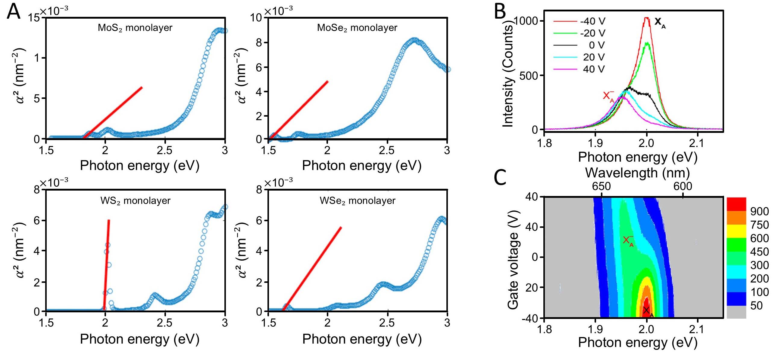
Figure 4. (A) Optical band gap graphs for monolayer TMDs (MoS2, MoSe2, WS2, and WSe2), adapted with permission[70]. Copyright 2017, Licensed under CC BY 4.0; (B) photoluminescence spectra of WS2 at back gate voltages between -40 and +40 V and (C) mapping of photoluminescence intensity as a function of photon energy and gate voltage. (B and C) are adapted with permission[74]. Copyright 2015, American Chemical Society.
Spin-orbit coupling further enriches optical structure by splitting valence (and to a lesser extent conduction) bands, giving rise to A and B excitons with characteristic energy separation (e.g., smaller for Mo-based compounds and larger for W-based compounds)[75]. Depending on spin alignment and valley selection rules, excitonic transitions may be optically bright or dark, and the population of excitonic species can be tuned by temperature, carrier density, and dielectric environment. Temperature-dependent red shifts and intensity reductions in photoluminescence can be understood in terms of band-gap renormalization and increased non-radiative channels mediated by phonons[76,77]. Carrier doping can stabilize charged excitonic complexes such as trions and biexcitons; for example, gate-dependent photoluminescence in monolayer WS2 demonstrates a transition between neutral exciton emission and trion-dominated emission as the Fermi level is shifted [Figure 4B and C][74,78]. In addition, valley-selective optical excitation under circularly polarized light provides access to coupled spin-valley degrees of freedom, highlighting the sensitivity of TMD optical response to polarization and symmetry[76].
Because excitons in monolayer TMDs are strongly affected by dielectric screening, substrate and encapsulation choices can substantially modify optical energies and quantum yields. Substrate engineering can reduce non-radiative losses, tune exciton binding, and improve measurement reproducibility by controlling charge traps and dielectric disorder[79-81]. These optical properties form a mechanistic bridge between fundamental materials physics and practical biosensing design, where excitation-wavelength selection, emission-band placement, and stability under illumination are major determinants of imaging depth, signal fidelity, and phototoxicity.
Mechanical properties
TMD monolayers exhibit high in-plane stiffness and strength due to strong covalent bonding within the X-M-X plane, while their out-of-plane compliance arises from weak van der Waals interlayer coupling.
Some elastic constants of TMDs are shown in the graph [Figure 5A-C]. Elastic moduli show mild thickness dependence as interlayer interactions become relevant in few-layer systems. Comparative analyses of elastic constants indicate that many TMDs satisfy stability criteria (e.g., Born-Huang conditions), but their hardness and ductility can vary significantly with composition and phase[82]. For example, disulfides often exhibit larger elastic constants than diselenides, consistent with shorter and stronger covalent bonds in S-containing lattices.
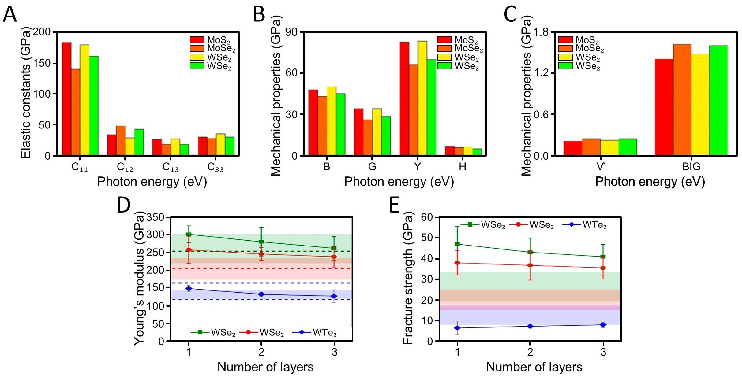
Figure 5. Mechanical properties of MoS2, MoSe2, WS2 and WSe2. (A) the elastic constants; (B) including bulk moduli (B), shear moduli (G), Young’s moduli (Y), microhardness (H); (C) including poisson’s ratio (ν) and B/G ratios. (A-C) are adapted with permission[82]. Copyright 2022, Beilstein-Institute; (D) the volumetric Young’s moduli; (E) fracture strengths of 1-3 L WS2, WSe2, and WTe2. Colored regions denote literature-reported theoretical ranges for 1 L materials, while dashed lines correspond to our reference reported density functional theory (DFT)-calculated Young’s moduli along two directions. (D and E) are adapted with permission[83]. Copyright 2021, American Chemical Society.
Interlayer interactions and in-plane covalent bonding jointly govern the mechanical performance of TMDs, making thickness control critical for high-strength applications. Falin et al.[83] demonstrated that increasing layer number leads to a reduction in tensile strength for WS2 and WSe2, while WTe2 shows the opposite trend, attributed to the reduced chemical stability of its monolayer form. Among these materials, WS2 exhibited the highest fracture strength, whereas WSe2 and WTe2 showed significantly lower values
Thickness, defects, and environmental exposure can influence fracture toughness through crack initiation and propagation pathways, whereas mild oxidation or aging can alter the elastic response, depending on the material and ambient conditions[84,85].
Mechanical strain affects not only strength but also the magnetic and optoelectronic properties of TMDs. Kansara et al. investigated the effects of 0%-9% strain on NbX2 (X = S, Se, Te) and observed strain-induced magnetic changes, including the emergence of ferromagnetism[86]. Beyond mechanical durability, strain serves as a functional tuning parameter because band structure and optical selection rules can shift under deformation. Strain engineering, implemented through flexible substrates or thermal-expansion mismatch, can modulate electronic structure and enable phenomena such as piezoelectric response in non-centrosymmetric monolayers, supporting the integration of TMDs into flexible sensors and optoelectronic devices[87]. These mechanical degrees of freedom are also relevant to biomedical implementations, where conformal contact, robustness under handling, and mechanically induced optical shifts can affect imaging reproducibility and device integration. Their surfaces can be designed to promote cellular adhesion and growth, making them suitable scaffolds for tissue engineering. By mimicking the mechanics of natural tissues, TMDCs support the regeneration of damaged skin, bone, or heart tissue.
Biocompatibility and toxicological evaluation of TMDs
The biocompatibility of TMD nanomaterials is not a fixed attribute but an outcome of their physicochemical state in biological media. Differences in membrane composition can significantly influence the interaction and internalization of TMD nanosheets. For instance, MoS2 nanosheets exhibit distinct insertion behaviors depending on the lipid composition of the membrane, highlighting the role of membrane physicochemical properties in regulating nanoparticle uptake[88]. In addition, the rich cholesterol content or different lipid rafts of some cell lines may facilitate or completely block uptake depending on the surface charge and hydrophobicity of the nanomaterial[89]. Another example is that professional phagocytes such as macrophages may rapidly engulf these materials, while uptake may be more restricted in less active cells; this directly affects the exposure dose[90,91]. Furthermore, ROS produced by TMDs, particularly through metal ion release, can cause oxidative stress and trigger apoptosis when cellular antioxidant defenses are insufficient. However, when ROS levels remain within cellular buffering capacity, cells can exhibit adaptive or transient stress responses depending on their redox status[92,93]. Many TMDs exhibit chemical stability because surface termination by chalcogen atoms suppresses dangling bonds and reduces spontaneous ion release, limiting direct membrane disruption under certain conditions[94]. However, toxicity and biological response are batch-based variability, where inconsistencies in layer number, lateral size, morphology, crystal phase, and defect density can significantly alter the physicochemical properties and consequently, biological responses. Current synthesis methods exhibit inherent trade-offs between material quality and scalability. For instance, mechanical exfoliation yields high-quality, defect-free nanosheets but is not suitable for large-scale production, whereas chemical vapor deposition enables scalable fabrication but requires strict control over process parameters to ensure uniformity. Such variability directly affects reproducibility in biomedical applications, including drug loading efficiency, photothermal performance, and toxicity profiles[95]. At higher concentrations or under prolonged exposure, unmodified or oxidized nanostructures may induce excessive ROS production and oxidative stress, leading to mitochondrial dysfunction and loss of membrane integrity. This risk can increase as lateral size and thickness decrease, due to a higher surface-to-volume ratio and a larger density of reactive sites[96,97].
Phase-dependent behavior can be particularly relevant in biomedical contexts. Metallic 1T-like phases typically exhibit higher electrical conductivity and can show enhanced photothermal conversion under NIR irradiation, which is advantageous for PTT and certain antibacterial strategies. In contrast, semiconducting 2H phases can favor longer-lived charge separation under illumination and may promote ROS generation depending on band alignment and carrier trapping, potentially enhancing antibacterial activity through oxidative stress pathways[98,99]. Karunakaran et al.[100] reported that ligand-mediated exfoliation and surface functionalization of 2H-phase TMDs can modulate both biocompatibility and antibacterial activity, with trends attributed to differences in bonding, atomic mass, and surface charge density across compositions.
Surface engineering is therefore essential for translating TMDs into safe biomedical platforms. Polymer coatings (e.g., PEGylation) and protein adsorption can improve colloidal stability, reduce direct membrane contact, and suppress aggregation-driven toxicity. Xu et al.[101] reported that combined polyethylene glycol (PEG) and transferrin coatings improved dispersion stability and preserved surface oxidation state, suggesting that surface passivation can mitigate oxidative degradation and reduce ROS-driven cytotoxicity while enabling targeting. For example, MoS2, after being functionalized with molecules such as PEG, exhibits enhanced stability in physiological environments and can be metabolized in vivo via the liver and kidneys. These properties make it particularly suitable for high-sensitivity biosensors (e.g., FETs, electrochemical sensors with detection limits reaching nM-pM levels) and drug delivery systems[102]. Once introduced into biological fluids, TMDs can also acquire a protein corona that alters their effective surface chemistry and governs biodistribution, cellular uptake, and immune response[103]. For example, adsorption of immunoglobulins and other serum proteins can enhance macrophage recognition and proinflammatory signaling, with downstream activation pathways, such as NF-κB, reported in certain systems[90]. These observations emphasize that surface functionalization shapes not only cytotoxicity but also biorecognition and immune tolerance—parameters that directly determine circulation, accumulation, and reproducibility in phototherapy and in vivo imaging.
Physicochemical behaviors are highly sensitive to experimental conditions. While in vitro systems provide controlled environments to evaluate cellular responses, they fail to capture critical biological processes such as protein corona formation, immune clearance by the mononuclear phagocyte system, and complex tissue barriers. In vivo, rapid adsorption of plasma proteins can mask surface functionalization, altering biodistribution, cellular uptake, and overall toxicity profiles[95]. For instance, TMD degradation in biological environments is often mediated by oxidative and enzymatic processes. MoS2 can be converted into soluble molybdate and sulfate species in the presence of peroxidase enzymes[94,104]. However, the long-term fate of degradation products and their potential toxicity remain insufficiently understood, representing a major barrier to clinical translation[105]. Currently, no universally accepted guidelines exist for evaluating the safety, quality, and reproducibility of TMD nanomaterials, leading to significant uncertainty in their approval pathway[106].
Collectively, the phase-, layer-, defect-, and environment-dependent properties discussed above determine how absorbed photons are redistributed into heat, charge carriers, or reactive intermediates in TMD nanostructures. Addressing these challenges will require the development of scalable synthesis strategies, standardized material descriptors, and robust in vivo evaluation pipelines to ensure reproducibility, safety, and regulatory compliance.
PHOTOTHERAPEUTIC APPLICATIONS
Fundamentals of phototherapy
Phototherapy is a therapeutic approach that selectively damages or modulates diseased tissues through localized photo-activation. PTT and PDT are widely used due to their non-invasive and controllable nature [Figure 6][107]. Compared with other chemotherapies or radiotherapies, phototherapy offers superior spatial and temporal selectivity, as light can be precisely confined to diseased tissues[21]. Furthermore, by integrating phototherapeutic agents with biocompatible nanocarriers and molecular targeting ligands, therapeutic efficiency can be significantly enhanced[108-110]. The combination of localized activation and multi-functional nanomaterial design makes phototherapy a powerful platform for precision medicine[111,112].
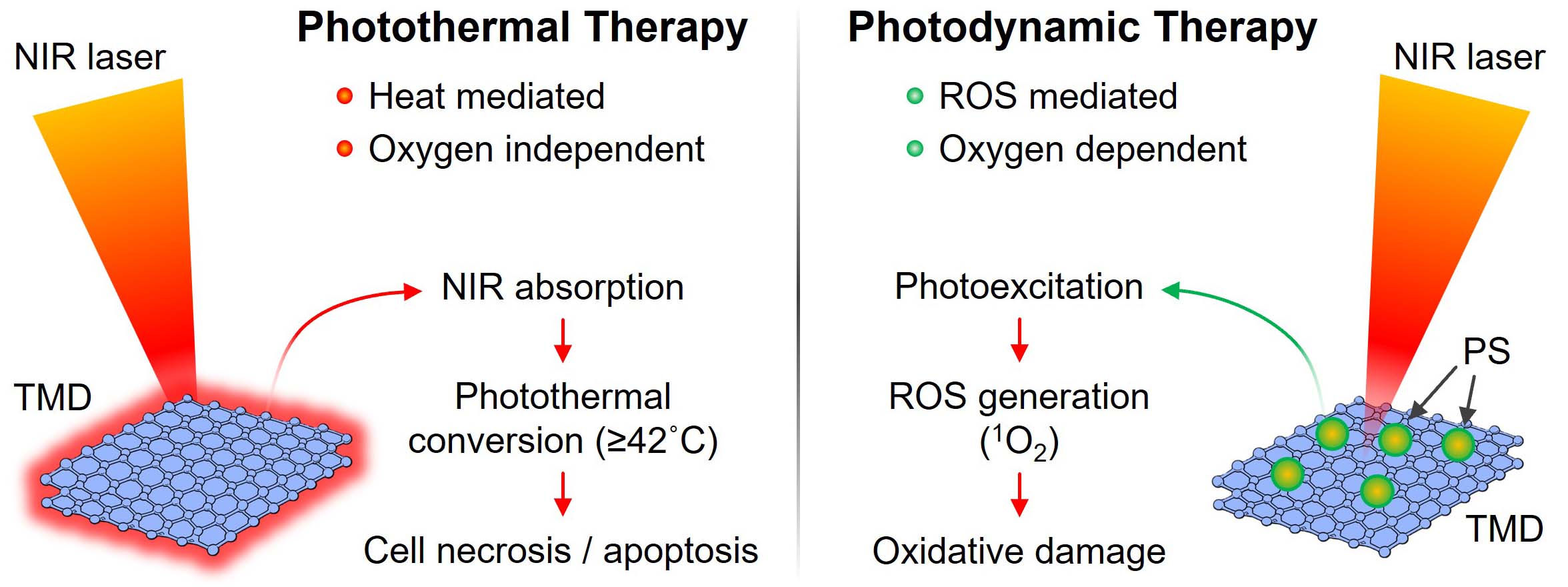
Figure 6. Schematic comparison of PTT and PDT mechanisms enabled by TMD nanoplatforms. PTT relies on NIR-induced heat generation, whereas PDT induces ROS-mediated oxidative damage through photo-excited PSs. PTT: Photothermal therapy; PDT: photodynamic therapy; TMD: transition metal dichalcogenide; PS: photosensitizer; ROS: reactive oxygen species; 1O2: singlet oxygen.
PTT is a treatment approach that eradicates abnormal cells by converting absorbed light energy into heat, leading to localized hyperthermia and cell necrosis. In PTT, NIR light is typically used to irradiate the target lesion after administration of photothermal agents that have high optical absorption and photothermal conversion efficiency[113]. These agents absorb photons and release heat, rapidly elevating the local temperature above 42 °C. The resulting hyperthermia induces protein denaturation, membrane disruption, and apoptosis in diseased cells. Because heat dissipation can affect neighboring regions, spatially confined delivery and retention of photothermal agents are essential for therapeutic precision. To achieve selective accumulation, functionalization with PEG, peptides, antibodies, or aptamers has been widely used to prolong systemic circulation and enable active binding to disease-specific receptors[114,115]. Furthermore, stimuli-responsive photothermal nanoplatforms have been designed to combine therapy and drug delivery within a single construct. In these systems, NIR-induced heating can trigger structural transformations of thermosensitive materials, thereby releasing encapsulated therapeutic agents[116]. This photo-triggered release enhances local drug concentration while reducing systemic toxicity.
In contrast, PDT relies on photochemical reactions to generate cytotoxic species that destroy diseased cells[117]. PDT employs light-activatable photosensitizers that are activated by illumination at an appropriate wavelength. Upon excitation, electrons within photosensitizer molecules are excited to the singlet state and then undergo intersystem crossing to the triplet state. From this high-energy state, two distinct photodynamic pathways can occur. In Type I processes, photoexcited charge carriers participate in electron transfer reactions to generate radical species such as superoxide (O2-) and hydroxyl radicals (OH). In contrast, Type II processes involve energy transfer from the triplet state of the photosensitizer to molecular oxygen, producing singlet oxygen (1O2)[118]. These ROS attack lipids, proteins, and nucleic acids, ultimately triggering apoptosis or necrosis.
In TMD-based systems, these processes can occur either intrinsically or through hybridization with external photosensitizers. Notably, the band alignment of TMDs plays a critical role in determining the dominant pathway. The conduction band position plays a key role in determining electron transfer to oxygen molecules, while the valence band influences oxidation reactions, thereby modulating the efficiency of ROS generation. While these photophysical and electronic factors govern ROS generation pathways, the overall therapeutic efficacy of PDT critically depends on both oxygen availability and the local photosensitizer concentration[119]. Consequently, nanocarrier formulations have been engineered to enhance photosensitizer delivery, improve solubility, and overcome tumor hypoxia.
Recently, hybrid nanoplatforms that integrate both PTT and PDT approaches have attracted considerable attention[120,121]. In such systems, mild photothermal heating can enhance local blood flow and oxygen diffusion, thereby promoting ROS production and reinforcing PDT effects. Conversely, ROS generation can sensitize tumor cells to thermal damage. When combined with photo-responsive or heat-sensitive drug carriers, these multifunctional systems enable synergistic multimodal phototherapy and controlled drug delivery. In this context, TMDs possess several unique physicochemical and surface-engineering features that make them attractive for phototherapy and drug-delivery integration. They exhibit strong optical absorption in the NIR region and high photothermal conversion efficiency when properly engineered, enabling efficient PTT. They can also be engineered to generate ROS upon light irradiation, either directly or via conjugated photosensitizers. In addition, their 2D geometry affords a high specific surface area and abundant active surface sites, which are ideal for loading therapeutic cargos, facilitating efficient and controlled drug delivery within a single theragnostic platform.
TMDs for photothermal therapy
TMDs have attracted considerable attention as next-generation photothermal platforms. Their unique structural, electronic, and optical properties, including broadband NIR absorption, tunable bandgap characteristics, large specific surface area, and versatile surface chemistry, enable powerful photothermal treatment compared to conventional metal nanoparticles and carbon-based photothermal materials[122]. In addition, the 2D morphology of TMDs enables intimate cellular interactions, efficient heat dissipation, and facile integration with targeting ligands, making them highly adaptable for PTT[123,124].
TMDs have emerged as a promising class of photothermal agents owing to their strong broadband absorption in the NIR region, high photothermal conversion efficiency, and structural robustness under prolonged laser irradiation. Unlike conventional plasmonic nanostructures or organic dyes, TMD-based photothermal platforms do not rely on localized surface plasmon resonance or fragile molecular chromophores, enabling stable heat generation with minimal photobleaching or morphological degradation[123,125,126]. These attributes make TMDs particularly attractive for PTT, where efficient and controllable temperature elevation is critical for effective tumor ablation.
Early work by Lei et al.[36] established the feasibility of TMD nanosheets as photothermal agents by introducing hydrophilic MoSe2 nanosheets with strong NIR absorption and efficient heat conversion under 808 nm laser irradiation. As depicted in Figure 7A, surface modification improved aqueous dispersibility while preserving the intrinsic photothermal response of the MoSe2 nanosheets. In vitro photothermal measurements demonstrated a temperature increase of approximately 29.3 °C after 20 min of
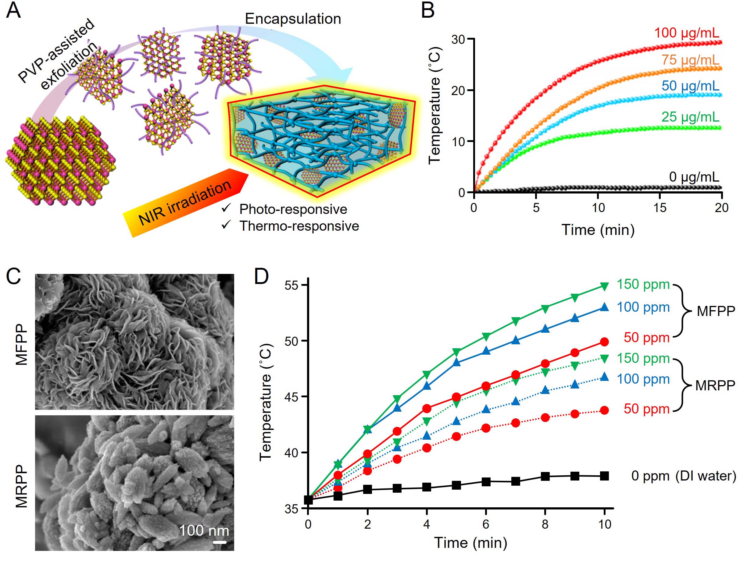
Figure 7. Representative TMD-based PTT platforms. (A) Schematic illustration for preparation of hydrophilic MoSe2 nanosheets; (B) temperature elevation of PVP-coated MoSe2 nanosheets with different concentrations under 808 nm irradiation (2.5 W/cm2). (A and B) are adapted with permission[36]. Copyright 2016, American Chemical Society; (C) field-emission scanning electron microscopy images of MFPP and MRPP; (D) temperature elevation of MFPP and MRPP solutions with different concentrations under 808 nm irradiation
Beyond pristine nanosheets, structural and optical hybridization has been employed to further enhance photothermal efficiency. Bagheri et al.[127] reported a hybrid core-shell architecture in which ultrathin TMD layers were integrated with silica-gold nanoshells, enabling tunable NIR absorption through plasmon-exciton coupling at the metal-TMD interface. By controlling shell thickness, TMD composition, and optical resonance conditions, the hybrid system achieved enhanced light confinement and superior heat generation efficiency. Numerical simulations combining near-field optical modeling and bioheat transfer analysis corroborated the experimentally observed improvements, highlighting how rational structural design can amplify photothermal performance across biologically relevant transparency windows.
Morphology-driven enhancement represents another effective strategy for improving TMD-based PTT. Sharma et al.[25] compared polymer-functionalized MoS2 nanoflowers and nanorods to elucidate the role of nanoscale architecture in photothermal behavior. As shown in Figure 7C, nanoflower structures exhibit a three-dimensional porous morphology that promotes light trapping and defect-mediated absorption. Consequently, nanoflowers produced more pronounced temperature elevation than nanorods under identical irradiation conditions [Figure 7D]. Dual polymer functionalization using PEG and PEI further improved colloidal stability, biocompatibility, and drug-loading capability, positioning morphology-engineered MoS2 nanostructures as versatile platforms for high-performance PTT and integrated therapeutic delivery. Notably, the comparative results in Figure 7D suggest that morphology plays a more dominant role than concentration alone in determining photothermal performance, highlighting the importance of structural design for efficient light absorption and heat generation.
These results demonstrate the photothermal performance of TMDs. Upon NIR excitation, generated excitons follow competing pathways, which are non-radiative relaxation and dissociation into free charge carriers. Non-radiative relaxation results in efficient heat generation through energy dissipation into lattice vibrations, while exciton dissociation enables charge-transfer processes that can contribute to ROS generation. This balance is influenced by material characteristics, including phase and defect density, which affect carrier lifetime and recombination behavior. In general, longer carrier lifetimes tend to lead to charge separation and ROS generation, while rapid non-radiative relaxation enhances photothermal conversion efficiency.
Despite these promising results, direct comparison between TMD-based photothermal systems remains challenging. Differences in the reported photothermal performance in this section are influenced by experimental conditions rather than intrinsic properties of the materials. While hybrid systems offer enhanced functionality, their complexity can make practical application difficult. Conversely, morphological control with appropriate surface functionalization is a highly scalable and promising strategy. Standardized evaluation and systematic validation are necessary for meaningful comparison and clinical translation.
Combined and multimodal phototherapy
Beyond monomodal PTT, TMDs have evolved into multifunctional nanoplatforms capable of integrating multiple therapeutic mechanisms within a single system. Their strong NIR absorption and tunable structure enable TMDs to function as photothermal transducers, photosensitizer carriers, and drug-delivery scaffolds. These characteristics directly address the intrinsic limitations of single-modality therapies, such as incomplete tumor ablation, hypoxia-limited PDT, or systemic toxicity associated with chemotherapy.
Several studies have demonstrated the effectiveness of coupling PTT and PDT using TMD-based platforms. Liu et al.[37] developed PEGylated single-layer MoS2 nanosheets capable of simultaneously delivering photosensitizers and generating efficient photothermal heating. The nanosheets exhibited strong NIR absorption and high loading capacity for chlorin e6 (Ce6), resulting in enhanced cellular uptake and synergistic therapeutic efficacy [Figure 8A]. Compared with either PTT or PDT alone, the combined treatment produced significantly improved tumor suppression in murine breast cancer models [Figure 8B], underscoring the advantage of TMD nanosheets as dual-functional phototherapeutic carriers. The results indicate that the combined PTT and PDT treatment does not simply produce an additive effect but rather alters the therapeutic response profile. While PTT and PDT individually exhibit distinct efficacy trends, their combination results in more sustained tumor suppression, suggesting a synergistic interaction between rapid thermal ablation and ROS-mediated cytotoxicity.
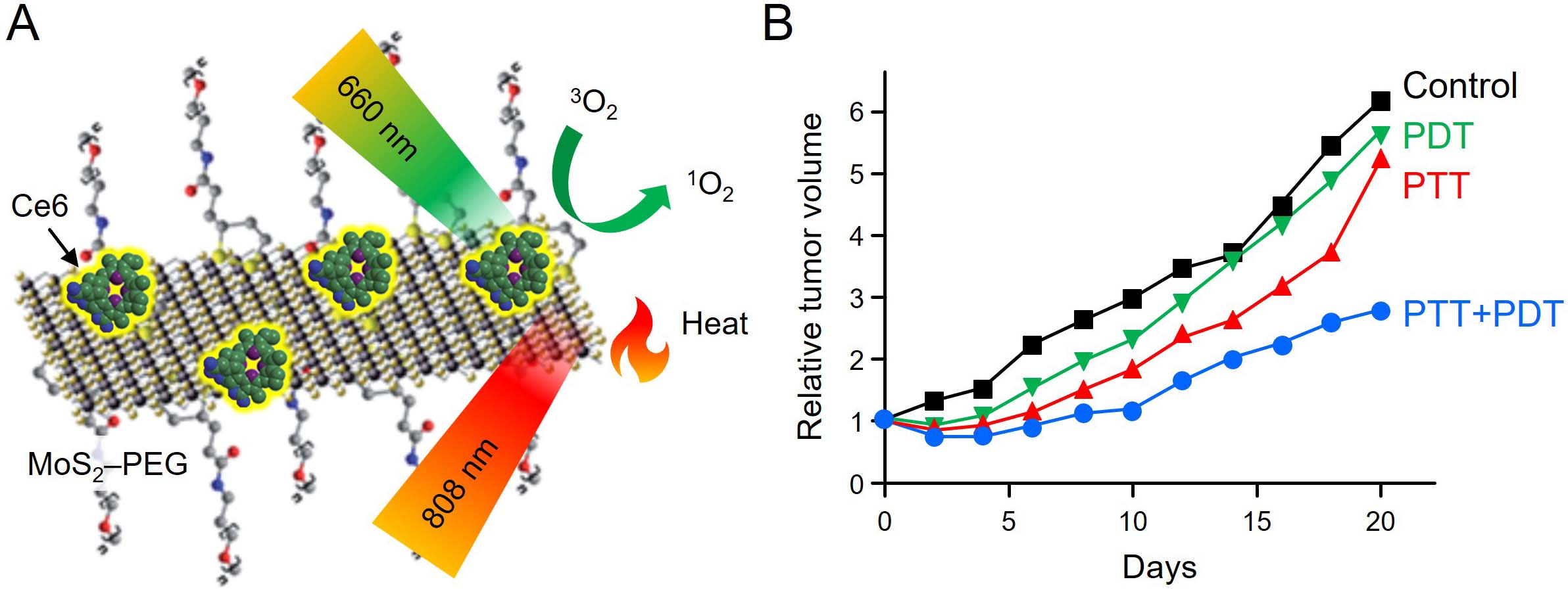
Figure 8. Representative combined PTT and PDT enabled by TMD nanoplatforms. (A) schematic illustration of single-layer MoS2 nanosheets simultaneously serving as photothermal agents and photosensitizer carriers; (B) Relative tumor volume changes of mice subjected to different treatment conditions. This figure is adapted with permission[37]. Copyright 2014, Royal Society of Chemistry. TMD: Transition metal dichalcogenide; PTT: photothermal therapy; PDT: photodynamic therapy; Ce6: chlorin e6.
More recently, Li et al.[38] introduced a MoS2-decorated red phosphorus nanoplatform that integrates photothermal heating with enhanced photocatalytic ROS generation. Under 808 nm laser irradiation, efficient charge separation within the hybrid system facilitated the production of multiple ROS species, enabling simultaneous PTT and PDT. In vivo experiments confirmed substantial tumor regression with minimal off-target toxicity, highlighting the therapeutic potential of TMD-based hybrid architectures.
TMDs have also been widely explored for synergistic chemo-PTT. Zhang et al.[22] reported a multifunctional MoS2 nanosheet platform functionalized with polyethyleneimine, polyethylene glycol, and folic acid to enhance stability, biocompatibility, and tumor-specific uptake. The platform exhibited a photothermal conversion efficiency of 54.2% under 808 nm irradiation and a high loading capacity for doxorubicin. In vitro studies demonstrated markedly enhanced cytotoxicity when photothermal heating was combined with chemotherapeutic drug release. Similarly, Zhu et al.[24] developed ultrathin Co-Fe-Mn multicomponent TMD nanosheets that exhibited exceptional NIR absorption and a photothermal conversion efficiency approaching 89%. In this system, NIR-induced heating not only directly ablated tumor cells but also accelerated Fenton-type reactions, generating cytotoxic hydroxyl radicals and enabling a potent photothermal-enhanced chemodynamic therapy.
Collectively, these studies demonstrate that TMDs serve not merely as passive photothermal agents but as integrative therapeutic platforms capable of coordinating multiple light-activated treatment pathways. Their modular structure and tunable physicochemical properties enable rational design of synergistic phototherapeutic systems with enhanced efficacy and reduced systemic toxicity. Despite these advantages, several challenges remain in the development of multimodal TMD-based phototherapeutic platforms, particularly in ensuring consistency across experimental conditions. Future work should focus on systematic evaluation of synergistic effects and standardized validation to facilitate clinical translation.
BIOSENSING APPLICATIONS
Biosensing modalities using TMDs
TMDs offer a distinctive optical platform in which absorption, emission, scattering, and non-radiative energy dissipation can be continuously tuned through layer thickness, excitonic structure, phase, and surface chemistry. Advanced optical imaging modalities such as multiphoton microscopy (MPM), Raman-based sensing, and PAI probe fundamentally different signal channels. However, they share a common physical origin: the interaction of incident photons with electronic states, followed by energy redistribution through radiative, coherent, vibrational, or thermal relaxation pathways. The relative balance among these pathways ultimately determines whether optical excitation manifests as fluorescence, nonlinear scattering, vibrational fingerprints, or acoustic emission[128-131].
This common physical framework is schematically summarized in Figure 9, which compares the light-matter interaction processes underlying different TMD-enabled biosensing modalities. As illustrated in Figure 9A, optical excitation of TMDs can relax through multiple competing channels, including exciton recombination, coherent nonlinear emission, vibrational energy exchange, and non-radiative heat generation. Figure 9B further organizes these processes using a Jablonski-type representation, clarifying how distinct excitation-relaxation pathways and detection schemes correspond to multiphoton imaging, Raman-based sensing, and PA signal generation. Figure 9C-E illustrate representative configurations of MPM, SERS, and PAI, highlighting how the TMD platform can be interfaced with different readout mechanisms by selectively emphasizing specific relaxation channels[128-131].
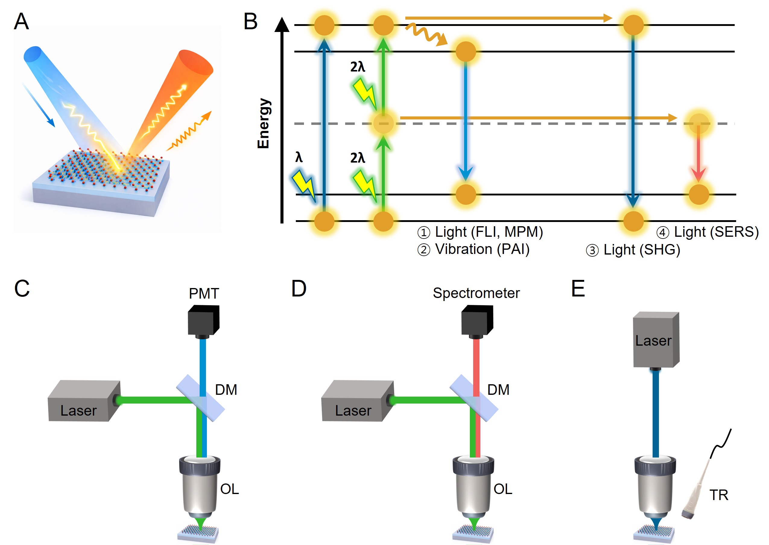
Figure 9. Schematic comparison of optical biosensing modalities using TMDs. (A) light-matter interactions in TMDs and the resulting optical and thermo-vibrational signals utilized for biosensing applications; (B) Jablonski diagram representing the underlying excitation-relaxation pathways for different optical sensing modalities; experimental configurations of (C) MPM and SHG based on nonlinear excitation and coherent emission process; (D) SERS sensing based on charge-transfer-mediated vibrational signal amplification; (E) PAI relying on optothermal energy conversion and subsequent ultrasonic signal generation. The colors of the incident and emitted beams indicate different excitation and detection wavelengths. FLI: Fluorescence imaging; MPM: multiphoton microscopy; PAI: photoacoustic imaging; SERS: surface-enhanced Raman scattering; SHG: second harmonic generation; PMT: photomultiplier tube; DM: dichroic mirror; OL: objective lens; TR: transducer.
In conventional bioimaging agents, this balance is often constrained by weak absorption cross-sections, limited photostability, inefficient non-radiative conversion, or narrow spectral tunability. TMDs overcome many of these limitations because their optical response is dominated by strongly bound excitons and highly tunable band structures, which govern both light absorption and subsequent relaxation dynamics[132,133]. As a result, TMDs provide a unifying materials framework in which distinct imaging modalities can be selectively activated or enhanced by engineering excitonic resonances, defect states, and interfacial energy alignment. In the following subsections, the operating principles of multiphoton and nonlinear optical imaging, SERS, and PAI are discussed, with an emphasis on how TMD-specific properties enable performance advantages that are not readily achievable with conventional contrast agents[5,7,14].
Nonlinear optical imaging
MPM relies on the near-simultaneous absorption of two or more photons to access an excited electronic state that would otherwise require higher-energy single-photon excitation. Because multiphoton absorption scales nonlinearly with photon flux, excitation is confined to a tightly focused focal volume under femtosecond pulsed illumination. This intrinsic spatial confinement suppresses out-of-focus excitation and photobleaching, enabling three-dimensional optical sectioning without the need for a confocal pinhole. The use of NIR excitation further reduces scattering and absorption in biological tissues, enabling imaging at depths inaccessible to conventional widefield or single-photon fluorescence microscopy[128,134]. However, the practical implementation of MPM is often limited by the relatively small multiphoton absorption cross-sections and photostability of traditional organic fluorophores, which necessitate high excitation powers and can introduce photodamage[135].
SHG microscopy represents a complementary nonlinear imaging modality based on coherent frequency doubling in non-centrosymmetric media. In SHG, two photons at a fundamental frequency interact coherently with a material to generate a photon at exactly twice the frequency. Because SHG does not involve population of real excited states, it is inherently non-bleaching and highly sensitive to local symmetry and structural order. In biological systems, endogenous SHG signals arise primarily from ordered fibrillar structures such as collagen and myosin, providing label-free contrast[136]. Nevertheless, SHG signals in biological tissues are typically weak and lack chemical specificity unless highly ordered or engineered nonlinear materials are introduced[137].
Monolayer TMDs provide a unique solution to these limitations by combining strong excitonic resonances with symmetry properties that intrinsically support nonlinear optical processes. Reduced dielectric screening in two-dimensional TMDs leads to large exciton binding energies and enhanced oscillator strengths, which significantly increase the probability of multiphoton absorption near excitonic resonances[138]. Two-photon excitation can efficiently access higher-lying excitonic states. These include states that are optically dark or weakly allowed under one-photon excitation but strongly coupled in two-photon processes. Consequently, TMD nanosheets and quantum dots exhibit TPA cross-sections that exceed those of many conventional fluorophores by orders of magnitude at comparable excitation wavelengths[28,139].
At the same time, the absence of inversion symmetry in monolayer TMDs directly enables a strong second-order nonlinear susceptibility, making them intrinsically efficient emitters for SHG. When the excitation energy approaches prominent excitonic resonances, the effective nonlinear susceptibility is strongly modified by exciton-photon coupling, leading to pronounced wavelength-dependent modulation of the SHG intensity. Experimental studies on monolayer MoS2 and WSe2 have established that SHG contrast in TMDs is governed not only by symmetry considerations but also by excitonic resonance conditions[26]. Excitonic effects similarly play a central role in higher-order nonlinear absorption processes. Degenerate TPA measurements have shown that the nonlinear absorption response of WS2 and MoS2 is dramatically enhanced in the monolayer limit, far exceeding expectations based on conventional interband transitions[27]. This enhancement reflects the emergence of exciton-assisted nonlinear pathways that are unique to two-dimensional semiconductors and evolve systematically with layer thickness[28].
These exciton-governed nonlinear responses underpin real-space nonlinear imaging in TMDs. MPM studies have demonstrated that both SHG and two-photon photoluminescence (TP-PL) from monolayer TMDs can be exploited as intrinsic contrast mechanisms, enabling spatially resolved visualization of excitonic features without external labels[29]. Moreover, the nonlinear response of TMDs can be further amplified through engineering of the photonic environment, demonstrating that exciton-mediated nonlinear processes in these materials are not only intrinsically strong but also highly tunable[42]. To elucidate how these excitonic effects manifest experimentally across different nonlinear pathways, representative studies are summarized below with emphasis on their underlying physical mechanisms and imaging implications.
The exciton-enhanced nonlinear response of monolayer TMDs is first manifested in their unusually strong SHG and TPA, which together establish the physical basis for multiphoton imaging. Figure 10 summarizes representative experimental evidence demonstrating how excitonic effects amplify second- and third-order nonlinear processes in monolayer TMDs. Early work by Malard et al.[26] provided direct experimental confirmation of intense SHG originating exclusively from monolayer MoS2. As depicted in Figure 10A, strong SHG contrast is observed only in monolayer regions, while bilayer and trilayer domains exhibit suppressed signals due to inversion symmetry restoration. The quadratic dependence of SHG intensity on excitation power, represented in Figure 10B, confirms the second-order nonlinear origin of the signal. Importantly, Figure 10C shows that the extracted second-order susceptibility χ2 exhibits pronounced resonant enhancement when the two-photon energy coincides with excitonic absorption features, establishing that SHG in monolayer TMDs is not merely symmetry-allowed but strongly reinforced by exciton-photon coupling.
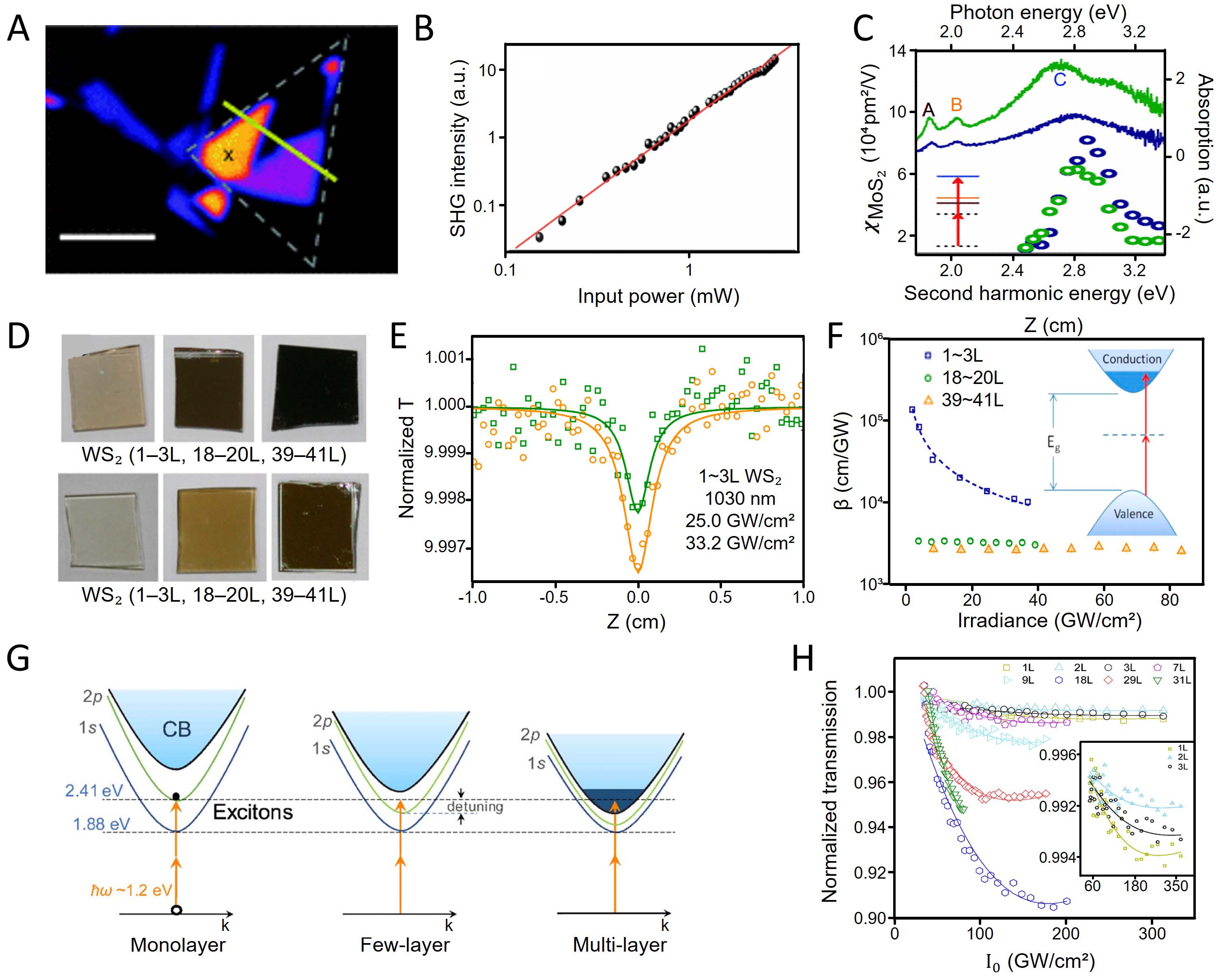
Figure 10. Exciton-mediated SHG and TPA in monolayer TMDs. (A) SHG image of a MoS2 thin film under 800 nm excitation; (B) SHG intensity as a function of excitation power measured at the marked position in (A), yielding a slope of approximately 2 and confirming the second-order nonlinear origin of the signal; (C) extracted second-order susceptibility χ2 of monolayer and trilayer MoS2 as a function of excitation photon energy, overlaid with linear absorption spectra, demonstrating resonant enhancement of SHG when the two-photon energy matches excitonic transitions. (A-C) are adapted with permission[26]. Copyright 2013, American Physical Society; (D) photographs of WS2 and MoS2 films with varying thicknesses (monolayer to multilayer), illustrating the systematic layer-dependent platform; (E) open-aperture Z-scan traces of monolayer WS2 films under 1030 nm femtosecond excitation, revealing pronounced TPA behavior below the linear bandgap; (F) extracted TPA coefficients as a function of layer number, highlighting the dramatic enhancement of nonlinear absorption in the monolayer limit. (D-F) are adapted with permission[27]. Copyright 2015, American Chemical Society; (G) schematic illustration of the exciton-assisted TPA mechanism at the K valley in monolayer MoS2, where the two-photon energy is nearly resonant with the two-photon dark excitonic state, enabling strong nonlinear absorption; (H) layer-resolved micro-I-scan measurements of MoS2. (G and H) are adapted with permission[28]. Copyright 2019, Optica Publishing Group.
Beyond second-order processes, the third-order nonlinear response of TMDs is likewise dramatically enhanced in the monolayer limit. Zhang et al.[27] investigated degenerate TPA in WS2 and MoS2 films with controlled thicknesses, as represented by the layer-dependent platform shown in Figure 10D. Open-aperture Z-scan measurements reveal pronounced nonlinear absorption under sub-bandgap excitation, as illustrated in Figure 10E, while the extracted TPA coefficients increase by several orders of magnitude as the layer number decreases, as summarized in Figure 10F. This pronounced enhancement cannot be explained by conventional interband transitions alone and instead reflects the dominant role of excitonic states in two-dimensional systems.
The microscopic origin of exciton-assisted TPA was clarified by Xie et al.[28], who identified the crucial contribution of dark excitonic states to nonlinear absorption. The dark excitonic states are optically inactive exciton states that do not couple to light under one-photon excitation but can be accessed through multiphoton or symmetry-breaking processes. Figure 10G schematically illustrates the exciton-assisted TPA mechanism in monolayer MoS2, where the two-photon excitation energy is nearly resonant with the 2p dark excitonic state at the K valley. Because this state is optically inaccessible under one-photon excitation yet strongly couples to two-photon transitions, it provides an efficient nonlinear absorption channel unique to the two-dimensional excitonic landscape. Layer-resolved micro-I-scan measurements, represented in
These exciton-enhanced nonlinear responses are directly relevant to biosensing applications, as they enable strong signal generation under relatively low excitation power and provide intrinsic contrast mechanisms without the need for external labels. Such properties are particularly advantageous for high-sensitivity detection in complex biological environments.
While these studies establish the intrinsic nonlinear optical advantages of pristine TMD monolayers, subsequent work has demonstrated that exciton-mediated nonlinear imaging can be extended beyond signal enhancement to direct real-space visualization of excitonic phenomena. Lin et al.[29] exploited MPM to image exciton complexes in monolayer WSe2 and WS2 without external fluorophores. As depicted in Figure 11A of the second integrated figure, wavelength-dependent SHG images of triangular WSe2 monolayers reveal spatially resolved modulation of SHG intensity as the excitation wavelength is tuned across different exciton-complex resonances. This behavior arises from the strong renormalization of the effective nonlinear susceptibility by excitonic transitions, causing small changes in excitation energy to produce a dramatic redistribution of SHG contrast across the flake. Such wavelength-selective contrast confirms that nonlinear imaging in TMDs is governed primarily by exciton-photon coupling rather than lattice symmetry or morphology alone.
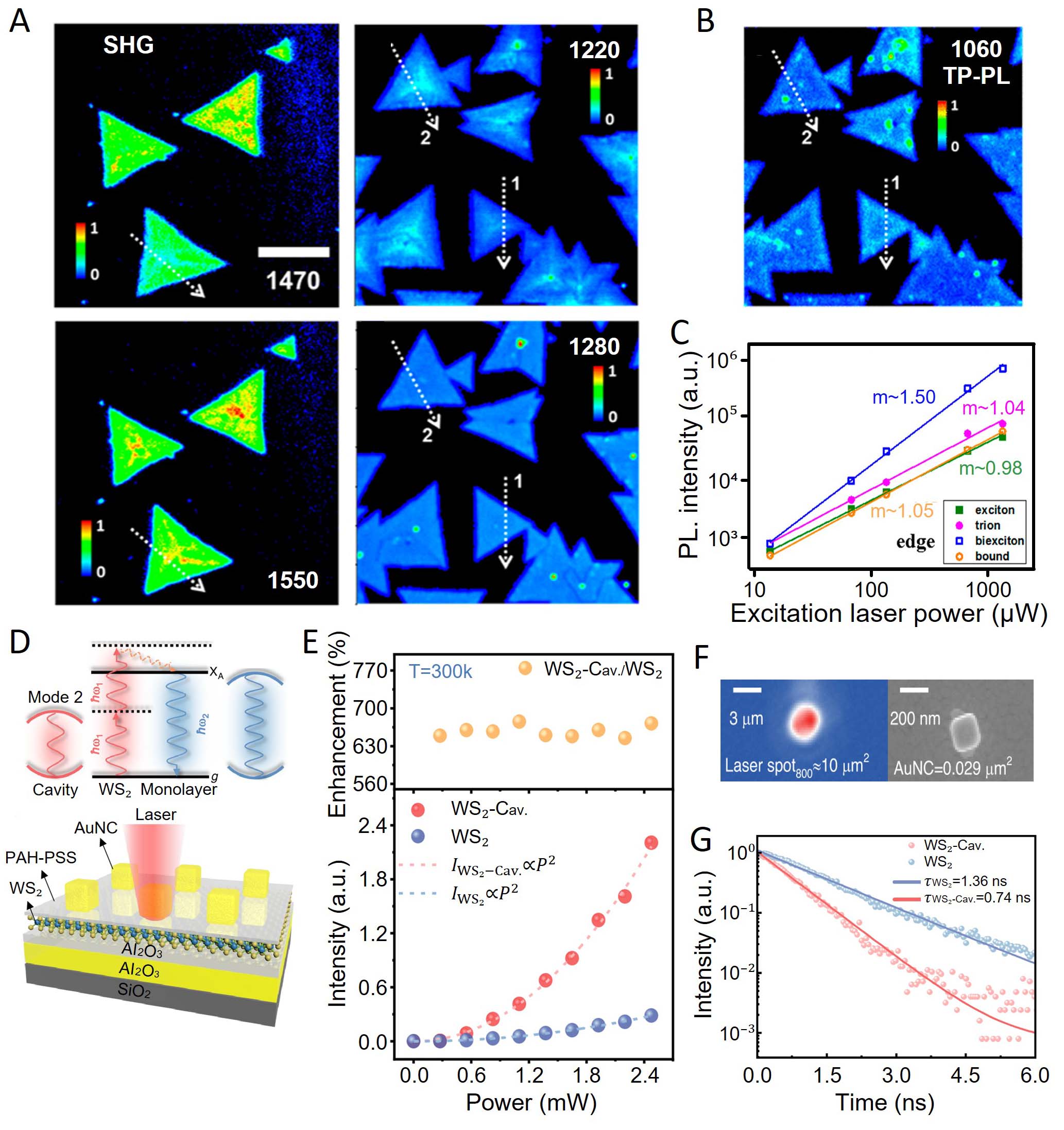
Figure 11. Real-space nonlinear imaging and ultimate performance of exciton-mediated nonlinear responses in TMDs. (A) wavelength-dependent SHG images of triangular monolayer WSe2 acquired under MPM, revealing spatially resolved modulation of SHG intensity as the excitation wavelength crosses different exciton-complex resonances; (B) TP-PL image of monolayer WS2 excited at
In addition to SHG, exciton-mediated TPA enables direct TP-PL imaging. Figure 11B represents a TP-PL image of monolayer WS2 excited at 1,060 nm, demonstrating real-space nonlinear imaging enabled by strong excitonic TPA. The emitted signal originates from exciton complexes that are inaccessible under linear excitation, highlighting a key advantage of TMDs for nonlinear bioimaging. The nonlinear origin of these signals is further corroborated by the power-dependent analysis shown in Figure 11C, where integrated photoluminescence intensities associated with excitons, trions, and biexcitons exhibit distinct power-law scaling behaviors. These different exponents directly reflect the multiparticle nature of each excitonic species, confirming that the observed signals arise from genuine nonlinear excitation pathways rather than thermal or defect-assisted processes.
The ultimate performance of exciton-mediated nonlinear responses can be further enhanced through photonic environment engineering. Liu et al.[42] demonstrated that embedding monolayer WS2 within a doubly resonant plasmonic nanocavity dramatically amplifies two-photon upconversion by simultaneously reinforcing excitation and emission processes. Figure 11D represents a schematic of the nanocavity-WS2 hybrid system, in which electromagnetic confinement enhances the local field at both the excitation and emission energies. As shown in Figure 11E, the upconverted emission intensity exhibits orders-of-magnitude enhancement compared to uncoupled WS2, accompanied by modified power-law scaling indicative of cavity-assisted nonlinear excitation. The representative nanocavity structure shown in Figure 11F highlights the engineered electromagnetic confinement that maximizes exciton-plasmon coupling. Time-resolved luminescence measurements, represented in Figure 11G, further reveal altered exciton dynamics in the cavity-coupled system, including accelerated radiative recombination and suppressed nonradiative loss channels.
Taken together, the studies delineate a clear progression from intrinsic exciton-enabled nonlinear optical responses in pristine TMD monolayers to engineered photonic systems that approach the ultimate limits of nonlinear performance. These results demonstrate that TMDs are not merely passive nonlinear materials but active excitonic platforms whose nonlinear response can be spatially resolved, spectrally tuned, and dramatically amplified[28,42]. Such capabilities establish a solid foundation for advanced nonlinear bioimaging and highlight the broader potential of exciton-based photonic technologies built upon two-dimensional semiconductors. These capabilities highlight the potential of TMD-based nonlinear imaging for biosensing, where the ability to spatially resolve and selectively enhance excitonic signals can enable sensitive detection and mapping of biological targets. The tunability of nonlinear responses further offers opportunities to optimize contrast and specificity in practical sensing platforms.
Despite these advances, several challenges remain in translating TMD-based nonlinear imaging into practical biosensing applications. The nonlinear response is highly sensitive to excitation conditions, local dielectric environment, and material quality, leading to variability across studies and limiting direct comparison. In addition, most demonstrations have been conducted under controlled conditions, and their robustness in complex biological environments remains to be fully established. While photonic structure engineering, such as plasmonic nanocavities, enables substantial signal enhancement, it often introduces additional complexity that may limit scalability and practical implementation. In this context, strategies that leverage intrinsic material properties—such as exciton engineering through layer control, strain, or dielectric tuning—are particularly promising. These approaches provide a more scalable and physically controllable pathway to enhance nonlinear response while maintaining system simplicity and biological compatibility.
Surface-enhanced Raman scattering
Raman spectroscopy provides molecularly specific information through inelastic light scattering, where incident photons exchange energy with the vibrational modes of molecules or lattices. The resulting Raman spectrum serves as a unique vibrational fingerprint that enables chemical identification without labeling[140]. However, Raman scattering is intrinsically weak, with only a minute fraction of incident photons undergoing inelastic scattering. This fundamental limitation leads to low sensitivity and long acquisition times, particularly in dilute biological environments[141].
SERS was developed to address this challenge by amplifying Raman signals near engineered substrates. Conventional SERS platforms rely primarily on noble-metal nanostructures that support localized surface plasmon resonances[142]. Under resonant excitation, these structures generate intense electromagnetic fields at nanoscale hotspots, thereby greatly enhancing the Raman scattering of nearby molecules. While this electromagnetic enhancement can achieve single-molecule sensitivity, metal-based SERS substrates often suffer from poor reproducibility, uncontrolled hotspot distributions, photothermal instability, and limited biocompatibility[143].
TMDs offer an alternative SERS enhancement mechanism that is predominantly chemical in origin. In semiconducting TMD monolayers, the electronic structure is highly sensitive to defects, edges, phase transitions, and electrostatic doping. Chalcogen vacancies, phase boundaries, and engineered edge sites introduce electronic states within or near the bandgap, increasing the density of states around the Fermi level. When analyte molecules adsorb onto such surfaces, these states facilitate photo-induced charge transfer between molecular orbitals and TMD bands under optical excitation. Resonant charge-transfer pathways effectively increase the change in polarizability during molecular vibrations, leading to significant Raman enhancement without relying on plasmonic field confinement[30].
Because TMDs are atomically flat and can be synthesized over large areas with controllable defect densities, their SERS enhancement is often spatially uniform and highly reproducible. Defect engineering, geometric control of edge exposure, and electrochemical modulation have collectively established TMDs as versatile, metal-free platforms in which charge-transfer-mediated Raman enhancement can be systematically tuned through electronic structure design[31,32,40]. In particular, defect-rich basal planes and edge-dominated architectures increase the density of accessible electronic states near the Fermi level, strengthening molecule-substrate coupling and enabling dynamic control over resonance conditions via external bias. More recently, atomic-scale engineering strategies have extended this framework beyond ensemble-averaged enhancement toward the single-molecule regime. By localizing charge at atomically defined features on otherwise flat two-dimensional surfaces, these approaches create spatially isolated Raman-active sites with a well-defined electronic origin. This progression—from defect-mediated chemical enhancement to atomic-scale charge localization—highlights the unique capability of TMDs to bridge macroscopic uniformity and ultimate molecular sensitivity within a single materials platform[41].
These characteristics address several limitations of conventional SERS in biological sensing. Chemical enhancement in TMD-based systems improves reproducibility and quantitative mapping over large areas. The absence of strong plasmonic heating reduces thermal damage to sensitive biological samples. Moreover, the chemically versatile surfaces of TMDs allow straightforward functionalization with biomolecular recognition elements while maintaining enhanced performance. Hybrid systems that combine TMDs with noble metals further exploit synergistic electromagnetic and chemical enhancement mechanisms, achieving high sensitivity while improving stability and control. Collectively, these features address key limitations of conventional SERS in biological sensing. Chemical enhancement improves reproducibility and enables quantitative Raman mapping, while reduced plasmonic heating minimizes thermal damage. The atomically flat surfaces of TMDs also allow straightforward functionalization with biomolecular recognition elements. In hybrid architectures, TMDs can combine chemical and electromagnetic enhancement to achieve high sensitivity with improved stability. As a result, the advanced electronic and optical properties of TMDs have enabled SERS to evolve into a metal-free, tunable, and even single-molecule-sensitive biosensing platform, as demonstrated by a series of representative experimental studies discussed below.
Sun et al.[31] provided one of the clearest experimental demonstrations that defect engineering in monolayer MoS2 can serve as a universal and metal-free SERS strategy. By introducing plasma-induced sulfur vacancies into monolayer MoS2, mid-gap electronic states were created near the Fermi level, substantially increasing the density of states available for photo-induced charge transfer between the substrate and adsorbed molecules, as schematically illustrated in Figure 12A. Raman measurements combined with electronic structure analysis revealed that defect-rich MoS2 exhibits markedly enhanced Raman signals across a broad range of analytes, while pristine MoS2 shows only weak responses [Figure 12B]. This study established defect-induced chemical enhancement as a dominant and controllable SERS mechanism in semiconducting TMDs, thereby eliminating the need for noble-metal plasmonic structures and improving signal uniformity and reproducibility.
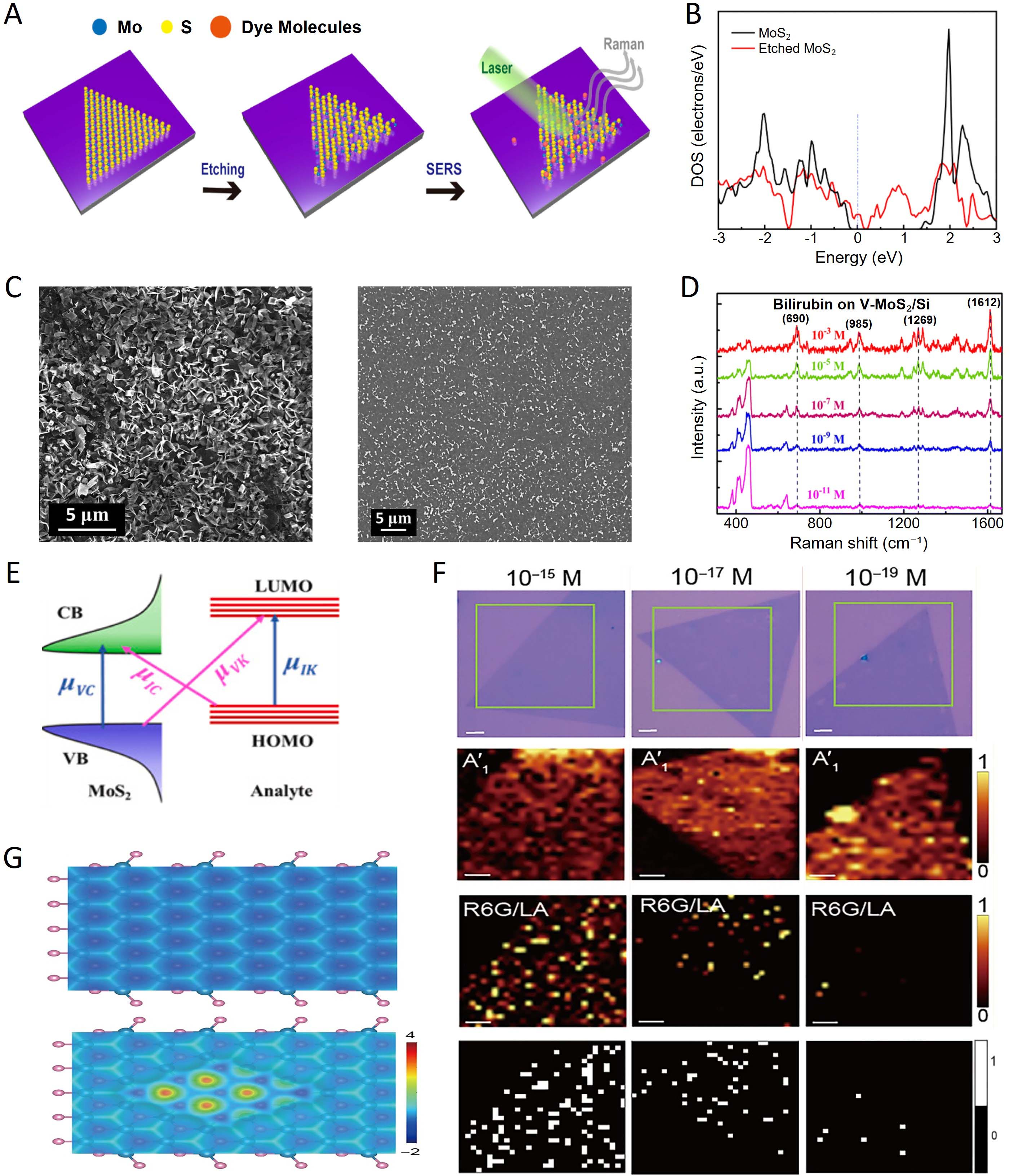
Figure 12. TMD-based SERS. (A) schematic illustration of SERS detection on defect-engineered monolayer MoS2; (B) comparison of Raman spectra and density of states between pristine and defect-rich MoS2. (A and B) are adapted with permission[31]. Copyright 2022, MDPI.; (C) representative SEM images of vertically and horizontally oriented MoS2 nanostructures; (D) SERS spectra of bilirubin on vertical MoS2/Si substrates. (C and D) are adapted with permission[32]. Copyright 2023, Royal Society of Chemistry; (E) electrochemically modulated band diagram illustrating tunable photon-driven charge transfer in MoS2-based EC-SERS systems, adapted with permission[40]. Copyright 2024, Elsevier; (F) combined single-molecule SERS mapping images at ultralow concentrations (10-15 to 10-19 M); (G) DFT electron-density maps comparing pristine and nanopore-engineered WS2, revealing the atomic-scale origin of the enhanced charge-transfer mechanism. (F and G) are adapted with permission[41]. Copyright 2025, American Association for the Advancement of Science. CB: conduction band; VB: valence band; HOMO: highest occupied molecular orbital; LOMO: lowest occupied molecular orbital.
Building upon defect-driven enhancement, Singh and Mishra demonstrated that the geometric orientation of MoS2 nanostructures offers an additional and orthogonal route to amplify SERS sensitivity. As shown by the SEM images in Figure 12C, vertically oriented MoS2 architectures expose a high density of edge sites, whose electronic structures differ substantially from those of basal planes. These edge-rich configurations promote stronger molecule-substrate coupling and increase the number of available charge-transfer pathways. Comparative SERS measurements using biologically relevant molecules, such as bilirubin, revealed significantly stronger Raman enhancement on vertically aligned MoS2 than on horizontally oriented counterparts [Figure 12D]. This work highlighted that morphology and edge engineering can be as critical as defect density in optimizing chemical enhancement in TMD-based SERS platforms.
While defect and structural engineering define static enhancement capabilities, Gupta et al.[40] introduced electrochemical modulation as a means to dynamically control SERS activity in MoS2-based systems. By growing MoS2 films directly on conducting substrates and applying an external bias, the Fermi level of MoS2 could be continuously shifted relative to molecular orbitals, as depicted in Figure 12E. This electrochemical gating enabled reversible tuning of charge-transfer resonance conditions, thereby allowing in situ modulation of SERS intensity. Such electrochemically controlled SERS represents a critical conceptual advance, demonstrating that TMD substrates can function as adaptive sensing platforms rather than fixed enhancers, with potential for quantitative and stimulus-responsive biosensing.
The ultimate sensitivity of TMD-based SERS was achieved through atomic-scale engineering strategies that push chemical enhancement into the true single-molecule regime. He et al.[41] demonstrated that nanopore-engineered monolayer WS2 supports discrete and spatially isolated SERS hotspots on an otherwise atomically flat surface. As shown by the SERS mapping images in Figure 12F, Raman signals at ultralow concentrations (10-15-10-19 M) appear as sparse, localized events, providing visually compelling evidence of single-molecule detection. Density functional theory calculations further revealed that the charge density becomes strongly localized at the nanopore rims, as illustrated in Figure 12G, providing an atomistic explanation for the enhanced charge-transfer efficiency. This study represents a conceptual culmination of TMD-based SERS, in which enhancement originates from well-defined atomic features rather than stochastic electromagnetic hotspots.
Figure 12 encapsulates the evolution of TMD-enabled SERS from defect-induced chemical enhancement and geometry-driven edge engineering to electrochemically tunable platforms and atomic-scale single-molecule detection. This progression underscores that TMDs are not merely passive Raman substrates but actively engineerable semiconducting systems whose electronic structure, surface chemistry, and interfacial energetics can be systematically tailored. Such versatility positions TMD-based SERS as a robust, reproducible, and fundamentally distinct alternative to conventional plasmonic platforms for advanced biosensing applications.
While TMD-based SERS platforms have demonstrated significant progress, several challenges remain in establishing their reliability as biosensing systems. The Raman enhancement is highly sensitive to defect density, edge structure, and adsorption configuration, leading to variability across studies and limiting quantitative reproducibility. Achieving precise and scalable control over defect formation, therefore, remains a key challenge. In addition, although chemical enhancement mechanisms offer improved uniformity compared to plasmonic substrates, their overall enhancement factors are often lower than those of optimized metal-based systems[31,142]. However, recent strategies that combine defect engineering with structural design or electrochemical modulation have shown promising routes toward enhanced sensitivity and tunability. In particular, approaches enabling dynamic control of charge-transfer resonance or atomic-scale engineering of active sites are expected to play a key role in advancing TMD-based SERS toward reproducible and quantitative biosensing applications.
Photoacoustic imaging
PAI is a hybrid optical-acoustic modality in which absorbed optical energy is converted into acoustic waves through rapid thermoelastic expansion, enabling imaging with optical contrast at depths beyond the reach of purely optical techniques[144]. From a biosensing perspective, the effectiveness of a PA contrast agent is governed by several key material parameters, including optical absorption strength, efficiency of non-radiative energy dissipation, and photothermal stability under repeated excitation. Conventional exogenous agents, such as small-molecule dyes, can enhance PA contrast but often suffer from photobleaching, aggregation-induced quenching, rapid clearance, and limited spectral tunability, which restrict their long-term and quantitative imaging performance[145].
TMDs offer a materials platform that naturally addresses many of these limitations. Optical absorption in monolayer and few-layer TMDs is dominated by excitonic resonances and interband transitions with large oscillator strengths, resulting in strong and broadband light harvesting across the visible and NIR regions[146]. Following excitation, strong exciton-phonon coupling promotes ultrafast non-radiative relaxation. As a result, a substantial fraction of the absorbed photon energy is converted into heat rather than radiative emission. This intrinsic bias toward non-radiative decay directly favors efficient thermoelastic signal generation, making TMDs particularly well-suited as PA transducers. In addition to efficient energy conversion, TMDs exhibit exceptional photothermal robustness owing to their layered crystal structure and strong in-plane covalent bonding. Unlike many organic chromophores, TMD nanosheets maintain stable absorption and photothermal behavior under repeated laser irradiation, enabling sustained PA signal generation during longitudinal imaging. The two-dimensional geometry further provides a large surface area for chemical modification and hybridization, allowing systematic tuning of optical and thermal properties without compromising structural integrity[33].
The PA performance of TMD-based systems can be further enhanced through compositional and interfacial engineering. Control over layer number and chalcogen composition enables modulation of absorption strength and spectral position within biologically relevant optical windows. Hybridization with NIR dyes, such as indocyanine green (ICG), provides an additional route to extend and intensify absorption at clinically relevant wavelengths. In such hybrid architectures, TMDs can stabilize dyes against photobleaching and aggregation while facilitating efficient non-radiative energy dissipation through interfacial charge or energy transfer. These synergistic effects yield PA responses that exceed those of either component alone[34,43].
Building on these principles, a growing body of experimental work has demonstrated how the intrinsic optothermal properties of TMDs translate into enhanced PA performance in biologically relevant settings. Representative studies have shown that reducing TMDs to the monolayer limit can significantly amplify PA signal generation, reflecting the transition to exciton-dominated absorption and more efficient non-radiative relaxation. Complementary approaches have employed hybrid architectures that combine TMD nanosheets with NIR dyes to further boost absorption strength, extend spectral coverage, and improve in vivo imaging sensitivity. Together, these studies illustrate how both intrinsic dimensional control and interfacial hybridization strategies can be exploited to tailor the PA response of TMD-based biosensing platforms, as exemplified by the works summarized in Figure 13[33,34,43].
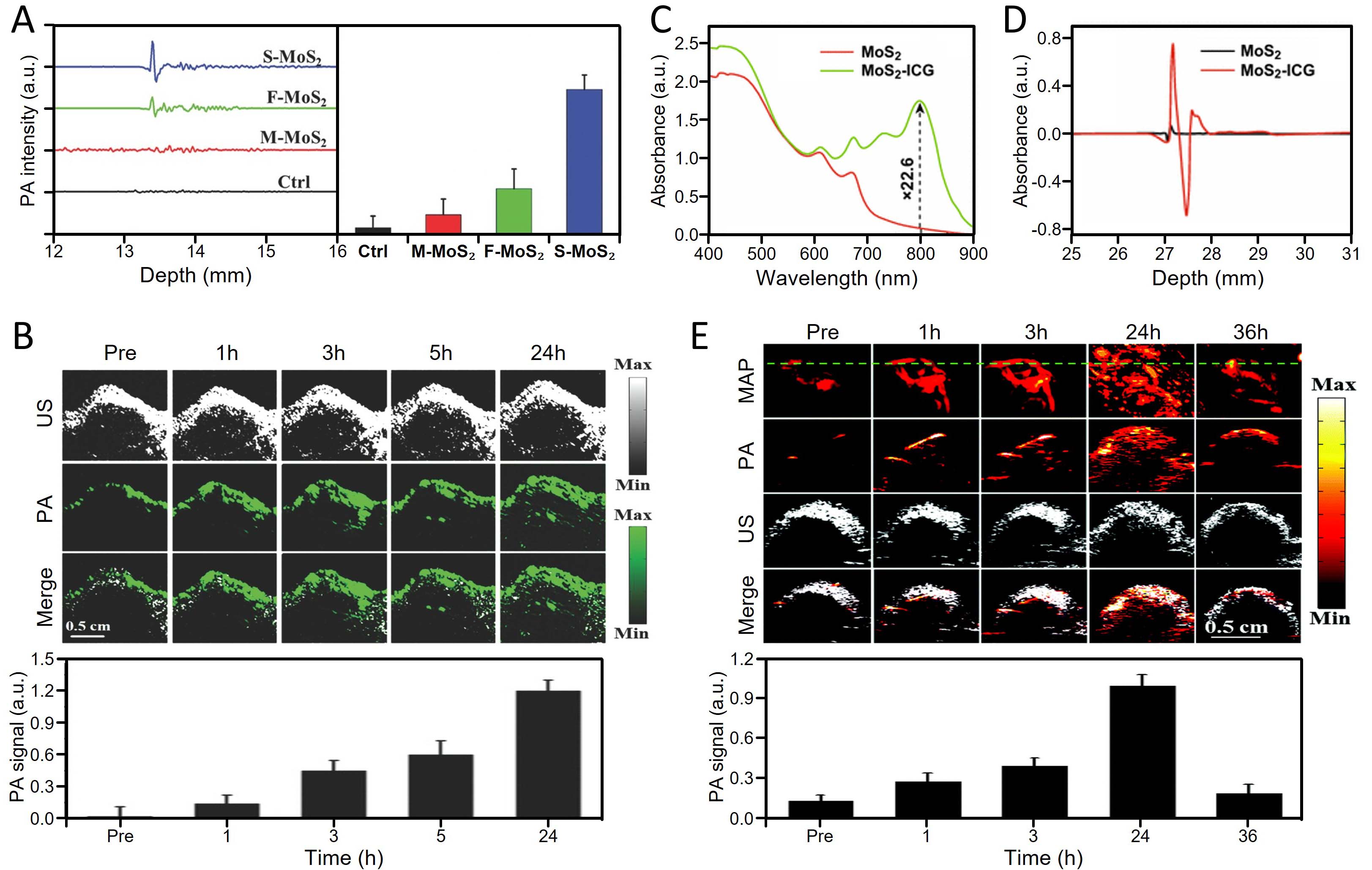
Figure 13. TMD-enabled PA biosensing and deep-tissue imaging. (A) quantitative comparison of PA signal amplitudes generated from single-layer (S-MoS2), few-layer (F-MoS2), and multilayer (M-MoS2) MoS2 nanosheets at identical concentrations; (B) in vivo orthotopic brain tumor PA imaging using single-layer MoS2 nanosheets, demonstrating robust signal enhancement at deep tissue depths beneath the skull and time-dependent accumulation within the tumor region. (A and B) are adapted with permission[33]. Copyright 2016, Wiley-VCH; (C) UV-VIS-NIR absorbance spectra of pristine MoS2 and MoS2-ICG hybrid nanosheets at identical MoS2 concentrations; (D) quantitative comparison of PA signal intensities generated by MoS2 and MoS2-ICG hybrids. (C and D) are adapted with permission[34,43]. Copyright 2018, Springer Nature; (E) in vivo tumor-targeted PA imaging and corresponding signal quantification using MoSe2-ICG hybrid nanosheets. PA: Photoacoustic; US: ultrasound.
TMDs offer a fundamentally different approach to PA contrast enhancement by leveraging their strong excitonic absorption, ultrafast nonradiative relaxation, and exceptional photothermal robustness. The intrinsic PA amplification capability of TMDs was demonstrated using MoS2 nanosheets with controlled layer thickness. As depicted in Figure 13A, single-layer MoS2 generates markedly stronger PA signals than few-layer or multilayer counterparts at identical concentrations, highlighting a pronounced layer-dependent amplification effect. This behavior reflects the emergence of exciton-dominated absorption and more efficient optothermal energy conversion in the monolayer limit, which together promote effective thermoelastic signal generation and establish monolayer TMDs as intrinsically amplified PA contrast agents.
The practical relevance of this intrinsic amplification was further validated through in vivo imaging of orthotopic brain tumors. As shown in Figure 13B, deep-tissue PA imaging following systemic administration of single-layer MoS2 nanosheets reveals robust signal enhancement within intracranial tumor regions, despite the presence of the skull and overlying tissue. Quantitative analysis confirms time-dependent accumulation and sustained contrast, demonstrating that TMD nanosheets can serve as effective PA contrast agents under physiologically relevant conditions, without relying on plasmonic or metallic components.
While pristine TMDs already provide strong PA contrast, hybridization strategies have been introduced to further enhance sensitivity and spectral compatibility with biological optical windows. A representative example is the MoS2-ICG hybrid system, in which the broadband absorption and photothermal stability of MoS2 are combined with the intense NIR absorption of indocyanine green. As illustrated in Figure 13C, conjugation with ICG substantially broadens and intensifies optical absorption in the NIR region, directly addressing the limited long-wavelength absorption of pristine MoS2. This enhancement translates into significantly increased PA signal generation, as quantified in Figure 13D, reflecting synergistic photon harvesting and efficient nonradiative energy dissipation. Importantly, the hybrid architecture mitigates common limitations of free ICG, including photobleaching and aggregation, by stabilizing the dye on the TMD surface and promoting optothermally favorable decay pathways.
The evolution of TMD-based PA biosensing culminates in hybrid systems designed for integrated imaging and therapy. As shown in Figure 13E, MoSe2-ICG hybrid nanosheets enable high-contrast, tumor-targeted PA imaging with clear time-dependent signal enhancement in vivo. Compared with MoS2-based systems, MoSe2 exhibits red-shifted excitonic absorption and a higher photothermal conversion efficiency, thereby further improving PA sensitivity at biologically relevant wavelengths[36,43]. The strong PA contrast observed in tumor tissue underscores the ability of TMD hybrids to accumulate at pathological sites and generate reliable deep-tissue signals, while their efficient photon-to-heat conversion establishes a direct mechanistic link between PAI and PTT.
Collectively, the studies summarized in Figure 13 delineate a coherent progression in TMD-enabled PA biosensing—from intrinsic, layer-dependent PA amplification in pristine monolayer TMDs, to hybrid-induced sensitivity enhancement through molecular dye coupling, and ultimately to multifunctional theranostic platforms capable of deep-tissue imaging and therapy.
While TMD-based photoacoustic systems have demonstrated strong potential for deep-tissue imaging and biosensing, several challenges remain for their broader application. The reported PA performance is often sensitive to material parameters such as layer thickness, lateral size, and surface functionalization, leading to variability across studies and complicating direct comparison of signal efficiency. In addition, most demonstrations have focused on short-term imaging; long-term biocompatibility, biodegradability, and clearance pathways remain to be fully established. Furthermore, although hybridization strategies using NIR dyes significantly improve absorption and sensitivity, they introduce additional complexity in stability, pharmacokinetics, and reproducibility. Differences in dye loading, binding stability, and interfacial interactions can result in inconsistent performance across systems. Nevertheless, compositional tuning and rational hybrid design have emerged as effective strategies to enhance PA response while maintaining biological compatibility. In particular, approaches that optimize excitonic absorption while controlling interfacial energy transfer are expected to play a key role in advancing TMD-based photoacoustic biosensing toward reliable, clinically relevant applications.
TMD-based systems demonstrate versatile functionalities across diverse phototherapeutic and biosensing applications. To provide a clearer overview, Table 2 summarizes the key features of TMD-based systems across major phototherapeutic and biosensing modalities. By consolidating the functional roles of TMDs, representative conventional materials, and the associated advantages and limitations, this overview highlights both the common strengths and the remaining challenges of TMD-based systems, offering a more unified perspective on their current status and future directions.
Comparative summary of TMD-based phototherapeutic and optical biosensing modalitie
| Modality | Role of TMDs | Conventional materials | Advantages of TMDs | Limtations/chllenges | Ref. |
| PTT | Photothermal agent | Plasmonic nanomaterials (e.g., Au, carbon); organic photothermal agents | Strong NIR absorption; high thermal stability; resistant to photobleaching; surface tunability | Dependence on experimental conditions; performance variability across studies | [16,123,125,126] |
| PDT & multimodal phototherapy | ROS generator; drug carrier | Organic photosensitizers | Combined photothermal and ROS-mediated therapeutic effects; high loading capacity | Oxygen dependence; system complexity; reproducibility challenges | [24,126,147] |
| MPM & SHG | Nonlinear imaging contrast agent | Organic fluorophores; label-free biological structures | Strong nonlinear signal; label-free imaging; high spatial resolution | Sensitivity to excitation conditions; environmental dependence; limited biological validation | [26,27,126] |
| SERS | Raman signal enhancer | Plasmonic metal substrates (e.g., Au, Ag) | Uniform signal; lower photothermal damage; tunable via defect control | Weaker electromagnetic enhancement compared to plasmonic substrates; reliance on chemical enhancement mechanisms | [31,32,133,142] |
| PAI | PA signal generator | Endogenous absorbers (e.g., hemoglobin); organic dyes (e.g., ICG) | Efficient light-to-heat conversion; strong and stable signals; high thermal stability | Sensitivity to material parameters; limited long-term validation; hybrid system complexity | [33,34,130] |
| Theranostics | Integrated sensing-therapy platform | Single-function therapeutic or sensing agents | Combined imaging and treatment; flexible design; multifunctionality | System complexity; reproducibility challenges | [24,124,148] |
CONCLUSIONS
This review has provided a mechanism-oriented perspective on TMDs as versatile material platforms for both phototherapeutic and optical biosensing applications. Rather than treating individual implementations in isolation, the discussion has emphasized how atomic structure, electronic band configuration, excitonic physics, nonradiative relaxation pathways, and surface chemistry collectively dictate the interactions of TMDs with light and biological environments. These shared physical foundations unify a broad spectrum of functionalities, including photothermal and photodynamic therapies, exciton-mediated nonlinear optical imaging, charge-transfer-driven SERS, and optothermal PA signal generation. Across these modalities, TMDs consistently overcome fundamental limitations of conventional agents—such as insufficient sensitivity, photo-instability, or limited penetration depth—by leveraging strong light-matter coupling and intrinsically efficient energy-dissipating channels.
A central conclusion emerging from the surveyed studies is that the performance of TMD-based systems is not fixed but is highly programmable through rational control of material parameters. Layer thickness, crystal phase, defect density, and surface functionalization critically regulate excitonic resonances, charge-transfer efficiency, and photothermal conversion, while hybridization with molecular dyes, plasmonic structures, or optical cavities further extends spectral reach and amplifies signal generation. This tunability enables the deliberate tailoring of TMD platforms toward specific objectives, ranging from single-molecule Raman detection and wavelength-selective nonlinear imaging to deep-tissue PA sensing coupled with image-guided therapy. Such modularity distinguishes TMDs from many existing nanomaterials and positions them as unifying building blocks for next-generation theranostic systems, in which imaging, sensing, and treatment are integrated through a common exciton-driven optophysical framework.
By organizing diverse TMD-enabled biomedical functions within a common physical framework, this review highlights general design principles that link material properties to multifunctional performance while also revealing several important challenges that remain to be addressed. In particular, a deeper understanding of exciton dynamics and energy dissipation pathways under realistic biological environments is required to ensure reliable and reproducible performance. In addition, achieving precise control over defects, interfaces, and material heterogeneity remains a key experimental challenge that directly impacts consistency and scalability across different studies. From a broader perspective, future progress will depend on the development of standardized evaluation protocols, improved biointerface engineering, and the integration of TMD-based systems into clinically relevant platforms. At the same time, emerging directions are beginning to expand the functional scope of TMD-based technologies. These include chiral TMD systems for polarization-sensitive biosensing, metasurface-integrated TMD architectures for enhanced nonlinear signal manipulation, and data-driven approaches such as machine learning for optimizing material properties and device performance. Together with advances in multimodal integration and system-level design, these developments point toward more intelligent, tunable, and application-specific platforms that are likely to define the next generation of TMD-enabled biomedical technologies.
DECLARATIONS
Acknowledgments
The graphical abstract includes elements quoted with permission[36], Copyright 2016, American Chemical Society; Copyright 2014, Royal Society of Chemistry[37]; Copyright 2013, American Physical Society[26]; Copyright 2019, Optica Publishing Group[29]; Copyright 2018, Springer Nature[43].
Authors’ contributions
Contributed to investigation, methodology, writing-original draft, writing-review, and editing: Kim, J.; Demiray, E. B.
Provided figure curation and editing across the entire manuscript: Kim, C. S.
Contributed to conceptualization, funding acquisition, investigation, project administration, supervision, writing-review, and editing: Kim, J.; Lee, H.; Han, D. W.
Availability of data and materials
Not applicable.
AI and AI-assisted tools statement
Not applicable.
Financial support and sponsorship
This work was supported by the National Research Foundation of Korea (NRF) grant funded by the Korea government (MSIT) (No. NRF-RS-2021-NR060086), and by the National Research Foundation of Korea (NRF) grant funded by the Korea government (RS-2024-00406152).
Conflicts of interest
All authors declared that there are no conflicts of interest.
Ethical approval and consent to participate
Not applicable.
Consent for publication
Not applicable.
Copyright
© The Author(s) 2026.
REFERENCES
1. Novoselov, K. S.; Geim, A. K.; Morozov, S. V.; et al. Electric field effect in atomically thin carbon films. Science 2004, 306, 666-9.
3. Butler, S. Z.; Hollen, S. M.; Cao, L.; et al. Progress, challenges, and opportunities in two-dimensional materials beyond graphene. ACS. Nano. 2013, 7, 2898-926.
4. Xu, M.; Liang, T.; Shi, M.; Chen, H. Graphene-like two-dimensional materials. Chem. Rev. 2013, 113, 3766-98.
5. Chhowalla, M.; Shin, H. S.; Eda, G.; Li, L.; Loh, K. P.; Zhang, H. The chemistry of two-dimensional layered transition metal dichalcogenide nanosheets. Nature. Chem. 2013, 5, 263-75.
6. Tan, C.; Cao, X.; Wu, X.; et al. Recent advances in ultrathin two-dimensional nanomaterials. Chem. Rev. 2017, 117, 6225-331.
7. Wang, Q. H.; Kalantar-Zadeh, K.; Kis, A.; Coleman, J. N.; Strano, M. S. Electronics and optoelectronics of two-dimensional transition metal dichalcogenides. Nat. Nanotechnol. 2012, 7, 699-712.
8. Mak, K. F.; Shan, J. Photonics and optoelectronics of 2D semiconductor transition metal dichalcogenides. Nat. Photonics. 2016, 10, 216-26.
9. Chernikov, A.; Berkelbach, T. C.; Hill, H. M.; et al. Exciton binding energy and nonhydrogenic rydberg series in monolayer WS2. Phys. Rev. Lett. 2014, 113, 076802.
10. Zavabeti, A.; Jannat, A.; Zhong, L.; Haidry, A. A.; Yao, Z.; Ou, J. Z. Two-dimensional materials in large-areas: synthesis, properties and applications. Nano. Micro. Lett. 2020, 12, 66.
11. Manzeli, S.; Ovchinnikov, D.; Pasquier, D.; Yazyev, O. V.; Kis, A. 2D transition metal dichalcogenides. Nat. Rev. Mater. 2017, 2, 17033.
12. Derakhshi, M.; Daemi, S.; Shahini, P.; Habibzadeh, A.; Mostafavi, E.; Ashkarran, A. A. Two-dimensional nanomaterials beyond graphene for biomedical applications. J. Funct. Biomater. 2022, 13, 27.
13. Splendiani, A.; Sun, L.; Zhang, Y.; et al. Emerging photoluminescence in monolayer MoS2. Nano. Lett. 2010, 10, 1271-5.
14. Mak, K. F.; Lee, C.; Hone, J.; Shan, J.; Heinz, T. F. Atomically thin MoS2: a new direct-gap semiconductor. Phys. Rev. Lett. 2010, 105, 136805.
15. Cheng, L.; Wang, C.; Feng, L.; Yang, K.; Liu, Z. Functional nanomaterials for phototherapies of cancer. Chem. Rev. 2014, 114, 10869-939.
16. Yin, W.; Yan, L.; Yu, J.; et al. High-throughput synthesis of single-layer MoS2 nanosheets as a near-infrared photothermal-triggered drug delivery for effective cancer therapy. ACS. Nano. 2014, 8, 6922-33.
17. Shi, J.; Liu, M.; Wen, J.; et al. All chemical vapor deposition synthesis and intrinsic bandgap observation of MoS2/graphene heterostructures. Adv. Mater. 2015, 27, 7086-92.
19. Park, K. H.; Yang, J. Y.; Jung, S.; et al. Metallic phase transition metal dichalcogenide quantum dots as promising bio-imaging materials. Nanomaterials 2022, 12, 1645.
20. Chen, Q.; Xu, L.; Liang, C.; Wang, C.; Peng, R.; Liu, Z. Photothermal therapy with immune-adjuvant nanoparticles together with checkpoint blockade for effective cancer immunotherapy. Nat. Commun. 2016, 7, 13193.
21. Lucky, S. S.; Soo, K. C.; Zhang, Y. Nanoparticles in photodynamic therapy. Chem. Rev. 2015, 115, 1990-2042.
22. Zhang, X.; Wu, J.; Williams, G. R.; Niu, S.; Qian, Q.; Zhu, L. Functionalized MoS2-nanosheets for targeted drug delivery and chemo-photothermal therapy. Colloid. Surf. B. Biointerfaces. 2019, 173, 101-8.
23. Liu, J.; Lu, K.; Gao, F.; Zhao, L.; Li, H.; Jiang, Y. Multifunctional MoS2 composite nanomaterials for drug delivery and synergistic photothermal therapy in cancer treatment. Ceram. Int. 2022, 48, 22378-86.
24. Zhu, Y.; Wang, Y.; Williams, G. R.; et al. RETRACTED: multicomponent transition metal dichalcogenide nanosheets for imaging-guided photothermal and chemodynamic therapy. Adv. Sci. 2020, 7, 2000272.
25. Sharma, A.; Bhalothia, D.; Vankayala, R.; Bagaria, A. Morphology-driven photothermal performance of polymer-functionalized mos₂ nanoplatforms for targeted therapeutic applications. Sci. Rep. 2025, 15, 26028.
26. Malard, L. M.; Alencar, T. V.; Barboza, A. P. M.; Mak, K. F.; De Paula, A. M. Observation of intense second harmonic generation from MoS2 atomic crystals. Phys. Rev. B. 2013, 87, 201401.
27. Zhang, S.; Dong, N.; Mcevoy, N.; et al. Direct observation of degenerate two-photon absorption and its saturation in WS2 and MoS2 monolayer and few-layer films. ACS. Nano. 2015, 9, 7142-50.
28. Xie, Y.; Zhang, S.; Li, Y.; et al. Layer-modulated two-photon absorption in MoS2: probing the shift of the excitonic dark state and band-edge. Photon. Res. 2019, 7, 762.
29. Lin, K.; Shiau, S.; Liu, S.; et al. Fast real-space imaging of the exciton complexes in WSe2 and WS2 monolayers using multiphoton microscopy. J. Phys. Chem. C. 2020, 124, 7979-87.
30. Ling, X.; Xie, L.; Fang, Y.; et al. Can graphene be used as a substrate for Raman enhancement? Nano. Lett. 2009, 10, 553-61.
31. Sun, S.; Zheng, J.; Sun, R.; et al. Defect-rich monolayer MoS2 as a universally enhanced substrate for surface-enhanced raman scattering. Nanomaterials 2022, 12, 896.
32. Singh, A.; Mishra, A. K. Large area CVD-grown vertically and horizontally oriented MoS2 nanostructures as SERS biosensors for single molecule detection. Nanoscale 2023, 15, 16480-92.
33. Chen, J.; Liu, C.; Hu, D.; et al. Single-layer MoS2 nanosheets with amplified photoacoustic effect for highly sensitive photoacoustic imaging of orthotopic brain tumors. Adv. Funct. Mater. 2016, 26, 8715-25.
34. Liu, C.; Chen, J.; Zhu, Y.; et al. Highly sensitive MoS2-indocyanine green hybrid for photoacoustic imaging of orthotopic brain glioma at deep site. Nano. Micro. Lett. 2018, 10, 48.
35. Chen, H.; Liu, T.; Su, Z.; Shang, L.; Wei, G. 2D transition metal dichalcogenide nanosheets for photo/thermo-based tumor imaging and therapy. Nanoscale. Horiz. 2018, 3, 74-89.
36. Lei, Z.; Zhu, W.; Xu, S.; Ding, J.; Wan, J.; Wu, P. Hydrophilic MoSe2 nanosheets as effective photothermal therapy agents and their application in smart devices. ACS. Appl. Mater. Interfaces. 2016, 8, 20900-8.
37. Liu, T.; Wang, C.; Cui, W.; et al. Combined photothermal and photodynamic therapy delivered by PEGylated MoS2 nanosheets. Nanoscale 2014, 6, 11219-25.
38. Li, T.; Guan, C.; Xu, L.; et al. Facile synthesis of MoS2@red phosphorus heterojunction for synergistically photodynamic and photothermal therapy of renal cell carcinoma. Colloid. Surf. B. Biointerfaces. 2024, 241, 114031.
39. Zhou, F.; Kua, J. H.; Lu, S.; Ji, W. Two-photon absorption arises from two-dimensional excitons. Opt. Express. 2018, 26, 16093.
40. Gupta, R. K.; Singh, A.; Mishra, A. K. Hydrothermally in-situ grown MoS2 films over different conducting substrates as electrochemical-SERS biosensors. Opt. Mater. 2024, 155, 115866.
41. He, H.; Zhao, M.; Xu, H.; et al. Engineering nanopore in monolayer WS2 for single-molecule imaging on the atomic-scale flat 2D surfaces. Sci. Adv. 2025, 11, eady2148.
42. Liu, F.; Liu, H.; Chi, C.; et al. Giant two-photon upconversion from 2D exciton in doubly-resonant plasmonic nanocavity. Light. Sci. Appl. 2025, 14, 312.
43. Chen, J.; Li, X.; Liu, X.; et al. Hybrid MoSe2-indocyanine green nanosheets as a highly efficient phototheranostic agent for photoacoustic imaging guided photothermal cancer therapy. Biomater. Sci. 2018, 6, 1503-16.
44. Liu, G. Theoretical study of Ti and Cr as candidate assisted metals for mechanical exfoliation of monolayer transition metal dichalcogenides. Sci. Rep. 2025, 15, 613.
45. Samadi, M.; Sarikhani, N.; Zirak, M.; Zhang, H.; Zhang, H.; Moshfegh, A. Z. Group 6 transition metal dichalcogenide nanomaterials: synthesis, applications and future perspectives. Nanoscale. Horiz. 2018, 3, 90-204.
46. Han, S. A.; Bhatia, R.; Kim, S. Synthesis, properties and potential applications of two-dimensional transition metal dichalcogenides. Nano. Converg. 2015, 2, 17.
47. Lasek, K.; Li, J.; Kolekar, S.; et al. Synthesis and characterization of 2D transition metal dichalcogenides: recent progress from a vacuum surface science perspective. Surf. Sci. Rep. 2021, 76, 100523.
48. Toh, R. J.; Sofer, Z.; Luxa, J.; Sedmidubský, D.; Pumera, M. 3R phase of MoS2 and WS2 outperforms the corresponding 2H phase for hydrogen evolution. Chem. Commun. 2017, 53, 3054-7.
49. Radisavljevic, B.; Radenovic, A.; Brivio, J.; Giacometti, V.; Kis, A. Single-layer MoS2 transistors. Nat. Nanotechnol. 2011, 6, 147-50.
50. Naik, M. H.; Jain, M. Ultraflatbands and shear solitons in moiré patterns of twisted bilayer transition metal dichalcogenides. Phys. Rev. Lett. 2018, 121, 266401.
51. Zubair, M.; Lee, D.; Kang, D. J. Harnessing the power of 2D materials for flexible energy harvesting applications. Carbon. Energy. 2025, 7, e70083.
52. Tongay, S.; Sahin, H.; Ko, C.; et al. Monolayer behaviour in bulk ReS2 due to electronic and vibrational decoupling. Nat. Commun. 2014, 5, 3252.
53. Kwon, S.; Yun, T. K.; Ma, P. J.; Nam, S. Harnessing in-plane optical anisotropy in WS2 through ReS2 crystal. Nanophotonics 2025, 14, 1553-61.
54. Li, Z.; Attanayake, N. H.; Blackburn, J. L.; Miller, E. M. Carbon dioxide and nitrogen reduction reactions using 2D transition metal dichalcogenide (TMDC) and carbide/nitride (MXene) catalysts. Energy. Environ. Sci. 2021, 14, 6242-86.
55. Gu, M.; Rao, A. M.; Zhou, J.; Lu, B. Molecular modulation strategies for two-dimensional transition metal dichalcogenide-based high-performance electrodes for metal-ion batteries. Chem. Sci. 2024, 15, 2323-50.
56. Dai, B.; Su, Y.; Guo, Y.; Wu, C.; Xie, Y. Recent strategies for the synthesis of phase-pure ultrathin 1T/1T′ transition metal dichalcogenide nanosheets. Chem. Rev. 2023, 124, 420-54.
57. Voiry, D.; Mohite, A.; Chhowalla, M. Phase engineering of transition metal dichalcogenides. Chem. Soc. Rev. 2015, 44, 2702-12.
58. Yang, Z. J.; Wang, Y.; Chhowalla, M. Metallic-phase two-dimensional transition-metal dichalcogenides for electrochemical applications. MRS. Bull. 2025, 50, 1243-51.
59. Zhao, W.; Ribeiro, R. M.; Eda, G. Electronic structure and optical signatures of semiconducting transition metal dichalcogenide nanosheets. Acc. Chem. Res. 2014, 48, 91-9.
60. Liu, H.; Huang, Z.; Chen, X.; Xue, W.; Zhang, S.; Qi, X. Interlayer coupling dependent spin and valley polarization in a two-dimensional metal-semiconductor contact heterostructure. Phys. Rev. B. 2025, 111, 075302.
61. Mahdouani, M.; Oudir, A.; Gardelis, S.; Bourguiga, R. Theoretical investigation of auger and electron-surface optical phonon processes near the K-points in monolayer PtSe2 and PtS2 on polar dielectric substrates. Materials 2026, 19, 1280.
62. Villaos, R. A. B.; Crisostomo, C. P.; Huang, Z.; et al. Thickness dependent electronic properties of Pt dichalcogenides. NPJ. 2D. Mater. Appl. 2019, 3, 2.
63. Liu, X.; Zhang, Y. Thermal properties of transition-metal dichalcogenide. Chin. Phys. B. 2018, 27, 034402.
64. Farris, R.; Hellman, O.; Zanolli, Z.; et al. Microscopic understanding of the in-plane thermal transport properties of 2H transition metal dichalcogenides. Phys. Rev. B. 2024, 109, 125422.
65. Zhang, D.; Ren, W.; Wang, K.; et al. A thermal conductivity switch via the reversible 2H-1T′ phase transition in monolayer MoTe2. Chin. Phys. B. 2023, 32, 050505.
67. Giri, A.; Park, G.; Jeong, U. Layer-structured anisotropic metal chalcogenides: recent advances in synthesis, modulation, and applications. Chem. Rev. 2023, 123, 3329-442.
68. Xiao, Y.; Zhou, M.; Liu, J.; Xu, J.; Fu, L. Phase engineering of two-dimensional transition metal dichalcogenides. Sci. China. Mater. 2019, 62, 759-75.
69. Tripathi, P.; Kumar, A.; Bankar, P. K.; Singh, K.; Gupta, B. K. Large-scale production and optical properties of a high-quality SnS2 single crystal grown using the chemical vapor transportation method. Crystals 2023, 13, 1131.
70. Tang, W.; Rassay, S. S.; Ravindra, N. M. Electronic & optical properties of transition-metal dichalcogenides. Madridge. J. Nanotechnol. Nanosci. 2017, 2, 58-64.
71. Chaves, A.; Azadani, J. G.; Alsalman, H.; et al. Bandgap engineering of two-dimensional semiconductor materials. NPJ. 2D. Mater. Appl. 2020, 4, 29.
72. Nguyen-truong, H. T. Exciton binding energy and screening length in two-dimensional semiconductors. Phys. Rev. B. 2022, 105, L201407.
73. Tang, W. Electrical, electronic and optical properties of MoS2 & WS2. Newark, NJ: New Jersey Institute of Technology; 2017.
74. Shang, J.; Shen, X.; Cong, C.; et al. Observation of excitonic fine structure in a 2D transition-metal dichalcogenide semiconductor. ACS. Nano. 2015, 9, 647-55.
75. Patra, C.; Mondal, S.; Mukherjee, R.; Nandakishora, Y. Advanced synthesis and unique properties of 2D transition metal dichalcogenides for realizing next-generation applications. ACS. Mater. Au. 2025, 5, 745-66.
76. Guddala, S.; Bushati, R.; Li, M.; Khanikaev, A. B.; Menon, V. M. Valley selective optical control of excitons in 2D semiconductors using a chiral metasurface. Opt. Mater. Express. 2019, 9, 536.
77. Ali, W.; Liu, Y.; Huang, M.; Xie, Y.; Li, Z. Temperature-dependent phonon scattering and photoluminescence in vertical MoS2/WSe2 heterostructures. Nanomaterials 2023, 13, 2349.
78. Burke, L. J.; Greenaway, M. T.; Betouras, J. J. Brightening dark excitons and trions in systems with a Mexican-hat energy dispersion: example of InSe. NPJ. 2D. Mater. Appl. 2025, 9, 63.
79. Cao, X.; Zhang, D.; Ye, W.; et al. Dielectric screening effects in the decoherence of excitons and exciton-phonon scattering in atomical monolayer WS2 semiconductors. Phys. Rev. B. 2025, 112, 045420.
80. Li, H.; Lu, G.; Yin, Z.; et al. Optical identification of single- and few-layer MoS2 sheets. Small 2012, 8, 682-6.
81. Papageorgiou, D. G.; Dong, M.; Zhang, H.; Liu, M.; Young, R. J. Mechanical properties of transition metal dichalcogenides: towards high-performance polymer nanocomposites. 2D. Mater. 2024, 12, 012002.
82. Kazemi, S. A.; Imani Yengejeh, S.; Wang, V.; Wen, W.; Wang, Y. Theoretical understanding of electronic and mechanical properties of 1T′ transition metal dichalcogenide crystals. Beilstein. J. Nanotechnol. 2022, 13, 160-71.
83. Falin, A.; Holwill, M.; Lv, H.; et al. Mechanical properties of atomically thin tungsten dichalcogenides: WS2, WSe2, and WTe2. ACS. Nano. 2021, 15, 2600-10.
84. Gao, J.; Li, B.; Tan, J.; Chow, P.; Lu, T.; Koratkar, N. Aging of transition metal dichalcogenide monolayers. ACS. Nano. 2016, 10, 2628-35.
85. Kang, K.; Godin, K.; Kim, Y. D.; et al. Graphene-assisted antioxidation of tungsten disulfide monolayers: substrate and electric-field effect. Adv. Mater. 2017, 29, 1603898.
86. Kansara, S.; Gupta, S. K.; Sonvane, Y. Effect of strain engineering on 2D dichalcogenides transition metal: a DFT study. Comput. Mater. Sci. 2018, 141, 235-42.
87. Yu, S.; Rice, Q.; Neupane, T.; Tabibi, B.; Li, Q.; Seo, F. J. Piezoelectricity enhancement and bandstructure modification of atomic defect-mediated MoS2 monolayer. Phys. Chem. Chem. Phys. 2017, 19, 24271-5.
88. Ye, R.; Song, W.; Ou, X.; Gu, Z.; Zhang, D. Membrane insertion of MoS2 nanosheets: fresh vs. aged. Front. Chem. 2021, 9, 706917.
89. Havelikar, U.; Ghorpade, K. B.; Kumar, A.; et al. Comprehensive insights into mechanism of nanotoxicity, assessment methods and regulatory challenges of nanomedicines. Discover. Nano. 2024, 19, 165.
90. Baimanov, D.; Wu, J.; Chu, R.; et al. Immunological responses induced by blood protein coronas on two-dimensional MoS2 nanosheets. ACS. Nano. 2020, 14, 5529-42.
91. Harries, R. W.; Brown, C. J.; Woodbine, L.; et al. Cell-substrate interactions lead to internalization and localization of layered MoS2 nanosheets. ACS. Appl. Nano. Mater. 2021, 4, 2002-10.
92. Xu, S.; Zheng, H.; Ma, R.; et al. Vacancies on 2D transition metal dichalcogenides elicit ferroptotic cell death. Nat. Commun. 2020, 11, 3484.
93. Park, M. N.; Choi, M.; Syahputra, R. A.; Delfino, D. V.; Ko, S.; Kim, B. Redox homeostasis as a therapeutic target in chronic oxidative diseases: implications for cancer treatment. Antioxidants 2026, 15, 203.
94. Wang, Z.; Von, Dem. Bussche. A.; Qiu, Y.; et al. Chemical dissolution pathways of MoS2 nanosheets in biological and environmental media. Environ. Sci. Technol. 2016, 50, 7208-17.
95. Tang, Y.; Huang, Z.; Tian, K.; Luo, F. Transition metal dichalcogenides as platforms for biosensing, phototherapy, and drug delivery. Sens. Diagn. 2026, 5, 649-73.
96. Kaur, N.; De, M. Shape and size dependent antimicrobial and anti-biofilm properties of functionalized MoS2. ACS. Infect. Dis. 2024, 11, 249-61.
97. Wang, Y.; Zhang, X.; Yue, H. Two-dimensional nanomaterials induced nano-bio interfacial effects and biomedical applications in cancer treatment. J. Nanobiotechnol. 2024, 22, 67.
98. Mutalik, C.; Okoro, G.; Chou, H.; et al. Phase-dependent 1T/2H-MoS2 nanosheets for effective photothermal killing of bacteria. ACS. Sustain. Chem. Eng. 2022, 10, 8949-57.
99. Karunakaran, S.; Pandit, S.; Basu, B.; De, M. Simultaneous exfoliation and functionalization of 2H-MoS2 by thiolated surfactants: applications in enhanced antibacterial activity. J. Am. Chem. Soc. 2018, 140, 12634-44.
100. Karunakaran, S.; Sahoo, S.; Sahoo, J.; De, M. Ligand-mediated exfoliation and antibacterial activity of 2H transition-metal dichalcogenides. ACS. Appl. Bio. Mater. 2022, 6, 126-33.
101. Xu, S.; Liang, S.; Wang, B.; et al. Bi-functionalized transferrin@MoS2-PEG nanosheets for improving cellular uptake in HepG2 cells. Materials 2023, 16, 2277.
102. Gaba, S.; Sahu, M.; Chauhan, N.; Jain, U. Unlocking the potential of low-dimensional MoS2 as a smart nanoplatform for environmental technologies, therapeutic strategies, and biomedical sensing. Talanta. Open. 2025, 12, 100498.
103. Baimanov, D.; Wang, J.; Zhang, J.; et al. In situ analysis of nanoparticle soft corona and dynamic evolution. Nat. Commun. 2022, 13, 5389.
104. Chen, X.; Shinde, S. M.; Dhakal, K. P.; et al. Degradation behaviors and mechanisms of MoS2 crystals relevant to bioabsorbable electronics. NPG. Asia. Mater. 2018, 10, 810-20.
105. Nandy, S. K.; Das, S.; Pandey, S.; et al. The futuristic applications of transition metal dichalcogenides for cancer therapy. Luminescence 2024, 39, e4771.
106. Zhou, X.; Sun, H.; Bai, X. Two-dimensional transition metal dichalcogenides: synthesis, biomedical applications and biosafety evaluation. Front. Bioeng. Biotechnol. 2020, 8, 236.
107. Yang, Z.; Sun, Z.; Ren, Y.; et al. Advances in nanomaterials for use in photothermal and photodynamic therapeutics. Mol. Med. Rep. 2019, 20, 5-15.
108. Wang, Y.; Yang, Y.; Yang, L.; et al. Gold nanostar@polyaniline theranostic agent with high photothermal conversion efficiency for photoacoustic imaging-guided anticancer phototherapy at a low dosage. ACS. Appl. Mater. Interfaces. 2022, 14, 28570-80.
109. Jeong, W. Y.; Kang, M. S.; Lee, H.; et al. Recent trends in photoacoustic imaging techniques for 2D nanomaterial-based phototherapy. Biomedicines 2021, 9, 80.
110. Pekkanen, A. M.; Dewitt, M. R.; Rylander, M. N. Nanoparticle enhanced optical imaging and phototherapy of cancer. J. Biomed. Nanotechnol. 2014, 10, 1677-712.
111. Li, X.; Lovell, J. F.; Yoon, J.; Chen, X. Clinical development and potential of photothermal and photodynamic therapies for cancer. Nat. Rev. Clin. Oncol. 2020, 17, 657-74.
112. Rosal B, Jia B, Jaque D. Beyond phototherapy: recent advances in multifunctional fluorescent nanoparticles for light-triggered tumor theranostics. Adv. Funct. Mater. 2018, 28, 1803733.
113. Zhao, L.; Liu, Y.; Chang, R.; Xing, R.; Yan, X. Supramolecular photothermal nanomaterials as an emerging paradigm toward precision cancer therapy. Adv. Funct. Mater. 2018, 29, 1806877.
114. Wang, K.; Xiang, Y.; Pan, W.; Wang, H.; Li, N.; Tang, B. Dual-targeted photothermal agents for enhanced cancer therapy. Chem. Sci. 2020, 11, 8055-72.
115. Gupta, N.; Malviya, R. Understanding and advancement in gold nanoparticle targeted photothermal therapy of cancer. Biochim. Biophys. Acta. Rev. Cancer. 2021, 1875, 188532.
116. Cheng, X.; Sun, R.; Yin, L.; Chai, Z.; Shi, H.; Gao, M. Light-triggered assembly of gold nanoparticles for photothermal therapy and photoacoustic imaging of tumors in vivo. Adv. Mater. 2016, 29, 1604894.
117. Celli, J. P.; Spring, B. Q.; Rizvi, I.; et al. Imaging and photodynamic therapy: mechanisms, monitoring, and optimization. Chem. Rev. 2010, 110, 2795-838.
118. Donnelly, R. F.; Mccarron, P. A.; Tunney, M. M. Antifungal photodynamic therapy. Microbiol. Res. 2008, 163, 1-12.
119. Jerjes, W.; Upile, T.; Hamdoon, Z.; Alexander Mosse, C.; Morcos, M.; Hopper, C. Photodynamic therapy outcome for T1/T2 N0 oral squamous cell carcinoma. Lasers. Surg. Med. 2011, 43, 463-9.
120. Wang, Q.; Dai, Y.; Xu, J.; et al. All-in-one phototheranostics: single laser triggers NIR-II fluorescence/photoacoustic imaging guided photothermal/photodynamic/chemo combination therapy. Adv. Funct. Mater. 2019, 29, 1901480.
121. Overchuk, M.; Weersink, R. A.; Wilson, B. C.; Zheng, G. Photodynamic and photothermal therapies: synergy opportunities for nanomedicine. ACS. Nano. 2023, 17, 7979-8003.
122. Tyagi, N.; Arya, R. K. K.; Bisht, D.; et al. Mechanism and potentialities of photothermal and photodynamic therapy of transition metal dichalcogenides (TMDCs) against cancer. Luminescence 2024, 39, e4770.
123. Chou, S. S.; Kaehr, B.; Kim, J.; et al. Chemically exfoliated MoS2 as near-infrared photothermal agents. Angew. Chem. Int. Ed. 2013, 125, 4254-8.
124. Liu, T.; Shi, S.; Liang, C.; et al. Iron oxide decorated MoS2 nanosheets with double PEGylation for chelator-free radiolabeling and multimodal imaging guided photothermal therapy. ACS. Nano. 2015, 9, 950-60.
125. Huang, X.; Neretina, S.; El-Sayed, M. A. Gold nanorods: from synthesis and properties to biological and biomedical applications. Adv. Mater. 2009, 21, 4880-910.
126. Robinson, J. T.; Tabakman, S. M.; Liang, Y.; et al. Ultrasmall reduced graphene oxide with high near-infrared absorbance for photothermal therapy. J. Am. Chem. Soc. 2011, 133, 6825-31.
127. Bagheri, S.; Farokhnezhad, M.; Esmaeilzadeh, M. Transition metal dichalcogenide coated gold nanoshells for highly effective photothermal therapy. Phys. Chem. Chem. Phys. 2023, 25, 33038-47.
128. Denk, W.; Strickler, J. H.; Webb, W. W. Two-photon laser scanning fluorescence microscopy. Science 1990, 248, 73-6.
129. Ferraro, J. R.; Nakamoto, K.; Brown, C. W. Introductory raman spectroscopy; Elsevier; 2003. Available from: https://www.sciencedirect.com/book/monograph/9780122541056/introductory-raman-spectroscopy [Last accessed on 30 Jun 2026].
130. Wang, L. V.; Hu, S. Photoacoustic tomography: in vivo imaging from organelles to organs. Science 2012, 335, 1458-62.
131. Boyd, R. W.; Gaeta, A. L.; Giese, E. Springer handbook of atomic, molecular, and optical physics. New York, NY: Springer New York; 2006.
132. Zrazhevskiy, P.; Gao, X. Quantum dot imaging platform for single-cell molecular profiling. Nat. Commun. 2013, 4, 1619.
133. Le Ru, E.; Etchegoin, P. Principles of surface-enhanced Raman spectroscopy and related plasmonic effects; Elsevier, 2008. Available from: https://ijarst.in/public/uploads/paper/604741750498084.pdf [Last accessed on 30 Jun 2026].
134. Xu, C.; Zipfel, W.; Shear, J. B.; Williams, R. M.; Webb, W. W. Multiphoton fluorescence excitation: new spectral windows for biological nonlinear microscopy. Proc. Natl. Acad. Sci. USA. 1996, 93, 10763-8.
135. Hoover, E. E.; Squier, J. A. Advances in multiphoton microscopy technology. Nat. Photonics. 2013, 7, 93-101.
136. Campagnola, P. J.; Loew, L. M. Second-harmonic imaging microscopy for visualizing biomolecular arrays in cells, tissues and organisms. Nat. Biotechnol. 2003, 21, 1356-60.
137. Zipfel, W. R.; Williams, R. M.; Webb, W. W. Nonlinear magic: multiphoton microscopy in the biosciences. Nat. Biotechnol. 2003, 21, 1369-77.
138. Wang, G.; Chernikov, A.; Glazov, M. M.; et al. Colloquium: excitons in atomically thin transition metal dichalcogenides. Rev. Mod. Phys. 2018, 90, 021001.
139. Li, Y.; Dong, N.; Zhang, S.; et al. Giant two-photon absorption in monolayer MoS2. Laser. Photonics. Rev. 2015, 9, 427-34.
140. Keresztury, G. Raman spectroscopy: theory. In: Handbook of vibrational spectroscopy; John Wiley & Sons Ltd; 2002.
141. Kneipp, K.; Wang, Y.; Kneipp, H.; et al. Single molecule detection using surface-enhanced Raman scattering (SERS). Phys. Rev. Lett. 1997, 78, 1667-70.
143. Camden, J. P.; Dieringer, J. A.; Wang, Y.; et al. Probing the structure of single-molecule surface-enhanced raman scattering hot spots. J. Am. Chem. Soc. 2008, 130, 12616-7.
144. Wang, L. V.; Yao, J. A practical guide to photoacoustic tomography in the life sciences. Nat. Methods. 2016, 13, 627-38.
145. Fu, Q.; Zhu, R.; Song, J.; Yang, H.; Chen, X. Photoacoustic imaging: contrast agents and their biomedical applications. Adv. Mater. 2018, 31, 1805875.
146. Xu, X.; Yao, W.; Xiao, D.; Heinz, T. F. Spin and pseudospins in layered transition metal dichalcogenides. Nat. Phys. 2014, 10, 343-50.
147. Lan, M.; Zhao, S.; Liu, W.; Lee, C. S.; Zhang, W.; Wang, P. Photosensitizers for photodynamic therapy. Adv. Healthc. Mater. 2019, 8, 1900132.
Cite This Article
 Open Access
How to Cite
Download Citation
Export Citation File:
Type of Import
Tips on Downloading Citation
Citation Manager File Format
Type of Import
Direct Import: When the Direct Import option is selected (the default state), a dialogue box will give you the option to Save or Open the downloaded citation data. Choosing Open will either launch your citation manager or give you a choice of applications with which to use the metadata. The Save option saves the file locally for later use.
Indirect Import: When the Indirect Import option is selected, the metadata is displayed and may be copied and pasted as needed.
About This Article
Copyright

Data & Comments
Data
0










Comments
Comments must be written in English. Spam, offensive content, impersonation, and private information will not be permitted. If any comment is reported and identified as inappropriate content by OAE staff, the comment will be removed without notice. If you have any queries or need any help, please contact us at [email protected].