

fig13
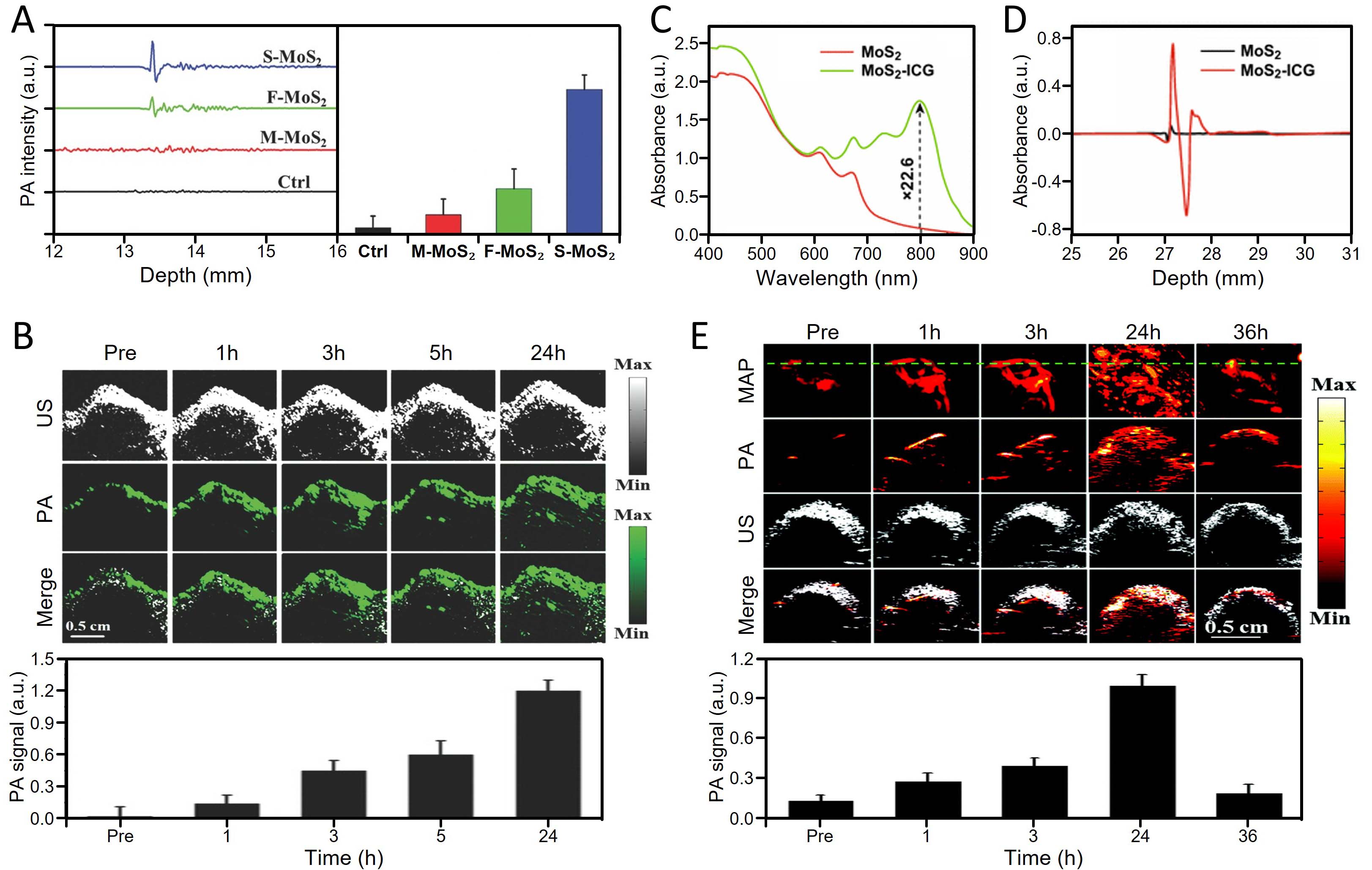
Figure 13. TMD-enabled PA biosensing and deep-tissue imaging. (A) quantitative comparison of PA signal amplitudes generated from single-layer (S-MoS2), few-layer (F-MoS2), and multilayer (M-MoS2) MoS2 nanosheets at identical concentrations; (B) in vivo orthotopic brain tumor PA imaging using single-layer MoS2 nanosheets, demonstrating robust signal enhancement at deep tissue depths beneath the skull and time-dependent accumulation within the tumor region. (A and B) are adapted with permission[33]. Copyright 2016, Wiley-VCH; (C) UV-VIS-NIR absorbance spectra of pristine MoS2 and MoS2-ICG hybrid nanosheets at identical MoS2 concentrations; (D) quantitative comparison of PA signal intensities generated by MoS2 and MoS2-ICG hybrids. (C and D) are adapted with permission[34,43]. Copyright 2018, Springer Nature; (E) in vivo tumor-targeted PA imaging and corresponding signal quantification using MoSe2-ICG hybrid nanosheets. PA: Photoacoustic; US: ultrasound.





