

fig9
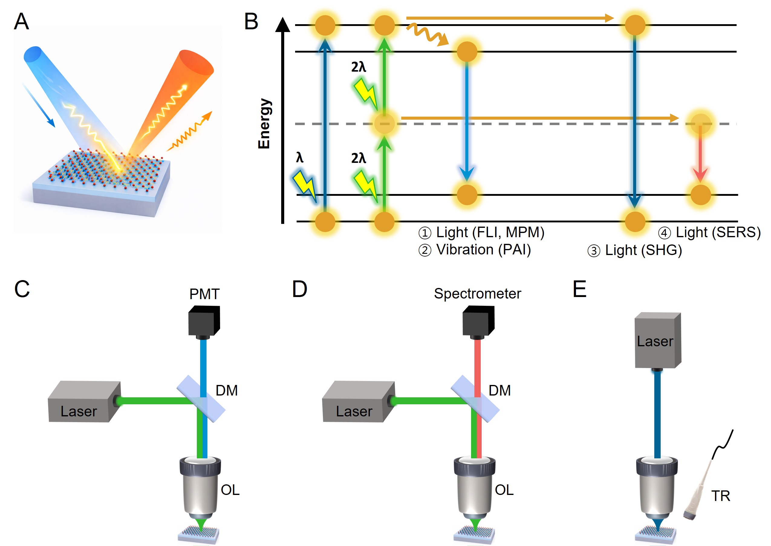
Figure 9. Schematic comparison of optical biosensing modalities using TMDs. (A) light-matter interactions in TMDs and the resulting optical and thermo-vibrational signals utilized for biosensing applications; (B) Jablonski diagram representing the underlying excitation-relaxation pathways for different optical sensing modalities; experimental configurations of (C) MPM and SHG based on nonlinear excitation and coherent emission process; (D) SERS sensing based on charge-transfer-mediated vibrational signal amplification; (E) PAI relying on optothermal energy conversion and subsequent ultrasonic signal generation. The colors of the incident and emitted beams indicate different excitation and detection wavelengths. FLI: Fluorescence imaging; MPM: multiphoton microscopy; PAI: photoacoustic imaging; SERS: surface-enhanced Raman scattering; SHG: second harmonic generation; PMT: photomultiplier tube; DM: dichroic mirror; OL: objective lens; TR: transducer.





