



MASLD epidemiology, natural history and diagnosis
 0
0 
Abstract
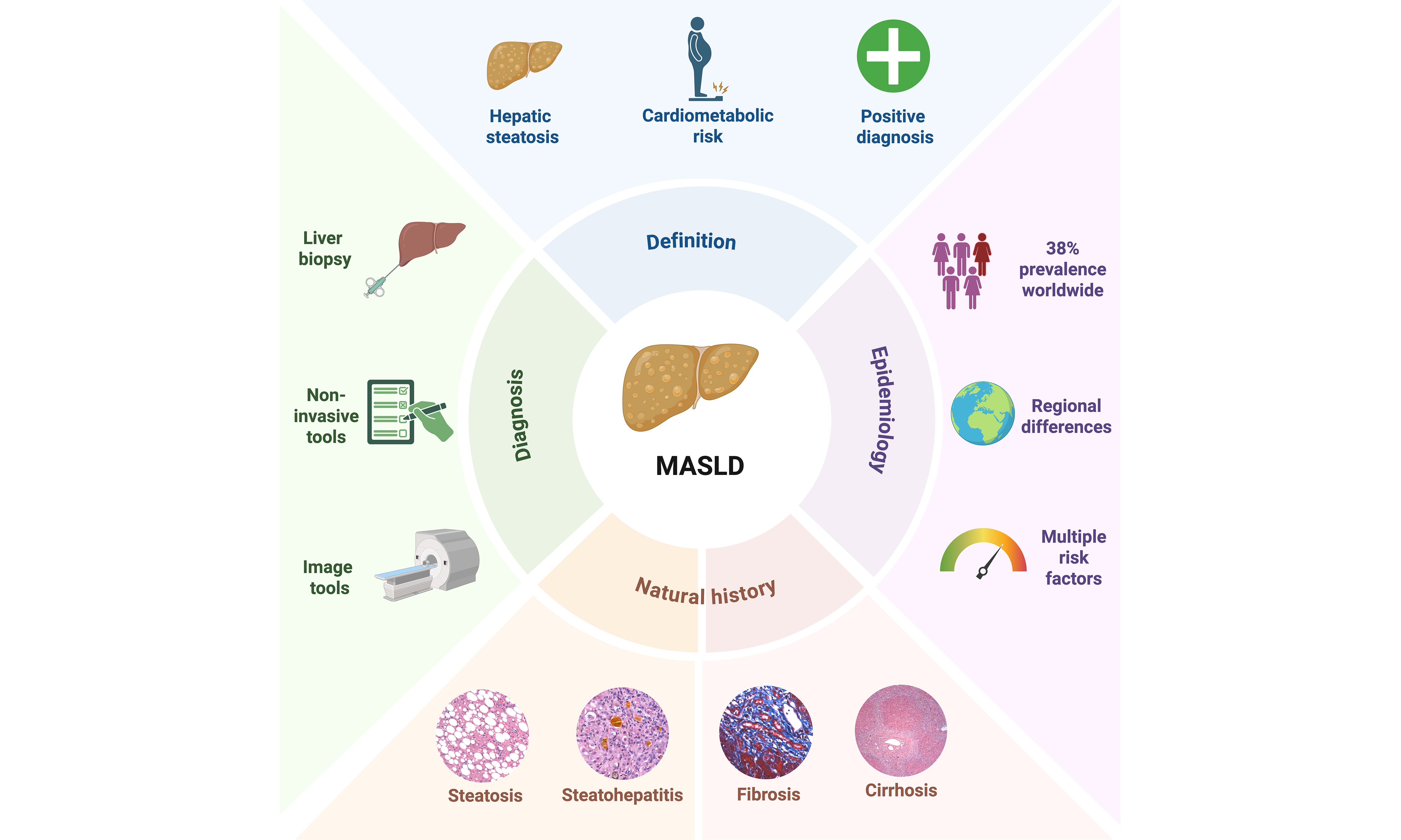
Metabolic dysfunction-associated steatotic liver disease (MASLD) has become the leading cause of chronic liver disease worldwide, with an estimated prevalence of 38%. Its burden varies considerably across regions, populations and demographic groups, influenced by genetic, biological, environmental and social factors. The disease follows a progressive histopathological continuum ranging from simple steatosis, through steatohepatitis, advanced fibrosis and cirrhosis, to hepatocellular carcinoma. The stage of fibrosis is the most important determinant of liver-related outcomes, while extrahepatic manifestations, particularly cardiovascular disease, represent the leading cause of mortality. For diagnosis, the focus has shifted from liver biopsy to non-invasive strategies that combine serum markers and imaging studies. These tools have already been incorporated into international clinical guidelines, although access to them remains uneven across different healthcare settings. This review aims to provide an updated and comprehensive overview of the epidemiology, natural history, and diagnostic strategies for MASLD, with an emphasis on recent epidemiological data, factors influencing disease progression, and the increasingly important role of non-invasive diagnostic tools in clinical practice.
Keywords
INTRODUCTION
Chronic liver disease (CLD) remains a major public health problem nowadays. Metabolic dysfunction-associated steatotic liver disease (MASLD) is currently the most common cause of CLD worldwide[1]. For decades, its diagnosis relied on ruling out other causes of CLD, especially excessive alcohol consumption. This changed when MASLD was recognized as a multisystemic metabolic disease. As a result, its definition was updated to acknowledge that multiple factors are associated with its development and progression, including genetic, biological, environmental and cardiometabolic factors[2,3]. The 2023 Delphi consensus proposed steatotic liver disease (SLD) as a broader term that covers various etiologies of the disease. This new term includes MASLD, defined as the presence of hepatic steatosis accompanied with at least one cardiometabolic criteria and limited alcohol consumption (< 140 g/week in women, < 210 g/week in men); metabolic dysfunction and alcohol-associated liver disease (MetALD), which applies to individuals who meet the cardiometabolic criteria for MASLD but have moderate alcohol consumption (140-350 g/week for women, 210-420 g/week for men) and alcohol-related liver disease (ALD), in which alcohol is the primary etiological factor at higher levels of consumption (> 350 g/week in women, > 420 g/week in men)[2].
According to the Global Burden of Disease (GBD) Study, approximately 1.3 billion people worldwide had MASLD in 2023. Projections indicate that this represents a 142.7% increase since 1990 and that by 2050, the number of people living with MASLD is projected to reach 1.8 billion[4]. The natural history of MASLD includes its evolution from simple steatosis (SS) to steatohepatitis [metabolic dysfunction-associated steatohepatitis (MASH)], advanced fibrosis, cirrhosis, and hepatocellular carcinoma (HCC), with fibrosis being the most important predictor of liver-related mortality[5]. While the main outcomes of MASLD are hepatic, we must remember that this disease is associated with extrahepatic outcomes, such as cardiovascular disease (CVD), type 2 diabetes (T2D), chronic kidney disease, polycystic ovary syndrome (PCOS) and extrahepatic cancers[6,7]. Timely diagnosis is essential for early intervention and to prevent or delay the progression of the disease.
The diagnosis of MASLD is based on the presence of hepatic steatosis in combination with the presence of cardiometabolic risk factors[2,3]. To assess more advanced stages, liver biopsy is still considered the gold standard. However, we are now in an era in which non-invasive alternatives are becoming more widespread. Non-invasive tests (NITs), which include calculated scores such as Fibrosis-4 index (FIB-4) and the enhanced liver fibrosis (ELF) test, the blood biomarker N-terminal pro-peptide of type III collagen (PRO-C3) and imaging-based tools, have facilitated the detection and staging of the disease without the need to take a tissue sample[8-10]. Composite scores designed specifically for fibrotic MASH, such as the FibroScan-AST (FAST) score and the HOMA, Ast, CK18 score (MACK-3), have provided greater accuracy[11], and both the European Association for the Study of the Liver (EASL) and the American Association for the Study of Liver Disease (AASLD) guidelines now incorporate them into their clinical guidelines[12,13]. Although no test has yet completely replaced biopsy, the trend is clear.
This review aims to provide an updated and comprehensive overview of the epidemiology, natural history, and diagnostic strategies for MASLD, with an emphasis on recent epidemiological data, factors influencing disease progression, and the increasingly important role of non-invasive diagnostic tools in clinical practice.
EPIDEMIOLOGY OF MASLD
Global epidemiology
The global epidemiology of MASLD reflects the interplay of multiple factors, including ethnicity, genetics, sex, nutritional transition and access to health care systems. A 2024 meta-analysis, which included more than 78 million people from 38 countries, confirmed that the prevalence of the disease varies considerably[14]. It is estimated that MASLD currently affects 38% of the global adult population[1]. But higher prevalence rates have been reported in specific population groups. For example, it has been estimated that among people with T2D, the prevalence may range from 28% to 69%[15], and among people living with obesity, rates ranging from 60% to 90% have been reported[16].
Differences in geographic distribution
North Africa and the Middle East have been found to have the highest age-standardized prevalence globally (29,246 cases per 100,000 population), while the high-income Asia-Pacific region has the lowest (8,653 cases per 100,000 population)[4]. Regarding mortality and disability, Andean and Central American countries top the rankings. Mexico recorded the highest national rate of disability-adjusted life years (DALYs) in 2023 at 192.1 per 100,000 inhabitants[4]. This reflects a combination of rapid nutritional transition, high rates of obesity, and the elevated frequency of the patatin-like phospholipase domain-containing protein 3 (PNPLA3) rs738409 variant, which results in an isoleucine-to-methionine substitution at position 148 (I148M), in Latin American populations[4,17].
In Europe, prevalence estimates range from 20% to 30% in most countries. However, southern and eastern Europe consistently report higher rates, partly due to dietary patterns and rising rates of metabolic syndrome (MetS)[14]. In the United States, MASLD affects approximately one in three adults, with marked disparities by ethnicity. Hispanic populations have the highest prevalence, followed by non-Hispanic whites and non-Hispanic blacks[18]. In Asia, MASLD develops at lower body mass index (BMI) thresholds due to higher levels of visceral adiposity, making MASLD particularly common among lean individuals and often undiagnosed[19]. Sub-Saharan Africa remains the region with the least available data; the most recent evidence points to a growing burden driven by urbanization that is systematically underestimated in global studies[4].
MASLD IN SPECIAL POPULATIONS
Pediatric population
Nowadays, 7% to 14% of children and adolescents have MASLD. As in adults, the prevalence rises to over 47% in the population living with obesity[20]. This increase closely mirrors trends in childhood obesity and is no longer limited to high-income countries; in fact, low- and middle-income countries are seeing some of the most pronounced increases. Hispanic, Indigenous and low-income children bear a disproportionate burden, due to genetic susceptibility, the food environment and limited access to healthcare[21].
Lean MASLD
MASLD in lean individuals is increasingly recognized as a clinically distinct entity. A recent meta-analysis involving more than 10 million people revealed that the combined prevalence of MASLD in lean individuals is 1.94% in the general population[22]. Despite not having excess weight, lean patients share metabolic risk profiles similar to those of patients with obesity and may have comparable rates of diabetes, cardiovascular events, and liver-related outcomes[22]. The lean phenotype often delays diagnosis, making this group particularly vulnerable to not receiving treatment or receiving inadequate treatment.
Age and sex differences
Over time, it has become evident that MASLD affects men and women differently due to differences in fat distribution and hormones. In men, a higher incidence of MASLD is observed from youth through middle age, declining after the age of 50-60[23,24]. In contrast, young women have a lower incidence of MASLD, which increases after menopause and then decreases after age 70. In addition, women with PCOS, Turner syndrome, or who have had early menarche are also at high risk of developing MASLD[25].
RISK FACTORS ASSOCIATED WITH MASLD
Cardiometabolic risk factors
MASLD can be considered the hepatic manifestation of an underlying systemic metabolic disorder. Its diagnosis requires at least one of the five cardiometabolic criteria, displacing alcohol as the primary determinant for its diagnosis [Table 1][2,12]. Insulin resistance (IR) is the primary determining factor. It promotes de novo lipogenesis while impeding fatty acid oxidation and the export of very low-density lipoproteins (VLDL), leading to a net accumulation of hepatic lipids. More than 60% of people with T2D have hepatic steatosis and IR has been shown to be a stronger predictor of MASLD than obesity itself[26,27]. Additionally, visceral fat releases free fatty acids (FFAs) directly into the portal circulation and produces a profile of proinflammatory adipokines, with elevated levels of tumor necrosis factor-alpha (TNF-α), interleukin-6 (IL-6) and leptin, along with reduced adiponectin, leading to hepatocellular injury and fibrogenesis[7,28].
Diagnostic criteria for MASLD
| MASLD (Rinella et al., 2023)[12] | |
| Hepatic steatosis | Evidence of hepatic steatosis in adults (By imaging techniques or histological techniques) |
| Cardiometabolic factor | At least 1 of the 5 cardiometabolic criteria: • BMI ≥ 25 kg/m2 (≥ 23 kg/m2 in Asians) or waist circumference ≥ 94/80 cm in men and women or ethnicity-adjusted thresholds • Fasting serum glucose ≥ 5.6 mmol/L (100 mg/dL) or 2-h post-load glucose ≥ 7.8 mmol/L (≥ 140 mg/dL) or HbA1c ≥ 5.7% (39 mmol/L) or type 2 diabetes or treatment for type 2 diabetes • Blood pressure > 130/85 mmHg or specific antihypertensive drug treatment • Plasma triglycerides ≥ 1.70 mmol/L (150 mg/dL) or lipid lowering treatment • Plasma HDL-cholesterol ≤ 1.0 mmol/L (40 mg/dL) in men and ≤ 1.3 mmol/L (50 mg/dL) in women or lipid lowering treatment |
| Alcohol | < 140 g/week in women, < 210 g/week in men, exceeding these thresholds classifies as MetALD or ALD |
Sociodemographic determinants
The epidemiology of MASLD cannot be fully understood without considering its sociodemographic dimensions. Lower socioeconomic status, limited education and food insecurity are associated with higher prevalence and poorer outcomes. This is not only due to their effect on diet and physical activity, but also because they limit access to screening and healthcare[29,30]. A cross-sectional analysis of NHANES revealed that higher educational attainment, but not higher income, was independently associated with a lower likelihood of having MASLD only among food-secure individuals. Among those who were food insecure, the association disappeared entirely[30]. Migrant populations face a similar disadvantage, as they must cope with changes in diet, occupational exposures, and systemic barriers to healthcare that exacerbate their risk[29]. Paradoxically, high-income countries are also experiencing a rise in MASLD driven by sedentary lifestyles and the consumption of ultra-processed foods, reflecting the different ways in which the burden can manifest depending on the context[29].
Genetic susceptibility
Genome-wide association studies (GWAS) have identified several high-impact variants that have been consistently replicated across different populations[31,32]. The most well-validated is PNPLA3 (rs738409, I148M). It has been observed that individuals who are homozygous for the risk allele have a 3- to 4-fold increased risk of developing MASLD, MASH, advanced fibrosis and HCC. This variant is particularly common in Hispanic populations, which partly explains their disproportionate burden of the disease[33]. TM6SF2 (rs58542926, E167K) reduces VLDL secretion and retains triglycerides within the hepatocyte, which paradoxically lowers plasma triglycerides and low-density lipoprotein cholesterol (LDL), a phenotype that complicates cardiovascular risk assessment in these patients[31]. MBOAT7 (rs641738) alters phosphatidylinositol remodeling, promoting steatosis and inflammation[31]. HSD17B13 (rs72613567) acts in the opposite direction; its splicing variant is protective, as it reduces hepatic inflammation and the risk of progression to MASH and HCC. Its presence also modified the response to Resmetirom in the MAESTRO-NASH trial, establishing a proof of concept for genotype-guided therapy[34].
Gut microbiome and dietary factors
The gut-liver axis is a central and modifiable component of the pathogenesis of MASLD. Gut dysbiosis, along with increased intestinal permeability, amplifies hepatic exposure to lipopolysaccharide (LPS), microbial metabolites, pathogen-associated molecular patterns and damage-associated molecular patterns[35,36]. Among the microbial metabolites implicated, three stand out. Short-chain fatty acids (SCFAs), produced by the fermentation of dietary fiber, promote intestinal barrier integrity and modulate hepatic lipid metabolism; their depletion promotes fat accumulation. Trimethylamine N-oxide (TMAO), generated from dietary choline and L-carnitine, has been linked to the severity of MASH, the progression of fibrosis, and cardiovascular risk. Bile acids, extensively metabolized by gut bacteria, regulate signaling of the farnesoid X receptor (FXR) and Takeda G protein-coupled receptor 5 (TGR5) in the liver, pathways that are now the target of active pharmacological treatment[35-37]. Diet modulates all these axes simultaneously; consumption of ultra-processed foods drives de novo lipogenesis, promotes lipotoxicity, alters the composition of the microbiota and depletes SCFAs[38,39].
Environmental pollutants
Chronic exposure to environmental pollutants represents an emerging risk factor for the development of MASLD. A recent meta-analysis involving more than 48 million adults showed that exposure to various classes of environmental pollutants is significantly associated with an increased risk of SLD including MASLD. Air pollutants such as fine particulate matter, coarse particulate matter, ultrafine particles and nitrogen dioxide; endocrine disruptors such as bisphenol A and phthalates: mono-(2-ethyl-5-carboxypentyl), mono-(2-ethyl-5-hydroxyhexyl), and mono-(2-ethyl-5-oxohexyl) and heavy metals such as mercury, lead and cadmium showed independent and significant associations with SLD[40]. Additional evidence from a prospective cohort study involving more than 244,000 participants from the UK Biobank suggests that these associations are mediated by alterations in lipids, lipoproteins and amino acids metabolism induced by exposure to the pollutants themselves[41].
In addition, these contaminants interact within the gut-liver axis. Bisphenol A compromises epithelial integrity by reducing Akkermansia muciniphila and increasing Proteobacteria, thereby promoting the translocation of LPS into the portal circulation[42]. Heavy metals act more directly on the intestinal epithelium, amplifying endotoxemia and the hepatic inflammatory response[43]. Microplastics, in turn, synergistically enhance these effects when combined with a high-fat diet; notably, the microbial disruption they induce persists even after exposure is removed[44].
Alcohol and MetALD
One of the most clinically significant contributions to the field of MASLD is the recognition of MetALD. MetALD is a distinct epidemiological and pathophysiological entity[45,46]. The coexistence of metabolic dysfunction and alcohol consumption is not uncommon. Both conditions share overlapping pathogenic mechanisms, such as IR, gut dysbiosis, disruption of the intestinal barrier, and oxidative stress, and their combination synergistically accelerates disease progression[45,46]. Emerging evidence challenges the notion of a “safe” threshold for alcohol consumption in patients with preexisting metabolic risk. It has been demonstrated that even moderate consumption appears to exacerbate liver injury through CYP2E1 induction, mitochondrial dysfunction, and micronutrient depletion[46].
The multiple parallel hits model
For years, the progression of MASLD was explained using a “two-hit” model, with hepatic steatosis representing the first hit and oxidative stress or inflammation the second[47]. While this was a useful framework, it never fully captured the complexity of the disease. The current framework, the “multiple parallel hits” hypothesis [Figure 1], conceptualizes MASLD as the result of simultaneous and synergistic insults. IR, gut dysbiosis, lipotoxicity, genetic susceptibility, epigenetic modifications and environmental exposures converge to cause oxidative stress, hepatocellular injury and immune-mediated fibrogenesis[48,49].
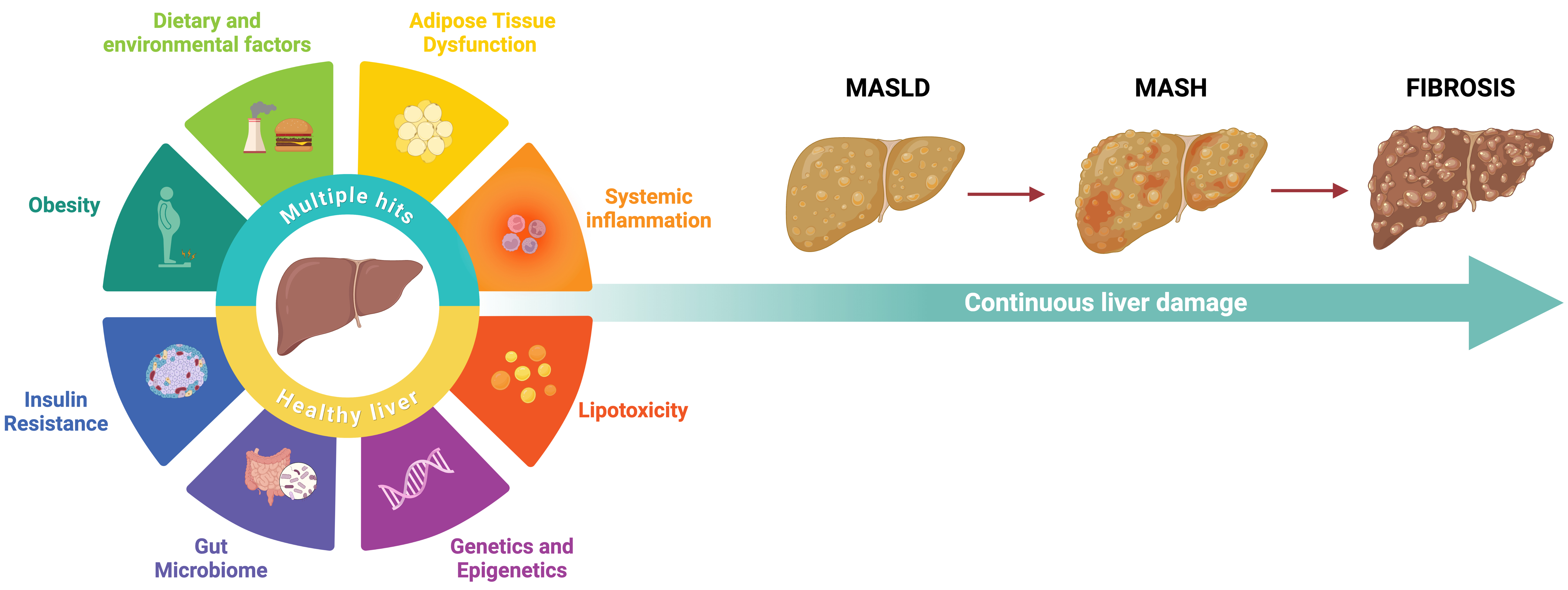
Figure 1. The multiple parallel hits model in progressive liver damage in MASLD. The pathogenesis of MASLD is best explained by the “multiple parallel hits” hypothesis, according to which simultaneous and synergistic insults converge on the liver rather than acting sequentially. These include insulin resistance, which drives de novo lipogenesis and disrupts fatty acid oxidation; obesity and adipose tissue dysfunction, which generate a profile of pro-inflammatory adipokines; lipotoxicity, mediated by the accumulation of free fatty acids and their derivatives; gut microbiota dysbiosis, which increases intestinal permeability and hepatic exposure to pathogen-associated molecular patterns; systemic inflammation, driven by activation of the innate immune system; and genetic and epigenetic susceptibility variants that modulate individual risk and disease severity; and dietary and environmental factors, including excessive consumption of ultra-processed foods, sedentary lifestyle, and chronic exposure to environmental pollutants such as fine particulate matter, bisphenol A, phthalates, and heavy metals. Created in BioRender. Ramírez, M. (2026) https://BioRender.com/2vcrroy. MASLD: Metabolic dysfunction-associated steatotic liver disease; MASH: metabolic dysfunction-associated steatohepatitis.
NATURAL HISTORY OF MASLD
Disease spectrum: from steatosis to cirrhosis and HCC
MASLD is a disease with a dynamic and bidirectional natural history [Figure 2]. Its onset is characterized by the presence of SS, defined as the excessive accumulation of triglycerides in > 5% of hepatocytes[49]. The chronic accumulation of lipids triggers inflammatory and lipotoxic mechanisms that drive the development of MASH. MASH, in turn, is characterized by the presence of steatosis, hepatocellular ballooning and lobular inflammation; in some cases, it may be accompanied by fibrosis[50]. The persistence of liver damage and pro-inflammatory signals leads to increased synthesis and deposition of the extracellular matrix (ECM), thereby increasing the extent of fibrosis. Finally, if fibrosis persists, it can lead to a distortion of the entire architecture of the hepatic parenchyma as well as its functionality, known as cirrhosis. Cirrhosis is associated with the development of clinical complications, primarily portal hypertension (PH)[51]. Notably, MASLD-associated HCC can develop at any point along this spectrum, even in the absence of cirrhosis[52].
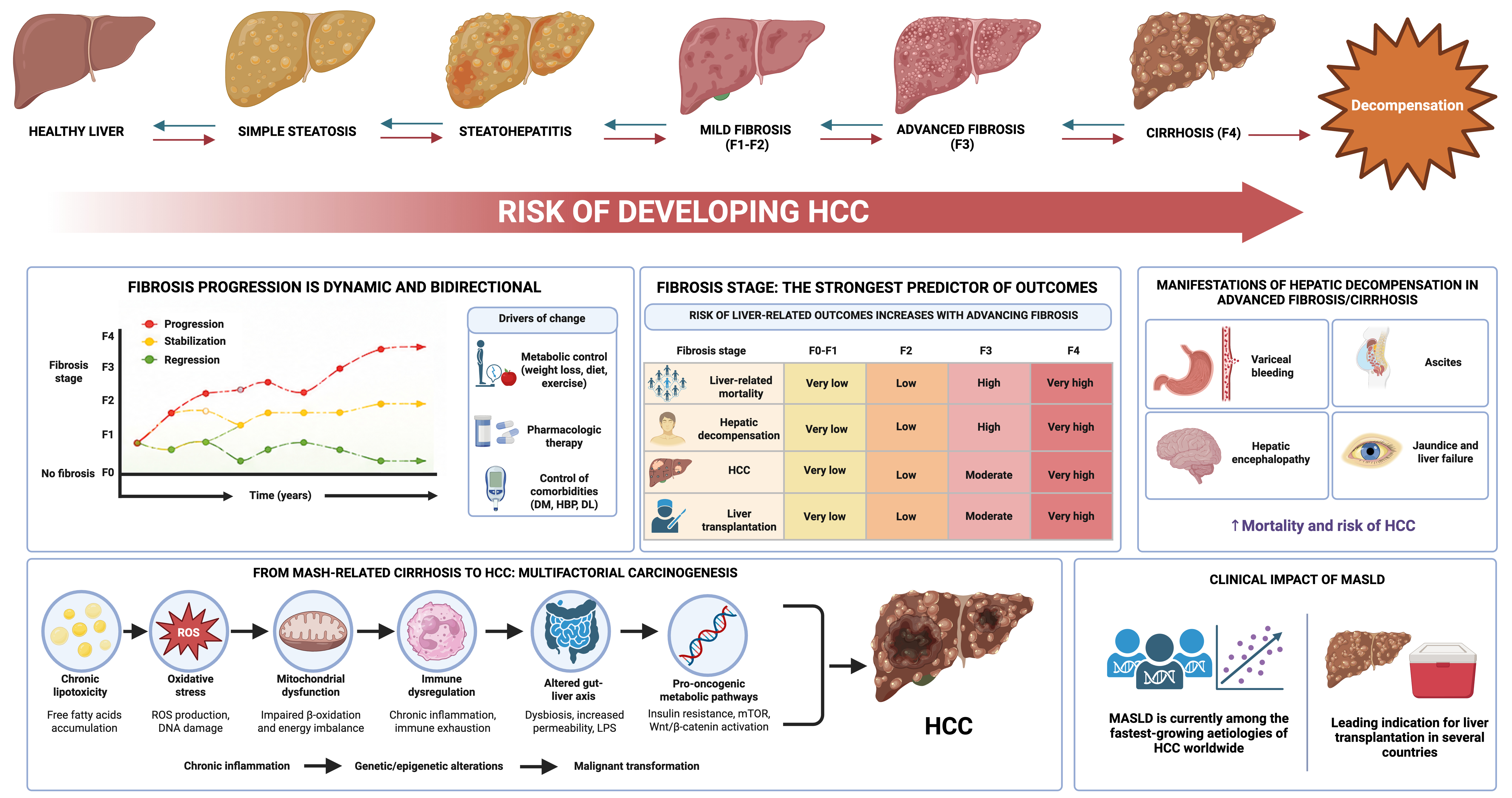
Figure 2. Natural history and clinical impact of MASLD. MASLD progresses from SS to steatohepatitis (MASH), where the coexistence of hepatocellular ballooning, lobular inflammation, and ECM deposition triggers the transdifferentiation of hepatic stellate cells into myofibroblasts that produce collagen types I and III. This establishes a profibrotic microenvironment that progresses to perisinusoidal and portal fibrosis (F1-F2), bridging fibrosis with nodular formation (F3) and cirrhosis (F4). Cirrhosis is characterized by architectural distortion, regenerative nodularity and sinusoidal capillarization. Ultimately, the disease can progress to stages of decompensation associated with portal hypertension. The progression of the disease is dynamic and bidirectional, modulated by metabolic control, pharmacological intervention and the management of comorbidities. The stage of fibrosis is the most robust independent predictor of liver-related mortality, decompensation, HCC, and the need for transplantation, with risk increasing progressively from F0-F1 to F4. It is important to note that the risk of HCC is present across the entire spectrum of the disease; up to 50% of HCC cases associated with MASLD develop without prior cirrhosis. The transition to HCC reflects the convergence of multiple mechanisms. Chronic lipotoxicity generates ROS, leading to oxidative damage to DNA and mitochondrial dysfunction with impaired β-oxidation; chronic inflammation and immune exhaustion promote genetic and epigenetic alterations; gut dysbiosis increases intestinal permeability and hepatic exposure to LPS via the gut-liver axis, amplifying inflammatory signaling; and pro-oncogenic pathways such as insulin resistance, mTOR activation, and Wnt/β-catenin signaling converge to drive malignant transformation. MASLD is currently one of the fastest-growing causes of HCC worldwide and a leading indication for liver transplantation in several countries. Created in BioRender. Ramírez, M. (2026) https://BioRender.com/wgvhgrt. MASLD: Metabolic dysfunction-associated steatotic liver disease; SS: simple steatosis; MASH: metabolic dysfunction-associated steatohepatitis; ECM: extracellular matrix; HCC: hepatocellular carcinoma; ROS: reactive oxygen species; LPS: lipopolysaccharide; mTOR: mechanistic target of rapamycin.
Rates of disease progression and regression
Not all individuals with MASLD will experience disease progression, and those who do will not follow a uniform course. Most patients with SS do not develop significant inflammation or progressive fibrosis. However, approximately 20% progress to MASH with significant histological activity[50]. Once MASH has been established, the risk of progression to fibrosis increases significantly. Among patients with MASH and stage F0-F2 fibrosis, 14% progressed to F3 and 2% to F4 over an average of 4.5 years[53]. A meta-analysis of 54 studies involving 26,738 patients found that 31% of patients developed MASH after a median of 4.7 years, while 29% of those who already had MASH showed resolution after a median of 3.5 years. Regarding fibrosis progression, the estimated time to advance one stage was 9.9 years at F0, 10.3 years at F1, 13.3 years at F2 and 22.2 years at F3[54]. Finally, progression to cirrhosis occurs in 3%-5% of patients and often takes more than 20 years[50].
On the other hand, fibrosis regression is recognized as an achievable therapeutic endpoint. Sustained weight reduction, improved glycaemic control, reduction in hepatic inflammation and emerging pharmacological therapies may induce histological improvement and partial reversal of fibrosis. Regression is most pronounced in earlier fibrosis stages but can also occur in compensated cirrhosis under favorable metabolic conditions[55].
Predictors of disease progression
Several clinical, metabolic, and genetic factors determine the rate of progression in MASLD, often acting synergistically. The stage of fibrosis is the most robust independent predictor of liver-related mortality, HCC and all-cause mortality[56]. Among modifiable factors, T2D is one of the most significant. Persistent hyperglycemia generates advanced glycation end products that increase oxidative stress, activate Kupffer cells and hepatic stellate cells (HSCs), promoting fibrogenesis[57]. A multicentre study of 2,000 patients with T2D found that 20.4% had advanced fibrosis, with diabetes duration > years [odds ratio (OR) 2.105], fasting glucose
Liver-related mortality
Liver-related complications represent the most feared hepatic consequence of MASLD, but CVD remains the leading cause of death in this group of patients[1,61]. Importantly, cardiovascular mortality exceeds liver-related mortality across most MASLD stages, particularly in non-cirrhotic disease[61]. However, as fibrosis advances, liver-related mortality rises and becomes dominant in cirrhotic populations[56,62]. Notably, it independently increases overall and cardiovascular mortality and incident cardiovascular events even after adjustment for metabolic risk factors[1,61].
MASLD-associated HCC: cirrhotic and non-cirrhotic pathways
Beyond fibrosis, HCC represents the most serious outcome on the spectrum. It can develop in both cirrhotic and non-cirrhotic livers. Up to 50% of HCC cases in MASLD occur without prior cirrhosis, with larger nodules and a delayed diagnosis, since these patients often do not qualify for current surveillance programs[63,64]. In patients with MASLD-related cirrhosis, genetic susceptibility, chronic inflammation and regenerative nodular hyperplasia drive malignant transformation[65,66]. On the other hand, non-cirrhotic HCC appears through mechanisms linked to lipotoxicity, IR, hyperinsulinemia, adipokine dysregulation, oxidative stress, mitochondrial dysfunction and altered innate immunity[52,66].
Extrahepatic manifestations
MASLD is a systemic disorder with important extrahepatic involvement[67] [Figure 3]. The pathophysiological basis underlying these extrahepatic complications includes IR, chronic low-grade inflammation, lipotoxicity, oxidative stress, endothelial dysfunction, altered adipokines, mitochondrial injury, gut microbiota dysbiosis, and activation of profibrogenic and proatherogenic pathways[1,68]. Also, the burden of extrahepatic disease often exceeds liver-related complications, particularly during early and intermediate stages.
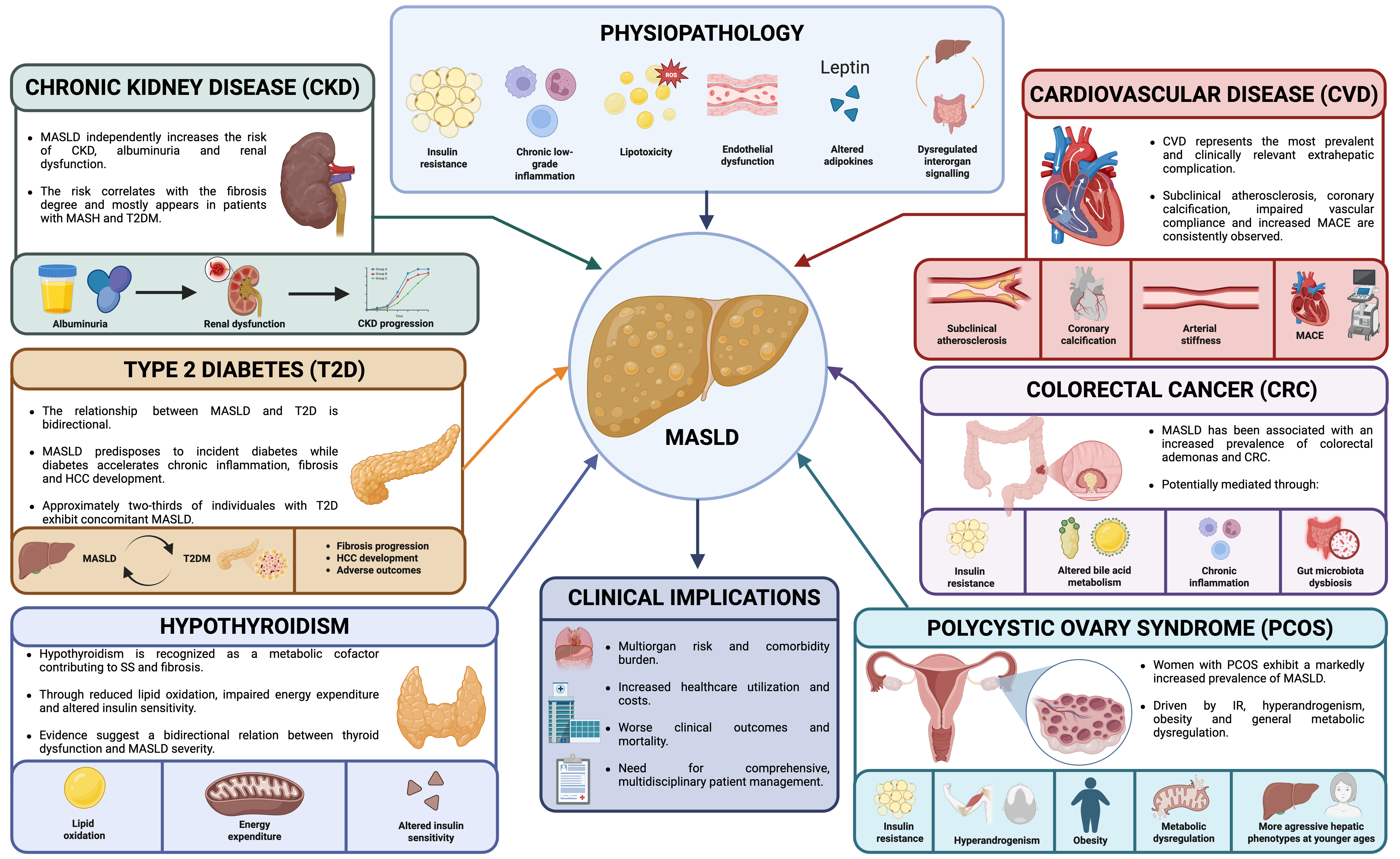
Figure 3. Extrahepatic manifestations of MASLD. Created in BioRender. Ramírez, M. (2026) https://BioRender.com/ce9bge2. MASLD: Metabolic dysfunction-associated steatotic liver disease; CKD: chronic kidney disease; CVD: cardiovascular disease; MACE: major adverse cardiovascular events; T2D: type 2 diabetes; CRC: colorectal cancer; MAFLD: metabolic-associated fatty liver disease; HCC: hepatocellular carcinoma; PCOS: polycystic ovary syndrome; SS: simple steatosis; IR: insulin resistance.
CVD is the leading cause of death among patients with MASLD. These include accelerated atherosclerosis, coronary artery disease, heart failure, atrial fibrillation, valvular calcification and an increased risk of serious adverse cardiovascular events. Steatosis contributes to endothelial dysfunction, arterial stiffness, inflammation, and impaired myocardial energy metabolism, thereby establishing MASLD as an independent cardiometabolic risk amplifier beyond traditional MetS components[1,61]. CKD is another major extrahepatic manifestation, with MASLD being independently associated with albuminuria, progressive decline in estimated glomerular filtration rate and increased risk of end-stage renal disease. The interaction between these two is mediated by shared mechanisms, such as IR, renin-angiotensin-aldosterone system activation, systemic inflammation, oxidative stress and ectopic lipid accumulation[68].
As previously discussed, the bidirectional relationship between MASLD and T2D represents one of the most clinically relevant metabolic interactions. Approximately one-third of patients with T2D have advanced fibrosis, emphasizing the importance of fibrosis screening in diabetic populations[13,69]. MASLD also increases the risk of incident T2D, while the coexistence of diabetes accelerates disease progression and liver-related mortality. MASLD has also been associated with increased prevalence of colorectal adenomas and colorectal cancer (CRC), potentially related to chronic inflammatory signaling, altered bile acid metabolism, hyperinsulinemia and dysfunction of the gut-liver axis[1]. Furthermore, endocrine manifestations are increasingly recognized and include hypothyroidism and PCOS, both of which may exacerbate hepatic steatosis and fibrosis progression through impaired lipid metabolism, IR, androgen excess and adipose tissue dysfunction. Additionally, MASLD has been linked to sarcopenia, obstructive sleep apnoea, psoriasis, osteoporosis and other extrahepatic malignancies[70].
DIAGNOSIS-NON-INVASIVE STRATEGIES
The diagnostic tools available for MASLD span from simple biochemical markers to advanced imaging modalities and novel biomarkers [Table 2][8,12,13,71-81]. The diagnostic algorithms for MASLD have evolved over the last decade, shifting from a liver biopsy-centered approach toward the widespread implementation of NITs[12,82,83]. International guidelines emphasize a risk-stratified diagnostic method integrating metabolic risk assessment, serum-based fibrosis scores and elastography-based techniques to identify patients at risk for advanced fibrosis, cirrhosis and HCC[12,82,83].
Non-invasive diagnostic tools for MASLD
| Biomarker | Diagnostic target | Cutoff | Guideline endorsement | Population notes |
| ALT/AST/GGT [12,13] | Hepatocellular injury | Variable by lab | EASL, AASLD, APASL | Universal; normal values do not exclude MASLD |
| Ultrasound[71] | Steatosis detection | Echogenic liver pattern | EASL, AASLD, APASL | Universal; sensitivity 84.8%, specificity 93.6%, AUROC 0.93 for moderate-severe steatosis vs. histology; limited sensitivity for mild steatosis (< 33%); operator-dependent |
| VCTE[72] | Fibrosis staging (LSM) + steatosis quantification (CAP) | Cut-offs vary by BMI and region | EASL, AASLD, APASL | Validated globally; XL probe improves performance in obesity; AUROC for CAP 0.924 (S ≥ S1), 0.794 (S ≥ S2); AUROC for LSM 0.897 (F ≥ F3), 0.925 (F = F4); LSM cut-offs higher in Europe/America vs. Asia; LSM unaffected by steatosis or probe type |
| MRI-PDFF[73] | Steatosis quantification | ≥ 5% hepatic fat fraction | EASL, AASLD (clinical trials preferred) | AUC 0.98 (S0 vs. S1-3), 0.92 (S0-1 vs. S2-3), 0.90 (S0-2 vs. S3); sensitivity 0.77-0.92, specificity 0.87-0.94 vs. biopsy; most accurate NIT for steatosis; reference standard in clinical trials; limited by cost and availability |
| 2D-SWE[74] | Fibrosis staging | Variable by platform | EASL (alternative to VCTE) | Validated in European and Asian cohorts; useful in patients with obesity; AUROC 0.82 (≥ F2), 0.86 (≥ F3), 0.89 (F4); integrated into conventional ultrasound platforms; cut-offs not yet universally standardized |
| MRE[8] | Fibrosis staging (all stages) | Low risk: < 2.5 kPa (sensitivity ~90%) | AASLD (preferred); EASL (specialized centers) | Validated across BMI ranges and ethnicities; most accurate NIT; sensitivity 88%-91% at 2.32 kPa threshold; whole-liver assessment; limited by cost and availability |
| FIB-4[12,13] | Advanced fibrosis (≥ F3) | Low risk: < 1.3; High-risk: ≥ 2.67 | EASL, AASLD, APASL | Validated globally; free calculation from routine biochemistry; reduced specificity in patients > 65 years; performance limited in patients < 35 years |
| NFS[75] | Advanced fibrosis | Low-risk: < -1.455; High-risk: > 0.676 | EASL, AASLD (alternative to FIB-4) | Validated in Western cohorts; sensitivity 70%, specificity 61%, AUC 0.74 for ≥ F3; lower performance than FIB-4; limited in obesity, T2D and younger patients |
| APRI[76] | Significant and advanced fibrosis, cirrhosis | Significant fibrosis: > 0.7-1.0; Cirrhosis: > 1.5-2.0 | Not recommended for MASLD | Developed for viral hepatitis; AUROC 0.78 (advanced fibrosis), 0.76 (significant fibrosis), 0.72 (cirrhosis); lower accuracy than FIB-4 and ELF; useful in resource-limited settings |
| ELF[76,77] | Advanced fibrosis | 7.7 (low); 7.7-9.8 (intermediate); > 9.8 (high) | EASL, AASLD (second line) | Validated in European and North American cohorts; limited data in Latin America and Asia; AUROC 0.81-0.87 for advanced fibrosis - highest among serum biomarkers in MASLD; requires specific immunoassay platform; affected by age and renal disease; limited data in primary care and resource-limited settings |
| ADAPT[78] | Advanced fibrosis (≥ F3) | Derived cutoff | Emerging; not yet endorsed | Validated in Asia-Pacific and European cohorts; AUROC 0.865 for advanced fibrosis in MASLD; sensitivity 82.2%, NPV 96.1%; outperforms PRO-C3 alone, APRI, FIB-4, BARD and NFS; no indeterminate zone; requires PRO-C3 immunoassay |
| MACK-3[79] | Fibrotic MASH (MASH + NAS ≥ 4 + fibrosis ≥ F2) | Rule-out: < 0.135 (sensitivity 91%); Rule-in: > 0.549 (specificity 85%) | Not yet endorsed | Validated in European and Asian cohorts; limited external validation; AUROC 0.791; sensitivity 91% at rule-out threshold; specificity 85% at rule-in threshold; performance unaffected by age, sex, diabetes or BMI; comparable accuracy to FAST score; requires CK-18 immunoassay |
| FAST score[80] | Fibrotic MASH (MASH + NAS ≥ 4 + fibrosis ≥ F2) | Rule-out: ≤ 0.35 (sensitivity 88%, NPV 91%); Rule-in: ≥ 0.67 (specificity 87%, PPV 60%) | EASL (exploratory) | Validated in European and Middle Eastern cohorts; better performance for ruling out than ruling in fibrotic MASH; requires VCTE |
| MAST score[81] | Fibrotic MASH (MASH + NAS ≥ 4 + fibrosis ≥ F2) | Derived cutoff | Emerging; not endorsed | Validated in North American clinical trial cohorts; sensitivity 55.9%, specificity 88.1%, AUC 0.84 (0.81-0.88); similar overall diagnostic accuracy to FAST but higher sensitivity and more consistent results; NPV 96.5-98.1%; requires MRE + MRI-PDFF + AST; limited by cost and MRI availability |
Clinical suspicion and screening in at-risk populations
MASLD is asymptomatic during early stages and is commonly identified incidentally through abnormal liver biochemistry or imaging studies performed for unrelated indications[82,83]. Consequently, active case-finding strategies in high-risk populations have become a major focus in the clinical practice. International guidelines recommend targeted screening among individuals with obesity, T2D, MetS, dyslipidemia, hypertension, PCOS, obstructive sleep apnoea and established CVD[12,82,83].
Clinical suspicion should arise in the presence of central adiposity, IR, persistently elevated aminotransferases or imaging evidence of hepatic steatosis after exclusion of secondary causes of liver disease, including significant alcohol consumption, viral hepatitis, autoimmune liver disorders, hereditary hemochromatosis, Wilson disease, and hepatotoxic medications[82,83]. However, normal liver enzyme levels do not exclude MASLD because a considerable proportion of patients with advanced fibrosis or even cirrhosis may exhibit aminotransferase levels within normal laboratory reference ranges[51,83].
Biochemical markers
Routine biochemical studies remain an essential component of MASLD evaluation, although conventional liver enzymes demonstrate limited sensitivity and specificity for disease staging[82,83].
Alanine aminotransferase (ALT) is the most frequently elevated enzyme in MASLD and often reflects hepatocellular injury. However, ALT concentrations correlate poorly with histopathological severity, and normal ALT levels may coexist with advanced fibrosis[51,83]. Furthermore, normal laboratory reference ranges may underestimate clinically relevant liver injury because traditional upper limits frequently exceed metabolically healthy thresholds[82].
Aspartate aminotransferase (AST) is generally less elevated during early stages but increases with fibrosis progression. Consequently, the AST/ALT ratio may provide indirect information regarding fibrosis severity[83]. An AST/ALT ratio > 1 is frequently associated with advanced fibrosis or cirrhosis and may indicate worsening of the liver architecture[83].
Gamma-glutamyl transferase (GGT) is commonly elevated and is associated with oxidative stress, IR, and high cardiometabolic risk; therefore, elevated GGT levels have been linked to increased liver-related and cardiovascular mortality[61].
Biochemical assessment should always include fasting glucose, glycated hemoglobin (HbA1c), fasting insulin and lipid profile. Hypertriglyceridemia, low high-density lipoprotein cholesterol (HDL-C), impaired fasting glucose, and IR strongly support the diagnosis of MASLD[82].
Although individual biochemical markers lack adequate diagnostic performance for fibrosis degree staging, they constitute important components of most fibrosis scores and longitudinal disease monitoring strategies[83].
Steatosis assessment
Controlled attenuation parameter (CAP)
CAP, incorporated into vibration-controlled transient elastography (VCTE), represents one of the most widely used NITs for hepatic steatosis quantification[68,83]. It demonstrates reasonable diagnostic accuracy for detecting steatosis across various grades and is particularly useful during screening because of its accessibility, reproducibility, and simultaneous assessment of liver stiffness[83,84]. However, diagnostic performance may be reduced in severe obesity, narrow intercostal spaces or marked hepatic inflammation[83].
Magnetic resonance imaging-proton density fat fraction (MRI-PDFF)
MRI-PDFF is currently considered the most accurate NIT for quantitative hepatic fat assessment[85]. MRI-PDFF enables precise measurement of liver fat content across the entire parenchyma and exhibits excellent reproducibility and sensitivity to longitudinal changes in steatosis[85,86]. MRI-PDFF has become the preferred imaging study in clinical trials evaluating pharmacological therapies for MASLD and MASH. However, widespread implementation is limited by cost, accessibility, and technical requirements[85].
Ultrasound
Conventional ultrasonography remains the most used first-line imaging study for hepatic steatosis detection due to its low cost and broad availability[49]. However, ultrasonography has limited sensitivity for mild steatosis (< 20%-30% hepatocyte involvement) and operator dependence[83]. Novel ultrasound attenuation-based technologies, including attenuation imaging and ultrasound-guided attenuation parameter techniques, provide improved quantitative assessment of steatosis while preserving the accessibility advantages of conventional ultrasonography[87].
Fibrosis scores: FIB-4, NAFLD fibrosis score (NFS), AST-to-platelet ratio index (APRI), age, diabetes, PRO-C3 and platelets (ADAPT) and ELF
FIB-4
The FIB-4 index, calculated using age, AST, ALT, and platelet count, is currently the most widely recommended first-line fibrosis assessment tool[12,13]. FIB-4 demonstrates excellent negative predictive value (NPV) for excluding advanced fibrosis and is particularly useful in primary care[82,83]. Low FIB-4 values identify patients at minimal risk who may remain under longitudinal follow-up, whereas indeterminate or elevated values need a second risk stratification using elastography or specialized serum biomarkers[82,83].
Nevertheless, FIB-4 has significant limitations that affect its clinical interpretation. Because age is included in the numerator of its formula, patients over 65 years of age consistently obtain higher values regardless of their actual degree of fibrosis[88]. Although the 2024 EASL and 2023 AASLD guidelines recommend raising the low-risk cutoff to 2.0 in individuals over 65 years of age to reduce false positives, recent evidence calls this recommendation into question[12,13]. A prospective study of 985 patients referred from primary care demonstrated that a threshold of 1.3 maintains a NPV of 100% for liver stiffness measurement (LSM)
NFS
The NFS incorporates age, BMI, diabetes diagnosis, ALT/AST levels, platelet count, and albumin concentration[83]. Although NFS has been widely used, recent guidelines favor FIB-4 because NFS performance may be limited in younger individuals and patients with obesity or diabetes[12,83].
APRI
APRI is less accurate than FIB-4 for MASLD-related fibrosis assessment but may provide additional information in some other clinical contexts. APRI demonstrated greater utility in viral hepatitis than in metabolic liver diseases and is less frequently used in modern MASLD algorithms[83].
ELF test
ELF test incorporates serum markers of ECM, including hyaluronic acid, procollagen type III N-terminal peptide (PIIINP) and tissue inhibitor of metalloproteinases 1 (TIMP-1). It has demonstrated strong diagnostic performance for advanced fibrosis and may predict liver-related clinical outcomes independently of liver biopsy findings[90]. Recent guidelines increasingly incorporate ELF test as a second-line assessment tool following indeterminate FIB-4 results[12,13,91].
However, its implementation in clinical practice is limited by its high cost and limited accessibility. It requires specific automated immunoassay platforms that are not universally available, which restricts its use outside of specialized settings and poses a significant barrier in resource-limited settings[92,93].
Elastography
Elastography techniques provide non-invasive quantification of liver stiffness as a surrogate marker of fibrosis severity and have become central components of MASLD evaluation[69,83].
LSM
LSM reflects the biomechanical properties of the liver and correlates strongly with fibrosis stage. Increased stiffness values are associated with advanced fibrosis, PH, hepatic decompensation, and HCC risk[69,83].
VCTE
VCTE is currently the most widely used elastography modality in MASLD[12,82,83]. It combines LSM with CAP-based steatosis assessment and provides rapid bedside evaluation without radiation. It has excellent diagnostic performance for advanced fibrosis and cirrhosis, although accuracy may be influenced by obesity, hepatic congestion, inflammation, and food intake. However, the development of extra large probes has significantly improved feasibility in obese populations[83].
Two-dimensional shear wave elastography (2D-SWE)
2D-SWE enables real-time visualization and quantitative stiffness assessment integrated into conventional ultrasound platforms. Compared with VCTE, 2D-SWE offers improved spatial resolution and greater operator flexibility while maintaining high diagnostic accuracy for advanced fibrosis[94].
Magnetic resonance elastography (MRE)
MRE is considered the most accurate NIT for fibrosis assessment in MASLD. MRE demonstrates superior diagnostic performance for differentiating fibrosis degrees, particularly in obese individuals and patients with intermediate fibrosis[95]. It provides whole-liver assessment with lower sampling variability than liver biopsy. However, high cost and limited availability restrict its use outside tertiary referral centers and research institutes[86].
Novel biomarkers
Some biomarkers seek to improve diagnostic precision for MASH activity and fibrosis progression beyond traditional biochemical and elastography tools.
Cytokeratin-18 (CK-18) and MACK-3
CK-18 fragments reflect hepatocyte apoptosis and have been extensively investigated as biomarkers for MASH. Elevated CK-18 levels correlate with hepatocellular ballooning and necroinflammatory activity, although variability in diagnostic thresholds has limited routine clinical implementation[96]. The homeostatic model assessment of insulin resistance (HOMA-IR), AST, and CK-18-3 (MACK-3) score combines these biomarkers for the non-invasive diagnosis of fibrotic MASH. It achieved an area under the receiver operating characteristic curve (AUROC) of 0.847 in a multicenter cohort of 846 patients, outperforming NFS and FIB-4, with a sensitivity of 90% and a specificity of 94.2%[97].
N-terminal PRO-C3
PRO-C3, a marker of type III collagen formation, directly reflects active fibrogenesis and demonstrates promising diagnostic accuracy for advanced fibrosis and progression. Its integration into non-invasive scores may improve identification of patients with active MASH, fibrosis or cirrhosis[78]. The ADAPT score identifies advanced fibrosis in MASLD. In a validation cohort of 150 patients and a multinational validation cohort of 281 patients, it achieved AUROCs of 0.86 and 0.87, respectively, outperforming APRI, FIB-4, and NFS, with a NPV consistently above 90% in all subgroups. The absence of liver enzymes in its formula eliminates the confounding effect of age on ALT, and the absence of an indeterminate zone reduces diagnostic uncertainty[78].
FAST score
FAST score combines VCTE LSM, CAP, and AST levels to identify patients with fibrotic MASH [MASH + NAFLD activity score (NAS) ≥ 4 + fibrosis ≥ F2]. FAST score is particularly useful for selecting patients for clinical trial enrolment in research settings or hepatology referral[98].
MRI-AST score (MAST)
MAST score integrates MRI-PDFF, MRE, and AST values to identify patients with fibrotic MASH[99]. It demonstrates high diagnostic accuracy for fibrotic MASH and may facilitate non-invasive identification of patients requiring different therapeutic interventions[100].
Omics approaches
Advances in omics technologies have transformed the search for non-invasive biomarkers for MASLD across the entire disease spectrum. For example, genomics has identified risk variants associated with MASLD. It has been observed that combining these variants into polygenic risk scores can help identify individuals with distinct phenotypes who will follow clinically distinct disease courses[101,102]. Regarding transcriptomics, a systematic review of 1,149 studies identified miR-122 as the most studied microRNA in MASLD, followed by miR-21, miR-34a, and miR-192-5p. For the detection of MASLD, miR-200 and miR-298 demonstrated area under the curve (AUCs) of 0.96 and 0.98, respectively. In MASH, miR-200, miR-298 and miR-342 achieved AUCs of up to 0.99, while miR-122 showed values ranging from 0.81 to 1.0[103]. Prognostically, elevated levels of miR-122 correlated with disease severity and fibrosis progression, and miR-21 and miR-223 were specifically linked to obesity-associated MASH[103]. In clinical studies, combined miRNA panels with metabolic parameters have shown AUCs of up to 0.95 for steatosis when combining miR-126-5p with leptin, and 0.81 for hepatic fat content[104]. However, variability across cohorts, a lack of standardization in quantification and the absence of validation still limit its clinical translation[105]. Meanwhile, high-resolution proteomics using mass spectrometry has identified plasma panels with significant diagnostic potential. A panel of three proteins IGFBP7, SSc5D and Sema4D, linked to ECM remodeling and hepatic inflammation, achieved an AUC of 0.87-0.89 for the detection of significant fibrosis in MASLD[106]. Five-protein panels including ALDOB, IGF1, and IGF2, associated with hepatocellular metabolic dysfunction, outperformed FIB-4 and APRI in the diagnosis of MASLD and fibrosis ≥ F2[107]. Finally, metabolomics and lipidomics have identified plasma signatures with AUCs of up to 0.94 for advanced fibrosis, including decreased levels of lysophospholipids and sphingomyelins and increased levels of unsaturated triglycerides[108]. Specific lipid profiles also allow for differentiation between MASLD and MetALD[109].
Single-cell RNA sequencing and spatial transcriptomics
Single-cell RNA sequencing (scRNA-seq) and spatial transcriptomics have redefined our cellular understanding of MASLD with unprecedented resolution. scRNA-seq has revealed functional heterogeneity in subpopulations of HSCs, myofibroblasts and monocyte-derived macrophages, with variations in ECM production and cytokine release among subtypes of the same cell type[110]. The integration of both technologies in MASH liver demonstrated that early infiltration of Kupffer cells and T lymphocytes triggers HSCs activation, identifying ADAMTSL2, PTGDS and S100A6 as genes spatially correlated with the degree of fibrosis[111]. A spatial multi-omic atlas of 61 human livers identified the microphthalmia-associated transcription factor as a key regulator of lipid-laden macrophages and delineated profibrotic communication between central vein endothelial cells and stellate cells in advanced MASH. Meanwhile, multiplexed fluorescent in situ hybridization was used to characterize two hepatocyte populations that expand with fibrotic lesions without a defined zonal distribution[112,113]. These findings pave the way for the molecular subtyping of MASLD and the identification of cell-type-specific biomarkers to complement current non-invasive diagnostic tools.
MASLD-specific diagnostic algorithms
International guidelines, such as those from the EASL, AASLD and the Asian Pacific Association for the Study of the Liver (APASL), suggest sequential non-invasive diagnostic algorithms designed to maximize accessibility while minimizing unnecessary liver biopsies [Table 3][12,13,82,91,114-118].
Stepwise non-invasive diagnostic algorithm for MASLD based on international guidelines consensus
| Step | Clinical objective | Recommended tool | Interpretation | Alternative in low-resource settings |
| 1 | Identification of at-risk population | Clinical history + cardiometabolic criteria (T2D, abdominal obesity + ≥ 1 risk factor, or persistently elevated liver enzymes) | Presence of ≥ 1 criterion triggers diagnostic workup | Identical |
| 2 | Steatosis detection | Abdominal ultrasound | Echogenic liver pattern suggestive of steatosis | Identical |
| 3 | First-line fibrosis stratification | FIB-4 index | < 1.3: low risk → follow-up in primary care; 1.3-2.67: indeterminate → second-line assessment; ≥ 2.67: high risk → hepatology referral | Identical |
| 4 | Second-line fibrosis assessment (indeterminate FIB-4) | VCTE | LSM < 8 kPa: no advanced fibrosis; 8-12 kPa: indeterminate; > 12 kPa: advanced fibrosis | If VCTE unavailable: ELF test; if neither VCTE nor ELF available: NFS, APRI or serial FIB-4[114,115] |
| 5 | Confirmation of advanced fibrosis/MASH activity | ELF test or PRO-C3 | Complementary to elastography; high ELF indicates active fibrogenesis | If neither available: serial FIB-4 monitoring for fibrosis progression assessment[114] |
| 6 | Complex or discordant cases | MRE or liver biopsy | MRE: most accurate NIT for fibrosis staging; biopsy reserved for diagnostic uncertainty or suspected alternative aetiology | If MRE unavailable: VCTE as surrogate, acknowledging lower accuracy in complex cases[116]; biopsy only when strictly necessary[117] |
| 7 | Advanced fibrosis/cirrhosis management | Hepatology referral + LSM assessment (VCTE) for PH evaluation + HCC surveillance (ultrasound + AFP every 6 months) | LSM > 20 kPa: high risk of clinically significant PH | If LSM unavailable: clinical signs + platelet count as indirect proxy for PH[118]; ultrasound + AFP |
The EASL recommends a stepwise approach that begins with FIB-4 in at-risk populations, followed by elastography or specialized serum biomarkers in individuals with indeterminate or high scores. Patients with evidence of advanced fibrosis should subsequently undergo hepatology referral and HCC surveillance evaluation[13]. Similarly, the AASLD endorses FIB-4 as a tool for initial risk stratification in primary care, followed by a secondary evaluation using elastography or specialized serological tests, depending on local availability and the clinical context[12,83]. Finally, the APASL emphasizes the regional adaptation of diagnostic algorithms, prioritizing non-invasive assessment in populations at high metabolic risk, and highlighting the importance of integrating cardiometabolic risk management into these algorithms[91].
LIVER BIOPSY AND HISTOPATHOLOGY IN MASLD
Despite the evolution of NITs, liver biopsy continues to be the gold standard for the histopathological diagnosis and staging of MASLD, particularly in patients with suspected MASH and advanced fibrosis[82,83]. Histopathological evaluation provides direct visualization of steatosis, hepatocellular injury, lobular inflammation, fibrosis, and/or concomitant diseases that may not be identified using NITs alone[51]. Nevertheless, the role of liver biopsy has progressively shifted from routine diagnostic use toward selective application in complex clinical scenarios, prognostic stratification, and research[82,83]. Notably, clinical practices for liver biopsy vary considerably from one center to another due to the lack of international standards, which influences both the indication and the technique used[119].
When is biopsy still needed? Current indications
Currently, biopsy is reserved for clinical contexts in which diagnostic uncertainty persists, NITs have discordant results, or confirmation of MASH and fibrosis degree is considered essential for treatment[82,83]. The main indications include the identification and staging of MASH in patients at high risk for advanced fibrosis, suspected MetALD, autoimmune hepatitis, cholestasis, hereditary hemochromatosis, Wilson’s disease, drug-induced liver injury, viral coinfection or unexplained laboratory abnormalities inconsistent with the metabolic phenotype[51,82]. In clinical trials, liver biopsy continues to be the gold standard for defining therapeutic endpoints in MASH[120].
Limitations of liver biopsy
Sampling variability represents one of the most important limitations of liver biopsy[51]. A standard liver biopsy specimen samples only approximately 1/50,000 of the parenchyma, creating a substantial risk for underestimation or overestimation of disease severity due to heterogeneous lesion distribution[117]. In addition, histopathological interpretation is partially subjective and may be influenced by the pathologist’s experience and the scoring methodology used, with hepatocellular ballooning and low-grade inflammatory activity being the findings with the greatest interobserver variability[121]. Beyond these technical and interpretive limitations, a liver biopsy is an invasive procedure associated with discomfort, pain, the risk of bleeding, infection, and rare but serious complications such as hemoperitoneum[12,46]. In this context, for focal lesions in anatomically complex locations, such as those adjacent to the gallbladder, hilar liver masses, subdiaphragmatic lesions, or lesions of the caudate lobe, laparoscopic biopsy offers significant advantages over conventional percutaneous biopsy. It allows for direct visualization of the liver surface and complements percutaneous biopsy in high-risk cases or those with difficult access[119]. To address these limitations, advances in digital pathology and artificial intelligence (AI)-assisted analysis aim to reduce observer-dependent variability and improve diagnostic reproducibility[122].
Scoring systems
The NAS, developed by the NASH Clinical Research Network (NASH-CRN), is one of the most widely used histopathological grading systems in both clinical research and therapeutic trials. The NAS score is derived from the assessment of steatosis degree (0-3), lobular inflammation (0-3), and hepatocellular ballooning (0-2), making a score ranging from 0-8[123]. On the other hand, the steatosis-activity-fibrosis score (SAF) was specifically designed to improve diagnostic reproducibility and classification of MASH[124]. It independently evaluates steatosis degree (S0-S3), inflammatory activity (A0-A4), and fibrosis (F0-F4) [Figure 4]. Activity scoring incorporates both ballooning degeneration and lobular inflammation, facilitating a more standardized characterization of the injury[124].
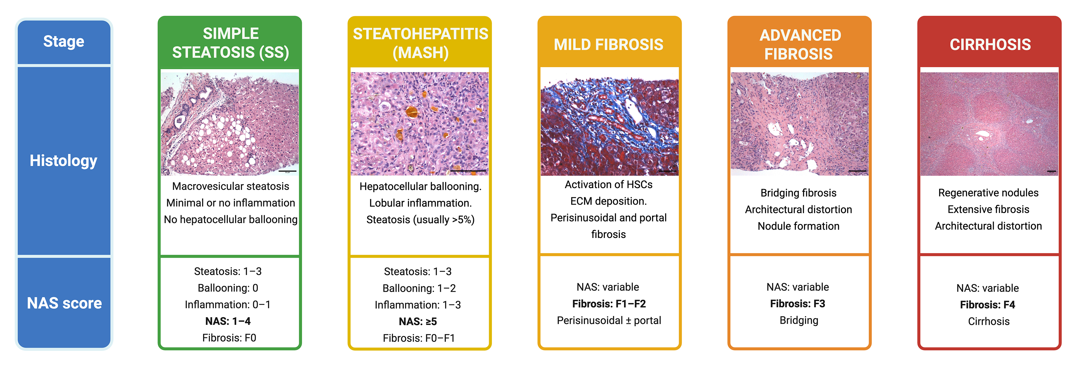
Figure 4. Histopathological spectrum of MASLD and corresponding parameters of the NASH-CRN scoring system. Representative histology and main characteristics of each stage of the disease, including the components of the NAS score (steatosis, lobular inflammation, and hepatocellular ballooning) and the corresponding stage of fibrosis (F0-F4). Micrographs were obtained at ×100 (cirrhosis), ×200 (SS, mild and advanced fibrosis), and ×400 (MASH); scale bars represent 50 µm. Created in BioRender. Ramírez, M. (2026) https://BioRender.com/hthbzdp. SS: Simple steatosis; MASH: metabolic dysfunction-associated steatohepatitis; HSCs: hepatic stellate cells; ECM: extracellular matrix; NAS: NAFLD Activity Score; NASH-CRN: Nonalcoholic Steatohepatitis Clinical Research Network.
Toward biopsy-free endpoints in clinical trials
The recognition of biopsy limitations has accelerated efforts to establish validated non-invasive biomarkers as surrogate endpoints for therapeutic trials in MASLD and MASH, with clinical trials increasingly incorporating multimodal combinations of serum biomarkers, elastography, and imaging studies to select patients with active fibrosis and monitor treatment response without serial liver biopsies[69,120].
MetALD: AN EMERGING CLINICAL ENTITY
MetALD represents a clinically distinct entity within the spectrum of SLD. Regarding its natural history, MetALD has been shown to carry a worse prognosis than pure MASLD. A meta-analysis involving more than 9.8 million participants demonstrated that MetALD is associated with higher cancer mortality [hazard ratio (HR) 2.10; 95%CI: 1.35-3.28] and cardiovascular mortality (HR 1.17; 95%CI: 1.12-1.22) compared with individuals without SLD[125]. Furthermore, a more recent meta-analysis involving 11.5 million adults confirmed that MetALD, compared with MASLD, is associated with a higher risk of hepatic events (HR 1.62) and HCC (HR 1.33), with no significant differences in cardiovascular risk between the two conditions[6]. From a clinical perspective, MetALD requires a multidisciplinary approach that integrates hepatological evaluation with interventions targeting alcohol consumption and cardiometabolic control. Abstinence or reduction in alcohol consumption is the primary therapeutic goal. The EASL and AASLD guidelines recommend restriction or abstinence for all individuals with SLD and metabolic risk factors, regardless of their level of consumption[12,13].
Measuring alcohol consumption in MetALD
Accurately measuring alcohol consumption is the main diagnostic challenge in MetALD, as it relies on self-reporting, which systematically underestimates actual consumption due to stigma and memory bias[126]. In clinical practice, standardized questionnaires such as the alcohol use disorders identification test (AUDIT), are the first-line tool for estimating consumption. However, their agreement with objective biomarkers is moderate[127]. Phosphatidylethanol (PEth) is an ethanol metabolite incorporated into the erythrocyte membrane; it is detectable in whole blood for up to four weeks after consumption and has high sensitivity and specificity for recent alcohol intake[128,129]. Its use in conjunction with self-reporting increases the diagnosis of MetALD by up to four times and that of ALD by up to three times compared to self-reporting alone[129]. PEth levels between 35 and 210 ng/mL identify approximately 20% of patients with presumed MASLD who actually have MetALD[130]. The thresholds most commonly used in clinical practice are < 20 ng/mL to confirm abstinence and
GAPS AND FUTURE DIRECTIONS IN MASLD RESEARCH
Despite the progress made, there are still significant gaps in our understanding of MASLD. Epidemiological data from low- and middle-income countries are scarce, which limits the validity of global estimates and risk models[1]. Beyond the epidemiological data, there are certain populations that remain systematically underserved. MASLD in lean patients is no longer a diagnostic gray area, as there are now BMI-independent criteria that have improved its recognition[2,3]. The real gap lies in how often their risk of progression and complications is underestimated. Despite the absence of obesity, lean patients can develop significant fibrosis and suffer from liver complications at rates comparable to those of individuals with obesity. However, they are frequently excluded from high-risk screening programs and receive inadequate treatment[22,134]. Pediatric MASLD is similarly poorly characterized. Currently, there are no validated non-invasive biomarkers of fibrosis, clinical trials are limited and long-term outcomes are poorly defined. MetALD, in turn, is an emerging but still poorly understood condition. There is a lack of specific diagnostic algorithms, longitudinal studies characterizing its natural history and rates of fibrosis progression, and clinical trials targeting this population[126].
Stigma is another gap that rarely receives enough attention. Defining the disease as “fatty” and “non-alcoholic” carries implicit moral judgments that influence how patients are treated and their decision to seek medical care. The change in nomenclature to MASLD helped, but the stigma surrounding obesity and metabolic diseases persists in clinical settings. Language reform alone is not enough, clinical culture and medical training must also change[62,135].
Regarding diagnosis, NITs have transformed clinical practice, but access remains profoundly unequal. Sequential algorithms are costly, the FIB-4 lacks specificity in intermediate-risk populations and MRE and MRI-PDFF are beyond the reach of most healthcare systems. These disparities disproportionately affect regions with the highest burden of MASLD, including Latin America and sub-Saharan Africa[12,82]. Beyond access, most available biomarkers have failed to identify active fibrogenesis. Omics-based approaches have identified promising candidate biomarkers, but none have yet reached clinical implementation. Multi-omics integration represents the most transformative pathway for non-invasive diagnosis and molecular subtyping of the disease[105]. Machine-learning-assisted integration and international registries complement this landscape[136-138]. The approval of Resmetirom and the growing number of therapies in development mark the beginning of a new therapeutic era for MASLD. To take advantage of this progress, we need to close the gaps in our knowledge so we can identify the right patients early and accurately, before the disease progresses to irreversible stages[139,140].
CONCLUSION
MASLD represents a global public health problem. Its burden continues to rise, disproportionately affecting certain regions and populations. Its multisystemic nature, driven by metabolic dysfunction, genetic susceptibility, and environmental and sociodemographic factors, requires a comprehensive diagnostic approach. Advances in non-invasive tools have transformed the diagnostic paradigm, enabling more accessible and reproducible risk stratification. In this context, simple serum scores such as FIB-4 are recommended as first-line tools for fibrosis assessment, as they facilitate early identification of patients requiring specialized evaluation. However, their limitations must be considered in specific populations, and their implementation will depend on resource availability in each clinical setting. Furthermore, the approval of Resmetirom marks the beginning of a new therapeutic era, in which timely and accurate diagnosis becomes a clinical priority with direct implications for prognosis.
DECLARATIONS
Acknowledgments
The authors gratefully acknowledge Medica Sur Clinic and Foundation for institutional support throughout the preparation of this review. Ramírez-Mejía MM and Gómez-Camacho S are enrolled in the Plan of Combined Studies in Medicine (PECEM-MD/PhD) program at the National Autonomous University of Mexico, whose academic framework and commitment to research training were instrumental in the development of this work. The authors also wish to express their sincere gratitude to Dr. Carlos Ortiz-Hidalgo for generously providing the histological images included in Figure 4. The Graphical Abstract was created with BioRender.com [Created in BioRender. Ramírez, M. (2026) https://BioRender.com/3zkc1os].
Authors’ contributions
Conceptualization, writing-original draft, writing-review & editing, supervision: Méndez-Sánchez N
Writing-original draft, writing-review & editing, visualization, investigation: Ramírez-Mejía MM, Gómez-Camacho S
All authors read and approved the submitted version.
Availability of data and materials
Not applicable.
AI and AI-assisted tools statement
Not applicable.
Financial support and sponsorship
None.
Conflicts of interest
All authors declared that there are no conflicts of interest.
Ethical approval and consent to participate
Not applicable.
Consent for publication
Not applicable.
Copyright
© The Author(s) 2026.
REFERENCES
1. Younossi ZM, Kalligeros M, Henry L. Epidemiology of metabolic dysfunction-associated steatotic liver disease. Clin Mol Hepatol. 2025;31:S32-50.
2. Rinella ME, Lazarus JV, Ratziu V, et al. ; NAFLD Nomenclature consensus group. A multisociety Delphi consensus statement on new fatty liver disease nomenclature. Hepatology. 2023;78:1966-86.
3. Eslam M, Newsome PN, Sarin SK, et al. A new definition for metabolic dysfunction-associated fatty liver disease: an international expert consensus statement. J Hepatol. 2020;73:202-9.
4. GBD 2023 MASLD Collaborators. Global burden of metabolic dysfunction-associated steatotic liver disease, 1990-2023, and projections to 2050: a systematic analysis for the Global Burden of Disease Study 2023. Lancet Gastroenterol Hepatol. 2026;11:463-94.
5. Miao L, Targher G, Byrne CD, Cao YY, Zheng MH. Current status and future trends of the global burden of MASLD. Trends Endocrinol Metab. 2024;35:697-707.
6. Celsa C, Pennisi G, Tulone A, et al. Risk of hepatic and extrahepatic outcomes associated with metabolic dysfunction-associated steatotic liver disease and metabolic dysfunction and alcohol-associated steatotic liver disease: a systematic review and meta-analysis. Lancet Gastroenterol Hepatol. 2025;10:998-1012.
7. Targher G, Byrne CD, Tilg H. MASLD: a systemic metabolic disorder with cardiovascular and malignant complications. Gut. 2024;73:691-702.
8. Wilson MP, Singh R, Mehta S, Murad MH, Fung C, Low G. Comparing FIB-4, VCTE, pSWE, 2D-SWE, and MRE thresholds and diagnostic accuracies for detecting hepatic fibrosis in patients with MASLD: a systematic review and meta-analysis. Diagnostics. 2025;15:1598.
9. Dawod S, Brown K. Non-invasive testing in metabolic dysfunction-associated steatotic liver disease. Front Med. 2024;11:1499013.
10. Lai JC, Liang LY, Wong GL. Noninvasive tests for liver fibrosis in 2024: are there different scales for different diseases? Gastroenterol Rep 2024;12:goae024.
11. Fichez J, Mouillot T, Vonghia L, et al. Non-invasive tests for fibrotic MASH for reducing screen failure in therapeutic trials. JHEP Rep. 2025;7:101351.
12. Rinella ME, Neuschwander-Tetri BA, Siddiqui MS, et al. AASLD Practice Guidance on the clinical assessment and management of nonalcoholic fatty liver disease. Hepatology. 2023;77:1797-835.
13. European Association for the Study of the Liver (EASL), European Association for the Study of Diabetes (EASD), European Association for the Study of Obesity (EASO). EASL-EASD-EASO Clinical Practice Guidelines on the management of metabolic dysfunction-associated steatotic liver disease (MASLD). J Hepatol. 2024;81:492-542.
14. Amini-Salehi E, Letafatkar N, Norouzi N, et al. Global prevalence of nonalcoholic fatty liver disease: an updated review meta-analysis comprising a population of 78 million from 38 countries. Arch Med Res. 2024;55:103043.
15. Cao L, An Y, Liu H, et al. Global epidemiology of type 2 diabetes in patients with NAFLD or MAFLD: a systematic review and meta-analysis. BMC Med. 2024;22:101.
16. Grocic M, Nguyen M, Sanyal AJ. Current trends in the epidemiology of obesity and the association between obesity and metabolic liver disease (MASLD/MASH). Diabetes Obes Metab. 2026;28:3-18.
17. Bae JH. Racial and ethnic disparities in metabolic dysfunction-associated steatotic liver disease outcomes: a call for culturally sensitive interventions: Editorial on “Differences in liver and mortality outcomes of non-alcoholic fatty liver disease by race and ethnicity: a longitudinal real-world study”. Clin Mol Hepatol. 2024;30:665-8.
18. Rich NE, Oji S, Mufti AR, et al. Racial and ethnic disparities in nonalcoholic fatty liver disease prevalence, severity, and outcomes in the United States: a systematic review and meta-analysis. Clin Gastroenterol Hepatol. 2018;16:198-210.e2.
19. Sato-Espinoza K, Chotiprasidhi P, Huaman MR, Díaz-Ferrer J. Update in lean metabolic dysfunction-associated steatotic liver disease. World J Hepatol. 2024;16:452-64.
20. Ramírez-Mejía MM, Díaz-Orozco LE, Barranco-Fragoso B, Méndez-Sánchez N. A review of the increasing prevalence of metabolic-associated fatty liver disease (MAFLD) in children and adolescents worldwide and in mexico and the implications for public health. Med Sci Monit. 2021;27:e934134.
21. Lefere S, Koot BGP, Mann JP, Bufler P, De Bruyne R, Hudert CA. Update in clinical science: MASLD in children and adolescents. J Hepatol. 2026;84:1164-77.
22. Cheah MCC, Crane H, George J. Global prevalence, metabolic characteristics, and outcomes of lean-MAFLD: a systematic review and meta-analysis. Hepatol Int. 2025;19:607-18.
23. Cherubini A, Della Torre S, Pelusi S, Valenti L. Sexual dimorphism of metabolic dysfunction-associated steatotic liver disease. Trends Mol Med. 2024;30:1126-36.
24. Jamalinia M, Saeian S, Nikkhoo N, Nazerian A, Lankarani KB. Sex and gender differences in MASLD: pathophysiological mechanisms, clinical implications, and future directions. Metab Target Organ Damage 2025;5:60.
25. Eng PC, Forlano R, Tan T, Manousou P, Dhillo WS, Izzi-Engbeaya C. Non-alcoholic fatty liver disease in women - current knowledge and emerging concepts. JHEP Rep. 2023;5:100835.
26. Truong XT, Lee DH. Hepatic insulin resistance and steatosis in metabolic dysfunction-associated steatotic liver disease: new insights into mechanisms and clinical implications. Diabetes Metab J. 2025;49:964-86.
27. Dua A, Kumari R, Singh M, et al. Metabolic dysfunction-associated steatotic liver disease (MASLD): the interplay of gut microbiome, insulin resistance, and diabetes. Front Med. 2025;12:1618275.
28. Ma J, Ma Y, Wan X, et al. Metabolic and genetic mechanisms of metabolic dysfunction-associated steatotic liver disease: an integrative perspective from molecular pathways to clinical challenges. Front Endocrinol. 2025;16:1639064.
29. Lorek D, Łupina K, Bisaga W, et al. The socioeconomic and environmental determinants of metabolic dysfunction-associated steatotic liver disease: understanding inequalities in prevalence and outcomes. Korean J Fam Med. 2025;46:61-9.
30. Singh V, Cheng S, Velazquez A, Trivedi HD, Kwan AC. The impact of social determinants of health on metabolic dysfunction-associated steatotic liver disease among adults in the United States. J Clin Med. 2025;14:5484.
31. Kanda T, Sasaki-Tanaka R, Abe H, et al. Polygenic risk score for metabolic dysfunction-associated steatotic liver disease and steatohepatitis: a narrative review. Int J Mol Sci. 2025;26:5164.
32. Pei Y, Goh GB. Genetic risk factors for metabolic dysfunction-associated steatotic liver disease. Gut Liver. 2025;19:8-18.
33. Kozlitina J, Sookoian S. Global epidemiological impact of PNPLA3 I148M on liver disease. Liver Int. 2025;45:e16123.
34. Karapanagiotidi F, Boutari C, Sinakos E. Genetic predisposition to MASLD: potential for therapeutic management. Int J Mol Sci. 2026;27:1933.
35. Benedé-Ubieto R, Cubero FJ, Nevzorova YA. Breaking the barriers: the role of gut homeostasis in metabolic-associated steatotic liver disease (MASLD). Gut Microbes. 2024;16:2331460.
36. Schnabl B, Damman CJ, Carr RM. Metabolic dysfunction-associated steatotic liver disease and the gut microbiome: pathogenic insights and therapeutic innovations. J Clin Invest. 2025;135:e186423.
37. Ramírez-Mejía MM, Ponciano-Rodríguez G, Méndez-Sánchez N. Implications of the liver-gut axis in liver disease: from mechanisms to therapeutic targets. Arch Med Res. 2025;56:103335.
38. García S, Monserrat-Mesquida M, Ugarriza L, et al. Ultra-processed food consumption and metabolic-dysfunction-associated steatotic liver disease (MASLD): a longitudinal and sustainable analysis. Nutrients. 2025;17:472.
39. Geladari EV, Kounatidis D, Christodoulatos GS, et al. Ultra-processed foods and metabolic dysfunction-associated steatotic liver disease (MASLD): what is the evidence so far? Nutrients 2025;17:2098.
40. Li X, Li J, Zhou J, et al. Mapping the association between environmental pollutants and steatotic liver disease: a systematic review and meta-analysis. Environ Res. 2025;285:122623.
41. Ran S, Zhang J, Tian F, et al. Association of metabolic signatures of air pollution with MASLD: observational and Mendelian randomization study. J Hepatol. 2025;82:560-70.
42. Zhao Y, He H, Zeng J, Zhang J. Environmental endocrine-disrupting chemicals and metabolic dysfunction-associated steatotic liver disease. Liver Int. 2026;46:e70793.
43. Ghosh S, Nukavarapu SP, Jala VR. Effects of heavy metals on gut barrier integrity and gut microbiota. Microbiota and Host. 2023;2:e230015.
44. Wei G, Liu W, Shen F, et al. Polystyrene microplastics exposure increases the disruption of intestinal barrier integrity and gut microbiota homeostasis during obesity and aging. J Environ Sci. 2026;162:430-43.
45. Gao B, Arab JP, Liangpunsakul S, et al. Metabolic dysfunction and alcohol-associated liver disease (MetALD). eGastroenterology. 2025;3:e100319.
46. Acierno C, Barletta F, Caturano A, et al. Alcohol consumption and liver metabolism in the era of MASLD: integrating nutritional and pathophysiological insights. Nutrients. 2025;17:2229.
48. Li Y, Yang P, Ye J, Xu Q, Wu J, Wang Y. Updated mechanisms of MASLD pathogenesis. Lipids Health Dis. 2024;23:117.
49. Ramírez-Mejía MM, Qi X, Abenavoli L, Romero-Gómez M, Eslam M, Méndez-Sánchez N. Metabolic dysfunction: the silenced connection with fatty liver disease. Ann Hepatol. 2023;28:101138.
50. Hagström H, Shang Y, Hegmar H, Nasr P. Natural history and progression of metabolic dysfunction-associated steatotic liver disease. Lancet Gastroenterol Hepatol. 2024;9:944-56.
52. Huang DQ, Tran S, Barnett S, et al. Incidence and predictors of hepatocellular carcinoma in NAFLD without diagnosed cirrhosis: a nationwide real-world U.S. study. Hepatol Int. 2024;18:540-9.
53. Lekakis V, Papatheodoridis GV. Natural history of metabolic dysfunction-associated steatotic liver disease. Eur J Intern Med. 2024;122:3-10.
54. Le P, Payne JY, Zhang L, et al. Disease state transition probabilities across the spectrum of NAFLD: a systematic review and meta-analysis of paired biopsy or imaging studies. Clin Gastroenterol Hepatol. 2023;21:1154-68.
55. Sierra L, Liu L, Khurana A, et al. Reversing fibrosis in metabolic dysfunction-associated steatohepatitis-beyond telling patients to lose weight. Cleve Clin J Med. 2026;93:279-86.
56. Hagström H, Nasr P, Ekstedt M, et al. Fibrosis stage but not NASH predicts mortality and time to development of severe liver disease in biopsy-proven NAFLD. J Hepatol. 2017;67:1265-73.
57. Gancheva S, Roden M, Castera L. Diabetes as a risk factor for MASH progression. Diabetes Res Clin Pract. 2024;217:111846.
58. Martínez-Sánchez FD, Corredor-Nassar MJ, Feria-Agudelo SM, et al. Factors associated with advanced liver fibrosis in a population with type 2 diabetes: a multicentric study in Mexico city. J Clin Exp Hepatol. 2025;15:102536.
59. Julián MT, Arteaga I, Torán-Monserrat P, et al. The link between abdominal obesity indices and the progression of liver fibrosis: insights from a population-based study. Nutrients. 2024;16:1586.
60. Krawczyk M, Rau M, Schattenberg JM, et al. ; NAFLD Clinical Study Group. Combined effects of the PNPLA3 rs738409, TM6SF2 rs58542926, and MBOAT7 rs641738 variants on NAFLD severity: a multicenter biopsy-based study. J Lipid Res. 2017;58:247-55.
61. Mustafa A, Kite C, Lagojda L, et al. Cardiovascular disease in the context of metabolic dysfunction-associated steatotic liver disease (MASLD): a comprehensive narrative review. Int J Mol Sci. 2025;26:11275.
62. Lazarus JV, Mark HE, Anstee QM, et al. ; NAFLD Consensus Consortium. Advancing the global public health agenda for NAFLD: a consensus statement. Nat Rev Gastroenterol Hepatol. 2022;19:60-78.
63. Phoolchund AGS, Khakoo SI. MASLD and the development of HCC: pathogenesis and therapeutic challenges. Cancers. 2024;16:259.
64. Mi K, Ye T, Zhu L, Pan CQ. Risk-stratified hepatocellular carcinoma surveillance in non-cirrhotic patients with MASLD. Gastroenterol Rep. 2025;13:goaf018.
65. Huang DQ, El-Serag HB, Loomba R. Global epidemiology of NAFLD-related HCC: trends, predictions, risk factors and prevention. Nat Rev Gastroenterol Hepatol. 2021;18:223-38.
66. Rodriguez LA, Schmittdiel JA, Liu L, et al. Hepatocellular carcinoma in metabolic dysfunction-associated steatotic liver disease. JAMA Netw Open. 2024;7:e2421019.
67. Shenoy A, Ahmed A, Kim D. Extrahepatic manifestation of metabolic dysfunction-associated steatotic liver disease. Metab Target Organ Damage. 2025;5:46.
68. Bilson J, Mantovani A, Byrne CD, Targher G. Steatotic liver disease, MASLD and risk of chronic kidney disease. Diabetes Metab. 2024;50:101506.
69. Mantovani A, Petracca G, Beatrice G, Tilg H, Byrne CD, Targher G. Non-alcoholic fatty liver disease and risk of incident diabetes mellitus: an updated meta-analysis of 501 022 adult individuals. Gut. 2021;70:962-9.
70. Córdova-Gallardo CJ, Vargas-Beltran AM, Ramírez Mejía MM, Méndez-Sánchez N. MASLD as a multisystemic disease. In: Méndez-Sánchez N, Qi X, Editors. Metabolic dysfunction-associated steatotic liver disease: prevalence, research insights and the future directions. Singapore: Springer Nature Singapore; 2024. pp. 87-94. Available from https://link.springer.com/book/10.1007/978-981-97-9519-2. [accessed 26 August 2026].
71. Hernaez R, Lazo M, Bonekamp S, et al. Diagnostic accuracy and reliability of ultrasonography for the detection of fatty liver: a meta-analysis. Hepatology. 2011;54:1082-90.
72. Cao YT, Xiang LL, Qi F, Zhang YJ, Chen Y, Zhou XQ. Accuracy of controlled attenuation parameter (CAP) and liver stiffness measurement (LSM) for assessing steatosis and fibrosis in non-alcoholic fatty liver disease: a systematic review and meta-analysis. EClinicalMedicine. 2022;51:101547.
73. Zhang YX, Feng YP, You CL, Zhang LY. The diagnostic value of MRI-PDFF in hepatic steatosis of patients with metabolic dysfunction-associated steatotic liver disease: a systematic review and meta-analysis. BMC Gastroenterol. 2025;25:451.
74. Indre MG, Leucuta DC, Lupsor-Platon M, et al. Diagnostic accuracy of 2D-SWE ultrasound for liver fibrosis assessment in MASLD: a multilevel random effects model meta-analysis. Hepatology. 2025;82:454-69.
75. Han S, Choi M, Lee B, et al. Accuracy of noninvasive scoring systems in assessing liver fibrosis in patients with nonalcoholic fatty liver disease: a systematic review and meta-analysis. Gut Liver. 2022;16:952-63.
76. López Tórrez SM, Ayala CO, Ruggiro PB, et al. Accuracy of prognostic serological biomarkers in predicting liver fibrosis severity in people with metabolic dysfunction-associated steatotic liver disease: a meta-analysis of over 40,000 participants. Front Nutr. 2024;11:1284509.
77. Hinkson A, Lally H, Gibson H, et al. Meta-analysis: enhanced liver fibrosis test to identify hepatic fibrosis in chronic liver diseases. Aliment Pharmacol Ther. 2023;57:750-62.
78. Eslam M, Wong GL, Hashem AM, et al. A sequential algorithm combining ADAPT and liver stiffness can stage metabolic-associated fatty liver disease in hospital-based and primary care patients. Am J Gastroenterol. 2021;116:984-93.
79. Canivet CM, Zheng MH, Qadri S, et al. Validation of the blood test MACK-3 for the noninvasive diagnosis of fibrotic nonalcoholic steatohepatitis: an international study with 1924 patients. Clin Gastroenterol Hepatol. 2023;21:3097-106.e10.
80. Malandris K, Arampidis D, Mainou M, et al. FibroScan-AST score for diagnosing fibrotic MASH: a systematic review and meta-analysis of diagnostic test accuracy studies. J Gastroenterol Hepatol. 2024;39:2582-91.
81. Liu S, Zhang Q, Wang L, Tang Q, Hu B. MAST versus FAST for active fibrotic MASH: a meta-analysis supporting risk-stratified diagnostic pathways. Front Med. 2026;13:1779292.
82. European Association for the Study of the Liver, European Association for the Study of Diabetes, European Association for the Study of Obesity. EASL-EASD-EASO Clinical Practice Guidelines on the management of metabolic dysfunction-associated steatotic liver disease (MASLD): executive Summary. Diabetologia. 2024;67:2375-92.
83. Portincasa P, Khalil M, Mahdi L, et al. Metabolic dysfunction-associated steatotic liver disease: from pathogenesis to current therapeutic options. Int J Mol Sci. 2024;25:5640.
84. Petroff D, Blank V, Newsome PN, et al. Assessment of hepatic steatosis by controlled attenuation parameter using the M and XL probes: an individual patient data meta-analysis. Lancet Gastroenterol Hepatol. 2021;6:185-98.
85. Alsaqal S, Hockings P, Ahlström H, et al. The combination of MR elastography and proton density fat fraction improves diagnosis of nonalcoholic steatohepatitis. J Magn Reson Imaging. 2022;56:368-79.
86. Qu Y, Middleton MS, Loomba R, et al. Magnetic resonance elastography biomarkers for detection of histologic alterations in nonalcoholic fatty liver disease in the absence of fibrosis. Eur Radiol. 2021;31:8408-19.
87. Lee DH. Quantitative assessment of fatty liver using ultrasound attenuation imaging. J Med Ultrason. 2021;48:465-70.
88. McPherson S, Hardy T, Dufour JF, et al. Age as a confounding factor for the accurate non-invasive diagnosis of advanced NAFLD fibrosis. Am J Gastroenterol. 2017;112:740-51.
89. Sung S, Al-Karaghouli M, Tam M, et al. Age-dependent differences in FIB-4 predictions of fibrosis in patients with MASLD referred from primary care. Hepatol Commun. 2025;9:e0609.
90. Lichtinghagen R, Pietsch D, Bantel H, Manns MP, Brand K, Bahr MJ. The enhanced liver fibrosis (ELF) score: normal values, influence factors and proposed cut-off values. J Hepatol. 2013;59:236-42.
91. Eslam M, Fan JG, Yu ML, et al. The Asian Pacific association for the study of the liver clinical practice guidelines for the diagnosis and management of metabolic dysfunction-associated fatty liver disease. Hepatol Int. 2025;19:261-301.
92. Tsochatzis EA, Valenti L, Thiele M, et al. Use of non-invasive diagnostic tools for metabolic dysfunction-associated steatohepatitis: a qualitative exploration of challenges and barriers. Liver Int. 2024;44:1990-2001.
93. Yeramaneni S, Chang ST, Cheung RC, et al. Comparison of referral rates and costs using fibrosis-4 and enhanced liver fibrosis (ELF) testing strategies for initial evaluation of metabolic dysfunction-associated steatotic liver disease (MASLD) in a veteran population. J Appl Lab Med. 2025;10:593-604.
94. Li Q, Gao Y, Wang D, et al. Prospective comparison of 2D-shear wave elastography and vibration-controlled transient elastography in assessing liver fibrosis in metabolic dysfunction-associated steatotic liver disease. Acad Radiol. 2025;32:5100-11.
95. Singh S, Venkatesh SK, Loomba R, et al. Magnetic resonance elastography for staging liver fibrosis in non-alcoholic fatty liver disease: a diagnostic accuracy systematic review and individual participant data pooled analysis. Eur Radiol. 2016;26:1431-40.
96. Kawanaka M, Kamada Y, Takahashi H, et al. ; Japan Study Group of NAFLD (JSG-NAFLD). Serum cytokeratin 18 fragment is an indicator for treating metabolic dysfunction-associated steatotic liver disease. Gastro Hep Adv. 2024;3:1120-8.
97. Boursier J, Anty R, Vonghia L, et al. Screening for therapeutic trials and treatment indication in clinical practice: MACK-3, a new blood test for the diagnosis of fibrotic NASH. Aliment Pharmacol Ther. 2018;47:1387-96.
98. Newsome PN, Sasso M, Deeks JJ, et al. FibroScan-AST (FAST) score for the non-invasive identification of patients with non-alcoholic steatohepatitis with significant activity and fibrosis: a prospective derivation and global validation study. Lancet Gastroenterol Hepatol. 2020;5:362-73.
99. Truong E, Gornbein JA, Yang JD, et al. MRI-AST (MAST) score accurately predicts major adverse liver outcome, hepatocellular carcinoma, liver transplant, and liver-related death. Clin Gastroenterol Hepatol. 2023;21:2570-7.e1.
100. Noureddin M, Truong E, Gornbein JA, et al. MRI-based (MAST) score accurately identifies patients with NASH and significant fibrosis. J Hepatol. 2022;76:781-7.
101. Jamialahmadi O, De Vincentis A, Tavaglione F, et al. Partitioned polygenic risk scores identify distinct types of metabolic dysfunction-associated steatotic liver disease. Nat Med. 2024;30:3614-23.
102. Xiao L, Li Y, Hong C, et al. Polygenic risk score of metabolic dysfunction-associated steatotic liver disease amplifies the health impact on severe liver disease and metabolism-related outcomes. J Transl Med. 2024;22:650.
103. Tobaruela-Resola AL, Milagro FI, Mogna-Pelaez P, Moreno-Aliaga MJ, Abete I, Zulet MÁ. The use of circulating miRNAs for the diagnosis, prognosis, and personalized treatment of MASLD. J Physiol Biochem. 2025;81:589-609.
104. Tobaruela-Resola AL, Milagro FI, Elorz M, et al. Circulating miR-122-5p, miR-151a-3p, miR-126-5p and miR-21-5p as potential predictive biomarkers for metabolic dysfunction-associated steatotic liver disease assessment. J Physiol Biochem. 2025;81:1275-88.
105. Thiele M, Villesen IF, Niu L, et al. ; MicrobLiver consortium, GALAXY consortium. Opportunities and barriers in omics-based biomarker discovery for steatotic liver diseases. J Hepatol. 2024;81:345-59.
106. Boel F, Akimov V, Teuchler M, et al. Deep proteome profiling of metabolic dysfunction-associated steatotic liver disease. Commun Med. 2025;5:56.
107. Verschuren L, Mak AL, van Koppen A, et al. Development of a novel non-invasive biomarker panel for hepatic fibrosis in MASLD. Nat Commun. 2024;15:4564.
108. Tan EY, Muthiah MD, Sanyal AJ. Metabolomics at the cutting edge of risk prediction of MASLD. Cell Rep Med. 2024;5:101853.
109. Schneider KM, Cao F, Huang HYR, et al. The lipidomic profile discriminates between MASLD and MetALD. Aliment Pharmacol Ther. 2025;61:1357-71.
110. Ning M, Lu D, Liang D, Ren PG. Single-cell RNA sequencing advances in revealing the development and progression of MASH: the identifications and interactions of non-parenchymal cells. Front Mol Biosci. 2025;12:1513993.
111. Li JZ, Yang L, Xiao MX, et al. Spatial and single-cell transcriptomics reveals the regional division of the spatial structure of MASH fibrosis. Liver Int. 2025;45:e16125.
112. Watson BR, Paul B, Rahman RU, et al. Spatial transcriptomics of healthy and fibrotic human liver at single-cell resolution. Nat Commun. 2025;16:319.
113. Li Z, Luo G, Gan C, et al. Spatially resolved multi-omics of human metabolic dysfunction-associated steatotic liver disease. Nat Genet. 2025;57:3112-25.
114. Zhou XD, Li YT, Kim SU, et al. ; VCTE-Prognosis Study Group, Paired Liver Biopsy Group. Longitudinal changes in fibrosis markers: monitoring stiffness/fibrosis progression and prognostic outcomes in MASLD. Clin Gastroenterol Hepatol. 2026;24:394-406.
115. Vettor R, Petroni ML, Dotan I, et al. Overcoming the barriers in the screening, diagnosis, and follow-up of patients with metabolic dysfunction-associated steatotic liver disease (MASLD) and metabolic dysfunction-associated steatohepatitis (MASH). Rev Endocr Metab Disord. 2026;27:213-35.
116. Mathur A, Ozkaya E, Rosberger S, et al. Concordance of vibration-controlled transient elastography and magnetic resonance elastography for fibrosis staging in patients with metabolic dysfunction-associated steatotic liver disease. Eur Radiol. 2025;35:6507-15.
117. Borz-Baba L, Aydin A, Parvin R. Liver biopsy in metabolic-associated steatotic liver disease: accuracy, challenges, and alternatives. Cureus. 2026;18:e103284.
118. de Franchis R, Bosch J, Garcia-Tsao G, Reiberger T, Ripoll C. ; Baveno VII Faculty. Baveno VII - renewing consensus in portal hypertension. J Hepatol. 2022;76:959-74.
119. Lei SY, Han Y, Rockey DC, et al. ; International Obesity Society Working Group on Steatotic Liver Disease. An international multidisciplinary consensus statement on laparoscopic liver biopsy. Cell Rep Med. 2026;7:102831.
120. Lonardo A, Ballestri S, Mantovani A, Targher G, Bril F. Endpoints in NASH clinical trials: are we blind in one eye? Metabolites 2024;14:40.
121. Turan Gokce D, Ekelik M, Gumussoy M, et al. Histological disease progression in patients with metabolic dysfunction-associated steatotic liver disease using paired liver biopsy. Eur J Gastroenterol Hepatol. 2026;38:76-81.
122. Ratziu V, Hompesch M, Petitjean M, et al. Artificial intelligence-assisted digital pathology for non-alcoholic steatohepatitis: current status and future directions. J Hepatol. 2024;80:335-51.
123. Kleiner DE, Brunt EM, Wilson LA, et al. ; Nonalcoholic Steatohepatitis Clinical Research Network. Association of histologic disease activity with progression of nonalcoholic fatty liver disease. JAMA Netw Open. 2019;2:e1912565.
124. Nascimbeni F, Bedossa P, Fedchuk L, et al. ; LIDO (Liver Injury in Diabetes and Obesity) Study Group. Clinical validation of the FLIP algorithm and the SAF score in patients with non-alcoholic fatty liver disease. J Hepatol. 2020;72:828-38.
125. Ciardullo S, Mantovani A, Morieri ML, Muraca E, Invernizzi P, Perseghin G. Impact of MASLD and MetALD on clinical outcomes: a meta-analysis of preliminary evidence. Liver Int. 2024;44:1762-7.
126. Arab JP, Díaz LA, Rehm J, et al. Metabolic dysfunction and alcohol-related liver disease (MetALD): Position statement by an expert panel on alcohol-related liver disease. J Hepatol. 2025;82:744-56.
127. Grüner Nielsen D, Andersen K, Søgaard Nielsen A, Juhl C, Mellentin A. Consistency between self-reported alcohol consumption and biological markers among patients with alcohol use disorder - a systematic review. Neurosci Biobehav Rev. 2021;124:370-85.
128. Rady ED, Cotter TG. Integrating phosphatidylethanol to enhance alcohol quantification in steatotic liver disease and clinical trials. Liver Int. 2026;46:e70489.
129. Tavaglione F, Díaz LA, Ajmera V, et al. Clinical utility of phosphatidylethanol to detect underreported alcohol use and enhance steatotic liver disease subclassification. J Hepatol. 2025;83:1023-34.
130. Vaz J, Nasr P, Helander A, et al. Phosphatidylethanol levels distinguish steatotic liver disease subgroups and are associated with risk of major liver outcomes. J Hepatol. 2025;83:1011-22.
131. Loomba R, Hagström H, Díaz LA. Emerging role of phosphatidylethanol testing in clinical practice for the identification of steatotic liver disease subtypes: is this the new standard? Gastroenterology 2025;169:1103-7.
132. Vaz J, Hagström H. Reply to: “Why one PEth result cannot redraw the MASLD/ALD boundary: a single-snapshot misclassification” and “Phosphatidylethanol-guided reclassification of steatotic liver disease subgroups improves prognostic stratification: greater precision is required”: phosphatidylethanol in steatotic liver disease: real-world evidence. J Hepatol. 2025;83:e234-5.
133. Tampaki M, Tsochatzis E, Lekakis V, Cholongitas E. Prevalence, characteristics and outcomes of patients with metabolic and alcohol related/associated liver disease (MetALD): a systematic review and meta-analysis. Metabolism. 2025;163:156101.
134. Méndez-Sánchez N, Brouwer WP, Lammert F, Yilmaz Y. Metabolic dysfunction associated fatty liver disease in healthy weight individuals. Hepatol Int. 2024;18:884-96.
135. Ramírez-Mejía MM, Qi X, Abenavoli L, Méndez-Sánchez N. The myth of the stigma of fatty liver: what does the evidence show? Ann Hepatol 2024;29:101535.
136. Dunn N, Verma N, Dunn W. Artificial intelligence for predictive diagnostics, prognosis, and decision support in MASLD, hepatocellular carcinoma, and digital pathology. J Clin Exp Hepatol. 2026;16:103184.
137. Kanwal F, Shubrook JH, Younossi Z, et al. Preparing for the NASH epidemic: a call to action. Metabolism. 2021;122:154822.
138. Hernández-Almonacid PG, Marín-Quintero X. Artificial intelligence in metabolic dysfunction-associated steatotic liver disease: transforming diagnosis and therapeutic approaches. World J Gastroenterol. 2026;32:111737.
139. Barbhuiya PA, Ahmed A, Pathak MP. Advancing metabolic dysfunction associated steatotic liver disease (MASLD) treatment: exploring novel formulations and therapies. J Drug Deliv Sci Technol. 2026;115:107702.
Cite This Article
 Open Access
How to Cite
Download Citation
Export Citation File:
Type of Import
Tips on Downloading Citation
Citation Manager File Format
Type of Import
Direct Import: When the Direct Import option is selected (the default state), a dialogue box will give you the option to Save or Open the downloaded citation data. Choosing Open will either launch your citation manager or give you a choice of applications with which to use the metadata. The Save option saves the file locally for later use.
Indirect Import: When the Indirect Import option is selected, the metadata is displayed and may be copied and pasted as needed.
About This Article
Special Topic
Copyright

Data & Comments
Data
0










Comments
Comments must be written in English. Spam, offensive content, impersonation, and private information will not be permitted. If any comment is reported and identified as inappropriate content by OAE staff, the comment will be removed without notice. If you have any queries or need any help, please contact us at [email protected].