
Macrophage ferroptosis in atherosclerosis: unraveling novel antiatherogenic targets and pharmacological strategies
 0
Graphical Abstract
Abstract
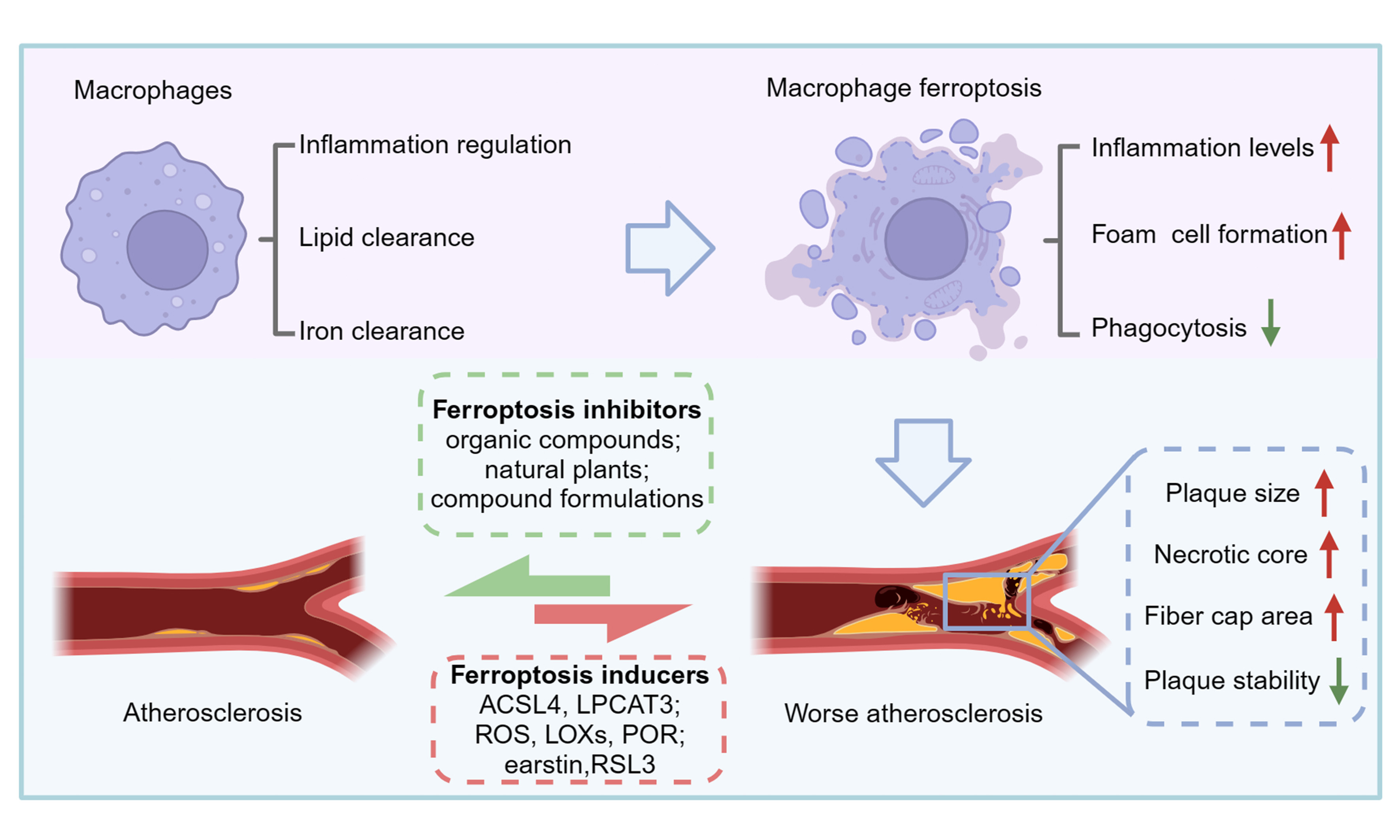
Atherosclerosis is the underlying cause of many cardiovascular diseases, and macrophages play a pivotal role in its intricate landscape, significantly regulating inflammation, lipid metabolism, and iron metabolism. Ferroptosis is an iron-dependent form of regulated cell death driven by lipid peroxidation, and its process is related to macrophage function in atherosclerosis. During atherosclerosis, macrophages contribute to the removal of harmful substances such as lipids and inhibit early development. However, macrophage ferroptosis promotes inflammatory progression, facilitates foam cell and necrotic core formation, promotes calcification, and increases plaque instability. This review first summarizes macrophage heterogeneity in atherosclerosis, then integrates their roles in lipid handling and iron metabolism, and finally focuses on how ferroptosis shapes plaque inflammation, necrotic core formation, calcification, and instability. Currently, the pharmacologic treatment of atherosclerosis is based on lipid-lowering agents, but it remains deficient. Therefore, this review encompasses a variety of potential pharmacological treatments, including organic compounds, natural plant extracts and derivatives, and compound formulations, which can inhibit macrophage ferroptosis and may constitute a novel approach for atherosclerosis treatment. Taken together, this review provides a breakthrough strategy for treating atherosclerosis and establishes a navigational pathway for future therapeutic interventions by targeting macrophage ferroptosis.
Keywords


 Download PDF
INTRODUCTION
Cardiovascular diseases (CVDs) are recognized globally for their high rates of mortality and disability[1]. Atherosclerosis, a chronic inflammatory disease, is a primary contributor to CVDs, characterized by lipid accumulation and immune system engagement[2]. Macrophages exert a dual effect during atherogenesis. In early lesions, they clear lipids and cellular debris, thereby providing protection[3]. However, when lipid uptake exceeds processing capacity, they transform into foam cells, amplify inflammation, and contribute to necrotic core formation[4]. Consequently, maintaining normal phagocytosis of macrophages and preventing macrophage death emerge as strategies to mitigate atherosclerosis.
Ferroptosis is a lipid peroxidation process driven by iron ions, distinct from other programmed cell death mechanisms[5], and studies have confirmed its role in accelerating atherosclerosis progression[6]. Excess intracellular iron promotes reactive oxygen species (ROS) generation via the Fenton reaction [in which Fe2+ catalyzes H2O2 to generate highly toxic hydroxyl radicals (·OH)], leading to lipid peroxidation of cellular membranes and ferroptosis. Macrophages play crucial roles in regulating inflammation, iron, and lipid metabolism in atherosclerosis due to their unique abilities in phagocytosis, cytotoxicity, and cytokine secretion[7]. During atherosclerosis, lipoproteins phagocytosed by macrophages increase the substrate for lipid peroxidation and facilitate the execution of ferroptosis. Furthermore, in this process, ROS production by macrophages is a key contributor to ferroptosis, pro-inflammatory polarization, and foam cell formation. During ferroptosis, the scavenging function of macrophages is diminished, leading to increased lipids and necrotic cells accumulation in atherosclerotic plaques. This, in turn, results in the formation and enlargement of necrotic cores[8]. In addition to ferroptosis, other types of cell death, such as apoptosis, autophagy, necroptosis, and pyroptosis, also occur in macrophages during the progression of atherosclerosis[9]. Regardless of the form of cell death, reduced clearance efficiency leads to the accumulation of dead cells and the formation of a necrotic core. This review focuses on ferroptosis, emphasizing its potential as a target for inhibiting the development of atherosclerosis.
The prevailing pharmacologic strategies for reducing atherosclerosis primarily involve lipid-lowering drugs, predominantly statins, and their combinations. In recent years, trials have successfully reduced cardiovascular events with non-statin lipid-lowering agents, highlighting the expansion of lipid-lowering strategies[10]. These include monoclonal antibodies, small interfering RNAs (siRNAs)[11], antisense oligonucleotides[12], vaccinations[13], and other lipid-lowering therapies[14], such as small-molecule inhibitors. However, these therapies are associated with adverse effects, such as myotoxicity, metabolic toxicity, nervous system damage, poor specificity, inadequate safety, and other possible side effects[15,16]. This led to an increased focus on identifying treatments with precise efficacy and minimal side effects in antiatherogenic research. Given that macrophages are involved in atherosclerosis and in ferroptosis, targeting macrophage ferroptosis represents a promising approach to treat atherosclerosis. Recent studies have identified various compound formulations and natural plant extracts that act on multiple pathways to attenuate macrophage ferroptosis, demonstrating their potential in atherosclerosis treatment.
Accordingly, this review explores the connection between macrophage ferroptosis and atherosclerosis, highlighting drugs with anti-ferroptotic effects on macrophages to provide new insights for the treatment of atherosclerosis. Although ferroptosis has attracted attention in other diseases, in the context of atherosclerosis, the specific mechanism of macrophage ferroptosis, its impact on plaque stability, and potential therapies targeting ferroptosis remain urgent issues to be addressed.
MACROPHAGES IN THE PROGRESSION OF ATHEROSCLEROSIS
Polarization and plasticity of macrophages affect the development of atherosclerosis
Macrophages are highly plastic innate immune cells that participate in nearly every stage of atherosclerosis, from monocyte recruitment and foam cell formation to plaque inflammation and repair[17]. Notably, macrophages are involved in almost all stages of atherosclerosis. Macrophages at atherosclerotic lesions are derived primarily from circulating recruited monocytes and vascular smooth muscle cells[18] [Figure 1]. Within plaques, macrophages display a spectrum of phenotypes rather than a binary M1/M2 state. Although M1 and M2 remain the best-characterized subsets, plaque microenvironments also induce oxidized phospholipid-induced macrophages (Mox), M4, hemoglobin-associated macrophages (M(Hb)), and heme-associated macrophages (Mhem) phenotypes, each with distinct inflammatory, lipid-handling, and iron-handling features. M1 macrophages promote inflammation and are activated by proinflammatory factors, including interferon-gamma (IFN-γ), tumor necrosis factor (TNF), oxidized low-density lipoprotein (ox-LDL), and lipopolysaccharide[19]. M2 macrophages are induced by interleukin-4 (IL-4) and IL-13, which play an anti-inflammatory and pro-tissue-repair role in the atherosclerotic process[20]. According to the differences in activators, M2 macrophages can be further categorized into four subtypes: M2a macrophages are activated by IL-13 and IL-4 and exert anti-inflammatory and pro-restorative effects; M2b macrophages are activated by immune complexes, IL-1β, and so on, and have immune regulatory effects; IL-10, transforming growth factor-beta (TGF-β), and glucocorticoids can activate M2c macrophages and promote tyrosine kinase Mer (MerTK)-mediated phagocytosis of apoptotic cells; Activation of M2d macrophages by ligands for Toll-like receptors (TLRs) and adenosine A2 receptor (A2a receptor) reduces pro-inflammatory factor production while also promote angiogenesis[21-23]. Mox macrophages exert dual roles in atherogenesis; they exhibit antioxidant properties via nuclear factor erythroid 2-related factor 2 (Nrf2)-mediated expression of antioxidant genes, but also promote plaque inflammation by secreting pro-inflammatory factors such as IL-1β. It was found that Mox has decreased chemotaxis and phagocytosis and high expression of antioxidant genes[24]. Mox macrophages were found to constitute up to 30% of macrophages in advanced atherosclerotic plaques[24]. Nrf2-mediated expression of a large number of antioxidant genes was found in Mox, indicating that Mox has an antioxidant effect. Mox also showed pro-inflammatory properties, and the expression of pro-inflammatory genes, such as IL-1β was elevated[24]. However, further research is needed to demonstrate Mox’s specific role in the chronic inflammatory process underlying atherosclerosis. Platelets are likewise thought to be involved in the atherosclerosis process, and platelet activation results in the release of chemokines stored in a-granules, which not only promote the adhesion of arterial endothelial cells and monocytes but also influence macrophage differentiation[25]. Notably, CXC chemokine ligand 4 (CXCL4), also known as platelet factor 4 (PF4), can activate M4 macrophages[26]. This protein inhibits CD163 expression, thereby reducing hemoglobin phagocytosis and processing by macrophages, and exhibits proatherogenic effects[27]. In contrast, macrophages exposed to hemoglobin-haptoglobin complexes (Hb-Hp) differentiate into M(Hb) and Mhem macrophages, which express relatively high levels of CD163 and heme oxygenase-1 (HO-1), promoting phagocytosis of aged or broken erythrocytes and iron recycling (discussed in detail in section “The role of macrophages in regulating lipid metabolism during atherogenesis”)[28] [Figure 1].
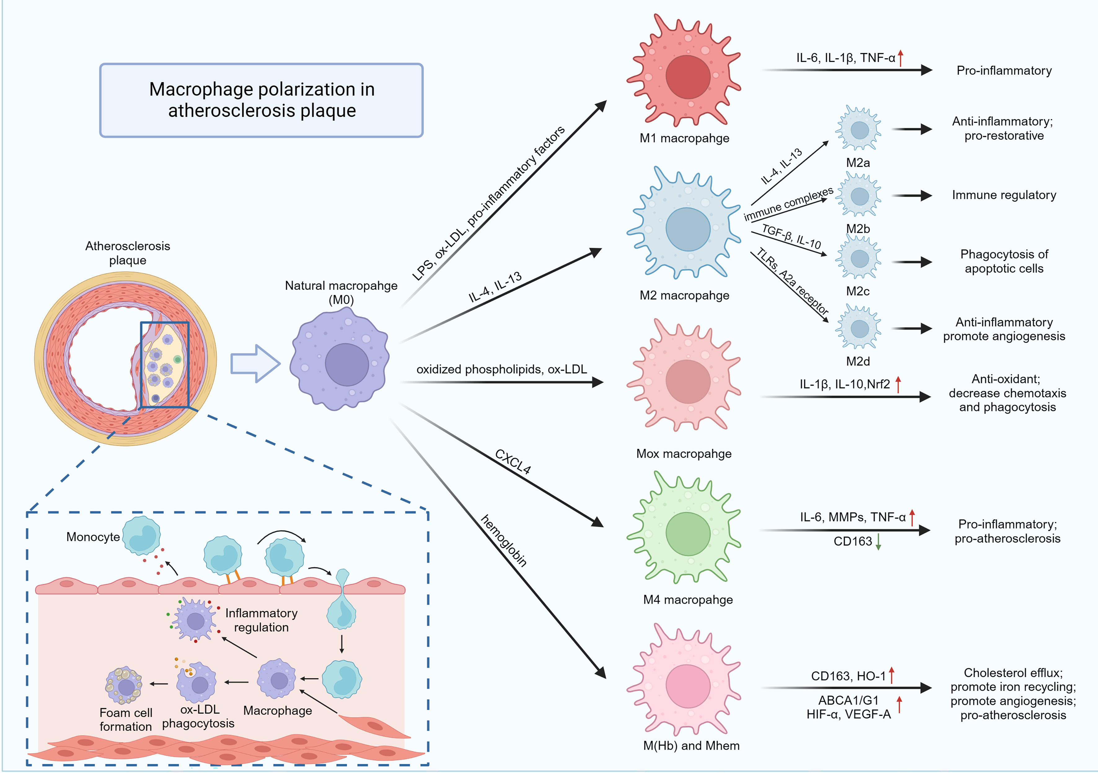
Figure 1. Macrophage polarization in atherosclerotic plaques. During atherosclerotic plaque formation, endothelial activation recruits monocytes that infiltrate and differentiate into macrophages. Macrophages play dual roles, releasing both inflammatory and anti-inflammatory factors and participating in the clearance of ox-LDL and the formation of foam cells. Additionally, vascular smooth muscle cells contribute to macrophage formation through migration and differentiation. Macrophages undergo polarization into distinct phenotypes. LPS, ox-LDL, and pro-inflammatory factors promote macrophage M1 polarization, which plays a pro-inflammatory role by releasing IL-6, IL-1β, and TNF-α. IL-4 and IL-13 induce M2 macrophage polarization, and M2-type macrophages are further subdivided into M2a, M2b, M2c, and M2d, playing various roles, such as anti-inflammatory, pro-tissue repair, immunomodulation, facilitating the phagocytosis of apoptotic cells, and promoting angiogenesis. Macrophages induced Mox polarization by oxidized phospholipids and ox-LDL and exhibited antioxidant activity, low migration, and low phagocytic activity by synthesizing IL-1β, IL-10, and Nrf2. CXCL4 activates M4 macrophages that exhibit pro-inflammatory and pro-atherosclerotic development by increasing the expression of IL-6, MMPs, and TNF-α, and inhibiting CD163 expression. In response to hemoglobin, macrophages differentiate into M(Hb) and Mhem macrophages, which express higher levels of CD163 and HO-1, ABCA1/G1, HIF-α and VEGF-A. These macrophages promote cholesterol efflux, iron recycling, and angiogenesis, and contribute to the progression of atherosclerosis. Red arrows indicate upregulation/positive regulation (e.g., promoting pro-inflammatory cytokine expression and macrophage polarization toward pro-inflammatory phenotypes). Green arrows indicate downregulation/negative regulation (e.g., CXCL4-mediated inhibition of CD163 expression and anti-inflammatory/protective pathways). Created in BioRender. Fan, F. (2026) https://BioRender.com/f63x996. ox-LDL: Oxidized low-density lipoprotein; LPS: lipopolysaccharide; IL: interleukin; TNF-α: tumor necrosis factor-alpha; TGF-β: transforming growth factor-beta; TLRs: Toll-like receptors; A2a receptor: adenosine A2 receptor; Nrf2: nuclear factor erythroid 2-related factor 2; CXCL4: CXC chemokine ligand 4; MMPs: matrix metalloproteinases; HO-1: heme oxygenase-1; ABCA1/G1: ATP-binding cassette transporter A1/G1; HIF-α: hypoxia-inducible factor-alpha; VEGF-A: vascular endothelial growth factor-A; CD163: cluster of differentiation 163; M(Hb): hemoglobin-associated macrophages.
The progression of atherosclerosis is primarily influenced by an imbalance in macrophage polarization, which leads to amplified pro-inflammatory effects and sustained inflammation[29,30]. Damage to the endothelium and the subsequent infiltration and accumulation of macrophages are the early features of atherosclerosis development[31,32]. Foam cell formation results from excessive lipid accumulation that exceeds the macrophage’s capacity for degradation and metabolism; if lipids are efficiently processed via pathways such as cholesterol efflux, foam cells do not form[18]. When anti-inflammatory macrophages are enriched, they stabilize plaques and promote plaque regression by synthesizing and releasing anti-inflammatory mediators and by facilitating collagen fiber synthesis[33]. Conversely, an increase in lipid deposition leads to an increase in the number of pro-inflammatory macrophages[34]. Pro-inflammatory macrophages maintain plaque inflammation by secreting pro-inflammatory factors such as IL-6, IL-1β, and tumor necrosis factor-alpha (TNF-α). When macrophages undergo ferroptosis within the atherosclerotic plaque, they become immunogenic, leading to the release of damage-associated molecular patterns (DAMPs), including high mobility group box 1 (HMGB1), adenosine triphosphate (ATP), and mitochondrial DNA[35]. These DAMPs act as endogenous stress signals that engage pattern recognition receptors on neighboring macrophages and vascular cells, prompting the assembly and activation of inflammasomes, most notably the NLR family pyrin domain containing 3 (NLRP3) inflammasome. Upon activation, caspase-1 cleaves pro-IL-1β and pro-IL-18 into their mature, secreted forms, unleashing potent inflammatory mediators IL-1β and IL-18, while also triggering pyroptosis and further DAMPs release[36]. This sustained inflammatory feedback loop leads to increased recruitment of inflammatory cells, expansion of the necrotic core through accumulation of non-cleared dysregulated foam cells[37]. Additionally, matrix metalloproteinases (MMPs) secreted by pro-inflammatory macrophages degrade collagen fibers, increasing the risk of plaque rupture[38]. In advanced plaques, angiogenesis is a key characteristic. These fragile and leaky neovessels facilitate the release of red blood cells and promote the phagocytosis of erythrocytes by macrophages[39]. Hemoglobin-induced macrophage polarization toward M(Hb) and Mhem subtypes promotes intraplaque angiogenesis. CD163+ macrophages increase Hb-Hp uptake and enhance the exocytosis of iron and lipids[40]. In vitro experiments indicate that this effect promotes atypical ferroptosis in macrophages, further contributing to plaque instability[41,42]. In contrast to the previously held view that CD163+ macrophages attenuate atherosclerosis, recent findings suggest that CD163+ macrophages promote intraplaque angiogenesis, thereby increasing vascular permeability and inflammatory cell recruitment[43]. Advanced plaques instability is characterized by a necrotic core formed by apoptotic macrophages, the thinning of the fibrous cap caused by MMPs, and sustained inflammation. These factors significantly contribute to plaque rupture and acute cardiovascular events[31]. Both M1 and M2 macrophages coexist in plaques, with M1 macrophages concentrated in the rupture-prone shoulder region and M2 macrophages located in the fibrous cap region and near newly formed blood vessels[7,44].
Throughout atherosclerosis development, polarized macrophages remain plastic, and the ratio of M1/M2 macrophages remains dynamic[45]. These macrophage subtypes can convert into each other and are influenced by cytokines in the plaque microenvironment. Specifically, M1 macrophages can undergo polarization toward M2 macrophages under the influence of IL-4, while M2 macrophages, when stimulated by lipopolysaccharide and IFN-γ, can also polarize into M1 macrophages[46]. These findings demonstrate that regulating the macrophage phenotype, such as promoting M1 macrophage polarization to the M2 phenotype, is a promising strategy for suppressing plaque inflammation and facilitating plaque regression.
The role of macrophages in regulating lipid metabolism during atherogenesis
In early atherosclerosis, endothelial activation recruits monocytes, which differentiate into macrophages and initiate lipid uptake, processing, storage, and efflux at the lesion site[47]. Monocytes adhere, infiltrate, and differentiate into macrophages, initiating the clearance function[48]. The primary targets of macrophage clearance within the plaque include lipids and necrotic erythrocytes[49,50]. They maintain metabolic stability at the plaque site through phagocytosis, transformation, storage, and efflux[19,51].
Macrophages express scavenger receptors, lectin-like ox-LDL receptor-1, and various other receptors on their surface, which enable the recognition and phagocytosis of lipids and reduce endothelial damage[52]. In the early stages of atherosclerosis, this effect is beneficial, as macrophages play a crucial role in removing harmful substances and preventing plaque formation, a process that is initially beneficial[18]. These macrophages utilize lysosomal acid lipase to transform ingested lipids into fatty acids and cholesterol[53]. Fatty acids are subsequently esterified into triglycerides in the endoplasmic reticulum, whereas cholesterol is esterified into cholesteryl esters[54,55]. During fatty acid oxidation (FAO), these fatty acids serve as mitochondrial fuel to generate acetyl coenzyme A, which produces ATP[56]. This process begins with the activation of fatty acids in the cytoplasm, converting them into the more water-soluble acyl-coenzyme A (acyl-CoA)[57]. Following activation, the carnitine palmitoyl transferase system, facilitates the shuttling of acyl-CoA into the mitochondrial matrix. Once inside, it undergoes beta-oxidation (β-oxidation) to produce acetyl-CoA, which is ultimately metabolized to H2O and CO2, releasing substantial energy[57,58]. This process runs in parallel with energy production from glucose oxidation, contributing to the energy requirements of macrophage activity and influencing macrophage function[59,60]. Studies have shown that M1 macrophages undergo aerobic glycolysis, whereas M2 macrophages engage in FAO[61]. Activation of the transcription factor signal transducer and activator of transcription 6 (STAT6) by IL-4 and IL-13 in M2 macrophages enhances CD36 expression, thereby increasing lipid uptake[4,62]. Furthermore, increases in both carnitine palmitoyl transferase expression and FAO have been observed. One study suggested that this upregulation contributed to the inhibition of atherosclerosis progression[63]. However, the necessity of FAO for M2 macrophage polarization is debated. Nomura et al. reported that inhibition of the FAO process did not impact IL-4-induced M2 macrophage polarization[64]. Additionally, emerging evidence suggests that dysregulated FAO in macrophages, particularly within atherosclerotic environments, can inadvertently promote NLRP3 inflammation through the release of DAMPs and subsequent activation of inflammasomes[65,66]. As previously described, NLRP3 inflammasome activation promotes the release of IL-1β and IL-18, thereby enhancing the inflammatory circuit involving neighboring macrophages, endothelial cells, and smooth muscle cells. [Figure 2].
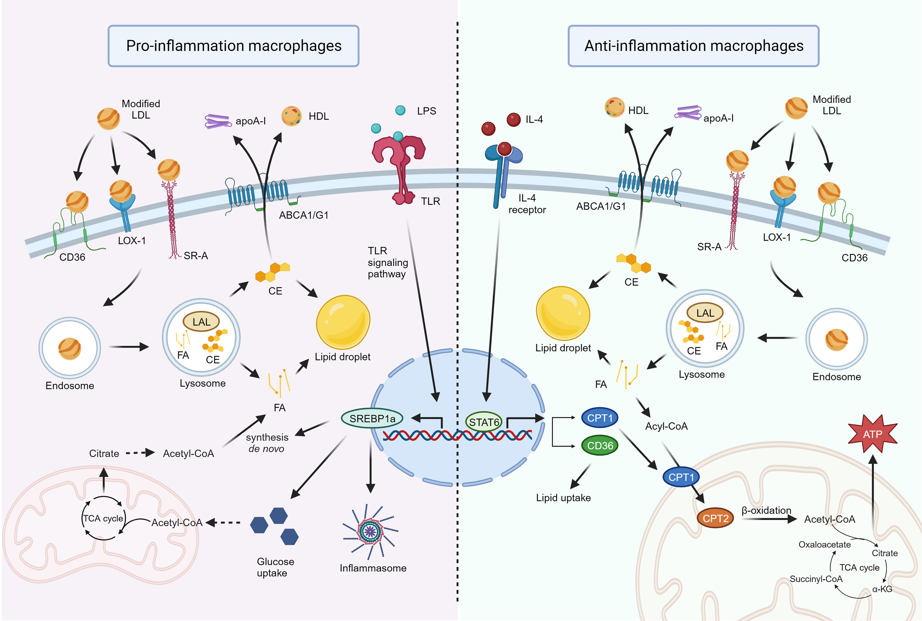
Figure 2. Inflammatory and anti-inflammatory types of macrophages in the regulation of lipid metabolism. The macrophages’ surface SR-A, LOX-1, and CD36 recognize and phagocytose modified LDL, which is converted to fatty acids and cholesterol in the presence of LAL. Surplus cholesterol is partly translocated to extracellular apoA-I and HDL via ABCA1/G1, and the remaining cholesterol and free fatty acids are stored in lipid droplets. In pro-inflammatory macrophages, lipopolysaccharide activation of the TLR signaling pathway promotes SREBP1a transcription, which facilitates glucose uptake and de novo fatty acid synthesis, as well as the release of inflammatory factors via inflammatory vesicles. In anti-inflammatory macrophages, the IL-4/STAT6 signaling pathway is activated, promoting the expression of FAO-related genes. Free fatty acids are activated to acyl-CoA and shuttled into the mitochondria by CPT1 and CPT2, where they are oxidized in the presence of β-oxidation to form acetyl-CoA and ultimately ATP via the tricarboxylic acid cycle. In addition, CD36 expression is elevated, increasing lipid uptake. Created in BioRender. Fan, F. (2026) https://BioRender.com/j64n485. SR-A: Scavenger receptors-A; LOX-1: lectin-like oxidized low-density lipoprotein receptor-1; LDL: low-density lipoprotein; LAL: lysosomal acid lipase; apoA-I: apolipoprotein A-I; HDL: high-density lipoprotein; ABCA1/G1: ATP-binding cassette transporter A1/G1; LPS: lipopolysaccharide; TLR: toll-like receptor; SREBP1a: sterol regulatory element binding transcription factor 1a; FAO: fatty acid oxidation; CPT: carnitine palmitoyl transferase; ATP: adenosine triphosphate; FA: fatty acids; CE: cholesterol; STAT6: signal transducer and activator of transcription 6; IL: interleukin; TCA: tricarboxylic acid cycle; CD36: cluster of differentiation 36; α-KG: alpha-ketoglutarate; acyl-CoA: acyl-coenzyme A; acetyl-CoA; acetyl-coenzyme A.
Triglycerides are predominantly stored within lipid droplets alongside other neutral lipids and surplus cholesterol esters[67]. Excess free cholesterol can be transported out of the cell and received by extracellular apolipoprotein A-I and high-density lipoprotein (HDL), facilitating its transfer via the ATP-binding cassette transporters ATP-binding cassette transporter A1 (ABCA1) and ATP-binding cassette transporter G1 (ABCG1)[68]. Ox-LDL has been identified as a primary contributor to foam cell formation[69]. During atherosclerosis progression, elevated ox-LDL levels prompt increased scavenger receptor and LOX-1 expression in macrophages, while the expression of ABCA1/G1 is suppressed. However, other studies suggest that low-concentration ox-LDL may promote cholesterol efflux by upregulating ABCA1, while high-concentration ox-LDL inhibits ABCA1/G1 expression to exacerbate foam cell formation. This imbalance results in a significant lipid influx and increased phagocytosis by macrophages[52]. Lipid accumulation in macrophages results in their transformation into foam cells characterized by an abundance of lipid droplets[70]. These foam cells are prone to apoptosis, which causes inflammation and the formation of a necrotic core in atherosclerotic plaques[71,72]. This process is indicative of the fundamental alterations that occur in atherosclerosis. Acetylated LDL (ac-LDL) is another modified lipoprotein that is taken up by macrophages through scavenger receptors rather than the classical LDL receptor, thereby promoting unregulated cholesterol ester accumulation and foam cell formation[73].
Macrophages, in addition to their roles in phagocytosis, catabolism, lipid storage, and exocytosis, have also been found to play a role in lipid synthesis[59] [Figure 2]. Lipopolysaccharide produced by gram-negative bacteria was found to activate Toll-like receptor 4 on macrophage surfaces, enhancing glucose uptake and de novo synthesis of fatty acids[72,74,75]. At the same time, one of the classic DAMPs proteins, HMGB1, can also activate this process[35]. This activation is mediated through the TLR signaling pathway, which in turn triggers the transcription of sterol regulatory element-binding protein (SREBP) family proteins[76]. SREBPs are anchored to the endoplasmic reticulum facilitate LDL receptor expression, and enhance glucose transport, especially under reduced cholesterol conditions[77]. Acetyl-CoA generated during the tricarboxylic acid cycle fuels fatty acid synthesis with an energy supply from nicotinamide adenine dinucleotide phosphate (reduced form) (NADPH)[78,79]. Research has shown that SREBP-1a deficiency leads to reduced macrophage lipid synthesis, and interactions between membrane phospholipid rafts with the actin cytoskeleton, thereby impairing phagocytosis[80]. Moreover, SREBP-1a is implicated not only in lipid synthesis but also in inflammasome activation, leading to IL-1β secretion and bolstering innate immunity[81]. This finding highlights the crucial role of lipid synthesis in supplying the necessary lipids and energy for membrane fusion during phagocytosis in macrophages, thereby enhancing their ability to combat pathogens. Conversely, during atherosclerosis progression, macrophages experience diminished lipid synthesis due to elevated cholesterol uptake and negative feedback regulation. This feedback involves the inhibition of SREBPs by the cholesterol metabolite 25-hydroxycholesterol[82]. Significantly, 25-hydroxycholesterol not only reduces lipid synthesis but also amplifies inflammation in lipid-rich macrophages, thereby promoting the progression of atherosclerosis[83]. In addition, recent studies have shown that 7-dehydrocholesterol (7-DHC), an intermediate in cholesterol synthesis, inhibits ferroptosis[84]. 7-DHC is converted to cholesterol in the presence of 7-DHC reductase, and macrophages are able to regulate 7-DHC reductase expression[85]. Since cholesterol accumulation is a crucial factor in macrophage foam cell formation and atherosclerosis, it is worth investigating whether macrophages regulate cholesterol synthesis and ferroptosis through 7-DHC in this context.
MACROPHAGES AND FERROPTOSIS DURING ATHEROSCLEROSIS DEVELOPMENT
The crucial role of macrophages in lipid and iron metabolism sets the stage for ferroptosis. Ferroptosis, identified as a novel form of non-caspase-dependent programmed cell death by Dixon et al. in 2012, has since been extensively investigated in various biological systems[5]. Accumulating evidence strongly suggests that macrophage ferroptosis contributes to the development of atherosclerosis.
Overview of ferroptosis
Ferroptosis is a process driven by iron-induced lipid peroxidation [Figure 3]. Macrophages generate significant amounts of Fe2+ following the phagocytosis of erythrocytes and transferrin-bound iron, leading to cellular iron overload and the production of massive amounts of ROS via the Fenton reaction. Normally, macrophages produce moderate levels of ROS to combat pathogens. However, excessive ROS can damage lipids, proteins, and DNA[86]. First, free polyunsaturated fatty acids (PUFAs) undergo esterification facilitated by acyl-CoA synthetase long-chain family member 4 (ACSL4) and lysophosphatidylcholine acyltransferase 3 (LPCAT3). This process results in the attachment of PUFA tails to phospholipids (PL-PUFAs) and their doping of the cell membrane[87]. This process is thought to drive ferroptosis. PL-PUFAs become susceptible to ROS attack, forming lipid peroxides (PL-PUFA-OOH) that accumulate in the cell membrane, ultimately leading to destruction of the cell membrane structure and subsequent cell death, a hallmark of ferroptosis[88]. In addition to the Fe2+-mediated Fenton reaction, which generates ROS that promote lipid peroxidation, iron-independent enzymatic reactions such as lipoxygenases and cytochrome p450 oxidoreductase have also been found to promote the accumulation of lipid peroxides in cell membranes[89,90].
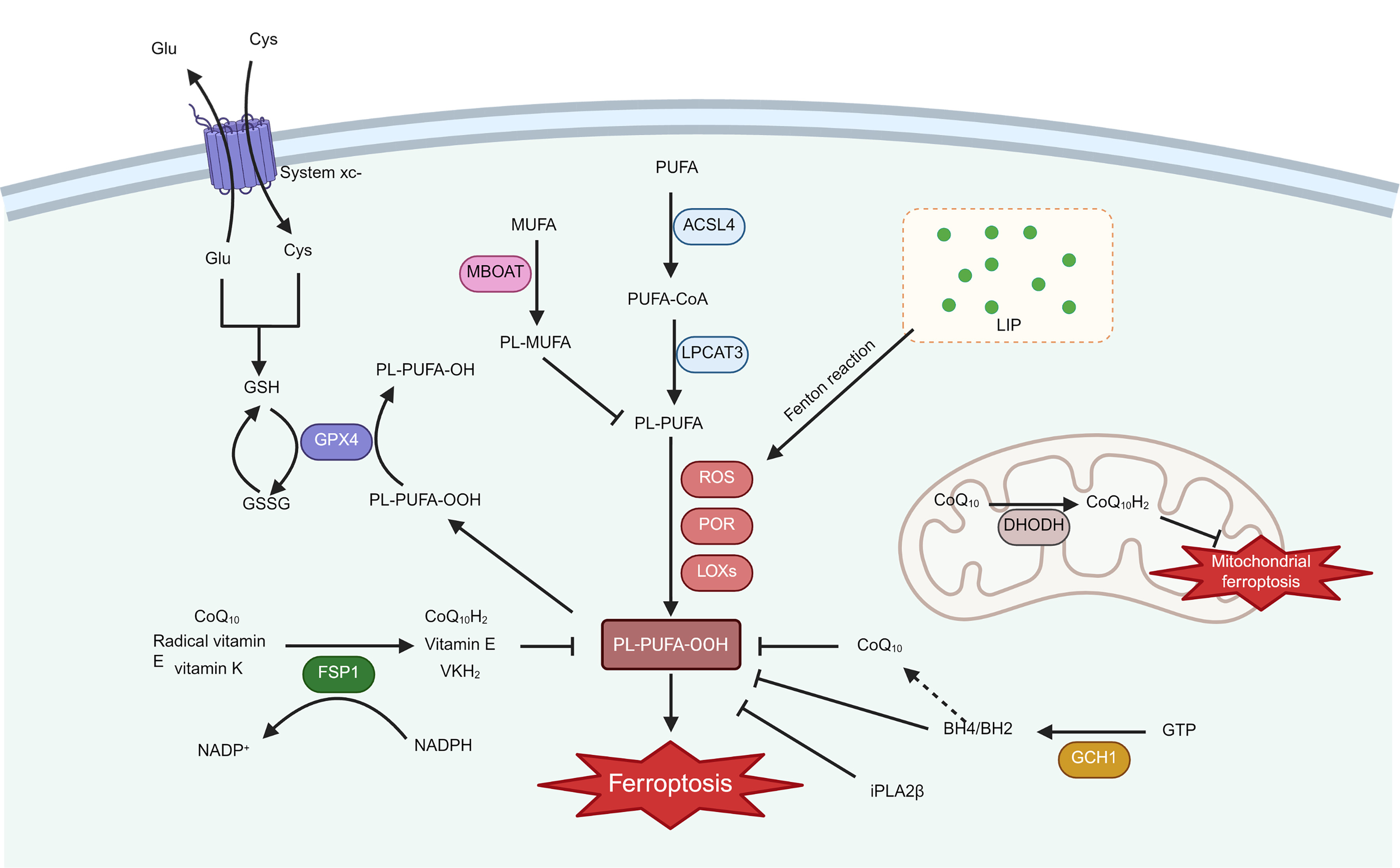
Figure 3. Mechanisms of ferroptosis inhibition in macrophages. PUFAs undergo esterification in the presence of ACSL4 and LPCAT3 to produce PL-PUFAs, which subsequently undergo peroxidation in the presence of ROS generated by the Fenton reaction or enzymatic reactions such as LOXs and POR to form PL-PUFA-OOH, which accumulates on the cell membrane and ultimately leads to ferroptosis. System xc- can exchange extracellular cystine and cellular glutamate for the synthesis of GSH, and GPX4 converts PL-PUFA-OOH to PL-PUFA-OH, while simultaneously oxidizing GSH to GSSG. Another ferroptosis mechanism is the FSP1/CoQ10/NAD(P)H axis, which reduces CoQ10 to CoQ10H2 using NADPH. Moreover, FSP1 exerts an anti-ferroptotic effect by regenerating vitamin E and promoting VKH2 production. Furthermore, GCH1 synthesizes GTP into BH4/BH2, which inhibits the oxidation of PL-PUFAs, and BH4 exerts an anti-lipid peroxidation effect by promoting the synthesis of CoQ10. In mitochondria, DHODH facilitates CoQ10H2 synthesis and inhibits ferroptosis in the inner mitochondrial membrane. MBOAT replaces PL-PUFAs with MUFAs incorporated into phospholipids to prevent PUFA oxidation. Additionally, iPLA2β inhibits ferroptosis by sheerly releasing oxidized PUFAs on phospholipids. Created in BioRender. Fan, F. (2026) https://BioRender.com/o50j697. PUFAs: Polyunsaturated fatty acids; ACSL4: acyl-CoA synthetase long-chain family member 4; LPCAT3: lysophosphatidylcholine acyltransferase 3; PL-PUFA: phospholipid containing polyunsaturated fatty acid chain; ROS: reactive oxygen species; LOXs: lipoxygenases; POR: p450 oxidoreductase; PL-PUFA-OOH: phospholipids with peroxidized polyunsaturated fatty acyl tail; GSH: glutathione; GPX4: glutathione peroxidase 4; GSSG: glutathione disulfide; Glu: glutamate; Cys: Cysteine; FSP1: ferroptosis suppressor protein 1; VKH2: vitamin K hydroquinone; GCH1: GTP cyclohydrolase 1; GTP: guanosine triphosphate; BH4: tetrahydrobiopterin; BH2: dihydrobiopterin; DHODH: dihydroorotate dehydrogenase; MBOAT: membrane-bound O-acyltransferase; MUFA: monounsaturated fatty acids; iPLA2β: calcium-independent phospholipase A2 beta; LIP: labile iron pool; PL-MUFA: phospholipid containing monounsaturated fatty acid chain; System xc-: cystine/glutamate antiporter; CoQ10: coenzyme Q10; CoQ10H2: ubiquinol (reduced coenzyme Q10); NADPH: nicotinamide adenine dinucleotide phosphate (reduced form); NADP+: nicotinamide adenine dinucleotide phosphate (oxidized form); PL-PUFA-OH: phospholipid with hydroxylated polyunsaturated fatty acyl tail; PUFA-CoA: polyunsaturated fatty acyl-coenzyme A.
Cells generally counteract ferroptosis through mechanisms, such as the cystine/glutamate antiporter (System xc-)/glutathione (GSH)/glutathione peroxidase 4 (GPX4) axis, a crucial component [Figure 3]. System xc- consists of two subunits, solute carrier family 7 member 11 (SLC7A11) and solute carrier family 3 member 2 (SLC3A2), exchanging extracellular cystine with intracellular glutamate at a 1:1 ratio[91,92]. GSH is synthesized from cystine with relevant enzymes. Subsequently, GPX4 oxidizes GSH to oxidize glutathione (GSSG) while reducing PL-PUFA-OOH to the corresponding alcohol (PL-PUFA-OH)[93]. However, in certain tumor cells, sensitivity to GPX4 inhibitors is not observed due to an anti-ferroptotic mechanism that runs in parallel with the System xc-/GSH/GPX4 axis, known as ferroptosis suppressor protein 1 (FSP1)/ubiquinone (CoQ10)/NAD(P)H axis [Figure 3]. FSP1, also known as apoptosis-inducing factor mitochondria-associated 2 (AIFM2), plays a central role in this axis. FSP1 reduces CoQ10 to ubiquinol (CoQ10H2) in the presence of NADPH, which is a lipophilic antioxidant that captures lipid peroxides and prevents their accumulation[94]. In addition, FSP1 exerts anti-ferroptosis effects through CoQ10 or direct promotion of a-tocopherol (vitamin E) regeneration[95]. Furthermore, studies have reported that vitamin K also has an anti-ferroptosis effect, while FSP1 can reduce vitamin K to vitamin K hydroquinone (VKH2) to attenuate lipid peroxidation[96].
Moreover, additional independent mechanisms of resistance to ferroptosis are gradually being uncovered [Figure 3]. For example, Kraft et al. reported that the GTP cyclohydrolase 1 (GCH1)/tetrahydrobiopterin (BH4) axis inhibits ferroptosis through lipid remodeling[97]. BH4 and dihydrobiopterin (BH2), synthesized by GCH1, protect the phospholipid PUFA tail from oxidation, thereby preventing cell membrane disruption. BH4 has also been shown to promote CoQ10 synthesis by facilitating the conversion of phenylalanine to tyrosine, thereby exerting an anti-lipid-peroxidation effect[97]. Dihydroorotate dehydrogenase (DHODH), which functions similarly to FSP1, promotes ubiquitin production in mitochondria and inhibits ferroptosis in the inner mitochondrial membrane[98]. Membrane-bound O-acyltransferase 1 and 2 (MBOAT1/2), which are regulated by sex hormones, perform ferroptosis surveillance functions independently of GPX4 and FSP1[99]. MBOAT2 transfers monounsaturated fatty acids (MUFAs) to phospholipid precursors and synthesizes MUFA-rich phospholipids to replace PL-PUFAs in preventing PUFA oxidation and effectively inhibiting cell damage from ferroptosis inducers[99]. Furthermore, calcium-independent phospholipase A2 beta (iPLA2β) was found to be induced by ROS and to cleave and release oxidized PUFAs from phospholipids, further inhibiting ferroptosis[100].
The role of macrophages in regulating iron metabolism
Macrophages are central to systemic iron recycling. They phagocytose senescent erythrocytes, process hemoglobin, import transferrin-bound iron, and either store excess iron in ferritin or export it via ferroportin. Macrophages recognize and phagocytose senescent erythrocytes and break down hemoglobin in the presence of HO-1 to generate Fe2+[101]. Another source of iron in macrophages is Fe3+ from the circulation. Assisted by transferrin (TF), the Fe3+-TF conjugate is recognized by transferrin receptor 1 (TFR1) on the macrophage surface and enters the cytoplasm via endocytosis[102,103]. The six-segment transmembrane epithelial antigen of prostate 3 (STEAP3), expressed on endosomes, plays a crucial role in reducing
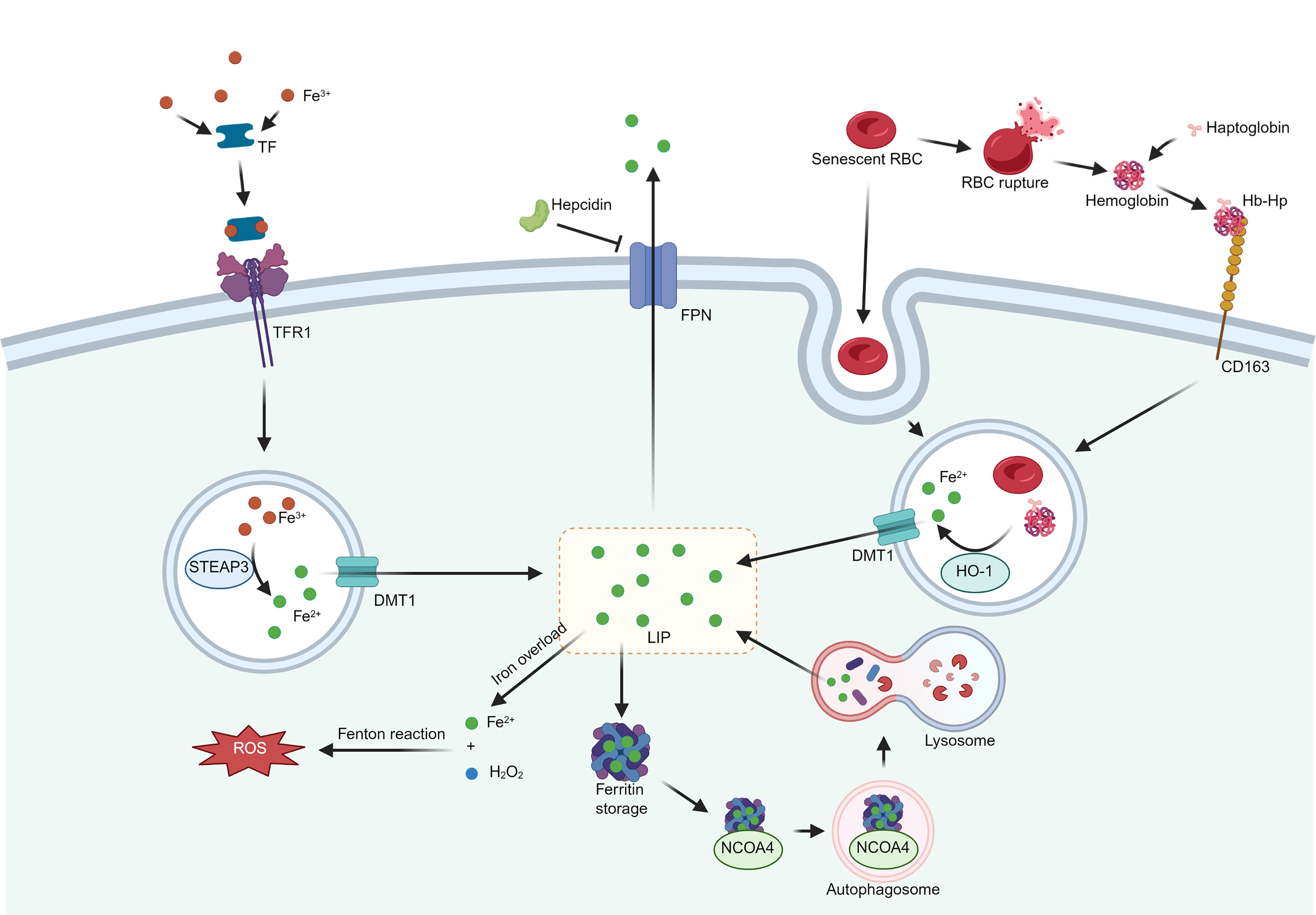
Figure 4. Origin and metabolic pathways of iron in macrophages. Two pathways contribute to the source of iron in macrophages. One of them is the recognition and phagocytosis of senescent erythrocytes or Hb-Hp formed after the rupture of erythrocytes via receptors such as CD163 on the surface of macrophages, which are catabolized by HO-1 to produce Fe2+, which is translocated to the LIP in the presence of DMT1. Another source is circulating Fe3+. Assisted by TF, the Fe3+-TF conjugate is recognized by TFR1 on the macrophage surface, reduces Fe3+ to Fe2+ via STEAP3, and finally translocates to the LIP via DMT1. Excess Fe2+ is excreted from macrophages via Ferroportin, which is controlled by hepcidin synthesized by the liver. Extra Fe2+ is bound to ferritin and stored. Under iron deficiency conditions, NCOA4 binds to ferritin and initiates the fusion of autophagosomes with lysosomes, leading to ferritin catabolism and subsequent release of Fe2+. Iron overload forms excess Fe2+, inducing a Fenton reaction that produces large amounts of ROS. Created in BioRender. Fan, F. (2026) https://BioRender.com/r45f260. Hb-Hp: Hemoglobin-haptoglobin; HO-1: heme oxygenase-1; LIP: labile iron pool; DMT1: divalent metal transporter 1; TF: transferrin; TFR1: transferrin receptor 1; STEAP3: six-segment transmembrane epithelial antigen of prostate 3; FPN: ferroportin; NCOA4: nuclear receptor coactivator 4; ROS: reactive oxygen species; CD163: cluster of differentiation 163; RBC: red blood cell.
Research indicates that iron overload can contribute to vascular endothelial disorders and exacerbate atherosclerosis[92,112]. Hereditary hemochromatosis, characterized by systemic iron overload due to abnormally increased iron absorption, is associated with an increased risk of CVD[113,114]. Similarly, mouse models with Ferroportin mutations exhibited elevated levels of non-transferrin-bound iron and displayed more severe atherosclerosis[114]. In addition, iron overload has been shown to promote M1 polarization in atherosclerotic macrophages and maintain inflammatory levels. This phenomenon might be attributed to iron-enhanced macrophage glycolysis[115]. In hemorrhagic areas of atherosclerotic plaques, hemoglobin-derived iron is a key factor in macrophage differentiation, inflammation, and lipid metabolism[116]. Aging and ruptured erythrocytes release substantial hemoglobin, which forms Hb-Hp with haptoglobin, as mentioned previously[117]. Through CD163, macrophages recognize and phagocytose Hb-Hp, further implicating iron in the intricate processes associated with atherosclerosis[118,119]. This action drives macrophage polarization into subtypes distinct from M1 and M2: hemoglobin-associated macrophages M(Hb) and Mhem, characterized by high expression of Ferroportin, resulting in lower intracellular iron and ROS levels. Moreover, increased expression of ABCA1/G1 promotes the efflux of intracellular cholesterol[9,23]. These distinct macrophage subtypes, characterized by diminished iron content and reduced lipid accumulation, replace foam cells and appear to attenuate atherosclerosis progression. However, further studies contradict this view. CD163+ macrophages induce hypoxia-inducible factor 1-alpha (HIF-1α) and vascular endothelial growth factor A (VEGF-A) expression, leading to increased plaque microvessel formation, enhanced vascular permeability, and recruitment of additional inflammatory cells[43]. Similarly, oxidation of hemoglobin to ferrylHb in atherosclerotic plaques promotes the polarization of the macrophage pro-inflammatory phenotype, thereby contributing to the development of atherosclerosis[120]. Furthermore, hemoglobin deposition due to intraplaque hemorrhage induced macrophage ferroptosis and expression of MMP2/9, which increased plaque instability. This effect is reversible by Ferrostatin-1 (Fer-1), a ferroptosis inhibitor[42]. These results suggest that M(Hb) and Mhem with low levels of iron may not have a reduced degree of ferroptosis.
In conclusion, the role of macrophages in maintaining systemic iron homeostasis is indisputable. Additionally, iron levels reciprocally influence macrophage function. During atherosclerosis progression, they play a pivotal role in hemoglobin- or transferrin-bound iron uptake, generating Fe2+ that can either be excreted extracellularly via Ferroportin or stored as ferritin-bound iron. However, excessive iron induces macrophage ferroptosis, leading to plaque destabilization in atherosclerosis.
Macrophage ferroptosis in atherosclerosis and its regulators
Because macrophages simultaneously control iron trafficking and lipid handling, they are especially vulnerable to ferroptosis; in turn, ferroptosis further promotes M1 polarization, lipid retention, and inflammatory amplification, creating a self-reinforcing loop in plaques[121]. Studies have shown that ferroptosis affects macrophage polarization. For example, ACSL4 was reported to promote macrophage ferroptosis and M1 polarization and regulate nasopharyngeal carcinoma tumorigenesis[122]. Microglia ferroptosis promotes M1 polarization in spinal cord injury[123]. Conversely, the inhibition of ferroptosis promotes macrophage M2 polarization[124,125]. In atherosclerosis, there is increasing evidence that macrophages undergo ferroptosis and promote plaque progression. Specifically, disrupted iron metabolism in macrophages leads to iron overload, triggering excessive ROS production and sustaining the M1 phenotype, thereby resulting in a persistent inflammatory state in atherosclerotic plaques[126]. In addition, the absorption of excess ox-LDL by macrophages contributes to foam cell formation and increases ferroptosis susceptibility[9]. The accumulation of foam cells forms a necrotic core, thus increasing plaque instability[127]. A previous study reported that macrophage iron retention enhances ox-LDL uptake and activates the Toll-like receptor 4 (TLR4)/nuclear factor kappa-B (NF-kB) pathway. Concurrently, it inhibits liver X receptor alpha (LXRα)- and ABCA1/G1-dependent cholesterol efflux, exacerbating cellular foaminess[128]. This evidence suggests a vicious cycle in atherosclerotic macrophages, in which iron and lipids continue to accumulate and interact, exacerbating metabolic imbalance.
Emerging evidence suggests that macrophage ferroptosis may also promote plaque calcification through extracellular vesicle release, extracellular matrix (ECM) remodeling, and iron-driven oxidative stress, although direct in vivo causal evidence remains limited. First, ferroptotic macrophages can generate death remnants, analogous to apoptotic bodies, that serve as nucleation sites for calcium-phosphate crystal formation, thereby promoting microcalcification and potentially accelerating plaque destabilization[129]. Second, evidence suggests that the death of macrophages, including apoptosis, pyroptosis, necrosis, and ferroptosis, generates extracellular vesicles (EVs), which serve as the starting point for microcalcification nodules[129]. EVs released when macrophages undergo ferroptosis not only contain factors such as Runt-related transcription factor 2 (Runx2), alkaline phosphatase (ALP), and osteocalcin, which promote bone differentiation, but also trigger the release of iron-containing EVs. Upon uptake by surrounding cells, these iron-laden EVs increase ROS/lipid peroxidation, stress signals that have been demonstrated to promote osteogenic transdifferentiation of vascular smooth muscle cells (VSMCs)[130,131]. The iron and oxidized lipids released from necrotic cores or potential ferroptotic remnants of macrophages within plaques can induce local oxidative stress and inflammation, disrupting the integrity of the surrounding ECM[132]. This exposure of phosphate-binding sites subsequently promotes the deposition of calcium salts. Current evidence supports the link between ferroptosis and plaque calcification through multiple pathways, including EV secretion, ECM remodeling, pro-inflammatory and pro-oxidative environments, and VSMC osteogenic transdifferentiation. However, no study yet has demonstrated a complete in vivo causal chain from macrophage ferroptosis to terminal plaque calcification, underscoring the need for systematic validation.
The connection between ferroptosis and other macrophage types in atherosclerosis is not well understood, and further research is needed to explore the sensitivity of Mox, M4, M(Hb), and Mhem cells to ferroptosis and whether ferroptosis influences their polarization. These subsets differ in their antioxidant/heme-handling programs, lipid uptake, and proteolytic profiles, and may therefore exhibit distinct vulnerabilities to iron-dependent lipid peroxidation. This has important implications for the release of DAMPs and inflammasome-mediated inflammation. Future work should therefore clarify which phenotypes are more prone to ferroptotic versus ferroptosis-resistant states, and whether ferroptosis acts upstream to skew polarization or downstream as a consequence of metabolism- and iron-handling-specific processes in subsets. Elucidating these relationships could reveal phenotype-specific therapeutic targets to limit DAMP-driven inflammation and stabilize vulnerable plaques.
Ferroptosis is a form of cell death characterized by an imbalance between oxidation and antioxidant defenses, intricately regulated by various mechanisms. Various ferroptosis regulators influence the atherosclerotic process by modulating macrophage ferroptosis. Among these, the transcription factor Nrf2 strongly contributes to the suppression of oxidative stress caused by ROS[133]. Its classical activation pathway is the Kelch-like ECH-associated protein 1 (Keap1)/Nrf2 pathway. Under low intracellular ROS conditions, Keap1 can bind, immobilize, and sequester Nrf2 in the cytoplasm. Moreover, Keap1 facilitates the ubiquitination of Nrf2 via the E3 ubiquitin ligase, thereby inhibiting its activation[134]. Conversely, when ROS levels are elevated, cysteine residues in specific structural domains in Keap1 are oxidized, altering Keap1 conformation and the release of Nrf2[134]. The nuclear translocation of Nrf2 promotes the transcription of antioxidant genes, including superoxide dismutase and catalase, promoting the metabolism of ROS. It also includes GPX4, GSH, and other inhibitors of ferroptosis, and HO-1, which promotes heme catabolism[135,136]. Studies on atherosclerosis have shown that Nrf2-deficient macrophages exhibit elevated inflammation, altered iron homeostasis, and increased transcription of cell-damage-related genes[137]. Targeting the Keap1/Nrf2 pathway upregulated macrophage GPX4 and system xc- levels, thereby inhibiting ferroptosis to attenuate atherosclerosis[138].
Another mechanism regulating ferroptosis in atherosclerotic macrophages is the hepcidin/Ferroportin pathway[139,140]. Hepcidin, which is synthesized by the liver, is the principal regulator of circulating iron levels and the maintenance of iron homeostasis[141]. When circulating iron levels are elevated, hepcidin is synthesized by the liver to regulate Ferroportin on the surface of enterocytes, macrophages, and other cells, decreasing cellular iron efflux to lower circulating iron levels[142,143]. The contribution of iron to atherosclerosis, a subject of debate for decades (referred to as the iron hypothesis), has garnered recent support[144]. Extensive evidence has identified hepcidin as a risk factor for atherosclerosis[145-147]. Moreover, intracellular iron homeostasis in macrophages is directly regulated by the hepcidin/Ferroportin pathway. Activation of the hepcidin/Ferroportin pathway results in an increase in intracellular iron levels within macrophages. This elevation in iron levels contributes to the maintenance of the M1 phenotype, promotes inflammation, induces ferroptosis, and facilitates atherosclerosis progression[140,148,149]. Furthermore, the inhibition of hepcidin increases macrophage cholesterol efflux, attenuating atherosclerosis[150]. Notably,
Tumor suppressor p53 (p53), a crucial transcription factor, regulates various cellular functions, including cell cycle, senescence, death, and so on[151]. In ferroptosis, the regulatory role of p53 is bidirectional, complex, and tissue-specific, influenced by the extent of cellular damage and ROS levels[152]. On one side, p53 directly inhibits SLC7A11 function through acetylation, promoting ferroptosis by inhibiting GSH synthesis and GPX4 activity[153]. Moreover, p53 can transcriptionally activate spermidine/spermine N1-acetyltransferase 1 (SAT1) and promote lipid peroxidation through its downstream arachidonate 15-lipoxygenase (ALOX5), thereby inducing ferroptosis[154]. On the other hand, p53 also regulates cyclin-dependent kinase inhibitor 1A (CDKN1A, also known as p21), which promotes the expression of GPX4 and exerts anti-ferroptosis effects[155]. Furthermore, p53 facilitates oxidized PUFA tail separation from phospholipid to protect cell membranes from ferroptosis attack by regulating iPLA2β expression[100].
In atherosclerosis, p53 has a bidirectional role, that may vary with disease stage. In advanced atherosclerotic plaques, elevated p53 expression inhibits vascular smooth muscle cell apoptosis while promoting macrophage apoptosis, and inhibiting atherosclerosis progression[156]. In contrast, increased ROS from iron overload induced M1 polarization in macrophage via p53 acetylation. Meanwhile, ox-LDL promotes macrophage p53-dependent apoptosis[157,158], likely exacerbating atherosclerosis. This complex, bidirectional nature of p53 in atherosclerosis and its impact on macrophage ferroptosis warrant further research for a comprehensive understanding.
Several regulators also play important roles in ferroptosis, including the Hippo/Yes-associated protein (YAP) signaling pathway. This pathway directly enhances the transcription of ACSL4 and TFR1, thereby facilitating cellular ferroptosis[159]. Additionally, hypoxia-inducible factors (HIFs), a group of proteins stably expressed under low-oxygen conditions, regulate the expression of metabolism-related genes. Research indicates that HIF-1α induced macrophage ferroptosis promotes atherosclerosis[160]. Similarly, HIF-2α induces lipid droplet-associated protein expression in hypoxic conditions, increasing polyunsaturated lipid accumulation[161]. This finding implies a potential role for HIF-2α in promoting macrophage foam cell formation and ferroptosis under hypoxic conditions, although further investigation is needed. In addition, the AMP-activated protein kinase (AMPK) pathway can prevent ox-LDL-induced ferroptosis in macrophages, mitigating the atherosclerotic process[162]. This protective mechanism may stem from the role of AMPK in the phosphorylation of acetyl coenzyme A carboxylase and the synthesis of PUFAs under energy stress or glucose deficiency[163].
THERAPEUTIC OPTIONS FOR ATTENUATING ATHEROSCLEROSIS BY INHIBITING FERROPTOSIS IN MACROPHAGES
Because macrophage ferroptosis sits at the intersection of iron overload, lipid peroxidation, and plaque inflammation, its pharmacological inhibition is a rational therapeutic strategy for atherosclerosis. However, such research is currently limited. In recent years, numerous studies have reported the use of drugs for treating various systemic diseases by inhibiting macrophage ferroptosis, including various compounds, natural plant extracts, and other agents. The potential value of such medications in treating atherosclerosis is also high. It is summarized as follows to provide new ideas for the treatment of atherosclerosis [Table 1].
The potential medicines inhibiting ferroptosis in macrophages
| Type | Medicine | Target | Diseases | Reference |
| Organic compounds | Metformin | AMPK/ERK | Atherosclerosis | [162] |
| Dexmedetomidine | Keap1/Nrf2/HO-1 | Ischemic-reperfusion injury | [165] | |
| PPARγ/STAT3 | Liver ischemia and reperfusion injury | [166] | ||
| Nrf2/GPX4 | Hepatic ischemia-reperfusion injury | [167] | ||
| Itaconate | Nrf2 via alkylation of Keap1 | Inflammatory diseases | [168] | |
| Nrf2 | Atherosclerosis | [169] | ||
| Nrf2 | Acute lung injury | [170] | ||
| Uridine | Nrf2 Activation and ACSL4 Inhibition | Induced acute lung injury | [171] | |
| Ferritin-1 and iron chelators | GPX4 | Mycobacterium tuberculosis | [172] | |
| Thymosin β4 | GPX4 | Non-alcoholic fatty liver | [173,174] | |
| Semaphorin 5A | PI3K-AKT-mTOR | Rheumatoid arthritis | [175] | |
| Natural botanical extracts and derivatives | Puerarin | ACSL4, PTGS, ALOX15, GPX4 | Macrophage inflammation | [177] |
| Micheliolide | Mtdh/BMP/MAPK | Renal fibrosis | [178] | |
| Keap1/Nrf2 | Atherosclerosis | [138] | ||
| Sulforaphane | Nrf2 | Atherosclerosis | [180] | |
| Naringenin | Nrf2/System xc-/GPX4 | Atherosclerosis | [181-185] | |
| Quercetin | GPX4, SLC7A11 | Neutrophilic airway inflammation | [187] | |
| Acute kidney injury | [188] | |||
| CeCH NPs | GPX4 | Sepsis-induced cardiac injury | [189] | |
| β-Caryophyllene | CB2R | Ulcerative colitis | [190] | |
| Baicalein | HMOX1, SLC40A1, FTH1 | Ovarian endometriosis | [191] | |
| Liensinine | Ferritin autophagy | Acute liver injury | [192,193] | |
| JFNE and JFNE-C | STAT3/p53/SLC7A11 | Inflammatory diseases | [194] | |
| Compound formulations | Maijitong granule | STAT6/DMT1 and STAT6/SOCS1/p53 | Atherosclerosis | [195] |
| Tongxinluo | GPX4, FSP1, ACSL4 | COPD | [196] | |
| Beclin-1-Induced Autophagy | Atherosclerosis | [197] | ||
| Qing-Xin-Jie-Yu Granule | System xc-/GPX4 | Atherosclerosis | [198] |
Organic compounds
Several small molecules and biologics have been reported to suppress macrophage ferroptosis, suggesting a broader therapeutic repertoire beyond conventional lipid-lowering drugs[164]. Recent studies have revealed that certain drug agents and proteins can hinder macrophage ferroptosis, thereby demonstrating their anti-atherosclerotic potential through the inhibition of this process.
One study has shown that Metformin inhibits foam cell formation, the inflammatory response, and ferroptosis in ox-LDL-treated macrophages via the AMPK/extracellular signal-regulated kinase (ERK) signaling pathway, thereby contributing to its anti-atherosclerotic effect[162]. Dexmedetomidine, an α-adrenergic receptor agonist, has shown promising pharmacological effects in CVDs, such as the enhancement of myocardial ischemia-reperfusion[165]. Further studies revealed that Dexmedetomidine promotes M2 macrophage polarization, reduces inflammation, and inhibits hepatic innate immune activation via the macrophage peroxisome proliferator-activated receptor gamma (PPARγ)/STAT3 pathway[166]. In addition, Dexmedetomidine has been reported to exhibit anti-ferroptotic and antioxidant effects through upregulation of the Nrf2/GPX4 pathway[167], suggesting its potential to inhibit macrophage ferroptosis and promote macrophage M2 polarization. Itaconate is a metabolite generated during inflammatory macrophage activation that activates Nrf2 via the alkylation of Keap1[168], and has been shown to inhibit atherosclerosis by inducing Nrf2-dependent inhibition of proinflammatory responses in macrophages[169]. Itaconate also alleviates lipopolysaccharide-induced acute lung injury by inhibiting macrophage ferroptosis in an Nrf2-dependent manner[170]. Uridine, recognized for its antioxidant properties, has been found to alleviate sepsis-induced acute lung injury by inhibiting macrophage ferroptosis[171]. Additionally, Fer-1 and iron chelators have been effective in preventing ferroptosis in macrophages infected with Mycobacterium tuberculosis[172]. Thymosin β4, an endogenous iron chelator involved in iron homeostasis, inhibits erastin and glutamate-induced ferroptosis in macrophage lines by upregulating GPX4 expression[173]. Moreover, the alleviation of non-alcoholic fatty liver disease (NAFLD) by inhibition of ferroptosis through up-regulation of GPX4 by Thymosin β4 has also been demonstrated[174]. Furthermore, the increase of semaphorin 5A in the synovial fluid of patients with rheumatoid arthritis, mainly originating from CD68+ synovial macrophages, has been shown to inhibit ferroptosis in synovial fibroblasts, ameliorating rheumatoid arthritis by the activation of the phosphoinositide 3-kinase (PI3K)/protein kinase B (AKT)/mammalian target of rapamycin (mTOR) signaling pathway and increasing GPX4 expression[175]. In summary, recent studies have shown that inhibiting macrophage ferroptosis has broad applications in anti-atherosclerotic therapy. The above evidence suggests that organic compounds such as dexmedetomidine, itaconate, uridine, and thymosin β4 can modulate macrophage ferroptosis through distinct signaling pathways and show great potential for anti-atherosclerotic therapy.
Natural botanical extracts and derivatives
Compounds derived from natural plants have increasingly garnered attention as prospective pharmaceuticals due to their accessibility, minimal side effects, and notable biological activities[176]. A study by Zeng et al. indicated that puerarin alleviates lipopolysaccharide-induced ferroptosis in RAW264.7 macrophages through pathways including tryptophan, arachidonic acid, and GSH metabolism, thereby exhibiting anti-inflammatory properties[177]. Similarly, Micheliolide, a compound derived from chamomile, has been identified for its antioxidant and anti-inflammatory effects[178]. Luo et al. reported that Micheliolide inhibits macrophage ferroptosis by targeting the Keap1/Nrf2 interaction, consequently reducing atherosclerosis severity[138]. Sulforaphane also inhibits ox-LDL-induced lipid accumulation in macrophages and facilitates ABCA1/G1-mediated cholesterol efflux, thereby alleviating the extent of atherosclerosis[179]. Furthermore, it was confirmed that Sulforaphane can stimulate Nrf2 in macrophages, forming the basis of its anti-atherosclerotic activity. Another study demonstrated that Sulforaphane is a potent Nrf2 activator that facilitates nuclear translocation of Nrf2, upregulates the levels of ferritin and SLC7A11, and inhibits lipid peroxidation and ferroptosis[180]. Naringenin, a natural flavonoid present in various plants, similarly prevents cholesterol-induced atherosclerosis in LDL receptor knockout (homozygous deficient mice, LDLR-/-) mice by reducing macrophage foam cell formation and inflammation[181]. In addition, Naringenin enhances macrophage M2 polarization, reduces macrophage recruitment, and inhibits inducible nitric oxide synthase (iNOS) expression[182,183]. In a subsequent study, researchers found that Naringenin not only inhibited ferroptosis but also alleviated myocardial ischemia/reperfusion injury through the Nrf2/System xc-/GPX4 pathway[184]. Furthermore, other studies also reported the anti-ferroptosis effect of Naringenin[185]. These findings collectively indicate that NAR has the potential to inhibit macrophage ferroptosis, thereby attenuating atherosclerosis.
Ferroptosis is responsible for the polarization of M1 macrophages[186]. Quercetin, a natural dietary flavonoid, exerts anti-inflammatory and anti-ferroptosis effects across various diseases. The study by Wang et al. developed a model of lipopolysaccharide-induced macrophage ferroptosis, revealing Quercetin’s effectiveness in counteracting ferroptosis during the inflammatory state in macrophages[187]. Furthermore, Quercetin mitigates neutrophil airway inflammation, inhibits M1 macrophage polarization, and acts as a potential macrophage ferroptosis inhibitor[187]. Additional research indicates that ferroptotic cells induce a pro-inflammatory state by triggering macrophage recruitment via C-C motif chemokine ligand 2 (CCL2), and that Quercetin inhibits ferroptosis-driven inflammation, thereby reducing inflammation-induced kidney damage[188]. Previous research has shown that combining Ceria Nanozyme Coordination with Curcumin to create Ceria Nanozyme Coordination with Curcumin Nanoparticles (CeCH NPs) effectively promotes M2 macrophage polarization and inhibits ferroptosis in a lipopolysaccharide-induced sepsis model, which significantly alleviates myocardial injury and thus restores cardiac function[189]. Wu et al. found that β-caryophyllene, a natural bicyclic sesquiterpene effectively reduces macrophage lipid peroxidation, decreases the levels of pro-ferritin factors, and increases the levels of anti-ferritin factors in RAS-selective lethal small molecule 3 (RSL3)-reduced macrophages, suggesting that β-caryophyllene had an anti-ferroptosis inhibitory effect on macrophage ferroptosis[190]. Additionally, baicalein enhances GPX4 expression in macrophages and inhibits ferroptosis, protecting macrophages from ferroptosis and rescuing phagocytosis in endometriosis[191]. Liensinine, a bioactive ingredient extracted from Lotus corniculatus[192], has emerged as a novel ferroptosis inhibitor that attenuates ferroptosis in acute liver injury by blocking autophagosome-lysosome fusion and inhibiting iron autophagy, thereby restoring macrophage phenotypic homeostasis and reducing acute liver injury[193]. Jing-Fang n-butanol extract (JFNE) and its active component (JFNE-C) inhibit lipopolysaccharide-induced ferroptosis in RAW264.7 macrophages via the STAT3/p53/SLC7A11 signaling pathway[194].
Compound formulations
In addition to natural phytomedicine monomers, some compound formulations have also been found to have anti-ferroptosis effects. Studies have shown that the traditional Chinese medicine Maijitong granule is effective in treating CVDs. Maijitong granule inhibits the progression of atherosclerosis by suppressing ferroptosis in peritoneal macrophages, thereby reducing lipid peroxidation and dysregulation of iron homeostasis. Mechanistically, STAT6 negatively regulates ferroptosis by transcriptionally inhibiting suppressor of cytokine signaling 1 (SOCS1)/p53 and DMT1, further highlighting the critical role of inhibiting macrophage ferroptosis in suppressing atherosclerosis[195]. Research has indicated that Tongxinluo inhibits ferroptosis by increasing the expression of the GPX4 and FSP1 mRNA and protein levels as well as decreasing the protein expression of ACSL4. This action helps delay the progression of atherosclerosis to prevent cardiovascular events[196]. In addition, Tongxinluo can increase plaque stability and exert anti-atherosclerotic effects by inhibiting ox-LDL-induced lipid deposition in macrophages[197]. This evidence suggests that Tongxinluo has multiple effects, inhibiting ferroptosis and attenuating macrophage lipid deposition. Thus, it has the potential to alleviate atherosclerosis by inhibiting ferroptosis and reducing macrophage foaming. Additionally, the regulation of macrophage ferroptosis is a critical mechanism through which Qing-Xin-Jie-Yu Granule stabilizes atherosclerosis-vulnerable plaques. Qing-Xin-Jie-Yu Granule has also been shown to alleviate atherosclerosis in aortic plaques and reduce macrophage ferroptosis by mediating the System xc-/GPX4 signaling pathway[198].
PERSPECTIVE AND CONCLUSION
Macrophage ferroptosis plays a key role in linking iron metabolism dysregulation, lipid peroxidation, and inflammatory amplification in atherosclerosis. Among the emerging mechanisms associated with disease progression, this finding provides deeper insights into atherosclerosis and reveals the interplay between iron metabolism, lipid peroxidation, and inflammatory amplification within the atherosclerotic microenvironment. This concept deepens our understanding of how macrophage dysfunction leads to plaque formation, growth, and instability.
However, the major gaps still remain: The subtype-specific susceptibility of plaque macrophages to ferroptosis. The causal relationship between ferroptosis and plaque stability. The translational feasibility of ferroptosis-targeted therapies.
Nevertheless, the therapeutic outlook is highly promising. In particular, agents targeting the GPX4 axis and the Nrf2 signaling pathway may offer substantial clinical potential by restoring redox balance, suppressing lipid peroxidation, and enhancing macrophage survival and function. Future work should define subtype-specific ferroptotic programs in macrophages, validate causal links in vivo, and evaluate the efficacy and safety of GPX4/Nrf2-based interventions.
DECLARATIONS
Acknowledgments
The Graphical Abstract was created in BioRender. Fan, F. (2026) https://BioRender.com/n02c554.
Authors’ contributions
Writing - original draft: Shi J, Fan F, Wang J
Writing - review and editing: Zheng G, Zhang R
Conceptualization, and project administration and supervision: Wang Y, Guo L, Zhang X, Ma C
Availability of data and materials
Not applicable.
AI and AI-assisted tools statement
During the preparation of this manuscript, the AI tool ChatGPT (version 3.0, released 2020-06-11) was used solely for language editing. The tools did not influence the study design, data collection, analysis, interpretation, or the scientific content of the work. All authors take full responsibility for the accuracy, integrity, and final content of the manuscript.
Financial support and sponsorship
This work was supported by the Scientific Research Program of Tianjin Education Commission (2024ZD006).
Conflicts of interest
All authors declared that there are no conflicts of interest.
Ethical approval and consent to participate
Not applicable.
Consent for publication
Not applicable.
Copyright
© The Author(s) 2026.
REFERENCES
1. Valk R, Hammill J, Grip J. Saturated fat: villain and bogeyman in the development of cardiovascular disease? Eur J of Prev Cardiol. 2022;29:2312-21.
2. Cafaro G, Perricone C, Gerli R, Bartoloni E. Cardiovascular risk in systemic autoimmune diseases. Lancet. 2023;401:21-2.
3. Moore KJ, Sheedy FJ, Fisher EA. Macrophages in atherosclerosis: a dynamic balance. Nat Rev Immunol. 2013;13:709-21.
4. Yan J, Horng T. Lipid metabolism in regulation of macrophage functions. Trends Cell Biol. 2020;30:979-89.
5. Dixon SJ, Lemberg KM, Lamprecht MR, et al. Ferroptosis: an iron-dependent form of nonapoptotic cell death. Cell. 2012;149:1060-72.
6. Ouyang S, You J, Zhi C, et al. Ferroptosis: the potential value target in atherosclerosis. Cell Death Dis. 2021;12:782.
7. Hou P, Fang J, Liu Z, et al. Macrophage polarization and metabolism in atherosclerosis. Cell Death Dis. 2023;14:691.
8. De Meyer GRY, Zurek M, Puylaert P, Martinet W. Programmed death of macrophages in atherosclerosis: mechanisms and therapeutic targets. Nat Rev Cardiol. 2024;21:312-25.
9. Li H, Cao Z, Wang L, et al. Macrophage subsets and death are responsible for atherosclerotic plaque formation. Front Immunol. 2022;13:843712.
10. Tokgözoğlu L, Libby P. The dawn of a new era of targeted lipid-lowering therapies. Eur Heart J. 2022;43:3198-208.
12. Liang XH, Sun H, Nichols JG, Crooke ST. RNase H1-dependent antisense oligonucleotides are robustly active in directing RNA cleavage in both the cytoplasm and the nucleus. Mol Ther. 2017;25:2075-92.
13. Landlinger C, Pouwer MG, Juno C, et al. The AT04A vaccine against proprotein convertase subtilisin/kexin type 9 reduces total cholesterol, vascular inflammation, and atherosclerosis in APOE*3Leiden.CETP mice. Eur Heart J. 2017;38:2499-507.
14. Lintner NG, McClure KF, Petersen D, et al. Selective stalling of human translation through small-molecule engagement of the ribosome nascent chain. PLoS Biol. 2017;15:e2001882.
15. Nordestgaard BG, Nicholls SJ, Langsted A, Ray KK, Tybjærg-Hansen A. Advances in lipid-lowering therapy through gene-silencing technologies. Nat Rev Cardiol. 2018;15:261-72.
16. Nishikido T, Ray KK. Non-antibody approaches to proprotein convertase subtilisin kexin 9 inhibition: siRNA, antisense oligonucleotides, adnectins, vaccination, and new attempts at small-molecule inhibitors based on new discoveries. Front Cardiovasc Med. 2019;5:199.
17. Locati M, Curtale G, Mantovani A. Diversity, mechanisms, and significance of macrophage plasticity. Annu Rev Pathol Mech Dis. 2020;15:123-47.
18. Tabas I, Bornfeldt KE. Macrophage phenotype and function in different stages of atherosclerosis. Circ Res. 2016;118:653-67.
19. Mantovani A, Garlanda C, Locati M. Macrophage diversity and polarization in atherosclerosis: a question of balance. Arterioscler Thromb Vasc Biol. 2009;29:1419-23.
20. Lafuse WP, Wozniak DJ, Rajaram MVS. Role of cardiac macrophages on cardiac inflammation, fibrosis and tissue repair. Cells. 2020;10:51.
21. Shapouri-Moghaddam A, Mohammadian S, Vazini H, et al. Macrophage plasticity, polarization, and function in health and disease. J Cell Physiol. 2018;233:6425-40.
22. Chistiakov DA, Bobryshev YV, Orekhov AN. Changes in transcriptome of macrophages in atherosclerosis. J Cell Mol Med. 2015;19:1163-73.
23. Liberale L, Dallegri F, Montecucco F, Carbone F. Pathophysiological relevance of macrophage subsets in atherogenesis. Thromb Haemost. 2017;117:7-18.
24. Kadl A, Meher AK, Sharma PR, et al. Identification of a novel macrophage phenotype that develops in response to atherogenic phospholipids via Nrf2. Circ Res. 2010;107:737-46.
25. Gleissner CA, von Hundelshausen P, Ley K. Platelet chemokines in vascular disease. Arterioscler Thromb Vasc Biol. 2008;28:1920-7.
26. Domschke G, Gleissner CA. CXCL4-induced macrophages in human atherosclerosis. Cytokine. 2019;122:154141.
27. Gleissner CA, Shaked I, Erbel C, Böckler D, Katus HA, Ley K. CXCL4 downregulates the atheroprotective hemoglobin receptor CD163 in human macrophages. Circ Res. 2010;106:203-11.
28. Finn AV, Nakano M, Polavarapu R, et al. Hemoglobin directs macrophage differentiation and prevents foam cell formation in human atherosclerotic plaques. J Am Coll Cardiol. 2012;59:166-77.
29. Leitinger N, Schulman IG. Phenotypic polarization of macrophages in atherosclerosis. Arterioscler Thromb Vasc Biol. 2013;33:1120-6.
30. Stöger JL, Goossens P, de Winther MP. Macrophage heterogeneity: relevance and functional implications in atherosclerosis. Curr Vasc Pharmacol. 2010;8:233-48.
31. Farahi L, Sinha SK, Lusis AJ. Roles of macrophages in atherogenesis. Front Pharmacol. 2021;12:785220.
32. Williams KJ, Tabas I. The response-to-retention hypothesis of early atherogenesis. Arterioscler Thromb Vasc Biol. 1995;15:551-61.
33. Rahman K, Fisher EA. Insights from pre-clinical and clinical studies on the role of innate inflammation in atherosclerosis regression. Front Cardiovasc Med. 2018;5:32.
34. Barrett TJ. Macrophages in atherosclerosis regression. Arterioscler Thromb Vasc Biol. 2020;40:20-33.
35. Feng Z, Meng F, Huo F, et al. Inhibition of ferroptosis rescues M2 macrophages and alleviates arthritis by suppressing the HMGB1/TLR4/STAT3 axis in M1 macrophages. Redox Biol. 2024;75:103255.
36. Kosmidou C, Efstathiou NE, Hoang MV, et al. Issues with the specificity of immunological reagents for NLRP3: implications for age-related macular degeneration. Sci Rep. 2018;8:461.
37. Colin S, Chinetti-Gbaguidi G, Staels B. Macrophage phenotypes in atherosclerosis. Immunol Rev. 2014;262:153-66.
39. Lancaster GI, Morgan PK, Murphy AJ. Iron macrophages: dance of death and MMP release in intraplaque hemorrhage. Thromb Haemost. 2024;124:581-3.
40. Moreno PR, Purushothaman M, Purushothaman KR. Plaque neovascularization: defense mechanisms, betrayal, or a war in progress. Ann N Y Acad Sci. 2012;1254:7-17.
41. Puylaert P, Roth L, Van Praet M, et al. Effect of erythrophagocytosis-induced ferroptosis during angiogenesis in atherosclerotic plaques. Angiogenesis. 2023;26:505-22.
42. Li B, Lu M, Wang H, et al. Macrophage ferroptosis promotes MMP2/9 overexpression induced by hemin in hemorrhagic plaque. Thromb Haemost. 2023;124:568-80.
43. Guo L, Akahori H, Harari E, et al. CD163+ macrophages promote angiogenesis and vascular permeability accompanied by inflammation in atherosclerosis. J Clin Invest. 2018;128:1106-24.
44. Stöger JL, Gijbels MJ, van der Velden S, et al. Distribution of macrophage polarization markers in human atherosclerosis. Atherosclerosis. 2012;225:461-8.
45. Peled M, Fisher EA. Dynamic aspects of macrophage polarization during atherosclerosis progression and regression. Front Immunol. 2014;5:579.
46. Khallou-Laschet J, Varthaman A, Fornasa G, et al. Macrophage plasticity in experimental atherosclerosis. PLoS One. 2010;5:e8852.
47. Jaipersad AS, Lip GY, Silverman S, Shantsila E. The role of monocytes in angiogenesis and atherosclerosis. J Am Coll Cardiol. 2014;63:1-11.
48. Epelman S, Lavine KJ, Randolph GJ. Origin and functions of tissue macrophages. Immunity. 2014;41:21-35.
49. Narasimhan PB, Marcovecchio P, Hamers AAJ, Hedrick CC. Nonclassical monocytes in health and disease. Annu Rev Immunol. 2019;37:439-56.
50. Pham TT, Le AH, Dang CP, et al. Endocytosis of red blood cell extracellular vesicles by macrophages leads to cytoplasmic heme release and prevents foam cell formation in atherosclerosis. J Extracell Vesicles. 2023;12:12354.
51. Chinetti-Gbaguidi G, Colin S, Staels B. Macrophage subsets in atherosclerosis. Nat Rev Cardiol. 2015;12:10-7.
52. Chistiakov DA, Melnichenko AA, Myasoedova VA, Grechko AV, Orekhov AN. Mechanisms of foam cell formation in atherosclerosis. J Mol Med. 2017;95:1153-65.
53. Li F, Zhang H. Lysosomal acid lipase in lipid metabolism and beyond. Arterioscler Thromb Vasc Biol. 2019;39:850-6.
54. Luo J, Yang H, Song BL. Mechanisms and regulation of cholesterol homeostasis. Nat Rev Mol Cell Biol. 2019;21:225-45.
55. Maxfield FR, Tabas I. Role of cholesterol and lipid organization in disease. Nature. 2005;438:612-21.
56. O’Neill LAJ, Kishton RJ, Rathmell J. A guide to immunometabolism for immunologists. Nat Rev Immunol. 2016;16:553-65.
57. Mehta MM, Weinberg SE, Chandel NS. Mitochondrial control of immunity: beyond ATP. Nat Rev Immunol. 2017;17:608-20.
58. Adeva-Andany MM, Carneiro-Freire N, Seco-Filgueira M, Fernández-Fernández C, Mouriño-Bayolo D. Mitochondrial β-oxidation of saturated fatty acids in humans. Mitochondrion. 2019;46:73-90.
59. Curi R, de Siqueira Mendes R, de Campos Crispin LA, Norata GD, Sampaio SC, Newsholme P. A past and present overview of macrophage metabolism and functional outcomes. Clin Sci. 2017;131:1329-42.
60. Yin B, Loike JD, Kako Y, et al. Lipoprotein lipase regulates Fc receptor-mediated phagocytosis by macrophages maintained in glucose-deficient medium. J Clin Invest. 1997;100:649-57.
61. Huang SC, Everts B, Ivanova Y, et al. Cell-intrinsic lysosomal lipolysis is essential for alternative activation of macrophages. Nat Immunol. 2014;15:846-55.
62. Vats D, Mukundan L, Odegaard JI, et al. Oxidative metabolism and PGC-1β attenuate macrophage-mediated inflammation. Cell Metab. 2006;4:13-24.
63. Nomura M, Liu J, Yu ZX, et al. Macrophage fatty acid oxidation inhibits atherosclerosis progression. J Mol Cell Cardiol. 2019;127:270-6.
64. Nomura M, Liu J, Rovira II, et al. Fatty acid oxidation in macrophage polarization. Nat Immunol. 2016;17:216-7.
65. Hohensinner PJ, Lenz M, Haider P, et al. Pharmacological inhibition of fatty acid oxidation reduces atherosclerosis progression by suppression of macrophage NLRP3 inflammasome activation. Biochem Pharmacol. 2021;190:114634.
66. Batista-Gonzalez A, Vidal R, Criollo A, Carreño LJ. New insights on the role of lipid metabolism in the metabolic reprogramming of macrophages. Front Immunol. 2020;10:2993.
67. Walther TC, Chung J, Farese RV. Lipid droplet biogenesis. Annu Rev Cell Dev Biol. 2017;33:491-510.
68. Rosenson RS, Brewer HB Jr, Ansell BJ, et al. Dysfunctional HDL and atherosclerotic cardiovascular disease. Nat Rev Cardiol. 2016;13:48-60.
69. Siegel-Axel D, Daub K, Seizer P, Lindemann S, Gawaz M. Platelet lipoprotein interplay: trigger of foam cell formation and driver of atherosclerosis. Cardiovasc Res. 2008;78:8-17.
70. Yuan Y, Li P, Ye J. Lipid homeostasis and the formation of macrophage-derived foam cells in atherosclerosis. Protein Cell. 2012;3:173-81.
71. Mclaren JE, Michael DR, Ashlin TG, Ramji DP. Cytokines, macrophage lipid metabolism and foam cells: Implications for cardiovascular disease therapy. Prog Lipid Res. 2011;50:331-47.
72. Galindo CL, Khan S, Zhang X, Yeh YS, Liu Z, Razani B. Lipid-laden foam cells in the pathology of atherosclerosis: shedding light on new therapeutic targets. Expert Opin Ther Targets. 2023;27:1231-45.
73. Lu W, Lin Y, Haider N, Moly P, Wang L, Zhou W. Ginsenoside Rb1 protects human vascular smooth muscle cells against resistin-induced oxidative stress and dysfunction. Front Cardiovasc Med. 2023;10:1164547.
74. Everts B, Amiel E, Huang SC, et al. TLR-driven early glycolytic reprogramming via the kinases TBK1-IKKɛ supports the anabolic demands of dendritic cell activation. Nat Immunol. 2014;15:323-32.
75. Cader MZ, Boroviak K, Zhang Q, et al. C13orf31 (FAMIN) is a central regulator of immunometabolic function. Nat Immunol. 2016;17:1046-56.
76. Jeon TI, Osborne TF. SREBPs: metabolic integrators in physiology and metabolism. Trends in Endocrinol Metab. 2012;23:65-72.
77. Shimano H, Sato R. SREBP-regulated lipid metabolism: convergent physiology - divergent pathophysiology. Nat Rev Endocrinol. 2017;13:710-30.
79. Metallo CM, Gameiro PA, Bell EL, et al. Reductive glutamine metabolism by IDH1 mediates lipogenesis under hypoxia. Nature. 2011;481:380-4.
80. Lee JH, Phelan P, Shin M, et al. SREBP-1a-stimulated lipid synthesis is required for macrophage phagocytosis downstream of TLR4-directed mTORC1. Proc Natl Acad Sci U S A. 2018;115:E12228-34.
81. Im SS, Yousef L, Blaschitz C, et al. Linking lipid metabolism to the innate immune response in macrophages through sterol regulatory element binding protein-1a. Cell Metab. 2011;13:540-9.
82. Adams CM, Reitz J, De Brabander JK, et al. Cholesterol and 25-hydroxycholesterol inhibit activation of SREBPs by different mechanisms, both involving SCAP and Insigs. J Biol Chem. 2004;279:52772-80.
83. Canfrán-Duque A, Rotllan N, Zhang X, et al. Macrophage-Derived 25-Hydroxycholesterol Promotes Vascular Inflammation, Atherogenesis, and Lesion Remodeling. Circulation. 2023;147:388-408.
84. Li Y, Ran Q, Duan Q, et al. 7-Dehydrocholesterol dictates ferroptosis sensitivity. Nature. 2024;626:411-8.
85. Xiao J, Li W, Zheng X, et al. Targeting 7-dehydrocholesterol reductase integrates cholesterol metabolism and IRF3 activation to eliminate infection. Immunity. 2020;52:109-122.e6.
86. Canton M, Sánchez-Rodríguez R, Spera I, et al. Reactive oxygen species in macrophages: sources and targets. Front Immunol. 2021;12:734229.
87. Liu J, Kang R, Tang D. Signaling pathways and defense mechanisms of ferroptosis. FEBS J. 2021;289:7038-50.
88. Fei W, Chen D, Tang H, et al. Targeted GSH-exhausting and hydroxyl radical self-producing manganese-silica nanomissiles for MRI guided ferroptotic cancer therapy. Nanoscale. 2020;12:16738-54.
89. Chrisnasari R, Hennebelle M, Vincken JP, van Berkel WJH, Ewing TA. Bacterial lipoxygenases: Biochemical characteristics, molecular structure and potential applications. Biotechnol Adv. 2022;61:108046.
90. Pandey AV, Flück CE. NADPH P450 oxidoreductase: structure, function, and pathology of diseases. Pharmacol Ther. 2013;138:229-54.
91. Rochette L, Dogon G, Rigal E, Zeller M, Cottin Y, Vergely C. Lipid peroxidation and iron metabolism: two corner stones in the homeostasis control of ferroptosis. Int J Mol Sci. 2022;24:449.
92. Xu X, Xu XD, Ma MQ, et al. The mechanisms of ferroptosis and its role in atherosclerosis. Biomed Pharmacother. 2024;171:116112.
93. Liu Y, Wan Y, Jiang Y, Zhang L, Cheng W. GPX4: The hub of lipid oxidation, ferroptosis, disease and treatment. Biochim Biophys Acta Rev Cancer. 2023;1878:188890.
94. Bersuker K, Hendricks JM, Li Z, et al. The CoQ oxidoreductase FSP1 acts parallel to GPX4 to inhibit ferroptosis. Nature. 2019;575:688-92.
95. Doll S, Freitas FP, Shah R, et al. FSP1 is a glutathione-independent ferroptosis suppressor. Nature. 2019;575:693-8.
96. Mishima E, Ito J, Wu Z, et al. A non-canonical vitamin K cycle is a potent ferroptosis suppressor. Nature. 2022;608:778-83.
97. Kraft VAN, Bezjian CT, Pfeiffer S, et al. GTP cyclohydrolase 1/tetrahydrobiopterin counteract ferroptosis through lipid remodeling. ACS Cent Sci. 2019;6:41-53.
98. Mao C, Liu X, Zhang Y, et al. Author Correction: DHODH-mediated ferroptosis defence is a targetable vulnerability in cancer. Nature. 2021;596:E13.
99. Liang D, Feng Y, Zandkarimi F, et al. Ferroptosis surveillance independent of GPX4 and differentially regulated by sex hormones. Cell. 2023;186:2748-64.e22.
100. Chen D, Chu B, Yang X, et al. iPLA2β-mediated lipid detoxification controls p53-driven ferroptosis independent of GPX4. Nat Commun. 2021;12:3644.
101. Ma C, Wu X, Zhang X, Liu X, Deng G. Heme oxygenase-1 modulates ferroptosis by fine-tuning levels of intracellular iron and reactive oxygen species of macrophages in response to Bacillus Calmette-Guerin infection. Front Cell Infect Microbiol. 2022;12:1004148.
102. Nairz M, Theurl I, Swirski FK, Weiss G. “Pumping iron” - how macrophages handle iron at the systemic, microenvironmental, and cellular levels. Pflugers Arch - Eur J Physiol. 2017;469:397-418.
103. Das BK, Wang L, Fujiwara T, et al. Transferrin receptor 1-mediated iron uptake regulates bone mass in mice via osteoclast mitochondria and cytoskeleton. ELife. 2022;11:e73539.
104. Koleini N, Shapiro JS, Geier J, Ardehali H. Ironing out mechanisms of iron homeostasis and disorders of iron deficiency. J Clin Invest. 2021;131:e148671.
105. Jiang L, Wang J, Wang K, et al. RNF217 regulates iron homeostasis through its E3 ubiquitin ligase activity by modulating ferroportin degradation. Blood. 2021;138:689-705.
106. Saad HKM, Abd Rahman AA, Ab Ghani AS, et al. Activation of STAT and SMAD Signaling Induces Hepcidin Re-Expression as a Therapeutic Target for β-thalassemia patients. Biomedicines. 2022;10:189.
107. Mleczko-Sanecka K, Silvestri L. Cell-type-specific insights into iron regulatory processes. Am J Hematol. 2020;96:110-27.
108. Orino K, Watanabe K. Molecular, physiological and clinical aspects of the iron storage protein ferritin. Vet J. 2008;178:191-201.
109. Mancias JD, Wang X, Gygi SP, Harper JW, Kimmelman AC. Quantitative proteomics identifies NCOA4 as the cargo receptor mediating ferritinophagy. Nature. 2014;509:105-9.
110. Ohshima T, Yamamoto H, Sakamaki Y, Saito C, Mizushima N. NCOA4 drives ferritin phase separation to facilitate macroferritinophagy and microferritinophagy. J Cell Biol. 2022;221:e202203102.
111. Xie LH, Fefelova N, Pamarthi SH, Gwathmey JK. Molecular mechanisms of ferroptosis and relevance to cardiovascular disease. Cells. 2022;11:2726.
112. Marques VB, Leal MAS, Mageski JGA, et al. Chronic iron overload intensifies atherosclerosis in apolipoprotein E deficient mice: Role of oxidative stress and endothelial dysfunction. Life Sci. 2019;233:116702.
113. Pietrangelo A. Hereditary hemochromatosis: pathogenesis, diagnosis, and treatment. Gastroenterology. 2010;139:393-408.e1.
114. Vinchi F, Porto G, Simmelbauer A, et al. Atherosclerosis is aggravated by iron overload and ameliorated by dietary and pharmacological iron restriction. Eur Heart J. 2020;41:2681-95.
115. Hu X, Cai X, Ma R, Fu W, Zhang C, Du X. Iron-load exacerbates the severity of atherosclerosis via inducing inflammation and enhancing the glycolysis in macrophages. J Cell Physiol. 2019;234:18792-800.
116. Boyle JJ, Harrington HA, Piper E, et al. Coronary intraplaque hemorrhage evokes a novel atheroprotective macrophage phenotype. Am J Pathol. 2009;174:1097-108.
117. Andersen CBF, Stødkilde K, Sæderup KL, et al. Haptoglobin. Antioxid Redox Signal. 2017;26:814-31.
118. Habib A, Finn AV. The role of iron metabolism as a mediator of macrophage inflammation and lipid handling in atherosclerosis. Front Pharmacol. 2014;5:195.
119. Buehler PW, Abraham B, Vallelian F, et al. Haptoglobin preserves the CD163 hemoglobin scavenger pathway by shielding hemoglobin from peroxidative modification. Blood. 2009;113:2578-86.
120. Potor L, Hendrik Z, Patsalos A, et al. Oxidation of hemoglobin drives a proatherogenic polarization of macrophages in human atherosclerosis. Antioxid Redox Signal. 2021;35:917-50.
121. Ross EA, Devitt A, Johnson JR. Macrophages: the good, the bad, and the gluttony. Front Immunol. 2021;12:708186.
122. Chen P, Wang D, Xiao T, et al. ACSL4 promotes ferroptosis and M1 macrophage polarization to regulate the tumorigenesis of nasopharyngeal carcinoma. Int Immunopharmacol. 2023;122:110629.
123. Xuan Y, Peng K, Zhu R, Kang Y, Yin Z. Hmox1 is identified as a ferroptosis hub gene and associated with the M1 type microglia/macrophage polarization in spinal cord injury: bioinformatics and experimental validation. Mol Neurobiol. 2023;60:7151-65.
124. Huang J, Pan H, Sun J, et al. TMEM147 aggravates the progression of HCC by modulating cholesterol homeostasis, suppressing ferroptosis, and promoting the M2 polarization of tumor-associated macrophages. J Exp Clin Cancer Res. 2023;42:286.
125. Dai E, Han L, Liu J, et al. Autophagy-dependent ferroptosis drives tumor-associated macrophage polarization via release and uptake of oncogenic KRAS protein. Autophagy. 2020;16:2069-83.
126. Ma J, Zhang H, Chen Y, Liu X, Tian J, Shen W. The role of macrophage iron overload and ferroptosis in atherosclerosis. Biomolecules. 2022;12:1702.
127. Bäck M, Yurdagul A, Tabas I, Öörni K, Kovanen PT. Inflammation and its resolution in atherosclerosis: mediators and therapeutic opportunities. Nat Rev Cardiol. 2019:16:389-406.
128. Xiao L, Luo G, Guo X, et al. Macrophage iron retention aggravates atherosclerosis: Evidence for the role of autocrine formation of hepcidin in plaque macrophages. Biochim Biophys Acta Mol Cell Biol Lipids. 2020;1865:158531.
129. Neels JG, Gollentz C, Chinetti G. Macrophage death in atherosclerosis: potential role in calcification. Front Immunol. 2023;14:1215612.
130. Brown CW, Amante JJ, Chhoy P, et al. Prominin2 drives ferroptosis resistance by stimulating iron export. Dev Cell. 2019;51:575-86.e4.
131. Yaker L, Tebani A, Lesueur C, et al. Extracellular vesicles from LPS-treated macrophages aggravate smooth muscle cell calcification by propagating inflammation and oxidative stress. Front Cell Dev Biol. 2022;10:823450.
132. Kawakami R, Katsuki S, Travers R, et al. S100A9-RAGE axis accelerates formation of macrophage-mediated extracellular vesicle microcalcification in diabetes mellitus. Arterioscler Thromb Vasc Biol. 2020;40:1838-53.
133. Kerins MJ, Ooi A. The roles of NRF2 in modulating cellular iron homeostasis. Antioxid Redox Signal. 2018;29:1756-73.
134. Kobayashi A, Kang M, Okawa H, et al. Oxidative stress sensor Keap1 functions as an adaptor for Cul3-based E3 ligase to regulate proteasomal degradation of Nrf2. Mol Cell Biol. 2023;24:7130-9.
135. Xiang Q, Zhao Y, Lin J, Jiang S, Li W. The Nrf2 antioxidant defense system in intervertebral disc degeneration: Molecular insights. Exp Mol Med. 2022;54:1067-75.
136. Ren S, Wang Y, Zhang Y, et al. Paeoniflorin alleviates AngII-induced cardiac hypertrophy in H9c2 cells by regulating oxidative stress and Nrf2 signaling pathway. Biomed Pharmacother. 2023;165:115253.
137. Gutiérrez-Cuevas J, Galicia-Moreno M, Monroy-Ramírez HC, et al. The role of NRF2 in obesity-associated cardiovascular risk factors. Antioxidants. 2022;11:235.
138. Luo X, Wang Y, Zhu X, et al. MCL attenuates atherosclerosis by suppressing macrophage ferroptosis via targeting KEAP1/NRF2 interaction. Redox Biol. 2024;69:102987.
139. Wan Q, Yang M, Liu Z, Wu J. Ambient fine particulate matter aggravates atherosclerosis in apolipoprotein E knockout mice by iron overload via the hepcidin-ferroportin axis. Life Sci. 2021;264:118715.
140. Bao X, Luo X, Bai X, et al. Cigarette tar mediates macrophage ferroptosis in atherosclerosis through the hepcidin/FPN/SLC7A11 signaling pathway. Free Radic Biol Med. 2023;201:76-88.
142. Billesbølle CB, Azumaya CM, Kretsch RC, et al. Structure of hepcidin-bound ferroportin reveals iron homeostatic mechanisms. Nature. 2020;586:807-11.
143. Camaschella C, Nai A, Silvestri L. Iron metabolism and iron disorders revisited in the hepcidin era. Haematologica. 2020;105:260-72.
144. Guo Q, Qian C, Qian ZM. Iron metabolism and atherosclerosis. Trends in Endocrinol Metab. 2023;34:404-13.
145. Malhotra R, Wunderer F, Barnes HJ, et al. Hepcidin Deficiency Protects Against Atherosclerosis. Arterioscler Thromb Vasc Biol. 2019;39:178-87.
146. Galesloot TE, Holewijn S, Kiemeney LA, de Graaf J, Vermeulen SH, Swinkels DW. Serum hepcidin is associated with presence of plaque in postmenopausal women of a general population. Arterioscler Thromb Vasc Biol. 2014;34:446-56.
147. Galesloot TE, Janss LL, Burgess S, et al. Iron and hepcidin as risk factors in atherosclerosis: what do the genes say? BMC Genet. 2015;16:79.
148. Sindrilaru A, Peters T, Wieschalka S, et al. An unrestrained proinflammatory M1 macrophage population induced by iron impairs wound healing in humans and mice. J Clin Invest. 2011;121:985-97.
149. Zanganeh S, Hutter G, Spitler R, et al. Iron oxide nanoparticles inhibit tumour growth by inducing pro-inflammatory macrophage polarization in tumour tissues. Nat Nanotechnol. 2016;11:986-94.
150. Saeed O, Otsuka F, Polavarapu R, et al. Pharmacological suppression of hepcidin increases macrophage cholesterol efflux and reduces foam cell formation and atherosclerosis. Arterioscler Thromb Vasc Biol. 2012;32:299-307.
151. Kruiswijk F, Labuschagne CF, Vousden KH. p53 in survival, death and metabolic health: a lifeguard with a licence to kill. Nat Rev Mol Cell Biol. 2015;16:393-405.
152. Xu R, Wang W, Zhang W. Ferroptosis and the bidirectional regulatory factor p53. Cell Death Discov. 2023;9:197.
153. Yang Y, Ma Y, Li Q, et al. STAT6 inhibits ferroptosis and alleviates acute lung injury via regulating P53/SLC7A11 pathway. Cell Death Dis. 2022;13:530.
154. Ou Y, Wang SJ, Li D, Chu B, Gu W. Activation of SAT1 engages polyamine metabolism with p53-mediated ferroptotic responses. Proc Natl Acad Sci U S A. 2016;113:E6806-12.
155. Tarangelo A, Magtanong L, Bieging-Rolett KT, et al. p53 suppresses metabolic stress-induced ferroptosis in cancer cells. Cell Rep. 2018;22:569-75.
156. Mercer J, Figg N, Stoneman V, Braganza D, Bennett MR. Endogenous p53 protects vascular smooth muscle cells from apoptosis and reduces atherosclerosis in ApoE knockout mice. Circ Res. 2005;96:667-74.
157. Zhou Y, Que KT, Zhang Z, et al. Iron overloaded polarizes macrophage to proinflammation phenotype through ROS/acetyl-p53 pathway. Cancer Med. 2018;7:4012-22.
158. Giovannini C, Varì R, Scazzocchio B, et al. OxLDL induced p53-dependent apoptosis by activating p38MAPK and PKCδ signaling pathways in J774A.1 macrophage cells. J Mol Cell Biol. 2011;3:316-8.
159. Wu J, Minikes AM, Gao M, et al. Intercellular interaction dictates cancer cell ferroptosis via NF2-YAP signalling. Nature. 2019;572:402-6.
160. Hu G, Yuan Z, Wang J. Autophagy inhibition and ferroptosis activation during atherosclerosis: hypoxia-inducible factor 1α inhibitor PX-478 alleviates atherosclerosis by inducing autophagy and suppressing ferroptosis in macrophages. Biomed Pharmacother. 2023;161:114333.
161. Zou Y, Palte MJ, Deik AA, et al. A GPX4-dependent cancer cell state underlies the clear-cell morphology and confers sensitivity to ferroptosis. Nat Commun. 2019;10:1617.
162. Zhao Y, Zhao Y, Tian Y, Zhou Y. Metformin suppresses foam cell formation, inflammation and ferroptosis via the AMPK/ERK signaling pathway in ox-LDL-induced THP-1 monocytes. Exp Ther Med. 2022;24:636.
163. Lee H, Zandkarimi F, Zhang Y, et al. Energy-stress-mediated AMPK activation inhibits ferroptosis. Nat Cell Biol. 2020;22:225-34.
165. Hou Z, Yang F, Chen K, Wang Y, Qin J, Liang F. hUC-MSC-EV-miR-24 enhances the protective effect of dexmedetomidine preconditioning against myocardial ischemia-reperfusion injury through the KEAP1/Nrf2/HO-1 signaling. Drug Deliv Transl Res. 2023;14:143-57.
166. Zhou H, Sun J, Zhong W, et al. Dexmedetomidine preconditioning alleviated murine liver ischemia and reperfusion injury by promoting macrophage M2 activation via PPARγ/STAT3 signaling. Int Immunopharmacol. 2020;82:106363.
167. Zhang Y, Wei H, Wang M, et al. Dexmedetomidine alleviates ferroptosis following hepatic ischemia-reperfusion injury by upregulating Nrf2/GPx4-dependent antioxidant responses. Biomed Pharmacother. 2023;169:115915.
168. Mills EL, Ryan DG, Prag HA, et al. Itaconate is an anti-inflammatory metabolite that activates Nrf2 via alkylation of KEAP1. Nature. 2018;556:113-7.
169. Song J, Zhang Y, Frieler RA, et al. Itaconate suppresses atherosclerosis by activating a Nrf2-dependent antiinflammatory response in macrophages in mice. J Clin Invest. 2023;134:e173034.
170. He R, Liu B, Xiong R, et al. Itaconate inhibits ferroptosis of macrophage via Nrf2 pathways against sepsis-induced acute lung injury. Cell Death Discov. 2022;8:43.
171. Lai K, Song C, Gao M, et al. Uridine alleviates sepsis-induced acute lung injury by inhibiting ferroptosis of macrophage. Int J Mol Sci. 2023;24:5093.
172. Amaral EP, Costa DL, Namasivayam S, et al. A major role for ferroptosis in Mycobacterium tuberculosis-induced cell death and tissue necrosis. J Exp Med. 2019;216:556-70.
173. Lachowicz JI, Pichiri G, Piludu M, et al. Thymosin β4 Is an endogenous iron chelator and molecular switcher of ferroptosis. Int J Mol Sci. 2022;23:551.
174. Zhu Z, Zhang Y, Huang X, et al. Thymosin beta 4 alleviates non-alcoholic fatty liver by inhibiting ferroptosis via up-regulation of GPX4. Eur J Pharmacol. 2021;908:174351.
175. Cheng Q, Chen M, Liu M, et al. Semaphorin 5A suppresses ferroptosis through activation of PI3K-AKT-mTOR signaling in rheumatoid arthritis. Cell Death Dis. 2022;13:608.
176. Li F, Weng JK. Demystifying traditional herbal medicine with modern approach. Nature Plants. 2017;3:17109.
177. Zeng J, Zhao N, Yang J, et al. Puerarin induces molecular details of ferroptosis-associated anti-inflammatory on RAW264.7 macrophages. Metabolites. 2022;12:653.
178. Peng F, Li H, Li S, et al. Micheliolide ameliorates renal fibrosis by suppressing the Mtdh/BMP/MAPK pathway. Lab Invest. 2019;99:1092-106.
179. Liu S, Zhang Y, Zheng X, et al. Sulforaphane inhibits foam cell formation and atherosclerosis via mechanisms involving the modulation of macrophage cholesterol transport and the related phenotype. Nutrients. 2023;15:2117.
180. Wang X, Chen X, Zhou W, et al. Ferroptosis is essential for diabetic cardiomyopathy and is prevented by sulforaphane via AMPK/NRF2 pathways. Acta Pharm Sin B. 2022;12:708-22.
181. Assini JM, Mulvihill EE, Sutherland BG, et al. Naringenin prevents cholesterol-induced systemic inflammation, metabolic dysregulation, and atherosclerosis in Ldlr-/- mice. J Lipid Res. 2013;54:711-24.
182. Ma K, Liu W, Liu Q, et al. Naringenin facilitates M2 macrophage polarization after myocardial ischemia-reperfusion by promoting nuclear translocation of transcription factor EB and inhibiting the NLRP3 inflammasome pathway. Environ Toxicol. 2023;38:1405-19.
183. Chao CL, Weng CS, Chang NC, Lin JS, Kao ST, Ho FM. Naringenin more effectively inhibits inducible nitric oxide synthase and cyclooxygenase-2 expression in macrophages than in microglia. Nutr Res. 2010;30:858-64.
184. Xu S, Wu B, Zhong B, et al. Naringenin alleviates myocardial ischemia/reperfusion injury by regulating the nuclear factor-erythroid factor 2-related factor 2 (Nrf2) /System xc-/ glutathione peroxidase 4 (GPX4) axis to inhibit ferroptosis. Bioengineered. 2021;12:10924-34.
185. Zhang X, Li M, Wu H, et al. Naringenin attenuates inflammation, apoptosis, and ferroptosis in silver nanoparticle-induced lung injury through a mechanism associated with Nrf2/HO-1 axis: In vitro and in vivo studies. Life Sci. 2022;311:121127.
186. Hao X, Zheng Z, Liu H, et al. Inhibition of APOC1 promotes the transformation of M2 into M1 macrophages via the ferroptosis pathway and enhances anti-PD1 immunotherapy in hepatocellular carcinoma based on single-cell RNA sequencing. Redox Biol. 2022;56:102463.
187. Wang Y, Wan R, Peng W, Zhao X, Bai W, Hu C. Quercetin alleviates ferroptosis accompanied by reducing M1 macrophage polarization during neutrophilic airway inflammation. Eur J Pharmacol. 2023;938:175407.
188. Wang Y, Quan F, Cao Q, et al. Quercetin alleviates acute kidney injury by inhibiting ferroptosis. J Adv Res. 2021;28:231-43.
189. Jiang C, Shi Q, Yang J, et al. Ceria nanozyme coordination with curcumin for treatment of sepsis-induced cardiac injury by inhibiting ferroptosis and inflammation. J Adv Res. 2024;63:159-70.
190. Wu YT, Zhong LS, Huang C, et al. β-caryophyllene acts as a ferroptosis inhibitor to ameliorate experimental colitis. Int J Mol Sci. 2022;23:16055.
191. Yi ZH, Li SQ, Ke JY, et al. Baicalein relieves ferroptosis-mediated phagocytosis inhibition of macrophages in ovarian endometriosis. Curr Issues Mol Biol. 2022;44:6189-204.
192. Zhang W, Chen H, Xu Z, et al. Liensinine pretreatment reduces inflammation, oxidative stress, apoptosis, and autophagy to alleviate sepsis acute kidney injury. Int Immunopharmacol. 2023;122:110563.
193. Li J, Huang Q, Lv M, et al. Role of liensinine in sensitivity of activated macrophages to ferroptosis and in acute liver injury. Cell Death Discov. 2023;9:189.
194. Li X, Qi H, Zhang X, Liang H, Zeng N. Jing-Fang n-butanol extract and its isolated JFNE-C inhibit ferroptosis and inflammation in LPS induced RAW264.7 macrophages via STAT3/p53/SLC7A11 signaling pathway. J Ethnopharmacol. 2023;316:116689.
195. Shi J, Yang MM, Yang S, et al. MaiJiTong granule attenuates atherosclerosis by reducing ferroptosis via activating STAT6-mediated inhibition of DMT1 and SOCS1/p53 pathways in LDLR-/- mice. Phytomedicine. 2024;128:155489.
196. Wang Y, Kuang X, Yin Y, et al. Tongxinluo prevents chronic obstructive pulmonary disease complicated with atherosclerosis by inhibiting ferroptosis and protecting against pulmonary microvascular barrier dysfunction. Biomed Pharmacother. 2022;145:112367.
197. Chen Y, Yu F, Zhang Y, et al. Traditional Chinese medication Tongxinluo attenuates lipidosis in ox-LDL-stimulated macrophages by enhancing Beclin-1-induced autophagy. Front Pharmacol. 2021;12:673366.
Cite This Article
 Open Access
How to Cite
Shi J, Fan F, Wang J, Zheng G, Zhang R, Guo L, Zhang X, Wang Y, Ma C. Macrophage ferroptosis in atherosclerosis: unraveling novel antiatherogenic targets and pharmacological strategies. Metab Target Organ Damage. 2026;6:69. https://dx.doi.org/10.20517/mtod.2025.190
Download Citation
If you have the appropriate software installed, you can download article citation data to the citation manager of your choice. Simply select your manager software from the list below and click on download.
Export Citation File
Type of Import
Tips on Downloading Citation
Citation Manager File Format
Type of Import
Direct Import: When the Direct Import option is selected (the default state), a dialogue box will give you the option to Save or Open the downloaded citation data. Choosing Open will either launch your citation manager or give you a choice of applications with which to use the metadata. The Save option saves the file locally for later use.
Indirect Import: When the Indirect Import option is selected, the metadata is displayed and may be copied and pasted as needed.
About This Article
Copyright

Data & Comments
Data
0










Comments
Comments must be written in English. Spam, offensive content, impersonation, and private information will not be permitted. If any comment is reported and identified as inappropriate content by OAE staff, the comment will be removed without notice. If you have any queries or need any help, please contact us at [email protected].