fig4
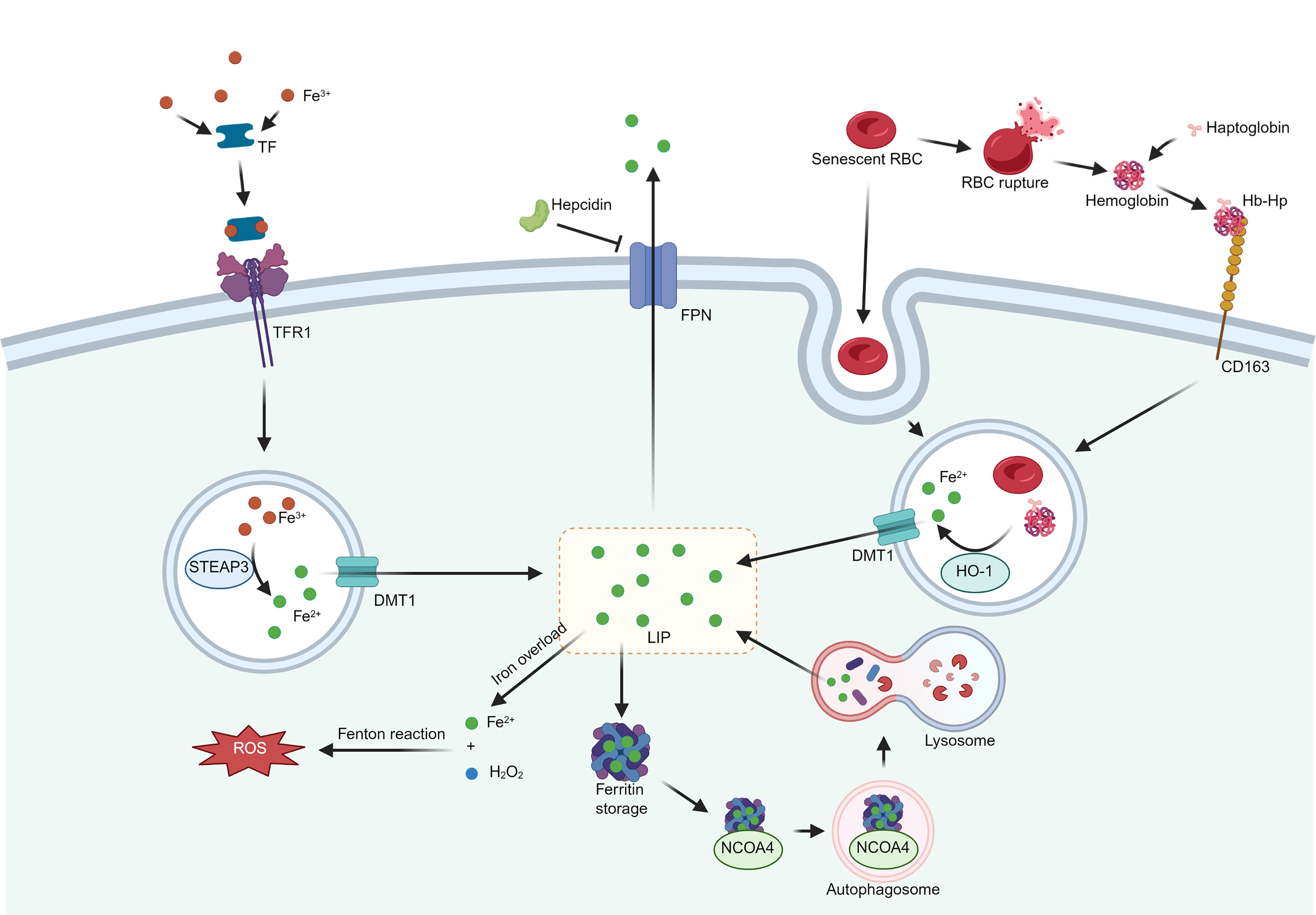
Figure 4. Origin and metabolic pathways of iron in macrophages. Two pathways contribute to the source of iron in macrophages. One of them is the recognition and phagocytosis of senescent erythrocytes or Hb-Hp formed after the rupture of erythrocytes via receptors such as CD163 on the surface of macrophages, which are catabolized by HO-1 to produce Fe2+, which is translocated to the LIP in the presence of DMT1. Another source is circulating Fe3+. Assisted by TF, the Fe3+-TF conjugate is recognized by TFR1 on the macrophage surface, reduces Fe3+ to Fe2+ via STEAP3, and finally translocates to the LIP via DMT1. Excess Fe2+ is excreted from macrophages via Ferroportin, which is controlled by hepcidin synthesized by the liver. Extra Fe2+ is bound to ferritin and stored. Under iron deficiency conditions, NCOA4 binds to ferritin and initiates the fusion of autophagosomes with lysosomes, leading to ferritin catabolism and subsequent release of Fe2+. Iron overload forms excess Fe2+, inducing a Fenton reaction that produces large amounts of ROS. Created in BioRender. Fan, F. (2026) https://BioRender.com/r45f260. Hb-Hp: Hemoglobin-haptoglobin; HO-1: heme oxygenase-1; LIP: labile iron pool; DMT1: divalent metal transporter 1; TF: transferrin; TFR1: transferrin receptor 1; STEAP3: six-segment transmembrane epithelial antigen of prostate 3; FPN: ferroportin; NCOA4: nuclear receptor coactivator 4; ROS: reactive oxygen species; CD163: cluster of differentiation 163; RBC: red blood cell.