

Gastric organoid/organ-on-a-chip platforms for disease modeling and personalized medicine: a review
 0
0 Graphical Abstract
Abstract
Mechanical, structural, and physiological changes in the stomach underlie both short- and long-term gastric pathologies, most of which are influenced by disruptions of acid secretion, mucosal barrier integrity, motility, or immune responses. Species-specific distinctions limit the study of the pathophysiological state of the human stomach in preclinical animal models. Organoid and organ-on-a-chip (OoC) technologies are emerging tools for recapitulating the compositional and functional properties of native gastric tissue, thus revolutionizing research in gastric health and disease as well as drug screening. The review summarizes recent progress in the development of organoids to model gastric development and disease states to investigate their underlying mechanisms and evaluate associated treatments. Furthermore, microfluidic-based OoC models of the human stomach with an epithelial lining that interfaces with adjacent tissues (including the endothelium and stroma) are described for their increased physiologic relevance owing to the presence of fluid flow and peristalsis-like motion. Finally, we discuss the engineering challenges of the present organoid/OoCsystems, including cell sourcing, experimental throughput, and scalable fabrication, followed by highlighting the future opportunities regarding the optimization of in vitro gastric models for studying gastric pathophysiology and developing personalized therapeutic agents.
Keywords


 Download PDF
INTRODUCTION
The stomach, a central organ in the human gastrointestinal tract, orchestrates the initial stages of digestion, nutrient processing, and host defense through a complex interplay of mechanical and biochemical processes[1]. Its specialized epithelium, characterized by distinct cellular lineages and a dynamic microenvironment, is susceptible to a wide array of pathologies, from chronic infections implicated in gastritis and peptic ulcer disease to the development of gastric cancer (GC)[2-4]. The burden of these diseases underscores the critical need for robust and predictive models that can accurately reflect gastric physiology and pathophysiology. Recent research has heavily relied on animal models that mimic human gastric diseases through genetic engineering or external interventions[5-7]. However, the translational accuracy of these studies remains a significant concern due to the inherent differences in gastric anatomy, physiology, and immune responses between animals and humans[8,9]. Thus, the development of human-relevant, preclinical gastric models that can resolve these translational challenges is urgently required.
Early in vitro investigations primarily utilized established human gastric cell lines, including adenocarcinoma cells or immortalized epithelial cells that provide convenient platforms for initial drug toxicity evaluation and basic mechanistic studies into host-microbiome interactions[10,11]. Despite these improvements, two-dimensional (2D) cell culture systems inherently lack the complex three-dimensional (3D) architecture, cellular heterogeneity, and precise microenvironment of the native gastric mucosa. Organoids represent a major advance in this area of research by enabling the development of 3D gastric models. Gastric organoids, derived from either adult gastric stem cells or induced pluripotent stem cells (iPSCs), can recapitulate key aspects of gastric tissue differentiation and self-organization within the extracellular matrix (ECM)[12,13]. These structures mimic the gastric gland architecture and contain multiple differentiated cell types, offering a more physiologically relevant platform for studying host-pathogen interactions and disease mechanisms[13,14].
Because most current gastric organoid cultures are static and lack crucial physical cues (for example, luminal fluid flow, dynamic mechanical stress from peristalsis, and a physiologically relevant pH gradient) to accurately model nutrient absorption, mucus barrier function, and infection dynamics, the introduction of microfluidic technology has emerged as a robust strategy to overcome these limitations. Microfluidic-based organ chips containing microscale channels can be lined with cells under fluid medium perfusion, thus reconstituting the structures and functions of tissue and organs in vitro[15,16]. Various types of human gastric chips have been engineered, ranging from single-channel devices with gastric epithelium maintained under continuous flow to those with two or more channels that enable epithelium to be interfaced with adjacent tissues, including perfusable vasculature, a collagen-rich stromal compartment, and diverse immune cell populations to recapitulate the physiology and pathological states of the stomach in vivo[17,18].
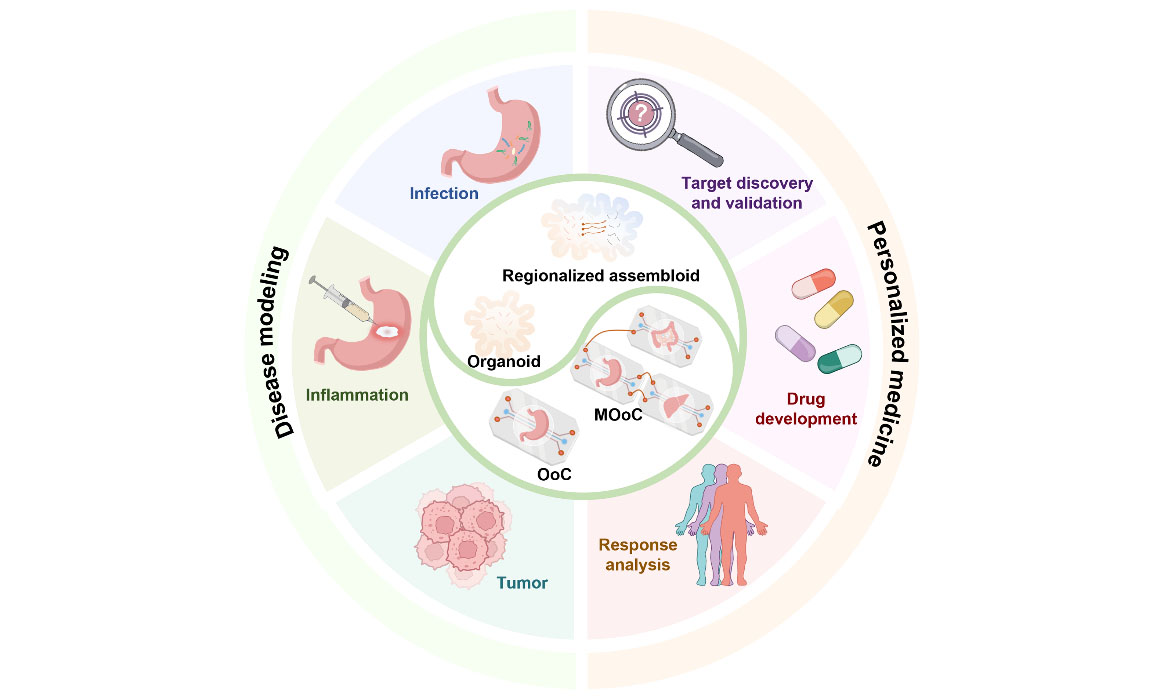
This review provides an overview of the progress made in the establishment of gastric pathophysiological models with organoid/organ-on-a-chip (OoC) technology [Figure 1]. In contrast to previous reviews that merely focus on gastric organoid technology, this article integrates the engineering dimensions of gastric modeling within a comparative and translational framework. Following the introduction of the basic characteristics of the stomach regarding health and disease, we discuss the development of organoids, emphasizing how they model gastric development and diseases, facilitate mechanistic studies, and aid in drug screening. Furthermore, we describe the advancements in designing OoC models to reconstitute the tissue architectures and microenvironment properties of the human stomach, and discuss their technical features considering the investigation of host-pathogen interactions, personalized treatment responses, and interorgan connections. Finally, we outline the remaining engineering challenges that must be overcome to extend the application of organoid/OoC technology and highlight promising strategies to optimize these models for advancing research into gastric diseases and personalized medicine.
GASTRIC PHYSIOLOGY AND PATHOLOGY
The stomach, a J-shaped hollow muscular organ situated between the esophagus and the small intestine, is a pivotal component of the gastrointestinal tract and is essential for the mechanical and chemical breakdown of food, sterilization of ingested material, regulation of nutrient delivery to the small intestine, and secretion of hormones for systemic regulation. The sophisticated structure and dynamic physiological functions of the stomach are maintained by a specialized epithelium organized into distinct regions, including cardia, fundus, corpus, and antrum (pylorus), with each characterized by unique cellular compositions and secretions [Figure 2A][12,25,26]. The gastric epithelium consists of deep invaginations named gastric pits, which lead into corpus and antrum glands harboring various differentiated cell types, including mucous neck cells that secrete mucus and bicarbonate to form a protective barrier against acid and mechanical damage; parietal cells that produce HCl and intrinsic factor crucial for digestion and nutrient absorption; chief cells that secrete pepsinogen to initiate protein digestion; enteroendocrine cells that secrete a variety of hormones such as gastrin, somatostatin, ghrelin, and serotonin to modulate gastric motility, acid secretion, and appetite; and progenitor cells located at the base of the gastric glands for continuous epithelial renewal and repair [Figure 2B][25-31].
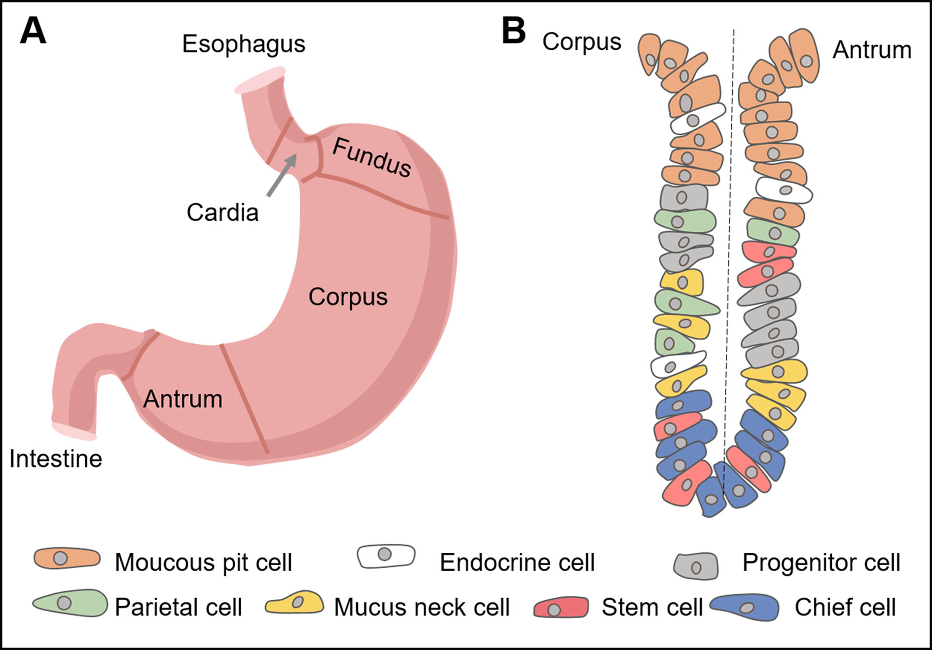
Figure 2. Human stomach structure and gland organization. (A) Structure of the human stomach; (B) schematic of the corpus and antrum glands with different cell compositions.
The gastric microenvironment is highly dynamic and influenced by the mechanical actions of peristalsis and the transit of a food bolus, as well as significant chemical gradients, most notably the highly acidic lumen maintained by parietal cells. Disruption of gastric homeostasis precipitates a spectrum of disorders. Helicobacter pylori (H. pylori) infection represents the primary etiological factor driving a spectrum of gastric pathologies, fundamentally altering host physiology[3]. The initial consequence of H. pylori colonization is the induction of chronic gastritis, a persistent inflammatory state of the gastric mucosa[3,32,33]. If left unchecked, this chronic inflammation can progress to more severe manifestations. Impaired mucosal defense mechanisms and sustained inflammatory damage are key contributors to the development of peptic ulcers, encompassing both gastric and duodenal ulcers[33-36]. GC, a leading cause of cancer-related mortality worldwide, arises from a complex interplay of H. pylori infection, host genetics, environmental factors, and chronic inflammation[37-40]. Notably, patients with gastritis and peptic ulcers are at higher risk of developing GC due to gastric mucosa alterations, excessive inflammatory cytokine production, and activation of proinflammatory pathways[39,40]. Thus, in vitro recapitulation of the human stomach in both health and disease is critical for improving our understanding of gastric physiology and pathology, as well as for developing potential therapeutics.
ORGANOID MODELS OF THE HUMAN STOMACH
Adult and induced pluripotent stem-cell-derived gastric organoids
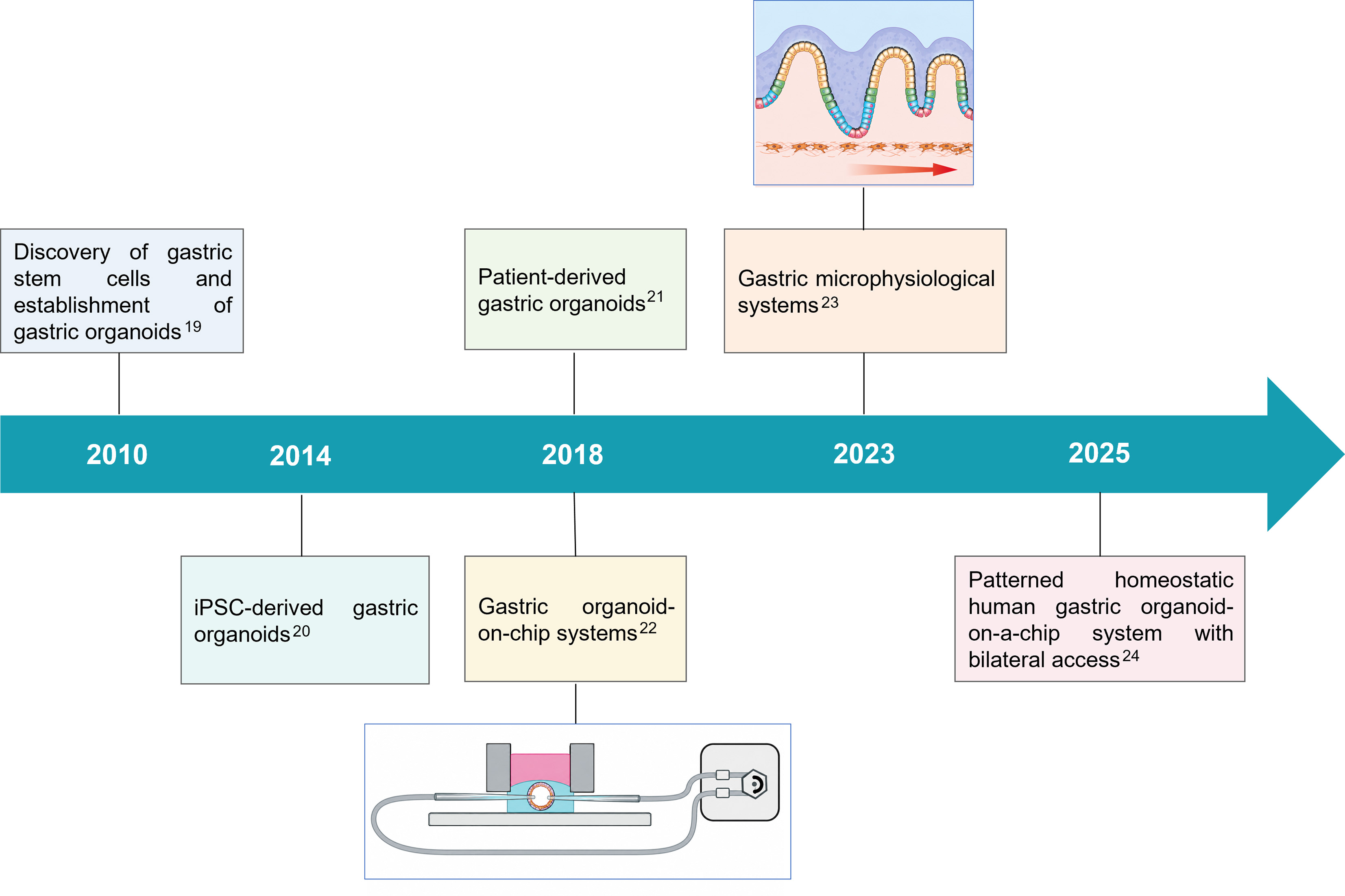
Gastric organoids are self-assembling, multicellular structures that can be classified into two categories based on the cell sources required for their generation: adult stem cell (AdSC)- and iPSC-derived organoids[12-14] [Table 1]. AdSC-derived organoids originate from stem or progenitor cells isolated from normal gastric tissue or patient biopsies and can maintain region-specific adult epithelial programs during long-term expansion[19,41,42]. They are therefore well suited to postnatal disease modeling, epithelial host-pathogen studies, and patient-specific drug testing. By contrast, iPSC-derived gastric organoids are generated through the staged specification of pluripotent cells toward definitive endoderm, foregut, and gastric lineages by manipulating defined signaling pathways[20,43-46]. iPSC-derived models can incorporate mesenchymal components and are especially valuable for developmental biology and congenital disease modeling, but their epithelial maturation generally remains more fetal-like than that of adult tissue.
Comparison of major gastric organoid preparation strategies
| Model | Cellular composition | Preparation strategies | Applications | Limitations | Ref. |
| AdSC gastric organoid | Predominantly mature adult epithelium; region-specific lineages | Biopsy/gland isolation; ECM-embedded expansion with Wnt/R-spondin niche support; corpus cultures require additional pathway control | Adult homeostasis, H. pylori responses, epithelial disease mechanisms | Biopsy dependence; donor variability; limited stromal/immune components | [19,41,42] |
| iPSC antral organoid | Epithelial and mesenchymal populations; immature/fetal-like | Stepwise endoderm, posterior foregut and antral specification | Embryonic development, congenital disease, lineage specification | Long differentiation time; batch variability; incomplete adult maturation | [43,45] |
| iPSC fundic organoid/gastroid | Fundic epithelial lineages; advanced gastroids can include mesenchymal/neural populations | Wnt/β-catenin activation for fundic fate; additional patterning and maturation cues | Regional patterning, parietal-cell maturation, developmental crosstalk | Protocol complexity; incomplete adult physiological function | [20,43,45,46] |
Gastric development modeling
Organoids have emerged as a powerful in vitro 3D model to study the fundamental mechanisms underlying gastric development. The first gastric organoid was established from murine antrum glands containing Lgr5+ AdSCs[19]. The same protocol has been leveraged to develop murine corpus organoids originating from Troy+ stem cells[47]. For humans, while the antral organoids can be established under the same conditions, inhibition of the tumor growth factor (TGF)-β signaling pathway is required to maintain the long-term growth of the corpus organoids[41,42].
Gastric organoids were further differentiated from iPSCs to impart them with both epithelial and mesenchymal cells. McCracken et al. successfully developed gastric organoids from iPSCs for the first time via the delicate manipulation of BMP, Wnt, FGF, and EGF pathways, thus recapitulating key stages of early human gastric development in vitro[20]. Subsequent investigations into the establishment of gastric fundic glands demonstrated that the activation of Wnt/β-catenin directs foregut progenitors to fundic-like organoids, while inhibition promotes the development of antral-like organoids[43,44]. Miao et al. identified the critical role of a metformin-responsive metabolic pathway in regulating the development and functional maturation of parietal cells, offering significant insights into how this epithelial lineage is specified[45]. To investigate the regionalized functions of the stomach along the anterior-posterior axis, Li et al. recently generated gastroids, self-organized multilineage gastric organoids featuring a fundic-antral patterned epithelial chamber with neural population annexation and a mesenchymal cell envelope[46]. Furthermore, neural populations of nonendodermal cells were revealed to instruct fundic-antral patterning in gastroids, with NR2F2 acting as a key mediator[46]. These studies have highlighted gastric organoids as a robust platform for extending our understanding of epithelial morphogenesis, regional specification, and the lineage-specific differentiation of the stomach, thereby elucidating the spatiotemporal regulation of human gastric development.
Disease modeling and mechanism investigation
Gastric organoids have been utilized to model and study H. pylori infection, which could induce chronic gastritis, result in peptic ulcers, and increase the risk of gastric carcinogenesis. To mimic host-pathogen interactions, McCracken et al. microinjected H. pylori into the lumen of iPSC-derived gastric organoids and demonstrated the critical role of the virulence factor cytotoxin-associated gene A (CagA) in the etiology of H. pylori-induced pathology[20]. CagA-positive H. pylori strains were found to induce aberrant epithelial cell proliferation and disrupt mucosal equilibrium via activation of the β-catenin pathway and downregulation of claudin-7[48]. Furthermore, gastric stem cell-derived organoids revealed that CD44 cooperates with CagA to interact with c-Met for the promotion of epithelial cell proliferation[49]. Beyond the direct interplay between H. pylori and the gastric epithelial cells, immune responses occur during H. pylori infection. pylori infection. A coculture of gastric organoids with autologous cytotoxic T cells showed that PD-L1 upregulation caused by H. pylori protected the organoids from T-cell cytotoxicity, while anti-PD-L1 treatment induced immunogenic organoid death[50]. The protective mechanism discovered during host-H. pylori interaction may explain how H. pylori facilitates the formation of premalignant lesions, which indicate the onset of GC. Thus, organoids are informative as experimentally tractable systems for epithelial and defined immune mechanisms.
Gene mutations drive the onset and progression of GC and greatly impact its drug response[51]. Gastroesophageal junction (GEJ) adenocarcinoma is a particularly aggressive subtype of GC, and a thorough understanding of its molecular basis is necessary for diagnosis and precision therapy. Zhao et al. generated AdSC-derived GEJ organoids, subsequently performed CRISPR/Cas9-mediated TP53 and CDKN2A knockout, and demonstrated PTAF and its receptor to be responsible for tumorigenesis in these organoids[52]. The inhibition of PTAF could suppress tumor growth, which represents a potential therapeutic pathway[52]. To determine how genes affect drug sensitivity, Lo et al. generated gastric tumor organoids by introducing TP53 and APC knockouts into normal gastric organoids[53]. Upon the integration of large-scale CRISPR screening with single-cell transcriptomics, the correlation between genetic alterations and cisplatin response was uncovered at the single-cell level, and TAF6L was demonstrated as a key regulator that promotes cell recovery from cisplatin treatment[53]. These studies highlight organoids as a robust system for investigating mechanisms involved in early neoplastic events and gene-drug interactions, and they may thereby guide drug discovery. However, it should be noted that they do not by themselves reproduce vascular, stromal, mechanical, or pharmacokinetic determinants of treatment responses.
Patient-derived disease organoids
GC is classified into four molecular subtypes by the Cancer Genome Atlas (TCGA), including Epstein-Barr virus (EBV)-positive, genomically stable (GS), microsatellite instability (MSI), and chromosomal instability (CIN)[54]. GC patient-derived organoids (PDOs) of these four subtypes have been generated[55]. Importantly, mutational spectrum overlap was observed between organoid cultures and parental tissue, indicating that PDOs could recapitulate the molecular patterns of the GC subtypes[55,56]. In these systems, PDOs have been used to analyze responses to targeted drugs against identified molecular alterations.
The most direct personalized-medicine workflow begins with an endoscopic biopsy or surgical specimen, followed by the rapid establishment of a patient-derived GC organoid, histologic and molecular quality control, and functional testing against clinically relevant drugs[57]. Molecular profiling can then be integrated with the phenotypic response to prioritize candidate therapies or identify resistance mechanisms. For instance, aberrant tumor cell proliferation induced by loss of tumor suppressor CDKN2A could be inhibited by the cyclin-dependent kinase 4/6 (CDK4/6) inhibitor[21]. As another example, trastuzumab has been demonstrated to be effective against PDOs with ERBB2 amplification, consistent with the response of patients harboring the same genetic alteration[55]. Intratumoral heterogeneity plays a key role in therapy response. GC PDOs established from normal, dysplastic, cancer, and lymph node metastases showed regional heterogeneity and different subclonal architectures, which resulted in sensitivity to different drugs[21,58].
Considering that drug resistance is a major clinical challenge affecting the prognosis and survival of patients with GC, organoids have been explored to clarify the underlying mechanisms of drug resistance. It was found that 5-fluorouracil (5-FU), a first-line drug for GC, led to tumor relapse upon therapy[59]. Ukai et al. developed GC PDOs resistant to 5-FU and identified that KHDRBS3 induced 5-FU resistance by modulating CD44 variant expression and promoting PDOs to acquire stem-like properties[60]. Because the commonly used post-surgery chemotherapy [the combination of 5-FU and cisplatin (CDDP)] frequently resulted in acquired resistance, Wong et al. generated 5-FU + CDDP-resistant GC subtype PDOs and revealed that adenosine deaminase acting on RNA 1 (ADAR1) induced chemoresistance by augmenting stearoyl-CoA desaturase (SCD1) mRNA stability, resulting in reduced ER stress and enhanced self-renewal [Figure 3A][61]. Zhao et al. established 57 GC PDOs from 73 patients (78% establishment rate) and reported concordance between the PDO drug-response results and actual clinical response in 91.7% (11/12) of evaluable patients[62]. As expected, the genes/pathways involved in proliferation and invasion are enriched in chemotherapy-resistant PDOs[62]. McDonald et al. further demonstrated the feasibility of rapidly generating organoids from endoscopic gastric cancer biopsies and performing drug testing within approximately two weeks in a proof-of-concept workflow[63]. These results are encouraging because they directly connect ex vivo testing to patient treatment response, but the sample sizes remain too small to establish routine clinical utility. While these models provide mechanistic and predictive information, the inadequate efficiency, assay turnaround time, tumor sampling, and absence of nonepithelial tumor microenvironment components are important constraints that must be overcome for clinical deployment.
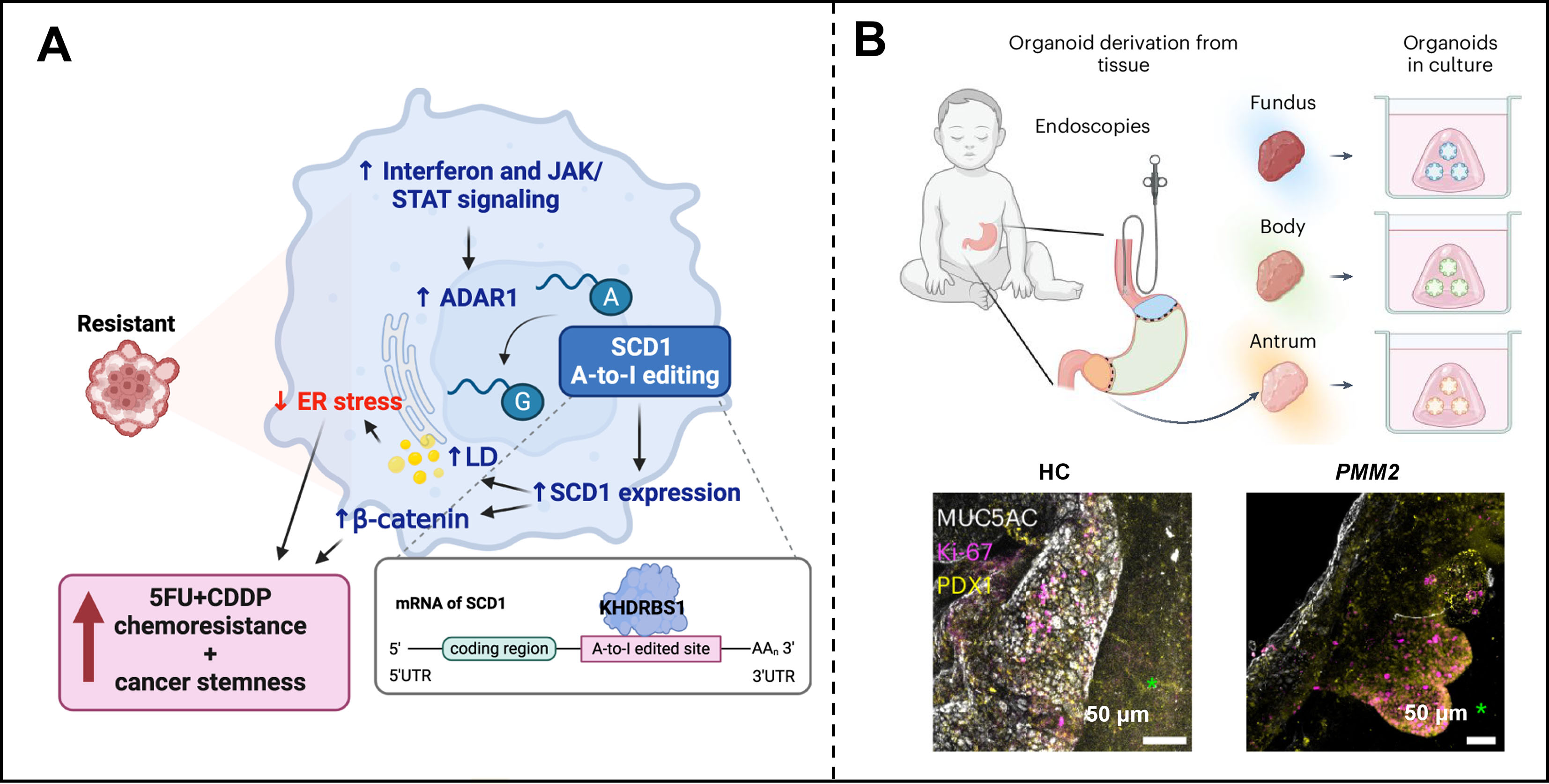
Figure 3. Gastric organoids used for drug response evaluation and disease development mechanism investigations. (A) GC PDO-mediated interpretation of resistance to 5-FU and CDDP combination therapy. Reproduced from Ref.[61] under the terms of the CC BY 4.0 License; (B) Generation of multiregional assembloids for the patient-specific recapitulation of antral foveolar hyperplasia. Reproduced from Ref.[64] under the terms of the CC BY 4.0 License. JAK-STAT: Janus Kinase-Signal Transducer and Activator of Transcription; ADAR1: adenosine deaminase acting on RNA 1; ER: endoplasmic reticulum; LD: lipid droplet; SCD1: stearoyl-CoA desaturase; 5FU: 5-fluorouracil; CDDP: cisplatin; GC: gastric cancer; PDO: patient-derived organoid; SRA: single-region assembloid; MRA: multiregional assembloid; HC: healthy control; MUC5AC: Mucin 5AC.
Considering that different regions of the human stomach show distinct morphological and functional properties, Jones et al. generated multiregional assembloids via the self-assembly of region-specific gastric organoids from pediatric patients with unusual antral foveolar hyperplasia [Figure 3B][64]. The assembloids efficiently recapitulate hyperplastic-like antral regions characterized by impaired gastric acid secretion due to decreased mucin secretion and glycosylated H+/K+ ATPase β[64]. Overall, these studies highlight PDOs as a potential tool for investigating individual treatment responses, as well as disease development and drug resistance mechanisms.
MICROFLUIDIC HUMAN STOMACH CHIPS
Microfluidic-based organ chips are an emerging technology that facilitates the culturing of different types of cells in tissue in close apposition within one or more hollow channels with fluid flow perfusion and physiologically relevant cue stimulation, offering a way to resolve the limitations of static culture systems[65-67]. The simplest design incorporates a single epithelial or mucus barrier channel to model gastric mucosa[68,69]. Peristalsis-like cyclic deformations have been further introduced to the epithelial structure to mimic gastric motility[22]. In addition, hydrogels with patterned surfaces have been integrated into the chip design to mimic the structural and cellular arrangement of native tissues[23,70]. Other chip designs incorporate vascular, stromal, and immune compartments to reconstitute the microenvironmental factors of the gastric epithelium[24,71-73]. The connection of the stomach to other organs has been further investigated through the establishment of multiorgan-on-chip models[74].
Gastric mucosa modeling
The gastric mucosa, the innermost layer of the stomach wall, serves as a protective barrier and contributes to acid, enzyme, and hormone production, immune surveillance, and epithelial renewal[75,76]. Several stomach chips have been developed to recapitulate the structure and functions of the gastric mucosa in vitro. To study how the mucosa protects the gastric epithelial cells against damage by the acidity in the lumen, a microfluidic device producing a continuous stream of mucus against incoming acid was developed to mimic the gastric mucus acid barrier, using a fluorescent pH reporter to monitor the dynamics of H+ penetration into the mucin layer [Figure 4A][68]. The study demonstrated proton sequestration and retarded diffusion within mucus, illustrating how a reductionist chip can provide a quantitative barrier readout that is difficult to obtain from closed 3D organoid lumens.
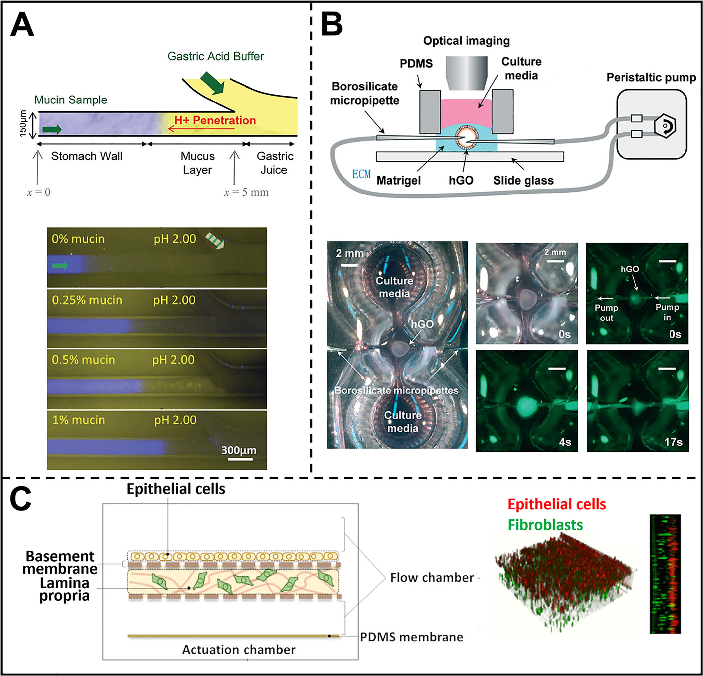
Figure 4. OoC for gastric mucosa modeling. (A) Microfluidic system for studying the barrier function of stomach mucin. Reproduced from Ref.[68]. Copyright 2012, Royal Society of Chemistry; (B) Stomach organoid-on-a-chip with luminal flow to induce cyclic mechanical deformation of the organoids, simulating gastric motility. Scale bars: 2 mm. Reproduced from Ref.[22]. Copyright 2018, Royal Society of Chemistry; (C) Stomach-on-a-chip with an epithelial cell monolayer, basement membrane, and the lamina propria to recapitulate human gastric mucosa architecture. Reproduced from Ref.[71]. Copyright 2023, Royal Society of Chemistry. PDMS: Polydimethylsiloxane; hGO: human gastric organoid; ECM: extracellular matrix; OoC: organ-on-a-chip.
To model gastric epithelium with lumen and rhythmical peristalsis, a stomach-on-a-chip (SoC) model was established by Lee et al. in which gastric organoids were encased within a microfluidic device and underwent cyclic expansion and contraction induced by a peristaltic pump [Figure 4B][22]. The robust 3D growth of gastric organoids in the presence of luminal flow could be recorded, providing a platform for the long-term delivery of agents into the gastric lumen for disease modeling and drug screening. To more authentically recapitulate the gastric mucosa, a SoC comprised of the epithelial cell monolayer, basement membrane, and lamina propria (gastric fibroblast-embedding collagen I gel) was developed [Figure 4C][71]. In the bioinspired SoC, a 2 μL min-1 perfusion rate in a 0.20 cm wide and 0.05 cm high channel corresponded to an estimated epithelial shear stress of approximately 0.003 dyne per cm2, which is deliberately within the physiological shear stress range experienced by gut cells in vivo (0.002-0.080 dyne per cm2). Peristalsis-like motion was realized by applying a vacuum to the microactuator at cyclical intervals. Sinusoidal actuation at 0.15 Hz produced surface stretching of approximately 10%. Next, the growth and integrity of the epithelial barrier were assessed by measuring the transepithelial electrical resistance (TEER). The results showed that the TEER values were above 100 Ω cm2 at two days post-seeding and reached an average of approximately 157 Ω cm2 after five days of culturing, characterizing a fully formed barrier. These studies identified that the mechanical actuation of the SoC plays a key role in the development of epithelial polarization and differentiation traits that are characteristic of the gastric mucosa, illustrating how device architecture can directly affect model performance rather than simply provide a culture container.
As the organoid-derived gastric epithelium shows apical-basal polarization, a bilaterally accessible patterned epithelial chip was designed to ensure the manipulation of both its apical and basal sides[70]. Basal stem cell niche growth factor delivery could be mimicked to enable proliferative and differentiation patterns of gastric epithelium organoids consistent with the stomach owing to the improved accessibility of the system. Compared with conventional enclosed 3D organoids, such open-access designs facilitate longitudinal imaging and cell-resolved perturbation, although some 3D gland geometry is sacrificed for experimental observability.
Host-pathogen models
Pathogen-mediated homeostatic disruption of the gastric mucosal barrier leads to various types of stomach disease pathogenesis, among which H. pylori is a representative pathogen[77]. The replication of host-pathogen interactions in vitro could improve the study of the gastric mucosal defense mechanisms against pathogens and the development of therapeutic strategies. A human stomach microphysiological system (hsMPS) was built by cultivating human antral organoids and gastric mesenchymal stromal cells in the luminal and abluminal channels of a compartmentalized microfluidic device[23]. H. pylori infection was modeled with the hsMPS, and trefoil factor peptide 1 (TFF1) was revealed as a critical element in gastroprotection against H. pylori [Figure 5]. Moreover, the introduction of peripheral blood mononuclear cells (PBMCs) demonstrated the immune cell recruitment and inflammation induction properties of H. pylori [Figure 5B], indicating the potential of hsMPS as a tool for uncovering the immune pathogenic mechanisms of H. pylori infection[23].
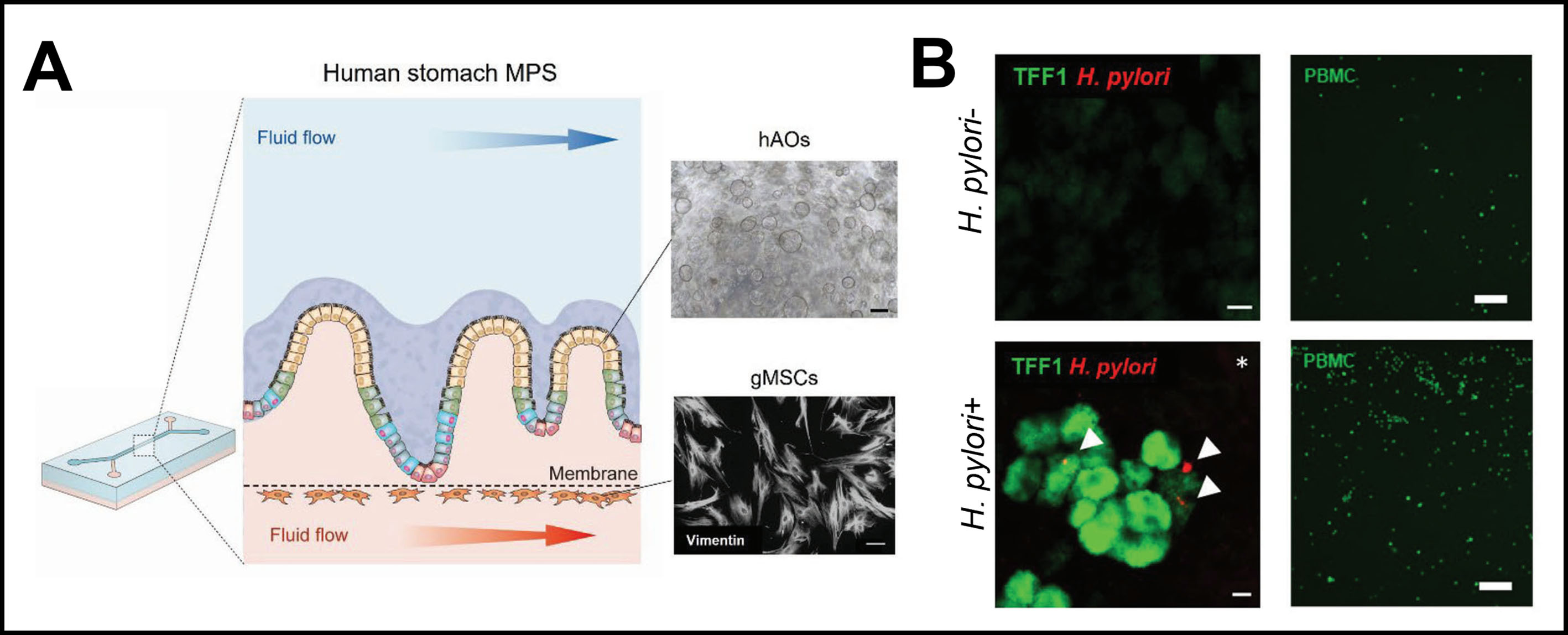
Figure 5. Stomach-on-a-chip for host-pathogen interaction modeling. (A) Human stomach microphysiological system for studying epithelial and immune responses to H. pylori infection. Scale bars: 250 μm (top) and 100 μm (bottom); (B) Impact of H. pylori infection on the expression of TFF1 and the ability to recruit PBMCs of the human gastric epithelium. Scale bars: 20 μm (left) and 250 μm (right). (A and B) is reproduced from Ref.[23] under the terms of the CC BY 4.0 License. MPS: Micro-physiological system; hAO: human antral organoids; gMSC: gastric mesenchymal stromal cell; PBMC: peripheral blood mononuclear cell; TFF1: trefoil factor peptide 1; H. pylori: Helicobacter pylori.
To explore cell-type-specific host-pathogen interactions, Hofer et al. proposed the generation of a patterned homeostatic human gastric organoid-on-a-chip model with bilateral access and an apical environment maintaining a pH of approximately 3. This can model H. pylori niche establishment and the persistent colonization of the gastric epithelium[24]. By incorporating a spatially confined culture and a dynamic acidity regulation system, this model overcomes the limitation of unipolar differentiation observed in conventional gastric organoids. The study demonstrates that under the physiological acidic luminal environment, the maturation of gastric pit cells on the chip is significantly enhanced. H. pylori colonization showed a graded response among different gastric epithelial cell subsets: basal progenitor cells predominantly secrete inflammatory mediators, while apical pit cells specifically activate tight junction remodeling and the lectin stress pathway. For the first time, this model enables the in situ observation of the dynamic formation of bacterial microcolonies and their authentic three-dimensional adhesion behavior, indicating that the nonuniform immune response of the host is the molecular basis for sustaining chronic infection. Overall, this study demonstrates why acid control and spatial epithelial patterning can qualitatively change the infection phenotype observed in vitro.
Clinical profiling indicates that dysbiotic communities enriched in taxa such as Fusobacterium and Neisseria can be associated with GC risk after H. pylori eradication, and mechanistic experiments have shown that selected gastric bacteria can induce distinct inflammatory and pro-oncogenic responses in gastric epithelial systems[78]. However, the non-H. pylori gastric microbiome remains substantially less developed as an in vitro model. Therefore, future gastric chips should introduce a defined microbial community rather than a single pathogen. In addition, apical access should be combined with controlled oxygen tension, low-pH gradients, mucus production, and defined microbial consortia while quantifying microbial abundance, epithelial functionality, and metabolite exchange to extend from single-pathogen infection models toward experimentally controlled gastric ecosystem models.
Cancer modeling
Although established cell lines are suboptimal for cancer studies, investigators have developed microfluidic chips to characterize GC growth and drug treatments in vitro. Bakhchova et al. proposed the establishment of a polarized gastric epithelial monolayer-on-a-chip with continuous and stable fluid flow for the imaging-based analysis of chemotherapeutic drug responses[69]. Considering that the tumor microenvironment fundamentally shapes drug response, a microfluidic tumor-on-chip platform encompassing endothelial cells and 3D-cultured GC cells was designed to mimic a vascularized tumor in vivo[72]. The chip contains the chamber layer [for the cultivation of human umbilical vein endothelial cells (HUVECs) and GC cells embedded in extracellular matrix (ECM) gel] and the gas layer (for oxygen concentration modulation) separated by a permeable membrane[72]. Upon hypoxic microenvironment modeling followed by CD8+ T cell perfusion, the system identified that hypoxia-induced forkhead box O3 (FOXO3a) upregulation mediates PD-L1 elevation, thereby contributing to immunotherapy resistance[72]. This model is particularly suitable for investigating the impact of oxygen gradients, immune-cell trafficking, and perfused drug exposure on tumors that are not captured by epithelial organoids alone.
As patient-derived organoids provide a more faithful, personalized, and physiologically relevant model of human cancer compared with cell lines, gastric tumor chips based on patient-derived organoids have been established for personalized drug screening and treatment. In particular, Meng et al. proposed a microcapsule-based OoC that incorporates gastric organoid-loaded microcapsules into a microfluidic chip, enabling the dynamic perfusion of various chemotherapeutic agents[79]. Heterogeneity in drug sensitivity was identified among organoids derived from different patients using this platform, which was in accordance with the responses of the corresponding patients. To further model tumor vasculature and the tissue-tissue interface of tumor and blood vessels, a vascularized tumor chip was built by integrating patient-derived gastric organoids with perfusable endothelium and tumor-blood vessel structures in interstitial tissues formed by stomach-derived decellularized ECM [Figure 6][73]. The clinical responses of patients treated with a Vascular Endothelial Growth Factor Receptor 2 (VEGFR2) inhibitor were reproduced using this platform. These studies highlight microfluidic chips as valuable platforms for the prediction of individual therapeutic responses and drug discovery, but they currently require a large quantity of tissue, long setup times, specialized fabrication, and assay-specific validation. Consequently, most current gastric tumor chips should be regarded as advanced preclinical or proof-of-concept platforms rather than clinically validated companion diagnostics. Translation will require predefined turnaround times, standardized drug screening metrics, blinded prospective comparison with clinical outcomes, and demonstration that chip-guided decisions improve outcomes beyond standard pathology and genomic biomarkers.
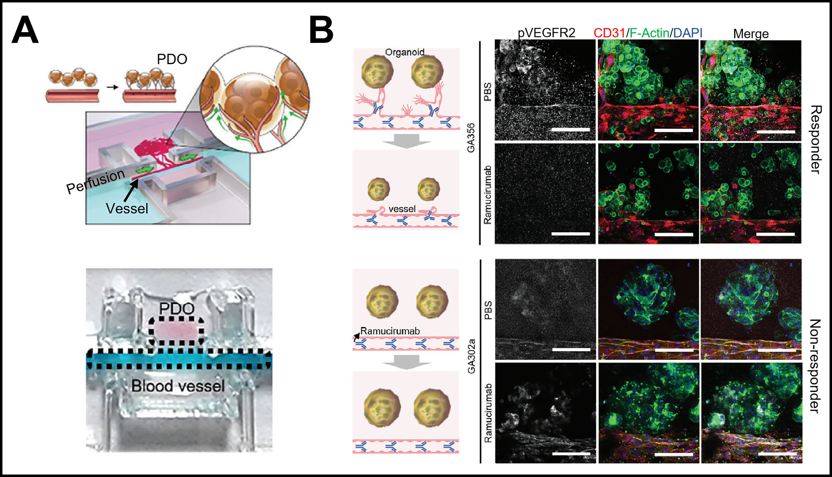
Figure 6. OoC for gastric cancer modeling. (A) Vascularized tumor-on-a-chip for the prediction of clinical responses to VEGFR2-targeted therapy; (B) Immunofluorescence staining images of pVEGFR2, F-actin, CD31, and DAPI in the PDOs of responders and nonresponders. Scale bars: 100 μm. (A) and (B) were reproduced from Ref.[73]. Copyright 2023, John Wiley & Sons. PDO: Patient-derived organoid; VEGFR2: Vascular Endothelial Growth Factor Receptor 2; CD31: Cluster of Differentiation 31; DAPI: 4',6-diamidino-2-phenylindole; OoC: organ-on-a-chip.
Integration into a multiorgan-on-a-chip
Modeling the body as a system of interconnected organs is a powerful strategy for gaining a holistic understanding of human physiology. Despite the ability of organoids to model individual organs, utilizing them to recapitulate organ-organ interactions remains challenging. With the advent of human-on-a-chip technology, various types of organoids have been integrated into a chip to simulate multiorgan interactions[65,80,81]. For example, Jin et al. developed a microfluidic array-based multiorgan chip model through the coculturing of stem cell-derived liver, intestinal, and stomach organoids [Figure 7][74]. The organoids cultivated in distinct chambers were allowed to communicate via rocker-induced media flow. This work aimed to simulate in vivo bile acid homeostasis, which is regulated by the interaction between the liver and intestine. The platform showed reduced expression of CYP7A1, a bile acid synthesis enzyme, upon exposure to bile acids in the liver organoids due to the paracrine effect of the intestinal organoid, demonstrating the physiological crosstalk between different organs[74].
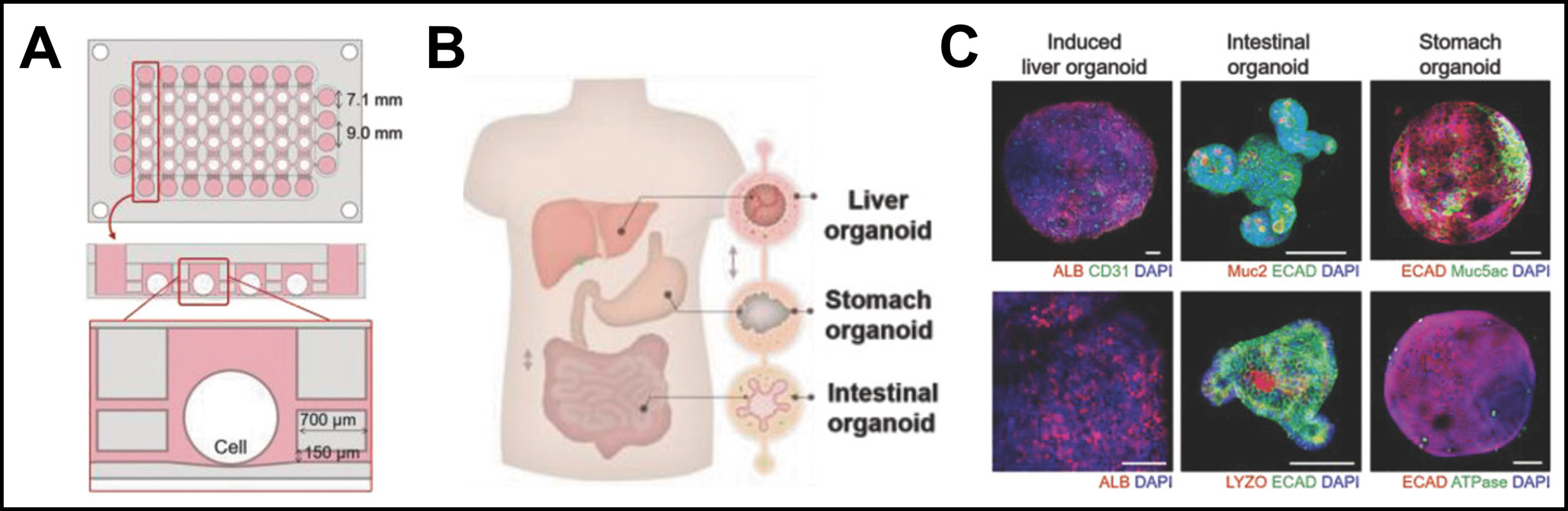
Figure 7. Multiorgan-on-a-chip. (A) Front and cross-sectional views of the microfluidic device for modeling the interactions of the stomach and other organs; (B) Schematic of the liver-stomach-intestine multiorgan model formed by organoids; (C) Immunostaining analysis of specific markers for different organs (albumin for liver; Muc2 and lysozyme for intestine; and Muc5ac and H+/K+ ATPase for stomach). Scale bars: 200 μm. (A-C) is reproduced from Ref.[74]. Copyright 2018, John Wiley & Sons. ALB: Albumin; ECAD: E-cadherin; Muc2: Mucin-2; DAPI: 4',6-Diamidino-2-phenylindole.
CONCLUSION AND OUTLOOK
In this review, recent advances in exploiting organoid and OoC models for studying gastric development, homeostasis, and diseases were summarized and discussed. A summary of the suitability of in vitro gastric models for disease modeling and personalized medicine is provided in Table 2. Normal gastric organoids enable the investigation of fundamental mechanisms underlying gastric development and H. pylori infection-induced inflammation. Further introduction of CRISPR/Cas technology facilitates modeling of the early onset of tumorigenesis and interpretation of gene-drug interactions in tumors. Conversely, patient-derived disease organoids allow phenotype and molecular characterization, as well as the evaluation of individualized drug response. Microfluidic-based stomach chips are a more physiologically relevant model owing to their ability to cultivate epithelial cells and cells from adjacent tissues, including endothelial cells, stromal fibroblasts, and immune cells, within different compartments and layers. Moreover, they can be used to introduce fluid flow, peristalsis-mimetic physical cues, and chemical gradients. Recently developed stomach chips may allow modeling of gastric mucosal, host-pathogen interactions, and cancer; identification of underlying molecular, cellular, and biophysical processes; patient-specific drug screening; and the interpretation of the relationships between the stomach and other organs.
Suitability of major gastric in vitro models for disease modeling and personalized medicine
| Model | Physiological relevance and complexity | Informative readouts | Applications | Clinical translation position | Ref. |
| 2D gastric cell line | Low to moderate; limited architecture and heterogeneity; low complexity | Signaling, viability, reporter assays | Mechanistic screening; early toxicity and drug discovery | Established research tool; weak patient specificity | [10,59] |
| AdSC-derived healthy gastric organoid | High adult epithelial relevance; limited nonepithelial tissues; moderate complexity | Lineage markers, secretion, infection response | Adult epithelial biology, host-pathogen mechanisms | Strong preclinical platform | [12-14,19] |
| iPSC-derived gastric organoid | High developmental relevance; maturation heterogeneity; high protocol complexity | Developmental patterning, lineage maturation | Development, congenital disease, cell-fate studies | Preclinical; maturation and standardization remain limited | [12-14,20] |
| Gastric cancer PDO | High patient-specific tumor epithelium; moderate complexity | Viability, genomic concordance, resistance signatures | Functional precision oncology | Most advanced patient-linked evidence; prospective validation still needed | [21,55] |
| Stomach-on-a-chip | Potentially high for barrier interfaces; high complexity | TEER, permeability, pH, mechanics, imaging | Barrier, infection, transport, tissue-interface biology | Predominantly preclinical/proof-of-concept | [22,71] |
| PDO-on-chip | Very high potential for patient-specific TME; very high complexity | Drug response, vascular, immune and hypoxia readouts | Mechanism-informed personalized therapy | Early translational stage | [73,79] |
| Multi-organ chip | System-level physiology; very high complexity | Interorgan metabolites and functional crosstalk | Drug metabolism, systemic interaction | Proof-of-concept | [74,80,81] |
Engineering challenges
Despite the progress that has been made, several challenges must be overcome to broaden the application of gastric organoid/OoC models. First, future models must maintain a stable apical pH gradient without nonspecific epithelial injury as the stomach combines a highly acidic lumen with a protected basolateral tissue compartment. Moreover, the glandular mucosa contains strong apical-basal and pit-gland spatial organization that is difficult to reproduce in flat monolayers, while closed spherical organoids restrict luminal access. Patterning, bilateral access, tubular/gland-like geometries, and controlled niche-factor gradients might provide solutions to some extent[24,70]. In addition, gastric mechanics are multiscale, considering slow-wave activity, local wall strain, luminal mixing, and antral contractions, and they should therefore not be reduced to a single “peristalsis” frequency. In terms of cell sources, although primary gastric organoids overcome the drawbacks of established cell lines by recapitulating gastric region-specific structure and function, their use is hindered by limited availability and a lack of standard quality control metrics and procedures. While iPSC-derived gastric organoids provide an alternative, they fail to become fully mature and precisely model adult tissues in vivo[82]. For stomach-on-a-chip systems, while polydimethylsiloxane (PDMS) is the most frequently used material for chip fabrication, it is not suitable for scalable production. In addition, its absorption of hydrophobic molecules might impair the accuracy of efficacy evaluation of drugs with high hydrophobicity[83,84]. The exploration of other materials or nonfouling coatings could resolve these challenges[84]. Furthermore, the experimental throughput of recent stomach chips must be increased to enable the high-throughput assessment of epithelial function in parallel and drug screening.
Future perspectives
While the incorporation of cells in addition to patient-derived epithelial cells, such as endothelium, stromal, and immune cells, improves the physiological relevance of gastric organoid and OoC models, these cells are commonly not obtained from the same patient. Therefore, the establishment of stomach-on-a-chip models containing all cells from the same patient could improve personalized medicine. These fully personalized stomach chips could be used to more authentically recapitulate individual disease phenotypes and drug responses, which might help to reveal the impact of sex, age, and race on interpatient variability[79]. Moreover, controlled microbial consortia should be incorporated to study how non-H. pylori communities interact with acid, mucus, and host tissue[78]. The integration of real-time monitoring tools and additional downstream analytical strategies should be further investigated. Gastric organoid/OoC technology would benefit from the introduction of biosensors that allow in situ and live-cell analysis of the dynamics of complicated biological processes and treatment responses without disrupting gastric architecture and cell viability. In addition to single-cell transcriptomics, other omics techniques (e.g., metabolomic, proteomic, and epigenomic approaches) should be included to identify the mechanisms involved in gene regulation, protein expression, epigenetic variation, and host-pathogen interactions[85]. Furthermore, the integration of an artificial-intelligence-based drug discovery platform into a human stomach-on-a-chip might accelerate therapeutics discovery for specific gastric disorders and aid in the development of drug delivery strategies with high gastric epithelium selectivity for minimized systemic toxicity[86,87]. In parallel, microfluidic manufacturing and delivery strategies may be developed to improve the reproducible control of materials, gradients, and dosing[88].
By contrast, standardization is required to convert technically sophisticated gastric models into reliable tools for clinical application. Recent human gastric organoid standards emphasize traceable cell sourcing, morphology, sterility, genomic stability, lineage-marker characterization, functional assays (such as acid or pepsinogen secretion where appropriate), and defined storage and quality-control procedures[89]. For microphysiological systems, reproducibility frameworks additionally recommend prespecified acceptance criteria and the reporting of device, biological, and analytical variables[90]. For stomach chips, a minimum reporting set should include the cell source and passage, ECM composition, channel dimensions, material identity, calculated shear stress, actuation frequency and strain, apical and basal pH, barrier baseline (e.g., TEER or permeability), assay timing, and interdevice variability. These metrics would allow laboratories to determine whether a model is reproducible and suitable for its intended regulatory or translational use.
DECLARATIONS
Authors’ contributions
Writing-original draft: Yin, P.
Visualization: Yin, P.; Zhang, W.; Geng, S.; Wang, Y.
Conceptualization: Yin, P.; Di, Z.; Wang, X.
Supervision: Di, Z.; Liu, Z.; Guo, G.; Wang, X.
Writing-review & editing: Di, Z.; Wang, X.
AI and AI-assisted tools statement
Not applicable.
Availability of data and materials
Not applicable.
Financial support and sponsorship
This work was supported by grants from the National Natural Science Foundation of China (No. 22327805 and No. 22476223).
Conflicts of interest
Wang, X. is an Associate Editor of the journal Micro Nano Science. Wang, X. was not involved in any steps of the editorial process, including reviewers’ selection, manuscript handling, and decision-making. The other authors declare that there are no conflicts of interest.
Ethical approval and consent to participate
Not applicable.
Consent for publication
Not applicable.
Copyright
© The Author(s) 2026.
REFERENCES
1. Willet, S. G.; Mills, J. C. Stomach organ and cell lineage differentiation: from embryogenesis to adult homeostasis. Cell. Mol. Gastroenterol. Hepatol. 2016, 2, 546-59.
2. Hunt, R. H.; Camilleri, M.; Crowe, S. E.; et al. The stomach in health and disease. Gut 2015, 64, 1650-68.
3. Malfertheiner, P.; Camargo, M. C.; El-Omar, E.; et al. Helicobacter pylori infection. Nat. Rev. Dis. Primers. 2023, 9, 19.
4. Bray, F.; Laversanne, M.; Sung, H.; et al. Global cancer statistics 2022: GLOBOCAN estimates of incidence and mortality worldwide for 36 cancers in 185 countries. CA. Cancer. J. Clin. 2024, 74, 229-63.
5. Khatoon, A.; Sachan, V.; Kori, P.; Khushtar, M.; Pujari, N. M. Current practices and emerging technologies in animal models for gastric ulcer research. JDDHS 2024, 1, 153-60.
6. Hou, N.; Du, X.; Wu, S. Advances in pig models of human diseases. Animal. Model. Exp. Med. 2022, 5, 141-52.
7. Ansari, S.; Yamaoka, Y. Animal models and Helicobacter pylori Infection. J. Clin. Med. 2022, 11, 3141.
8. Weingarden, A. R. Strengths and limitations of in vitro and animal models to advance understanding of human diet-microbiome interactions. Gut. Microbes. Rep. 2026, 3, 2636336.
9. Pan, Y.; Zhao, L.; Fang, J. Animal models and pathogenesis of gastric cancer: from premalignant conditions-to-metastasis. Cancer. Biol. Med. 2026, 23, 637-56.
10. Barranco, S. C.; Weintraub, B.; MacLean, K. K.; Beasley, E. G.; Jenkins, V. K.; Jr Townsend, C. M. Relationship between glutathione levels and drug or radiation sensitivities in human gastric cancer cell lines in vitro. Invest. New. Drugs. 1991, 9, 29-36.
11. Kim, K. J.; Zhong, H.; Tai, D.; et al. Microbiome signatures in advanced gastric cancer: emerging biomarkers for risk stratification, therapy guidance, and prognostic insight. Int. J. Mol. Sci. 2026, 27, 1452.
12. Seidlitz, T.; Koo, B. K.; Stange, D. E. Gastric organoids-an in vitro model system for the study of gastric development and road to personalized medicine. Cell. Death. Differ. 2021, 28, 68-83.
13. Lee, S.; Choi, J. H.; Park, S. Y.; Kim, J. Gastric organoid, a promising modeling for gastric stem cell homeostasis and therapeutic application. Int. J. Stem. Cells. 2024, 17, 337-46.
14. Mu, X.; Wang, L.; Pang, M.; et al. Exploring the applications of gastric organoids in disease modeling and personalized medicine. Gastrointestinal. Tumors. 2025, 12, e013.
16. Chang, Y.; Chen, T.; Geng, S.; et al. A scenario-adaptive microfluidic chip for constructing in vitro models of biological barriers. Anal. Chem. 2025, 97, 3816-21.
17. Jantaree, P.; Bakhchova, L.; Steinmann, U.; Naumann, M. From 3D back to 2D monolayer stomach organoids-on-a-chip. Trends. Biotechnol. 2021, 39, 745-8.
18. Carvalho, M. R.; Yan, L. P.; Li, B.; et al. Gastrointestinal organs and organoids-on-a-chip: advances and translation into the clinics. Biofabrication 2023, 15, 042004.
19. Barker, N.; Huch, M.; Kujala, P.; et al. Lgr5(+ve) stem cells drive self-renewal in the stomach and build long-lived gastric units in vitro. Cell. Stem. Cell. 2010, 6, 25-36.
20. McCracken, K. W.; Catá, E. M.; Crawford, C. M.; et al. Modelling human development and disease in pluripotent stem-cell-derived gastric organoids. Nature 2014, 516, 400-4.
21. Yan, H. H. N.; Siu, H. C.; Law, S.; et al. A comprehensive human gastric cancer organoid biobank captures tumor subtype heterogeneity and enables therapeutic screening. Cell. Stem. Cell. 2018, 23, 882-897.e11.
22. Lee, K. K.; McCauley, H. A.; Broda, T. R.; Kofron, M. J.; Wells, J. M.; Hong, C. I. Human stomach-on-a-chip with luminal flow and peristaltic-like motility. Lab. Chip. 2018, 18, 3079-85.
23. Jeong, H. J.; Park, J. H.; Kang, J. H.; Sabaté, Del. Río. J.; Kong, S. H.; Park, T. E. Organoid-based human stomach micro-physiological system to recapitulate the dynamic mucosal defense mechanism. Adv. Sci. (Weinh). 2023, 10, e2300164.
24. Hofer, M.; Kim, Y.; Broguiere, N.; et al. Accessible homeostatic gastric organoids reveal secondary cell type-specific host-pathogen interactions in Helicobacter pylori infections. Nat. Commun. 2025, 16, 2767.
25. Karam, S. M.; Leblond, C. P. Identifying and counting epithelial cell types in the “corpus” of the mouse stomach. Anat. Rec. 1992, 232, 231-46.
26. Lee, E. R.; Trasler, J.; Dwivedi, S.; Leblond, C. P. Division of the mouse gastric mucosa into zymogenic and mucous regions on the basis of gland features. Am. J. Anat. 1982, 164, 187-207.
27. Karam, S. M.; Leblond, C. P. Dynamics of epithelial cells in the corpus of the mouse stomach. I. Identification of proliferative cell types and pinpointing of the stem cell. Anat. Rec. 1993, 236, 259-79.
28. Karam, S. M.; Leblond, C. P. Dynamics of epithelial cells in the corpus of the mouse stomach. II. Outward migration of pit cells. Anat. Rec. 1993, 236, 280-96.
29. Karam, S. M.; Leblond, C. P. Dynamics of epithelial cells in the corpus of the mouse stomach. III. Inward migration of neck cells followed by progressive transformation into zymogenic cells. Anat. Rec. 1993, 236, 297-313.
30. Karam, S. M.; Leblond, C. P. Dynamics of epithelial cells in the corpus of the mouse stomach. V. Behavior of entero-endocrine and caveolated cells: general conclusions on cell kinetics in the oxyntic epithelium. Anat. Rec. 1993, 236, 333-40.
31. Lee, E. R.; Leblond, C. P. Dynamic histology of the antral epithelium in the mouse stomach: II. Ultrastructure and renewal of isthmal cells. Am. J. Anat. 1985, 172, 205-24.
32. Tsai, H. F.; Hsu, P. N. Interplay between Helicobacter pylori and immune cells in immune pathogenesis of gastric inflammation and mucosal pathology. Cell. Mol. Immunol. 2010, 7, 255-9.
33. Malfertheiner, P.; Link, A.; Selgrad, M. Helicobacter pylori: perspectives and time trends. Nat. Rev. Gastroenterol. Hepatol. 2014, 11, 628-38.
34. Yuan, Y.; Padol, I. T.; Hunt, R. H. Peptic ulcer disease today. Nat. Clin. Pract. Gastroenterol. Hepatol. 2006, 3, 80-9.
35. Watanabe, T.; Higuchi, K.; Tanigawa, T.; Tominaga, K.; Fujiwara, Y.; Arakawa, T. Mechanisms of peptic ulcer recurrence: role of inflammation. Inflammopharmacology 2002, 10, 291-302.
36. Almadi, M. A.; Lu, Y.; Alali, A. A.; Barkun, A. N. Peptic ulcer disease. Lancet 2024, 404, 68-81.
37. Polk, D. B.; Peek, R. M. Jr. Helicobacter pylori: gastric cancer and beyond. Nat. Rev. Cancer. 2010, 10, 403-14.
38. Wizenty, J.; Sigal, M. Helicobacter pylori, microbiota and gastric cancer - principles of microorganism-driven carcinogenesis. Nat. Rev. Gastroenterol. Hepatol. 2025, 22, 296-313.
39. Zavros, Y.; Merchant, J. L. The immune microenvironment in gastric adenocarcinoma. Nat. Rev. Gastroenterol. Hepatol. 2022, 19, 451-67.
40. Tan, Y.; Matsuzaki, J.; Saito, Y.; Suzuki, H. Environmental factors in gastric carcinogenesis and preventive intervention strategies. Genes. Environ. 2025, 47, 5.
41. Bartfeld, S.; Bayram, T.; van, de. Wetering. M.; et al. In vitro expansion of human gastric epithelial stem cells and their responses to bacterial infection. Gastroenterology 2015, 148, 126-136.e6.
42. Gifford, G. B.; Demitrack, E. S.; Keeley, T. M.; et al. Notch1 and Notch2 receptors regulate mouse and human gastric antral epithelial cell homoeostasis. Gut 2017, 66, 1001-11.
43. McCracken, K. W.; Aihara, E.; Martin, B.; et al. Wnt/β-catenin promotes gastric fundus specification in mice and humans. Nature 2017, 541, 182-7.
44. Broda, T. R.; McCracken, K. W.; Wells, J. M. Generation of human antral and fundic gastric organoids from pluripotent stem cells. Nat. Protoc. 2019, 14, 28-50.
45. Miao, Z. F.; Adkins-Threats, M.; Burclaff, J. R.; et al. A metformin-responsive metabolic pathway controls distinct steps in gastric progenitor fate decisions and maturation. Cell. Stem. Cell. 2020, 26, 910-925.e6.
46. Li, X.; Lin, F.; Cui, Q.; et al. Human gastroids to model regional patterning in early stomach development. Nature 2025, 646, 893-902.
47. Stange, D. E.; Koo, B. K.; Huch, M.; et al. Differentiated Troy+ chief cells act as reserve stem cells to generate all lineages of the stomach epithelium. Cell 2013, 155, 357-68.
48. Wroblewski, L. E.; Piazuelo, M. B.; Chaturvedi, R.; et al. Helicobacter pylori targets cancer-associated apical-junctional constituents in gastroids and gastric epithelial cells. Gut 2015, 64, 720-30.
49. Bertaux-Skeirik, N.; Feng, R.; Schumacher, M. A.; et al. CD44 plays a functional role in Helicobacter pylori-induced epithelial cell proliferation. PLoS. Pathog. 2015, 11, e1004663.
50. Holokai, L.; Chakrabarti, J.; Broda, T.; et al. Increased programmed death-ligand 1 is an early epithelial cell response to Helicobacter pylori infection. PLoS. Pathog. 2019, 15, e1007468.
51. Tan, P.; Yeoh, K. G. Genetics and molecular pathogenesis of gastric adenocarcinoma. Gastroenterology 2015, 149, 1153-1162.e3.
52. Zhao, H.; Cheng, Y.; Kalra, A.; et al. Generation and multiomic profiling of a TP53/CDKN2A double-knockout gastroesophageal junction organoid model. Sci. Transl. Med. 2022, 14, eabq6146.
53. Lo, Y. H.; Horn, H. T.; Huang, M. F.; et al. Large-scale CRISPR screening in primary human 3D gastric organoids enables comprehensive dissection of gene-drug interactions. Nat. Commun. 2025, 16, 7566.
54. Genome Atlas Research Network. Comprehensive molecular characterization of gastric adenocarcinoma. Nature 2014, 513, 202-9.
55. Seidlitz, T.; Merker, S. R.; Rothe, A.; et al. Human gastric cancer modelling using organoids. Gut 2019, 68, 207-17.
56. Vlachogiannis, G.; Hedayat, S.; Vatsiou, A.; et al. Patient-derived organoids model treatment response of metastatic gastrointestinal cancers. Science 2018, 359, 920-6.
57. Ma, J.; Liu, X.; Xu, H.; Jiang, C. Patient-derived organoids in gastric cancer: bridging the tumor microenvironment to functional precision oncology. Front. Bioeng. Biotechnol. 2026, 14, 1790902.
58. Nanki, K.; Toshimitsu, K.; Takano, A.; et al. Divergent routes toward Wnt and R-spondin niche independency during human gastric carcinogenesis. Cell 2018, 174, 856-869.e17.
59. Xu, Z. Y.; Tang, J. N.; Xie, H. X.; et al. 5-fluorouracil chemotherapy of gastric cancer generates residual cells with properties of cancer stem cells. Int. J. Biol. Sci. 2015, 11, 284-94.
60. Ukai, S.; Honma, R.; Sakamoto, N.; et al. Molecular biological analysis of 5-FU-resistant gastric cancer organoids; KHDRBS3 contributes to the attainment of features of cancer stem cell. Oncogene 2020, 39, 7265-78.
61. Wong, T. L.; Loh, J. J.; Lu, S.; et al. ADAR1-mediated RNA editing of SCD1 drives drug resistance and self-renewal in gastric cancer. Nat. Commun. 2023, 14, 2861.
62. Zhao, Y.; Li, S.; Zhu, L.; et al. Personalized drug screening using patient-derived organoid and its clinical relevance in gastric cancer. Cell. Rep. Med. 2024, 5, 101627.
63. McDonald, H. G.; Harper, M. M.; Hill, K.; et al. Creation of EGD-derived gastric cancer organoids to predict treatment responses. Cancers. (Basel). 2023, 15, 3036.
64. Jones, B. C.; Benedetti, G.; Calà, G.; et al. Human gastric multi-regional assembloids for functional parietal maturation and patient-specific modelling of antral foveolar hyperplasia. Nat. Biomed. Eng. 2026.
66. Zhang, B.; Korolj, A.; Lai, B. F. L.; Radisic, M. Advances in organ-on-a-chip engineering. Nat. Rev. Mater. 2018, 3, 257-78.
67. Zhao, Y.; Landau, S.; Okhovatian, S.; et al. Integrating organoids and organ-on-a-chip devices. Nat. Rev. Bioeng. 2024, 2, 588-608.
68. Li, L.; Lieleg, O.; Jang, S.; Ribbeck, K.; Han, J. A microfluidic in vitro system for the quantitative study of the stomach mucus barrier function. Lab. Chip. 2012, 12, 4071-9.
69. Bakhchova, L.; Jantaree, P.; Gupta, A.; Isermann, B.; Steinmann, U.; Naumann, M. On-a-chip-based sensitive detection of drug-induced apoptosis in polarized gastric epithelial cells. ACS. Biomater. Sci. Eng. 2021, 7, 5474-83.
70. Hofer, M.; Duque-Correa, M. A.; Lutolf, M. P. Patterned gastrointestinal monolayers with bilateral access as observable models of parasite gut infection. Nat. Biomed. Eng. 2025, 9, 1075-85.
71. Ferreira, D. A.; Conde, J. P.; Rothbauer, M.; Ertl, P.; Granja, P. L.; Oliveira, C. Bioinspired human stomach-on-a-chip with in vivo like function and architecture. Lab. Chip. 2023, 23, 495-510.
72. Xiang, H.; Chen, F.; Dong, Z.; et al. A microfluidic tumor-on-chip platform deciphers hypoxia-driven FOXO3a/PD-L1 signaling in gastric cancer immunotherapy resistance. Mater. Today. Bio. 2025, 33, 101925.
73. Kim, J.; Kim, J.; Gao, G.; et al. Bioprinted organoids platform with tumor vasculature for implementing precision personalized medicine targeted towards gastric cancer. Adv. Funct. Mater. 2024, 34, 2306676.
74. Jin, Y.; Kim, J.; Lee, J. S.; et al. Vascularized liver organoids generated using induced hepatic tissue and dynamic liver-specific microenvironment as a drug testing platform. Adv. Funct. Mater. 2018, 28, 1801954.
75. Laine, L.; Takeuchi, K.; Tarnawski, A. Gastric mucosal defense and cytoprotection: bench to bedside. Gastroenterology 2008, 135, 41-60.
76. Sáenz, J. B.; Mills, J. C. Acid and the basis for cellular plasticity and reprogramming in gastric repair and cancer. Nat. Rev. Gastroenterol. Hepatol. 2018, 15, 257-73.
78. Niikura, R.; Hayakawa, Y.; Nagata, N.; et al. Non-Helicobacter pylori gastric microbiome modulates prooncogenic responses and is associated with gastric cancer risk. Gastro. Hep. Adv. 2023, 2, 684-700.
79. Meng, L.; Fan, L.; Rao, S.; et al. Primary human gastric cancer cells integrated hydrogel microcapsules for personalized drug screening. ACS. Appl. Mater. Interfaces. 2026, 18, 7900-9.
80. Ingber, D. E. Human organs-on-chips for disease modelling, drug development and personalized medicine. Nat. Rev. Genet. 2022, 23, 467-91.
81. Huang, Y.; Liu, T.; Huang, Q.; Wang, Y. From organ-on-a-chip to human-on-a-chip: a review of research progress and latest applications. ACS. Sens. 2024, 9, 3466-88.
82. Pan, F. C.; Evans, T.; Chen, S. Modeling endodermal organ development and diseases using human pluripotent stem cell-derived organoids. J. Mol. Cell. Biol. 2020, 12, 580-92.
83. Berthier, E.; Young, E. W.; Beebe, D. Engineers are from PDMS-land, biologists are from Polystyrenia. Lab. Chip. 2012, 12, 1224-37.
84. van, Meer. B. J.; de, Vries. H.; Firth, K. S. A.; et al. Small molecule absorption by PDMS in the context of drug response bioassays. Biochem. Biophys. Res. Commun. 2017, 482, 323-8.
85. Vandereyken, K.; Sifrim, A.; Thienpont, B.; Voet, T. Methods and applications for single-cell and spatial multi-omics. Nat. Rev. Genet. 2023, 24, 494-515.
86. Deng, S.; Li, C.; Cao, J.; et al. Organ-on-a-chip meets artificial intelligence in drug evaluation. Theranostics 2023, 13, 4526-58.
87. Yong, Q.; Chen, C.; Zhang, X.; et al. The synergy of artificial intelligence and organ-on-a-chip: building a next-generation intelligent engine for drug development. Int. J. Pharm. 2026, 703, 127339.
88. Liu, Y.; Chen, X.; Tang, T.; Chen, X.; Fang, X. Smart delivery system based on microfluidic Nanomaterials. Prog. Mater. Sci. 2027, 163, 101771.
89. Hong, F.; Tan, R.; Wang, T.; et al. Standard: human gastric organoids. Cell. Regen. 2025, 14, 2.
Cite This Article
 Open Access
How to Cite
Yin, P.; Zhang, W.; Geng, S.; Wang, Y.; Di, Z.; Liu, Z.; Guo, G.; Wang, X. Gastric organoid/organ-on-a-chip platforms for disease modeling and personalized medicine: a review. Micro Nano Sci. 2026, 1, 16. https://dx.doi.org/10.20517/mns.2026.07
Download Citation
If you have the appropriate software installed, you can download article citation data to the citation manager of your choice. Simply select your manager software from the list below and click on download.
Export Citation File
Type of Import
Tips on Downloading Citation
Citation Manager File Format
Type of Import
Direct Import: When the Direct Import option is selected (the default state), a dialogue box will give you the option to Save or Open the downloaded citation data. Choosing Open will either launch your citation manager or give you a choice of applications with which to use the metadata. The Save option saves the file locally for later use.
Indirect Import: When the Indirect Import option is selected, the metadata is displayed and may be copied and pasted as needed.
About This Article
Copyright

Data & Comments
Data
0







Comments
Comments must be written in English. Spam, offensive content, impersonation, and private information will not be permitted. If any comment is reported and identified as inappropriate content by OAE staff, the comment will be removed without notice. If you have any queries or need any help, please contact us at [email protected].