

Metabolic reprogramming, ferroptosis, and tumor-associated macrophage states: mechanistic crosstalk and therapeutic prospects
 0
0 
Graphical Abstract
Abstract
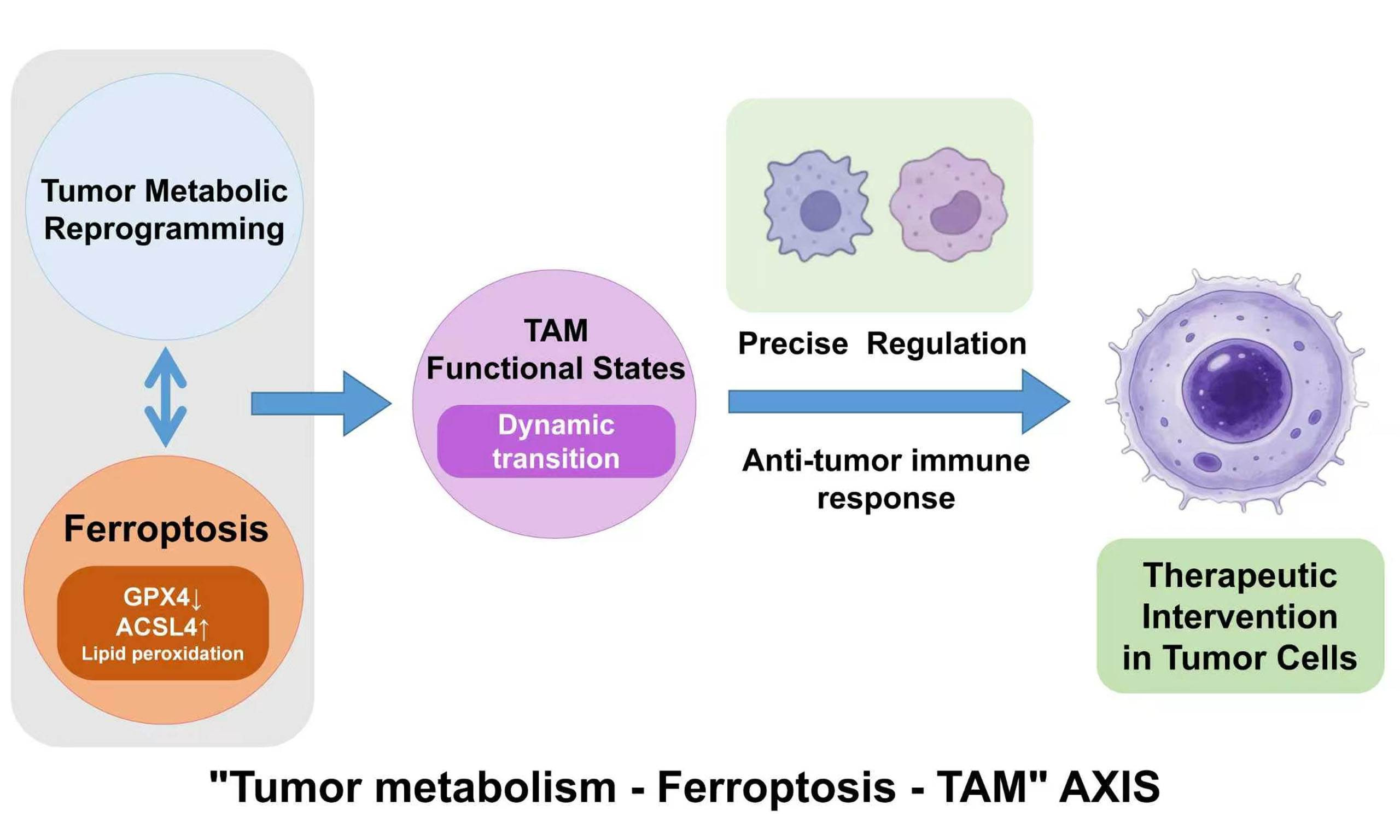
Tumor metabolic reprogramming serves as a fundamental driver of malignancy, fueling cancer cell growth while reshaping the immune landscape through metabolite accumulation. Ferroptosis, an iron-dependent form of regulated cell death, is intricately linked to these metabolic shifts. In the tumor microenvironment, tumor-associated macrophages (TAMs) are pivotal in modulating immune evasion and therapeutic resistance through diverse and context-dependent functional states. Emerging evidence suggests that metabolic alterations can dictate TAM functional plasticity by intersecting with ferroptosis-related pathways. However, the precisely orchestrated mechanisms within the “tumor metabolism-ferroptosis-TAM” axis remain to be fully integrated. This narrative review critically examines current evidence on tumor metabolic reprogramming and its impact on ferroptosis and TAM functional-state remodeling. We specifically focus on the molecular crosstalk through which metabolic and ferroptosis-associated signals may shape macrophage inflammatory, immunoregulatory, tissue-remodeling, and oxidative-stress-associated programs, and discuss potential combinatorial strategies targeting this regulatory axis.
Keywords


 Download PDF
INTRODUCTION
The proliferation of malignant tumor cells necessitates metabolic reprogramming, typified by the Warburg effect, dysregulated lipid metabolism, and aberrant amino acid catabolism. Rather than merely fulfilling the bioenergetic and synthetic demands of neoplastic cells, this metabolic rewiring establishes nutrient gradients and localized acidosis that alter the tumor microenvironment (TME)[1]. This microenvironment forms a “cold tumor” ecological niche through nutrient competition and accumulation of immunosuppressive metabolites, depleting effector immune cells, while immunosuppressive cells exhibit strong metabolic adaptability, thereby reinforcing the immunosuppressive environment[2]. Delineating the precise mechanisms by which metabolic aberrations orchestrate immune evasion remains an essential prerequisite for optimizing oncological interventions.
Meanwhile, ferroptosis functions as a distinct form of regulated cell death (RCD) driven by iron-dependent lipid peroxidation[3]. The initiation of this pathway is contingent upon a shift in the equilibrium between iron homeostasis, lipid remodeling, and cellular antioxidant networks. Beyond executing direct cytotoxicity, ferroptosis modifies the immune microenvironment via the extrusion of damage-associated molecular patterns (DAMPs)[4]. Consequently, ferroptosis functions as a mechanistic nexus intertwining metabolic alterations with immune remodeling, presenting a critical variable in assessing tumor immune heterogeneity.
Tumor-associated macrophages (TAMs) represent the predominant immunoregulatory infiltrating cell population in the TME of most solid tumors. This plasticity is also therapeutically relevant, because TAMs may either antagonize or support anticancer treatment depending on their functional state and microenvironmental context, providing a rationale for macrophage reprogramming rather than indiscriminate macrophage depletion[5]. Macrophages exhibit substantial functional plasticity. In response to local biochemical, cellular, spatial, and therapeutic cues, TAMs acquire a continuum of dynamically regulated functional states rather than two fixed phenotypes. In this review, the terms “M1-like” and “M2-like” are used only as operational descriptors of dominant inflammatory and immunoregulatory or tissue-remodeling programs, respectively; they do not denote discrete, stable, or mutually exclusive macrophage populations. Regulatory TAM states and oxidative stress-associated macrophage (Mox)-like states are likewise treated as distinct, context-dependent programs with different transcriptional, metabolic, and functional characteristics, rather than as synonyms for M2-like and M1-like states, respectively. In human tumors, these programs frequently overlap and vary according to tissue location, disease stage, and treatment exposure. In established malignancies, TME-derived signals often favor TAM programs associated with immunosuppression, angiogenesis, extracellular matrix remodeling, and impaired cytotoxic T-cell function. Tumor-derived metabolites are increasingly recognized as important mediators of this functional reprogramming. Accordingly, targeting TAM metabolism or modulating ferroptosis-associated signaling to alleviate immunosuppression and therapeutic resistance represents an emerging therapeutic strategy.
Although extant literature has independently characterized tumor metabolic reprogramming, the mechanisms of ferroptosis, and the immunometabolic dynamics of TAMs, an integrated and critical narrative synthesis of their tripartite interaction remains incomplete. Specifically, the proposed framework linking tumor metabolism, ferroptosis, and TAM functional-state remodeling requires critical evaluation, as the exact parameters dictating whether ferroptosis triggers immunogenic activation or paradoxically induces inflammation-associated immunosuppression remain poorly defined. Furthermore, the mechanistic variability of this axis across distinct histological subtypes introduces complexities that challenge its universal clinical applicability.
This narrative review critically evaluates representative mechanistic and translational studies concerning ferroptosis, TAMs, the tumor immune microenvironment, and immunometabolism. We examine how specific metabolic alterations influence ferroptotic susceptibility and assess the resulting immune consequences, with particular attention to the ability of metabolites, ferroptosis regulators, and signals released by ferroptotic tumor cells to reshape TAM functional programs. We further discuss the therapeutic potential and current pharmacological, safety, and biological barriers associated with targeting these interactions.
INTERPLAY BETWEEN TUMOR METABOLIC REPROGRAMMING AND FERROPTOSIS
The execution of ferroptosis is inherently linked to metabolic dysregulation, provided that the accumulation of lipid peroxides exceeds the threshold of cellular homeostatic buffering capacity. Consequently, the specific metabolic configurations of neoplastic cells determine their baseline susceptibility to ferroptotic induction.
Aberrant glucose metabolism and ferroptosis
Despite sufficient oxygen availability, malignant cells frequently maintain continuous lactate extrusion via aerobic glycolysis. In neoplastic populations overexpressing SLC7A11, maintaining cystine uptake necessitates substantial glucose consumption to support reduced nicotinamide adenine dinucleotide phosphate (NADPH) biosynthesis via the pentose phosphate pathway. Although this adaptation sustains glutathione (GSH) synthesis, it inadvertently establishes a strict glucose dependency[6]. Consequently, intense intercellular glucose competition within the TME limits NADPH regeneration rates. This microenvironmental deprivation, combined with metabolic acidosis, destabilizes redox equilibrium, facilitating the lipid peroxidation cascade when reactive oxygen species (ROS) accumulation surpasses antioxidant thresholds[7]. Evidence indicates that restricting glucose transport or inhibiting key glycolytic enzymes compromises tumor cells’ redox buffering capacity, thereby lowering the threshold for ferroptosis induction[8].
Lipid metabolic reprogramming and ferroptosis
Lipid metabolic rewiring functions as a primary determinant of ferroptotic vulnerability. While neoplastic cells frequently upregulate de novo fatty acid synthesis, polyunsaturated fatty acids (PUFAs) (the principal substrates for oxidation) determine cellular ferroptosis sensitivity based on their integration rate into cellular phospholipids[9]. Acyl-CoA synthetase long-chain family member 4 (ACSL4) regulates the esterification of PUFAs into membrane phospholipids, indicating that its relative expression alters the probability of ferroptotic lipid damage[10]. Conversely, the incorporation of exogenous monounsaturated fatty acids (MUFAs) or the upregulation of stearoyl-CoA desaturase 1 (SCD1) modulates membrane lipidomic profiles, elevating the MUFA/PUFA ratio and establishing a biochemical resistance mechanism that impedes lipid peroxide propagation[11].
Iron dyshomeostasis and ferroptosis
Disruption of cellular iron homeostasis is a core driver and key permissive factor for ferroptosis. Tumor cells frequently exhibit elevated iron reliance, increasing iron influx via transferrin receptor (TfR1) upregulation while restricting ferritin-mediated sequestration. This imbalance generates an expanded intracellular labile iron pool (Fe2+)[12]. The availability of free Fe2+ catalyzes lipid peroxidation via Fenton chemistry. In scenarios where endogenous antioxidant defenses are compromised, the accumulation of these oxidized lipids results in structural membrane destabilization, facilitating ferroptotic cell death[13].
MOLECULAR MECHANISMS OF FERROPTOSIS AND ITS DICHOTOMOUS ROLE IN TUMOR IMMUNOMODULATION
Ferroptosis operates under the regulation of distinct intracellular defense networks. The activation specificities of these networks introduce dichotomous effects on tumor immunity, indicating that therapeutic efficacy depends on resolving the temporal and spatial dynamics of these immunomodulatory signals.
Distinctive hallmarks of ferroptosis
Ferroptosis represents a regulated mechanism distinct from apoptosis, necroptosis, and autophagy regarding morphological, biochemical, and genetic criteria[14]. Its pathway relies on the accumulation of iron-dependent lipid peroxides exceeding cellular tolerance limits - a process subject to inhibition by specific iron chelators or radical-trapping antioxidants[15]. Morphologically, it involves condensed mitochondrial membrane densities and diminished cristae; biochemically, it requires iron-dependent ROS propagation and specific oxidized phospholipids[3]. The cellular response to this process is mediated by parallel antioxidant networks.
Principal intracellular ferroptosis defense networks
There are three core ferroptosis defense pathways within cells:
First, the System Xc--GSH-Glutathione Peroxidase 4 (GPX4) axis, where the cystine/glutamate antiporter regulates cystine import for GSH biosynthesis. GPX4 utilizes GSH to reduce phospholipid hydroperoxides to corresponding lipid alcohols, functioning as the primary mechanism for maintaining membrane integrity[16].
Second, the ferroptosis suppressor protein 1 (FSP1) - coenzyme Q10 (CoQ10) system, which operates independently in various membrane compartments. FSP1 interfaces with membrane-embedded CoQ10 to generate an extramitochondrial antioxidant rheostat, scavenging lipid peroxyl radicals[17].
Third, the guanosine triphosphate (GTP) cyclohydrolase 1 (GCH1)-tetrahydrobiopterin (BH4) signaling node, which synthesizes endogenous antioxidants [BH4 and dihydrobiopterin (BH2)] and specifically shields di-polyunsaturated fatty acid (di-PUFA)-tailed phospholipids from oxidative degradation, thus modifying the lipidome[18].
The dependency on these respective networks is highly lineage-specific and subject to microenvironmental pressure. This heterogeneity accounts for the differential sensitivities between neoplastic and immune populations under identical metabolic stress, highlighting the necessity for calibrated therapeutic targeting.
Ferroptosis as a conditional regulator of tumor immunity
Importantly, the immunological consequence of ferroptosis depends strongly on the cellular compartment in which it occurs. Ferroptosis of tumor cells may promote immunogenic signaling or generate macrophage-modulating cues, whereas ferroptosis of macrophages, myeloid-derived suppressor cells (MDSCs), or effector lymphocytes can produce distinct and sometimes opposing immune consequences[19]. Therefore, the cellular source of ferroptosis is specified below when interpreting its effects on the TME.
Initially, early-stage ferroptotic cancer cells release specific DAMPs capable of initiating immunogenic responses[20]. However, this effect is frequently counteracted as surviving neoplastic populations develop adaptive resistance through SLC7A11 up-regulation, NRF2 activation, or lipidomic restructuring, thereby maintaining tumor viability under persistent therapeutic pressure[21].
Additionally, the ferroptotic process generates secondary signaling cues. The release of oxidized phospholipids and metabolic intermediates like extracellular glutamate establishes a paracrine gradient that conditionally activates or suppresses adjacent leukocytes depending on localized concentrations[22]. In the context of the TME, these chemical outputs may alter inflammatory, immunoregulatory, and tissue-remodeling programs in infiltrating TAMs in a concentration- and context-dependent manner, complicating the assumption that ferroptosis acts uniformly as a tumor-suppressive event.
THE CONDITIONAL INTERPLAY AMONG TUMOR METABOLISM, FERROPTOSIS, AND TAM FUNCTIONAL-STATE REMODELING
Metabolic variation within the TME intersects continuously with ferroptosis-associated signaling and may shape the functional states of TAMs. Rather than operating as isolated occurrences, this remodeling relies on a tripartite regulatory structure: the direct modulation of macrophage ferroptotic thresholds by local metabolites, the intrinsic expression dependency of ferroptosis-regulating enzymes, and the paracrine re-education of macrophages by signaling cues extruded from ferroptotic neoplastic cells[23]. These mechanisms form an integrated, context-dependent network. Notably, the macrophage states in this axis-including M1-like, M2-like, regulatory (Reg-TAM), and Mox profiles-are context-driven functional states, not stable, interchangeable lineages. This network shows that signals from dying cells can reshape the local immune landscape under specific metabolic constraints [Figure 1].
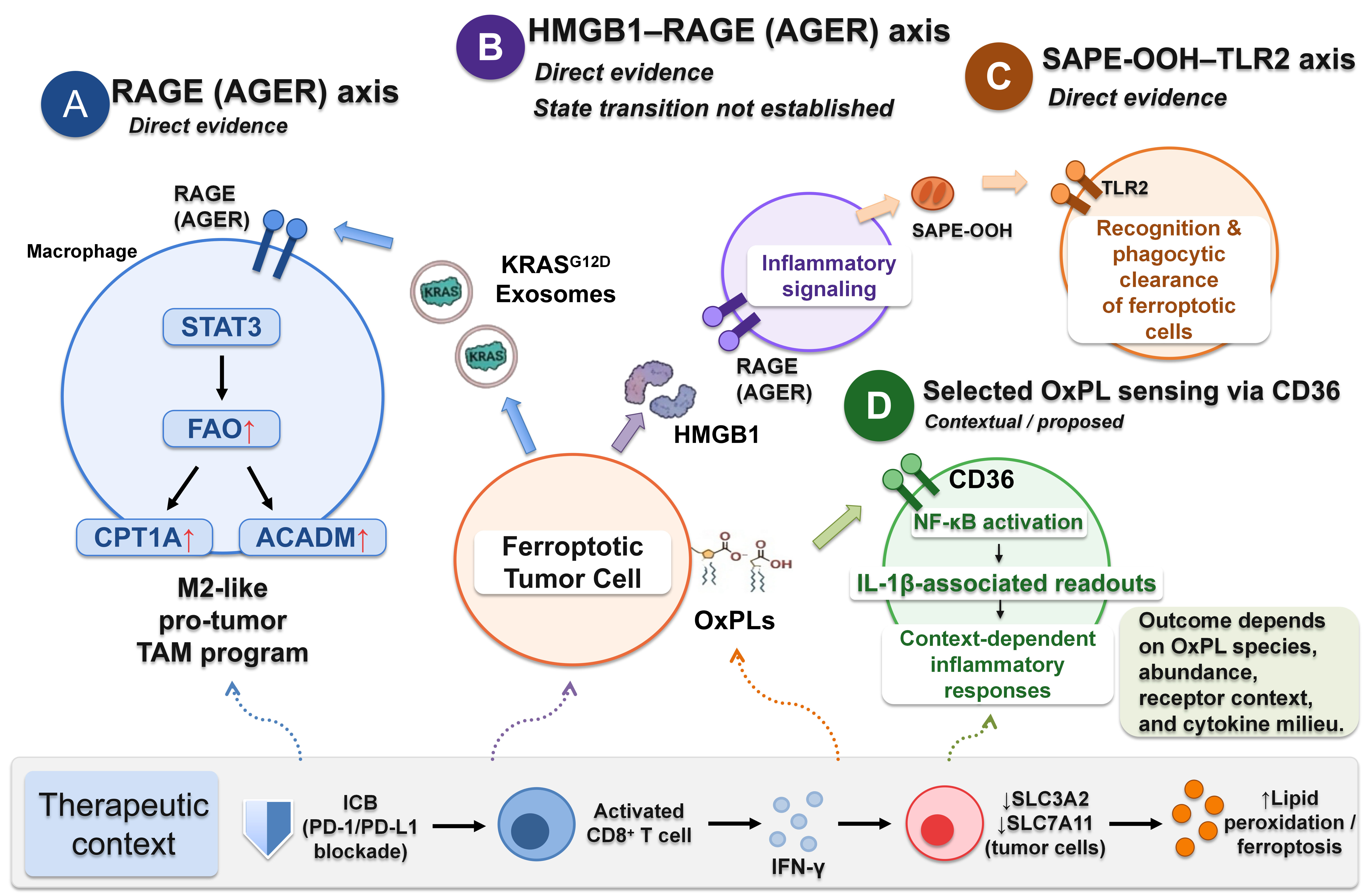
Figure 1. Context-dependent macrophage responses to signals released by ferroptotic tumor cells. Ferroptotic tumor cells release multiple classes of signals that can elicit distinct macrophage responses with different levels of experimental support. (A) In KRASG12D pancreatic ductal adenocarcinoma models, ferroptotic tumor cells release KRASG12D-containing exosomes that are taken up by macrophages in a RAGE/AGER-dependent manner, leading to STAT3 activation, increased FAO, upregulation of CPT1A and ACADM, and an M2-like pro-tumor TAM program; (B) Ferroptotic tumor cells can also release HMGB1, which engages RAGE (AGER) on macrophages and promotes inflammatory signaling; however, a defined macrophage-state transition has not been established; (C) SAPE-OOH exposed on the surface of ferroptotic cells is recognized by TLR2 on macrophages and functions as an “eat-me” signal that promotes recognition and phagocytic clearance of ferroptotic cells; (D) Other oxidized phospholipids (OxPLs) may be sensed through CD36 and are shown here as a contextual or proposed pathway associated with NF-κB activation, IL-1β-associated readouts, and context-dependent inflammatory responses. Importantly, this outcome is not uniform and may vary according to OxPL species, abundance, receptor context, and the surrounding cytokine milieu. FAO: Fatty acid oxidation; ACADM: acyl-CoA dehydrogenase medium chain; TAM: tumor-associated macrophage; SAPE-OOH: 1-stearoyl-2-(15-HpETE)-sn-glycero-3-phosphatidylethanolamine; TLR2: toll-like receptor 2; NF-κB: nuclear factor kappa-light-chain-enhancer of activated B cells; IL-1β: interleukin-1 beta; PD-1: programmed cell death protein 1; PD-1L: programmed cell death ligand 1; ICB: immune checkpoint blockade; RAGE (AGER): receptor for advanced glycation end products; STAT3: signal transducer and activator of transcription 3; CPT1A: carnitine palmitoyltransferase 1A; HMGB1: high mobility group box 1.
Metabolic drivers of ferroptosis-associated macrophage functional-state remodeling
The epigenetic and transcriptional modulation by lactate
Lactate, proceeding from aerobic glycolysis (the Warburg effect), accumulates sequentially within the TME, functioning less as a passive byproduct and more as a primary structural variable regulating TAM polarization[24]. Upon being internalized via monocarboxylate transporter (MCT)-mediated H+/lactate symport, lactate prevents the degradation of hypoxia-inducible factor-1α (HIF-1α), stabilizing its functional concentration. This stabilization increases the expression of M2-associated markers, including Arg1 and Fizz1, which is consistent with an immunoregulatory and tissue-remodeling program rather than a discrete or stable M2 macrophage identity. In the cited experimental model, these changes were associated with vascular endothelial growth factor (VEGF)-dependent angiogenesis and ARG1-related tumor-promoting activity[24].
In parallel, lactate-associated histone lactylation may influence ferroptosis resistance, although the available evidence is currently derived mainly from tumor cells rather than TAMs. In colorectal cancer cells, H3K18 lactylation upregulates insulin-like growth factor 2 mRNA-binding protein 2 (IGF2BP2) and enhances NRF2-associated ferroptosis resistance[25]. Whether an analogous epigenetic mechanism regulates ferroptotic susceptibility in TAMs remains to be established.
Given the central role of the NRF2-GPX4 pathway in ferroptosis defense, this epigenetic event may indirectly modulate macrophage ferroptotic sensitivity. Its phenotypic impact still lacks direct functional validation.
Furthermore, adjusting to lactate-rich conditions necessitates alternative metabolic processing, which potentially depletes requisite redox cofactors, including NADPH. This competitive expenditure neutralizes the baseline antioxidant buffering capacity of macrophages, lowering their defense threshold against sporadic lipid peroxidation events.
Lipid metabolic reprogramming and ferroptotic vulnerability
The spatial rewiring of lipid metabolism dictates tumor immune circumvention strategies. Specifically, the processing efficiency and integration rate of PUFAs stringently dictate cellular execution thresholds for ferroptosis[9]. Evidence corroborates that ambient PUFA availability alters macrophage survival parameters, primarily because PUFAs function as the obligate structural substrates for iron-dependent peroxidation. The efficiency of their oxidative modification establishes the rate-limiting step for this localized cell death modality[26]. Consequently, within the hypoxic and nutrient-restricted TME, intercellular competition for lipid precursors continually forces macrophages into severe metabolic adaptations. This environmental pressure may reshape macrophage metabolic, inflammatory, and tissue-remodeling programs while also altering their susceptibility to ferroptotic stress.
This metabolic rewiring further shapes their functional polarization profiles, fine-tunes their immunoregulatory capacity, and ultimately modulates their threshold for ferroptotic injury.
Potential intrinsic regulation of macrophage functional states by ferroptosis regulators
The abundance and activity of core ferroptosis regulators, including GPX4 and ACSL4, may influence macrophage survival, oxidative-stress responses, and inflammatory functions. Their effects should be interpreted according to the molecular and functional readouts measured in each experimental model, rather than as evidence of conversion into fixed or mutually exclusive macrophage phenotypes.
Ferroptosis regulators modulate macrophage homeostasis
GPX4 acts as a major defense against phospholipid peroxidation and is critical for maintaining macrophage redox homeostasis under inflammatory stress[27]. However, direct evidence that GPX4 restriction reprograms TAMs toward a stable M1-like state remains limited. In non-neoplastic inflammatory models, macrophage ferroptotic stress has been associated with increased M1-associated inflammatory features[28]. It suggests a possible connection between antioxidant capacity and macrophage activation rather than establishing a universal GPX4-dependent TAM transition.
ACSL4 promotes the incorporation of polyunsaturated fatty acids into membrane phospholipids and thereby influences ferroptotic susceptibility. In nasopharyngeal carcinoma models, tumor-cell ACSL4 overexpression or erastin treatment increased lipid peroxidation and ferroptosis and was accompanied by increased M1-associated macrophage features; these effects were attenuated by ferrostatin-1[29]. Because ACSL4 was manipulated primarily in tumor cells, these findings support an indirect ferroptosis-dependent tumor-cell-macrophage interaction rather than direct evidence that macrophage-intrinsic ACSL4 determines TAM state.
System Xc--mediated metabolic competition
Pharmacological inhibition of SLC7A11 can restrict cystine uptake, deplete GSH, and induce ferroptosis in tumor cells, as demonstrated in lung cancer models treated with gigantol[30]. By contrast, the proposed depletion of extracellular cystine and consequent impairment of GSH synthesis in adjacent TAMs remain a biologically plausible metabolic-competition model rather than a directly demonstrated mechanism in tumor tissues[31]. Direct measurements of extracellular cystine availability, macrophage GSH levels, ferroptotic injury, and functional-state changes in spatially defined tumor niches are still required.
The immunoglobulin superfamily member 9-interleukin-6/signal transducer and activator of transcription 3 axis and a senescence-associated TAM state
Beyond direct oxidative distress, tumor-derived signals can induce macrophage states that are not adequately captured by the M1-like/M2-like framework. Tumor-derived immunoglobulin superfamily member 9 (IGSF9) interacts with transmembrane and ubiquitin-like domain containing 1 (TMUB1) on TAMs and activates interleukin-6 (IL-6)/signal transducer and activator of transcription 3 (STAT3) signaling, inducing a senescence-associated TAM state characterized by a senescence-associated secretory phenotype (SASP)[32]. This state is accompanied by impaired phagocytic capacity and increased production of immunoregulatory factors that suppress local T-cell proliferation and activation. It should be discussed separately from M1-like and M2-like programs because it is defined by senescence-related transcriptional and functional alterations rather than by conventional polarization markers alone[32].
TME remodeling via ferroptotic neoplastic cues
Tumor cells undergoing ferroptotic disruption continually extrude a distinct sequence of DAMPs and oxidized lipid mediators. However, assuming these components function solely as immunological activators ignores their capacity to inadvertently drive immunosuppression depending on receptor interaction and microenvironmental conditions. In this section, ferroptosis occurs primarily in tumor cells, whereas TAMs act as recipient cells that sense ferroptosis-associated extracellular signals. Depending on the molecular cue and microenvironmental context, the resulting macrophage response may be immunostimulatory, immunosuppressive, or context-dependent.
Contextual DAMP signaling and paracrine immunosuppression
During continuous ferroptosis, compromised cells release distinct DAMPs and oxidized lipid mediators with different levels of experimental validation. HMGB1, a prototypical DAMP released by ferroptotic tumor cells, engages Receptor for advanced glycation end products (RAGE) on TAMs and triggers Nuclear factor kappa-light-chain-enhancer of activated B cells (NF-κB)-dependent inflammatory signaling and macrophage recruitment[33]. However, direct experimental evidence that HMGB1-RAGE signaling alone drives TAM transition toward a defined Reg-TAM or M2-like functional state remains limited. SAPE-OOH [1-stearoyl-2-(15-HpETE)-sn-glycero-3-phosphatidylethanolamine], exposed on the surface of ferroptotic cells, acts as a TLR2-dependent “eat-me” signal that mediates recognition and phagocytic clearance of ferroptotic cells[33,34]. This pathway has confirmed phagocytic function but has not been shown to induce M1-like, M2-like, Reg-TAM, or Mox-like macrophage states. Integrative synthesis of the available literature indicates that sustained accumulation of these and related mediators during chronic ferroptotic stress may collectively shape an immunosuppressive TAM program. This program is characterized by upregulated ARG1 and IL-10 expression and impaired CD8+ T-cell function[33].
Simultaneously, mutated KRASG12D (frequently associated with autophagy-dependent ferroptosis) becomes actively compartmentalized into exosomes prior to extracellular shedding[35]. Following exosomal internalization by TAMs, KRASG12D initiates localized STAT3 phosphorylation, transactivating critical fatty acid oxidation (FAO) genes [e.g., carnitine palmitoyltransferase 1A (CPT1A), acyl-CoA dehydrogenase medium chain (ACADM)]. In the cited model, this metabolic recalibration induces an FAO-associated, M2-like pro-tumor macrophage program rather than establishing a fixed M2 phenotype[35]. Ferroptosis occurring in other myeloid populations represents a distinct process. In particular, ferroptotic polymorphonuclear myeloid-derived suppressor cells (PMN-MDSCs) can generate immunosuppressive lipid mediators such as PGE2, thereby reinforcing local immune suppression rather than promoting tumor clearance[36]. This mechanism should be distinguished from tumor-cell ferroptosis that secondarily reprograms TAMs.
The dichotomous role of oxidized phospholipids
Evaluating the role of oxidized phospholipids (e.g., oxidized phosphatidylethanolamine) demands recognizing their dichotomous physiological capability. When recognized by scavenger receptors like CD36, these moieties precipitate metabolic shifts that, under precise conditions, direct TAMs toward an M1-like or Mox inflammatory functional state, marked by NF-κB engagement and localized IL-1β liberation[37].
The trajectory of TAM remodeling following ferroptosis remains heavily contingent upon multiple intersecting variables: the absolute concentration of released lipid signals, the spatial cytokine topography {baseline interferon-γ [IFN-γ] availability}, and the concurrent deployment of interventional therapies. For instance, when combined with immune checkpoint blockade, pro-ferroptotic interventions may increase tumor immunogenicity and promote IFN-γ production by activated T cells. Under appropriate cytokine conditions, IFN-γ may cooperate with ferroptosis-associated signals to counter pre-existing immunoregulatory macrophage programs and favor an M1-like inflammatory program, potentially supporting CD8+ T-cell cytotoxicity[38]. This terminology describes a dominant functional response rather than conversion into a stable M1 macrophage population. In the absence of such immune activation, ferroptosis-associated signals may instead reinforce tumor tolerance.
The lower panel summarizes an established therapeutic pathway in tumor cells: immune checkpoint blockade (ICB) activates CD8+ T cells, which produce IFN-γ, downregulate solute carrier family 3 member 2 (SLC3A2)/SLC7A11 in tumor cells, and thereby enhance lipid peroxidation and ferroptosis. Potential downstream effects of this ferroptosis-promoting context on macrophage programs are shown as indirect and outcome-variable. Solid arrows indicate direct experimental evidence, whereas dashed arrows indicate contextual, indirect, or proposed links.
Histotype-specific heterogeneity of the regulatory axis
Histotype-specific differences in lipid metabolism, glycolytic activity, antioxidant capacity, and macrophage composition may contribute to heterogeneous ferroptotic responses across tumor types. For example, Hepatocellular Carcinoma (HCC) and Pancreatic Ductal Adenocarcinoma (PDAC) exhibit distinct metabolic and immunometabolic features[39,40]. However, these differences should not be interpreted as establishing a fixed hierarchy of ferroptosis sensitivity among cancer types. Rather, they provide a biological rationale for investigating whether tumor-specific metabolic states influence ferroptosis susceptibility and TAM responses. Direct comparative studies across histotypes are still required.
Concurrently, the derivation pattern of TAM populations further dictates mechanistic susceptibility. In Glioblastoma (GBM), the TAM compartment involves dual ontogeny: sessile brain-resident microglia integrating via Lysyl oxidase (LOX) signals, countered by peripheral monocytes recruited via tumor-derived CCN1[2]. The differing hypoxic exposure histories and cytokine proximities associated with these differing ontogenies strictly restrict how each subset responds to induced metabolic stress. Similarly, breast carcinoma models suggest that spatially localized glycolytic variation correlates with regional differences in the density of macrophages expressing M2-associated markers[41].
First, sex hormones and receptor signaling establish organ-specific baseline immune landscapes. In urological malignancies, cell-type-specific expression and function of androgen and estrogen receptors drive divergent immune microenvironment phenotypes, classifying bladder cancer as immune-hot and prostate cancer as immune-cold. These hormone-driven differences directly shape TAM polarization states and overall responsiveness to immunotherapy[42]. Such baseline immune variation may further modify the activity of the metabolism-ferroptosis-TAM axis across organ sites.
Second, crosstalk between tumor metabolic reprogramming and CD8+ T-cell function contributes to tissue- and tumor-specific immune profiles. Tumor metabolic rewiring can impair CD8+ T-cell effector function through nutrient competition, the accumulation of immunosuppressive metabolites, and metabolic reprogramming of stromal and myeloid populations, including macrophages. Accordingly, targeting selected tumor metabolic adaptations may help alleviate immunosuppression and restore CD8+ T-cell activity[43]. The relative contribution of individual metabolic pathways varies across tumor types; for example, lipid metabolic abnormalities have been implicated in hepatocellular carcinoma, whereas glycolytic alterations and nutrient competition have been described in pancreatic ductal adenocarcinoma[43]. These observations suggest that tissue-specific metabolic-immune environments may also influence TAM functional states and, potentially, the local response to ferroptosis-associated stress.
Third, therapeutic regimens can dynamically remodel intratumoral immune-cell subsets in a context-dependent manner. In non-small cell lung cancer, neoadjuvant chemoimmunotherapy was associated with coordinated changes in bystander CD8+ and conventional CD4+ T-cell populations, some of which correlated with major pathological response[44]. Changes in HIF-1α expression also showed an association with changes in conventional CD4+ T-cell abundance, although the underlying mechanism remains to be established[44]. Such therapy-induced changes in the T cell compartment alter local cytokine signaling and TAM-T cell crosstalk, which further adjusts the output of the core metabolism-ferroptosis-TAM regulatory axis.
Accordingly, the efficacy of generalized pro-ferroptotic strategies is unlikely to be uniform across solid tumors. Future clinical translation will require prospective evaluation of tumor metabolic features, ferroptosis susceptibility, and TAM functional-state composition to determine whether these variables can support patient stratification. At present, such stratification remains investigational rather than clinically established. Clinical implementation therefore requires pretreatment stratification that considers the tumor-specific lipidomic profile together with the local composition and spatial distribution of TAM functional states. Calibrating ferroptosis induction alongside agents targeting discrete TAM signaling parameters remains a rigorous prerequisite for circumventing the functional limitations of existing monotherapies and securing durable combinatory responses[45] [Table 1].
Experimental evidence supporting interactions among tumor metabolism, ferroptosis, and macrophage functional states
| Disease model | Species | Pathway | Macrophage changes | Ferroptosis changes | Main result | Evidence strength | Study limitation | Reference |
| Lewis lung carcinoma; tumor-derived lactate | Murine syngeneic tumors; mouse bone-marrow-derived macrophages; ex vivo TAMs | Tumor-derived lactic acid; monocarboxylate-transporter-dependent uptake; macrophage HIF-1α | ARG1, VEGF, FIZZ1, macrophage galactose-type C-type lectin 1 (MGL1) and 2 (MGL2) expression; HIF-1α dependence; tumor-supporting activity | Not assessed | Tumor-derived lactic acid induced a HIF-1α-dependent immunoregulatory and angiogenic macrophage program | Partial/bridging | Supports tumor metabolism-to-macrophage signaling, but does not establish a ferroptosis-dependent mechanism | Colegio et al., 2014[24] |
| Colorectal cancer; lactate-associated ferroptosis resistance | Human colorectal cancer cell models; macrophage-related assays; murine xenograft models | Lactate/H3K18 lactylation-IGF2BP2-NRF2-GPX4 signaling | Macrophage-specific functional-state readouts were not the primary endpoint; no defined TAM-state or T-cell-suppression assay | Tumor-cell ferroptosis resistance; NRF2/GPX4 antioxidant defense; lipid-peroxidation-related assays | Lactate-driven lactylation increased IGF2BP2-NRF2 signaling and ferroptosis resistance in colorectal cancer | Contextual/adjacent | Does not directly demonstrate lactate-controlled macrophage ferroptosis or TAM functional-state remodeling in tumors | Zhu et al., 2025[25] |
| KRASG12D pancreatic ductal adenocarcinoma | Human and mouse PDAC cells; human peripheral-blood-monocyte-derived macrophages; mouse PDAC models; human tissue correlation | Autophagy-dependent ferroptosis; exosomal KRASG12D release and macrophage uptake; AGER/RAGE-STAT3-fatty acid oxidation signaling | KRASG12D uptake; STAT3 activation; CPT1A/ACADM expression; M2-associated and tumor-promoting macrophage functions | Ferroptotic tumor-cell death was experimentally induced and inhibited; autophagy related 5 (ATG5)-dependent release was assessed | Ferroptotic PDAC cells released KRASG12D, which induced a fatty-acid-oxidation-associated pro-tumor macrophage program | Direct - preclinical; human correlative support | Mechanism is strongly model- and genotype-specific; human evidence is correlative rather than interventional | Dai et al., 2020[35] |
| Mammary carcinoma and multiple ferroptotic-cell models | THP-1-derived, bone-marrow-derived and peritoneal macrophages; 4T1 mouse mammary tumor model; Tlr2-knockout mice | GPX4 dysfunction or RSL3-induced ferroptosis; SAPE-OOH; TLR2 inhibition, silencing or knockout; ACSL4 inhibition | Macrophage recognition, engulfment and phagocytic clearance of ferroptotic cells | GPX4 dysfunction/deficiency; RSL3 induction; phospholipidomics of SAPE-OOH; modulation by ACSL4 inhibition | SAPE-OOH on ferroptotic cells acted as an eat-me signal recognized by macrophage TLR2 and promoted phagocytic clearance | Direct - clearance mechanism | Demonstrates phagocytosis, not conversion to an M1-like, M2-like, regulatory or Mox-like macrophage state | Luo et al., 2021[34] |
| Prostate cancer; TAM-mediated ferroptosis resistance | Human prostate cancer cells; tumor cell-macrophage coculture; mouse tumor models; human single-cell RNA-seq analyses | Tumor extracellular-vesicle miR-181a-5p; macrophage taurine production/export; tumor-cell TauT-LXRα-SCD1 signaling | M2-associated macrophage features; taurine production and export; macrophage-conditioned effects on tumor cells | Tumor-cell viability, lipid ROS and malondialdehyde (MDA); specificity for ferroptotic rather than apoptotic, autophagic or necroptotic death | TAM-derived taurine suppressed prostate-cancer-cell ferroptosis through LXRα/SCD1 signaling in a reciprocal tumor-macrophage loop | Direct - reverse-direction mechanism | Establishes TAM-to-tumor ferroptosis regulation, but not ferroptotic-tumor-cell-induced TAM remodeling | Xiao et al., 2024[46] |
| Non-small-cell lung cancer and murine tumor models | Human NSCLC tissues; THP-1/U-937-derived macrophages; mouse BMDMs; LL/2 and MC38 mouse tumor models | Tumor-derived IGSF9-TMUB1-IL-6/STAT3 signaling; anti-IGSF9 intervention | Senescence-associated markers and SASP; reduced phagocytic capacity; T-cell suppression; spatial accumulation of senescence-like TAMs | Not assessed | Tumor-derived IGSF9 induced a senescence-associated, immunosuppressive TAM state through TMUB1-IL-6/STAT3 signaling | Partial/bridging | Supports tumor-to-TAM reprogramming but does not establish ferroptosis as the initiating process | Zhang et al., 2026[32] |
| Lung cancer; SLC7A11-GPX4-dependent ferroptosis | Lung cancer cell models; preclinical in vitro setting | Gigantol-mediated inhibition of the SLC7A11-GPX4 axis | Not assessed | SLC7A11/GPX4 suppression, redox disruption and ferroptosis-associated tumor-cell death | Gigantol induced ferroptosis in lung cancer cells through the SLC7A11-GPX4 pathway | Contextual/adjacent | Does not test cystine competition with macrophages, macrophage GSH depletion or any TAM functional outcome | Chen et al., 2024[30] |
THERAPEUTIC INTERVENTIONS TARGETING THE “TUMOR METABOLISM-FERROPTOSIS-TAM REPROGRAMMING” AXIS
Strategies evaluating this regulatory axis suggest theoretical utility for redefining TME interactions. However, the objective of precipitating tumor-specific ferroptosis while avoiding immunoregulatory interference relies heavily on maintaining rigorous target specificity. Most therapeutic strategies targeting the tumor metabolism-ferroptosis-TAM axis remain at the preclinical proof-of-concept stage. Although encouraging antitumor effects have been reported in cell-based and animal models, these findings should not be interpreted as evidence of clinical efficacy or readiness [Figure 2].
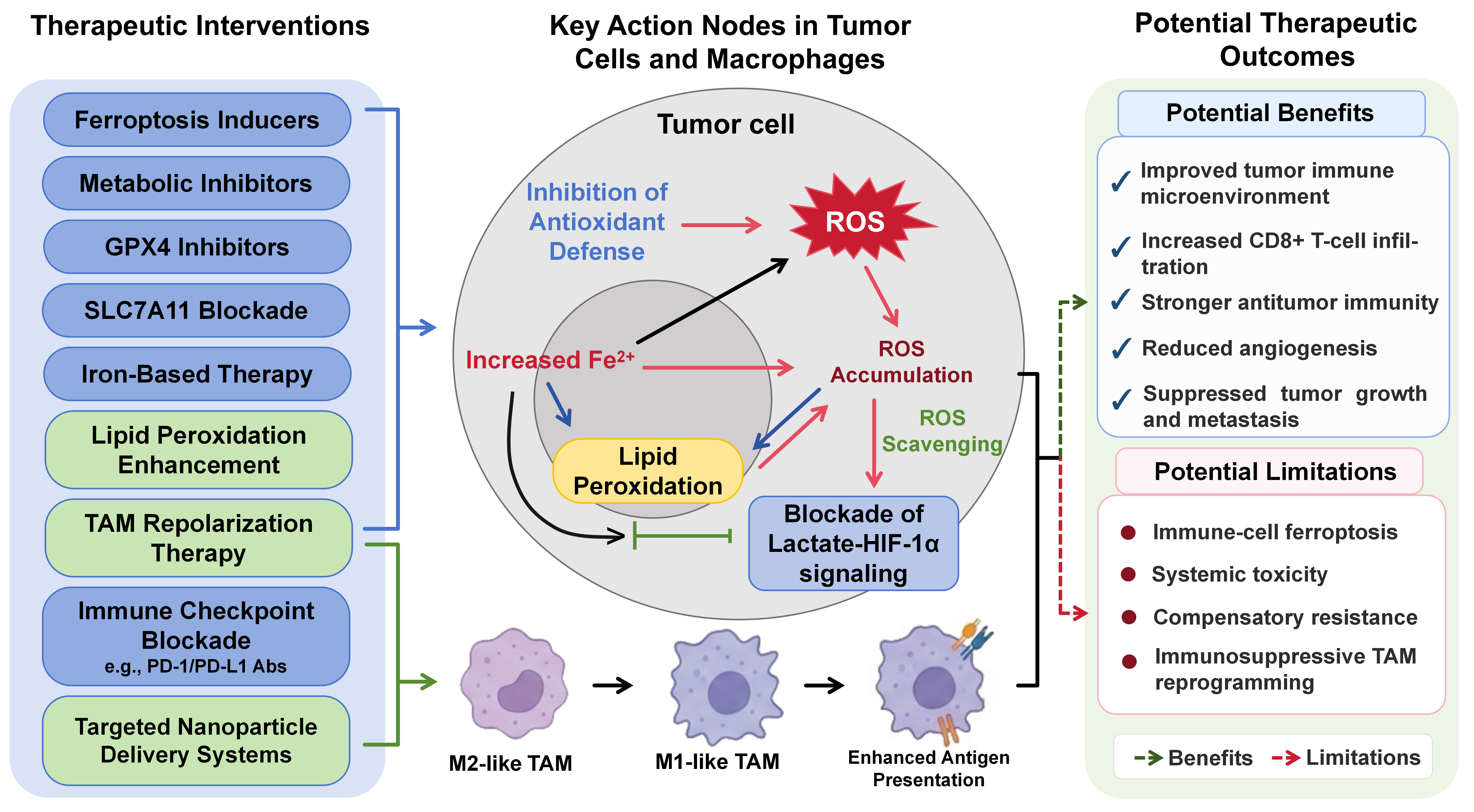
Figure 2. Proposed therapeutic interventions targeting the tumor metabolism-ferroptosis-TAM framework. This figure depicts the proposed mechanisms of action and potential therapeutic outcomes of ferroptosis inducers, metabolic inhibitors, GPX4/SLC7A11 inhibitors, iron-based therapies, lipid-peroxidation enhancement, TAM-directed therapy, immune checkpoint blockade (ICB), and targeted nano-delivery systems within the tumor microenvironment (TME). Although these interventions may enhance antitumor immunity, CD8+ T-cell infiltration, antigen presentation, and suppression of tumor growth and metastasis, they may also cause immune-cell ferroptosis, systemic toxicity, compensatory resistance, or immunosuppressive TAM reprogramming. Green dashed arrows indicate potential therapeutic benefits, whereas red dashed arrows indicate possible adverse, resistance-associated, or context-dependent effects. TAM: Tumor-associated macrophage; ROS: fatty acid oxidation; HIF-1α: hypoxia-inducible factor-1α.
Pharmacological interventions and efficacy bottlenecks
Current ferroptosis inducers (FINs) typically utilize three mechanisms: (1) System Xc- antagonism, (2) direct GPX4 inhibition, and (3) indirect orchestrators (e.g., Fenton reaction manipulation). While in vitro applications demonstrate efficient cytolysis, their clinical translation is challenged by critical limitations[19]. First, FINs display substantive pharmacokinetic restrictions, specifically regarding the metabolic instability and aqueous solubility limitations of agents such as erastin[19]; Second, the non-specific induction of lipid peroxidation corresponds to documented systemic toxicities in the central nervous system and renal parenchyma[47]; Third, neoplastic adaptation to FIN exposure frequently involves upregulation of compensatory mechanisms including the FSP1-CoQ10 system or SCD1 overexpression combined with reduced PUFA integration, progressively reducing susceptibility over successive treatment cycles[17,19].
Against this backdrop, natural product-derived ferroptosis modulators have attracted preclinical interest, although their pharmacological specificity and safety advantages remain unproven. For example, aqueous-soluble components of sporoderm-removed Ganoderma lucidum spore powder (A-GSP) promoted ferroptosis in oral squamous cell carcinoma cells, as indicated by Fe2+ accumulation, GSH depletion, increased lipid peroxidation, reduced GPX4 expression, and rescue by ferroptosis inhibitors, and also suppressed xenograft tumor growth without observable adverse reactions in mice[48]. Nevertheless, this complex extract remains far from clinically actionable because its active constituents and direct molecular targets are undefined, formal target-engagement and systemic pharmacokinetic/pharmacodynamic data are lacking, and its safety in immune cells and immunocompetent models has not been established.
Ferroptosis-associated immune checkpoint modulation and immune checkpoint blockade
The simultaneous application of FINs with immune checkpoint inhibitors (ICIs) seeks to link adaptive immunity activation with cytolytic ferroptosis. Mechanistically, ICI-activated CD8+ T cells release IFN-γ, which suppresses SLC3A2 and solute carrier family 7 member 11 (SLC7A11) expression in tumor cells, thereby increasing tumor-cell lipid peroxidation and ferroptotic susceptibility[49]. In this setting, CD8+ T cells act as the upstream immune effector, whereas tumor cells are the principal ferroptotic compartment, resulting in an antitumor effect. Preclinical melanoma models demonstrate numerical tumor suppression compared to monotherapies. However, this synergy assumes that ferroptosis-derived DAMPs act preferentially to sustain anti-tumor immunity without promoting compensatory immunoregulatory TAM programs[49].
Beyond combination with exogenous immune checkpoint inhibitors, ferroptosis-inducing platforms may themselves modulate tumor-cell immune checkpoint expression. A hydrogen peroxide-responsive gallium-releasing nanoplatform, Ga@MnO2@Alb, was reported to induce tumor-cell ferroptosis while suppressing CD47 and programmed cell death ligand 1 (PD-L1) expression through a mitochondria-AMP-activated protein kinase (AMPK)-c-MYC signaling axis[50]. Downregulation of the innate immune checkpoint CD47 enhanced macrophage-mediated phagocytosis of tumor cells, whereas reduced PD-L1 expression increased T-cell cytotoxicity. These findings suggest that ferroptosis-associated immune checkpoint modulation may reshape the tumor immune microenvironment by simultaneously relieving macrophage- and T-cell-directed inhibitory signals. However, the reported macrophage effect primarily involved enhanced phagocytic function rather than a directly demonstrated transition toward a stable M1-like or M2-like state. Whether such checkpoint modulation induces durable TAM functional-state remodeling remains to be established. Checkpoint-associated immune escape is further complicated by intertumoral and spatial heterogeneity in checkpoint expression. In melanoma, for example, PD-L1 expression has shown associations with response to programmed cell death protein 1 (PD-1) blockade under selected assessment conditions, but its prognostic and predictive value remains controversial[51]. Therefore, PD-L1 expression alone is unlikely to capture the broader immune context determining the efficacy of ferroptosis-checkpoint combinations.
Furthermore, within untreated niches, TAMs displaying M2-associated immunoregulatory features can transfer metabolites such as taurine, which subsequently limits oxidative collapse via liver X receptor alpha (LXRα)/SCD1 activation[46]. Establishing an effective FIN/ICI response requires specific disruption of this pathway to shift macrophage activity toward pro-inflammatory and tumor-restrictive programs rather than toward a discrete M1 phenotype[52]. By contrast, ferroptotic stress occurring within activated CD8+ T cells or Natural killer (NK) cells themselves can impair their survival and cytotoxic function, producing an immunosuppressive consequence rather than enhancing tumor-cell killing[22]. Current developmental strategies propose targeted nanomedicine platforms to isolate these variables[33,53]. Early evidence involving attenuated tumoral SLC3A2 expression in patients responsive to nivolumab provides initial correlations[49], yet achieving reliable clinical modulation remains contingent upon rigorous patient stratification.
Recent strategies for ferroptosis-guided TAM functional reprogramming
Recent studies have explored two principal approaches to modulating TAM functional states through ferroptosis. The first involves manipulating ferroptosis-related pathways in tumor cells or macrophages. In nasopharyngeal carcinoma models, ACSL4 overexpression or erastin treatment increased lipid peroxidation and tumor-cell ferroptosis and was accompanied by a shift from M2-associated toward M1-associated macrophage features; ferrostatin-1 reversed these effects, supporting a ferroptosis-dependent component[29]. In gastric cancer, tumor-derived exosomal calcium/calmodulin-dependent protein kinase II alpha (CaMK2A) promoted zinc finger DHHC-type palmitoyltransferase 3 (ZDHHC3)-dependent GPX4 palmitoylation and stabilization, thereby suppressing macrophage ferroptosis and favoring an M2-like immunoregulatory program. Conversely, macrophage-specific GPX4 deletion restrained tumor growth and enhanced the response to PD-1/PD-L1 blockade in murine models[54]. The second approach employs multifunctional delivery systems to coordinate tumor-cell ferroptosis with TAM remodeling. For example, an Fe3+- and erastin-containing nanomedicine induced ferroptotic stress, promoted M1-associated TAM changes, reduced fibroblast activation and collagen deposition, and improved nanoparticle penetration in pancreatic cancer models[55].
Collectively, these approaches may simultaneously enhance tumor-cell killing, alleviate myeloid immunosuppression, improve intratumoral drug penetration, and increase responsiveness to immunotherapy. Nevertheless, most of the available evidence remains preclinical. Moreover, multifunctional interventions often alter iron availability, oxidative stress, stromal organization, and immune signaling concurrently, making it difficult to determine whether the observed TAM remodeling is directly attributable to ferroptosis. Excessive ferroptosis may also damage beneficial macrophage subsets or other immune cells, whereas compensatory antioxidant responses, systemic toxicity, and formulation complexity may limit therapeutic efficacy and clinical translation. Importantly, ferroptosis does not uniformly promote antitumor macrophage activity. In KRASG12D pancreatic cancer models, ferroptotic tumor cells released exosomal KRASG12D, which was internalized by macrophages through RAGE/AGER and induced a STAT3-fatty acid oxidation-dependent M2-like pro-tumor program[35]. Thus, the therapeutic outcome of ferroptosis-guided TAM modulation is likely to depend on the targeted cell population, tumor genotype, ferroptotic signals released, treatment intensity, and local immune context.
Systemic safety and translational challenges
Translating these combined paradigms entails confronting the aforementioned dual-effect paradox: while inducing ferroptosis in tumor cells to exert tumoricidal activity, such approaches must spare tumor-infiltrating immune cells within the TME to preserve overall antitumor immunity[36]. Variations in baseline tumor lipid composition signify that lipid-enriched models (e.g., HCC) may demonstrate sensitivity, while tumors lacking sufficient PUFA densities (e.g., specific gliomas) inherently resist treatment due to substrate limitation alone[56]. Resolving these issues may require targeted delivery strategies together with candidate biomarkers reflecting ferroptosis susceptibility, such as ferritin-related iron status, 4-hydroxynonenal, or the ACSL4/GPX4 expression balance[56]. However, these markers should currently be regarded as hypothesis-generating or exploratory indicators rather than validated clinical stratification tools, and their predictive value requires prospective validation in well-defined patient cohorts. At present, the reliance on these proposed biomarkers requires rigorous multivariable prospective validation before stratifying clinical cohorts.
As a core component of the combined ferroptosis-immunotherapy paradigms, TAM polarization-targeted strategies also face translational constraints in safety and efficacy. Several focused improvements merit further development. Future regimens can shift from broad M1/M2 modulation toward subtype-specific functional reprogramming to improve therapeutic precision[5]. Pairing ferroptosis inducers with inhibitors of TAM fatty acid oxidation may block paracrine M2-like polarization and enhance combinatorial efficacy[35]. TAM-targeted nano-delivery systems also represent a viable direction to reduce off-target immune impairment.
CONCLUSION
Tumor metabolic reprogramming establishes the biochemical conditions that determine ferroptotic susceptibility. Ferroptosis-associated molecular cues may subsequently reshape the inflammatory, immunoregulatory, and tissue-remodeling programs of TAMs in a context-dependent manner, providing a potential explanation for heterogeneous therapeutic responses[23]. Evaluation of the proposed framework linking tumor metabolism, ferroptosis, and TAM functional-state remodeling must therefore account for substantial histotype-specific, spatial, and treatment-related heterogeneity. Accordingly, combinatorial therapeutic strategies should consider differences in baseline metabolic profiles, ferroptosis susceptibility, and macrophage-state composition rather than assuming a universally applicable M1/M2 polarization model.
Current evidence is derived predominantly from in vitro studies and preclinical murine models, with limited validation in human tumors. Future studies should first use single-cell and spatial multi-omics approaches to define which TAM states, anatomical niches, and ferroptosis-associated signals are linked to antitumor or immunosuppressive outcomes in human TMEs[57]; Second, TAM-directed therapies should move beyond indiscriminate macrophage depletion or binary M1/M2 repolarization toward state- and niche-specific functional reprogramming, while preserving macrophages with phagocytic and antigen-presenting activities; Third, tumor- or TAM-selective delivery systems, including microenvironment-responsive nanocarriers, optimized dosing, and rational sequencing with immune checkpoint blockade, may improve therapeutic specificity while limiting systemic toxicity and ferroptotic injury to beneficial immune cells. Finally, biomarker-guided patient stratification should integrate tumor lipid and iron status, ferroptosis-regulatory pathways, and TAM functional-state composition. Validation in immunocompetent models, patient-derived organoid-immune cell co-cultures, ex vivo tumor samples, and prospective clinical cohorts will be essential before ferroptosis-immunotherapy combinations can be considered clinically reliable.
DECLARATIONS
Authors’ contributions
Made substantial contributions to the conception and framework design of the review, literature retrieval, screening and comprehensive collation of relevant studies, evidence summary, mechanistic analysis, figure production, and original manuscript drafting: Hu H
Participated in study conception, literature organization and mechanistic analysis, made substantial contributions to content supplementation, manuscript revision and polishing, and revised and approved the manuscript: Ma Y
Availability of data and materials
Not applicable.
AI and AI-assisted tools statement
During the preparation of this manuscript, the AI tool ChatGPT (version 5.4, released 2026-03-05) was used solely for language editing. The tool did not influence the study design, data collection, analysis, interpretation, or the scientific content of the work. All authors take full responsibility for the accuracy, integrity, and final content of the manuscript.
Financial support and sponsorship
None.
Conflicts of interest
All authors declared that there are no conflicts of interest.
Ethical approval and consent to participate
Not applicable.
Consent for publication
Not applicable.
Copyright
© The Author(s) 2026.
REFERENCES
1. Liberti MV, Locasale JW. The warburg effect: how does it benefit cancer cells? Trends Biochem Sci. 2016;41:211-8.
2. Vijayanathan Y, Ho IAW. The impact of metabolic rewiring in glioblastoma: the immune landscape and therapeutic strategies. Int J Mol Sci. 2025;26:669.
3. Dixon SJ, Lemberg KM, Lamprecht MR, et al. Ferroptosis: an iron-dependent form of nonapoptotic cell death. Cell. 2012;149:1060-72.
4. Chen X, Kang R, Kroemer G, Tang D. Broadening horizons: the role of ferroptosis in cancer. Nat Rev Clin Oncol. 2021;18:280-96.
5. Mantovani A, Marchesi F, Malesci A, Laghi L, Allavena P. Tumour-associated macrophages as treatment targets in oncology. Nat Rev Clin Oncol. 2017;14:399-416.
6. Koppula P, Zhuang L, Gan B. Cystine transporter SLC7A11/xCT in cancer: ferroptosis, nutrient dependency, and cancer therapy. Protein Cell. 2021;12:599-620.
7. Koncošová M, Vrzáčková N, Křížová I, et al. Inhibition of mitochondrial metabolism leads to selective eradication of cells adapted to acidic microenvironment. Int J Mol Sci. 2021;22:10790.
8. Li H, Wu Y, Ma Y, Liu X. Interference with ENO2 promotes ferroptosis and inhibits glycolysis in clear cell renal cell carcinoma by regulating Hippo-YAP1 signaling. Oncol Lett. 2024;28:443.
9. Kagan VE, Mao G, Qu F, et al. Oxidized arachidonic and adrenic PEs navigate cells to ferroptosis. Nat Chem Biol. 2017;13:81-90.
10. Doll S, Proneth B, Tyurina YY, et al. ACSL4 dictates ferroptosis sensitivity by shaping cellular lipid composition. Nat Chem Biol. 2017;13:91-8.
11. Tesfay L, Paul BT, Konstorum A, et al. Stearoyl-CoA desaturase 1 protects ovarian cancer cells from ferroptotic cell death. Cancer Res. 2019;79:5355-66.
13. Hassannia B, Vandenabeele P, Vanden Berghe T. Targeting ferroptosis to iron out cancer. Cancer Cell. 2019;35:830-49.
14. Sharma A, Flora SJS. Positive and negative regulation of ferroptosis and its role in maintaining metabolic and redox homeostasis. Oxid Med Cell Longev. 2021;2021:9074206.
15. Kong Y, Li J, Lin R, et al. Understanding the unique mechanism of ferroptosis: a promising therapeutic target. Front Cell Dev Biol. 2023;11:1329147.
16. Friedmann Angeli JP, Schneider M, Proneth B, et al. Inactivation of the ferroptosis regulator Gpx4 triggers acute renal failure in mice. Nat Cell Biol. 2014;16:1180-91.
17. Doll S, Freitas FP, Shah R, et al. FSP1 is a glutathione-independent ferroptosis suppressor. Nature. 2019;575:693-8.
18. Kraft VAN, Bezjian CT, Pfeiffer S, et al. GTP cyclohydrolase 1/tetrahydrobiopterin counteract ferroptosis through lipid remodeling. ACS Cent Sci. 2020;6:41-53.
19. Lei G, Zhuang L, Gan B. Targeting ferroptosis as a vulnerability in cancer. Nat Rev Cancer. 2022;22:381-96.
20. Efimova I, Catanzaro E, Van der Meeren L, et al. Vaccination with early ferroptotic cancer cells induces efficient antitumor immunity. J Immunother Cancer. 2020;8:e001369.
21. Li Y, Liu Y, Zhao Y, et al. Ferroptosis in lung cancer: emerging mechanisms, therapeutic targeting, and immune modulation. Cell Signal. 2026;141:112403.
22. Li S, Li Z. The crosstalk between ferroptosis and the immune system in urological cancers: mechanisms, prognostic value, and therapeutic implications. Crit Rev Oncol Hematol. 2026;218:105070.
23. Chen X, Kang R, Kroemer G, Tang D. Ferroptosis in infection, inflammation, and immunity. J Exp Med. 2021:218.
24. Colegio OR, Chu NQ, Szabo AL, et al. Functional polarization of tumour-associated macrophages by tumour-derived lactic acid. Nature. 2014;513:559-63.
25. Zhu JF, Guo DP, Lv HN, Liang ZY, Song J, Zeng W. Histone lactylation-mediated up-regulation of IGF2BP2 enhances ferroptosis resistance via Nrf2 in colorectal cancer. Clin Transl Med. 2025;15:e70551.
26. Ma J, Zhang H, Chen Y, Liu X, Tian J, Shen W. The role of macrophage iron overload and ferroptosis in atherosclerosis. Biomolecules. 2022;12:1702.
27. Kang R, Zeng L, Zhu S, et al. Lipid peroxidation drives gasdermin d-mediated pyroptosis in lethal polymicrobial sepsis. Cell Host Microbe. 2018;24:97-108.e4.
28. Wang Y, Wan R, Peng W, Zhao X, Bai W, Hu C. Quercetin alleviates ferroptosis accompanied by reducing M1 macrophage polarization during neutrophilic airway inflammation. Eur J Pharmacol. 2023;938:175407.
29. Chen P, Wang D, Xiao T, et al. ACSL4 promotes ferroptosis and M1 macrophage polarization to regulate the tumorigenesis of nasopharyngeal carcinoma. Int Immunopharmacol. 2023;122:110629.
30. Chen P, Lv X, Zheng Z. Gigantol exerts anti-lung cancer activity by inducing ferroptosis via SLC7A11-GPX4 axis. Biochem Biophys Res Commun. 2024;690:149274.
31. Guo F, Zong S, Zhang X, et al. Ferroptosis and metastasis: molecular checkpoints, microenvironmental dynamics, and therapeutic opportunities. Mol Cancer. 2026:25.
32. Zhang J, Meng X, Zhao X, et al. Tumor-associated macrophages educated by IGSF9 exhibit a senescence-associated secretory phenotype to promote tumor immune escape. J Immunother Cancer. 2026;14:e012889.
33. Qian Z, Zhang Z, Bai W, et al. Immunometabolic crosstalk between tumor- associated macrophages and ferroptotic cancer cells: mechanisms, regulation, and therapeutic applications. Front Immunol. 2025;16:1628142.
34. Luo X, Gong HB, Gao HY, et al. Oxygenated phosphatidylethanolamine navigates phagocytosis of ferroptotic cells by interacting with TLR2. Cell Death Differ. 2021;28:1971-89.
35. Dai E, Han L, Liu J, et al. Autophagy-dependent ferroptosis drives tumor-associated macrophage polarization via release and uptake of oncogenic KRAS protein. Autophagy. 2020;16:2069-83.
36. Kim R, Taylor D, Vonderheide RH, Gabrilovich DI. Ferroptosis of immune cells in the tumor microenvironment. Trends Pharmacol Sci. 2023;44:542-52.
37. Srivastava N, Wan X. From damage signals to immune modulators: oxidized lipids in immunometabolic inflammation. J Lipid Res. 2026;67:100990.
38. Mo Y, Zou Z, Chen E. Research progress on ferroptosis regulation in tumor immunity of hepatocellular carcinoma. J Zhejiang Univ. ;53:715-25.
39. Cao J, Chen K, Hu K, et al. USP2-mediated PPARγ stabilization promotes hepatocellular carcinoma progression and M2 macrophage polarization via oleic acid. J Immunother Cancer. 2025;13:e012721.
40. Zhu LL, Wu Z, Li RK, et al. Deciphering the genomic and lncRNA landscapes of aerobic glycolysis identifies potential therapeutic targets in pancreatic cancer. Int J Biol Sci. 2021;17:107-18.
41. Zhang P, Li C, Li F, Wu J, Hu K, Huang H. Novel multi-omics analysis revealing metabolic heterogeneity of breast cancer cell and subsequent development of associated prognostic signature. Transl Oncol. 2025;59:102444.
42. Li D, Wang Z, Yu Q, et al. Tracing the evolution of sex hormones and receptor-mediated immune microenvironmental differences in prostate and bladder cancers: from embryonic development to disease. Adv Sci. 2025;12:e2407715.
43. Liu H, Yang W, Jiang J. Targeting tumor metabolism to augment CD8+ T cell anti-tumor immunity. J Pharm Anal. 2025;15:101150.
44. Wu L, Yang L, Sun J, et al. Synergistic changes in bystander CD8 and conventional CD4 T cells during neoadjuvant chemoimmunotherapy for non-small cell lung cancer reveal treatment response. Pathol Oncol Res. 2025;31:1612229.
45. Bhowmick S, Banerjee S, Shridhar V, Mondal S. Reprogrammed immuno-metabolic environment of cancer: the driving force of ferroptosis resistance. Mol Cancer. 2025;24:161.
46. Xiao H, Du X, Tao Z, et al. Taurine inhibits ferroptosis mediated by the crosstalk between tumor cells and tumor-associated macrophages in prostate cancer. Adv Sci. 2024;11:e2303894.
47. Zhao J, Xu B, Xiong Q, Feng Y, Du H. Erastin-induced ferroptosis causes physiological and pathological changes in healthy tissues of mice. Mol Med Rep. 2021;24:713.
48. Wu X, Wu Q, Wang Y, et al. Aqueous-soluble components of sporoderm-removed Ganoderma lucidum spore powder promote ferroptosis in oral squamous cell carcinoma. Chin J Cancer Res. 2023;35:176-90.
49. Wang W, Green M, Choi JE, et al. CD8+ T cells regulate tumour ferroptosis during cancer immunotherapy. Nature. 2019;569:270-4.
50. Zhou Z, Liu J, Li C, et al. Gallium nanoparticles‐mediated metalloimmunotherapy overcomes immunological tolerance induced by ferroptosis. Adv Funct Mater. 2026;36:e76579.
51. Fiorentino V, Pizzimenti C, Franchina M, et al. Programmed cell death ligand 1 immunohistochemical expression and cutaneous melanoma: a controversial relationship. Int J Mol Sci. 2024;25:676.
52. Zhan F, Hu Y, Jiang X, Fang Z. The progress of ferroptosis of immune cells in the tumor microenvironment and its impact on tumorigenesis and development. Immun Inflamm Dis. 2026;14:e70333.
53. Dai Y, Guo Z, Leng D, et al. Metal-coordinated NIR-II nanoadjuvants with nanobody conjugation for potentiating immunotherapy by tumor metabolism reprogramming. Adv Sci. 2024;11:e2404886.
54. Chen Z, Huang H, Shen Y, et al. Targeting the exosomal CaMK2A-ZDHHC3-GPX4 pathway reprograms tumor-associated macrophages and enhances anti-PD-1/PD-L1 immunotherapy in gastric cancer. Int J Biol Sci. 2026;22:5659-84.
55. Huang A, Li Q, Shi X, et al. An iron-containing nanomedicine for inducing deep tumor penetration and synergistic ferroptosis in enhanced pancreatic cancer therapy. Mater Today Bio. 2024;27:101132.
56. Chen X, Li J, Kang R, Klionsky DJ, Tang D. Ferroptosis: machinery and regulation. Autophagy. 2021;17:2054-81.
Cite This Article
 Open Access
How to Cite
Download Citation
If you have the appropriate software installed, you can download article citation data to the citation manager of your choice. Simply select your manager software from the list below and click on download.
Export Citation File
Type of Import
Tips on Downloading Citation
Citation Manager File Format
Type of Import
Direct Import: When the Direct Import option is selected (the default state), a dialogue box will give you the option to Save or Open the downloaded citation data. Choosing Open will either launch your citation manager or give you a choice of applications with which to use the metadata. The Save option saves the file locally for later use.
Indirect Import: When the Indirect Import option is selected, the metadata is displayed and may be copied and pasted as needed.
About This Article
Copyright

Data & Comments
Data
0










Comments
Comments must be written in English. Spam, offensive content, impersonation, and private information will not be permitted. If any comment is reported and identified as inappropriate content by OAE staff, the comment will be removed without notice. If you have any queries or need any help, please contact us at [email protected].