

Targeting senescence-mediated immune resistance for enhanced tumor immunotherapy
 0
0 Graphical Abstract
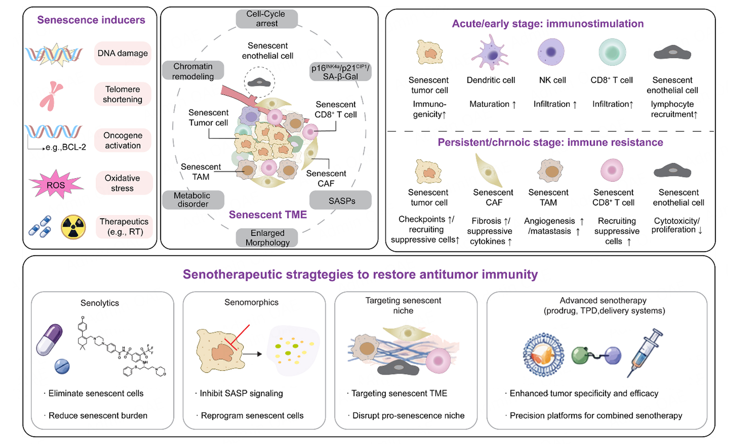
Abstract
Cellular senescence, a stable state of cell cycle arrest triggered by intrinsic and extrinsic stressors, has emerged as a pivotal regulator of tumor immunity and therapeutic resistance. Senescent cells exhibit pronounced temporal heterogeneity in immune modulation. Early-stage senescence enhances antitumor immunity by increasing immunogenicity and promoting the secretion of pro-inflammatory factors. In contrast, persistent senescence drives an immunosuppressive tumor microenvironment by recruiting myeloid-derived suppressor cells, upregulating immune checkpoints, and propagating a senescent niche. This temporal duality underlies the paradoxical role of therapy-induced senescence in both tumor suppression and immune evasion. Recent advances in senotherapy, including senolytics, senomorphics, and strategies targeting senescent stromal and immune compartments, offer promising avenues to therapeutically exploit senescence. Emerging approaches integrating prodrug designs, targeted protein degradation, and advanced drug delivery systems allow precise, context-specific modulation of senescence, minimizing off-target effects while potentiating immunotherapy. Here, we provide a comprehensive overview of tumor senescence hallmarks, its dynamic crosstalk with immunity, and emerging therapeutic strategies, highlighting current challenges and opportunities for harnessing senescence to overcome immune resistance and enhance cancer immunotherapy.
Keywords


 Download PDF
INTRODUCTION
Immunotherapy has fundamentally reshaped the cancer treatment landscape and emerged as a cornerstone of modern oncological care[1]. Its integration with conventional treatment modalities, including chemotherapy and radiotherapy, has further improved clinical outcomes across a broad range of malignancies[2]. Nevertheless, therapeutically refractory tumors, such as pancreatic ductal adenocarcinoma and triple-negative breast cancer, often derive limited benefit from these approaches, largely because of their highly immunosuppressive tumor microenvironments[3]. This limited therapeutic responsiveness is closely associated with the establishment and maintenance of an immunosuppressive tumor microenvironment (iTME). Accumulating evidence has identified multiple, interconnected determinants of the iTME, including low tumor immunogenicity[4], extensive stromal deposition[5], metabolic dysregulation[6], microbial colonization[7], and neural infiltration[8]. Among these factors, cellular senescence is increasingly recognized as a previously underappreciated hallmark of cancer and a potentially important regulator of immunosuppressive remodeling within the tumor microenvironment[9].
Cellular senescence is a state of stable cell-cycle arrest induced by diverse intrinsic and extrinsic stressors and serves as a protective mechanism that prevents the proliferation of damaged cells[10]. It can arise from intrinsic processes, such as telomere attrition leading to replicative senescence, or from extrinsic insults, such as ionizing radiation-induced DNA damage[11,12]. Accumulating evidence indicates that senescence exerts time-dependent and context-specific effects on tumor immunity. During the early phase of senescence, particularly following an acute DNA damage response, senescent cells may enhance tumor immunogenicity by upregulating antigen-presentation machinery, including major histocompatibility complex class I (MHC-I) molecules[13] and by secreting pro-inflammatory mediators that promote immune activation and facilitate immune surveillance[14]. By contrast, the persistent accumulation of senescent cells can establish a chronic inflammatory milieu that progressively reprograms the tumor immune microenvironment toward an immunosuppressive state, characterized in part by the recruitment and expansion of myeloid-derived suppressor cells (MDSCs)[15].
Accordingly, in clinical settings, therapy-induced senescence, particularly that induced by chemotherapy and radiotherapy, may paradoxically promote immune evasion and therapeutic resistance when senescent cells persist and accumulate. In this review, we first summarize the defining hallmarks and major inducers of cellular senescence in cancer. We then examine the current understanding of how senescence shapes tumor immunity, with particular emphasis on its temporally dynamic and context-dependent effects. Furthermore, we discuss emerging therapeutic approaches to targeting or modulating senescence, including senolytic, senomorphic, and other advanced senescence-directed strategies. Finally, we highlight the major challenges and future opportunities associated with harnessing cellular senescence for cancer immunotherapy, with the aim of providing a conceptual framework for overcoming resistance to immunotherapeutic interventions.
HALLMARKS, INDUCERS, AND CELLULAR HETEROGENEITY OF TUMOR SENESCENCE
Hallmarks and inducers of tumor senescence
Cellular senescence was first described in the 1960s, when normal human fibroblasts were observed to undergo a finite number of divisions before entering a state of permanent growth arrest, a phenomenon later termed replicative senescence[16]. Senescence was subsequently characterized as a stable cell-cycle arrest induced by diverse intrinsic and extrinsic stressors, serving as a critical safeguard against the propagation of damaged or potentially malignant cells[10,17]. Senescent cells have recently been incorporated into the expanded hallmarks-of-cancer framework as an important component of the tumor microenvironment[18], underscoring its complex and increasingly appreciated role in tumor biology.
Tumor senescence can be induced by a broad spectrum of cellular stresses, with persistent DNA damage representing a major initiating event [Figure 1A]. These senescence-inducing stimuli can be broadly classified as intrinsic or extrinsic. Intrinsic drivers include telomere attrition, oncogene activation, such as activation of RAS, BRAFV600E, or MYC, and genomic instability[19-21]. Alterations in tumor-suppressor pathways, including those involving PTEN, RB, and LKB1, may also trigger senescence through the compensatory activation of cellular stress-response programs[21,22]. Clinically relevant extrinsic inducers include DNA-damaging chemotherapy and radiotherapy[23,24]. Selected targeted regimens can also impose a senescence program in defined tumor contexts, whereas oxidative stress can induce premature senescence through persistent stress signaling[25]. These stimuli mediate sustained DNA damage responses and activate the p53-p21 and p16-Rb pathways[26], ultimately establishing durable cell-cycle arrest. Notably, the tumor microenvironment itself constitutes an important source of senescence-inducing stress. Hypoxia, nutrient deprivation, inflammatory signaling, and stromal interactions can all contribute to the accumulation of senescent cells within tumors[27-29], particularly following therapeutic intervention [Figure 1A]. The resulting enrichment of senescent cells may further amplify local stress signals and propagate senescence to neighboring cells, thereby reinforcing the senescent microenvironment through a feed-forward mechanism.
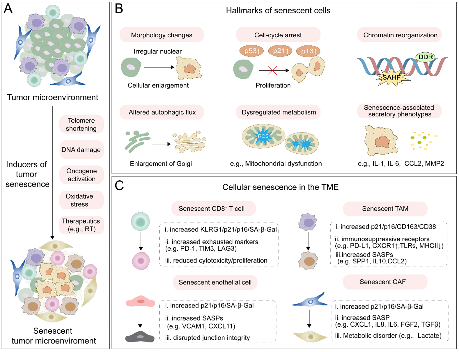
Figure 1. Inducers and hallmarks of senescence in the tumor microenvironment. (A) Intrinsic and extrinsic stressors, including telomere attrition, DNA damage, oncogene activation, oxidative stress, and anticancer therapy, induce cellular senescence across multiple tumor microenvironmental compartments. (B) Senescent features include morphological enlargement and nuclear abnormalities, cell-cycle arrest, persistent DNA damage responses and chromatin reorganization, altered autophagic flux and Golgi expansion, mitochondrial dysfunction, and development of the senescence-associated secretory phenotype. (C) Senescent or senescent-like CD8+ T cells, tumor-associated macrophages (TAMs), endothelial cells, and cancer-associated fibroblasts (CAFs) exhibit distinct combinations of molecular markers, secretory programs, and functional alterations that impair cytotoxic immunity, remodel the extracellular matrix and vasculature, and promote immune escape and metastasis.
Cellular senescence in the tumor microenvironment
In the tumor microenvironment, senescent cells undergo irreversible cell cycle arrest driven by sustained activation of cyclin-dependent kinase inhibitors, including p16INK4a, p21Cip1/Waf1, and p27Kip1[30]. These senescent cells undergo a broad spectrum of biological alterations involving cellular morphology, chromatin organization, and metabolic regulation [Figure 1B]. Morphologically, senescent cells are typically enlarged and flattened and exhibit increased lysosomal content and elevated senescence-associated β-galactosidase (SA-β-gal) activity[31]. At the chromatin level, they display extensive remodeling, including the formation of senescence-associated heterochromatin foci (SAHF)[32] and the accumulation of cytoplasmic chromatin fragments[33], linking genomic instability to innate immune signaling[34]. In parallel, sustained DNA damage signaling and cell-cycle arrest induce extensive transcriptional reprogramming[35], including the activation of pro-survival pathways that enable senescent cells to resist apoptosis and persist within the tumor microenvironment[36].
Among them, senescent tumor cells (STCs) undergo profound metabolic reprogramming [Figure 1B], characterized by mitochondrial dysfunction, increased production of reactive oxygen species (ROS), and dysregulated autophagy[37,38]. Moreover, a senescence-associated secretory phenotype (SASP) emerges as a coordinated secretory program driven by upstream transcriptional regulation [Figure 1B]. The SASP comprises a diverse array of bioactive factors, including pro-inflammatory cytokines (e.g., IL-6)[39], growth factors (e.g., TGF-β)[25], matrix-remodeling enzymes (e.g., MMPs)[40], as well as extracellular vesicles and their miRNA cargo[41,42]. Acting in autocrine and paracrine manners, the SASP reinforces senescence, remodels the tumor microenvironment, and modulates immune responses[43]. Notably, the composition and functional consequences of the SASP are highly context-dependent, varying with cell type, inducing stimulus, and temporal progression.
Cellular senescence within tumors is not confined to malignant cells but also arises across stromal, vascular, and immune compartments[44] [Figure 1C]. Senescent CD8+ T cells are characterized by increased expression of p16INK4a, p21Cip1/Waf1, CD57, and KLRG1, elevated senescence-associated β-galactosidase (SA-β-gal) activity, and loss of the costimulatory molecules CD27 and CD28[45]. Several exhaustion-associated inhibitory receptors, including PD-1, TIM-3, and LAG-3, may also be highly expressed in senescent T cells. Senescent and exhausted T cells also share functional features, including impaired proliferative capacity and reduced cytotoxicity[46]. This phenotypic and functional convergence raises the possibility that activation of senescence-associated signaling contributes to the initiation or stabilization of T-cell exhaustion, although the two states remain biologically distinct[47].
Tumor-infiltrating myeloid cells, particularly tumor-associated macrophages (TAMs), can similarly acquire senescent or senescence-like states[48] [Figure 1C]. Senescent TAMs are characterized by lysosomal alterations, metabolic reprogramming, and immunosuppressive activity[49]. These myeloid populations suppress CD8+ T-cell and natural killer (NK)-cell responses through mechanisms that include the upregulation of inhibitory molecules such as PD-L1 and the reduced expression or responsiveness of innate immune pattern-recognition receptors, including Toll-like receptors[49].
Senescent CAFs are generally characterized by combined p16/p21 expression, SA-β-Gal activity[50], and a SASP enriched in IL-8, CXCL1, TGF-β, MMPs, and FGF2[51] [Figure 1C]. Their excessive extracellular matrix deposition, induction of hypoxia, and lactate-mediated metabolic-epigenetic signaling restrict cytotoxic lymphocyte infiltration[50] and promote tumor plasticity, invasion[52], and lymphatic metastasis[53]. Senescent endothelial cells similarly exhibit canonical senescence-associated features, including increased expression of p16INK4a and p21Cip1/Waf1 and elevated SA-β-gal activity[54]. They also undergo substantial functional alterations characterized by increased oxidative stress and dysregulated adhesive and chemotactic signaling, including VCAM-1- and CXCL11-enriched secretory programs[54-56]. The resulting loss of endothelial junction integrity increases vascular permeability, disrupts immune-cell trafficking, and facilitates tumor-cell dissemination.
Despite substantial progress, the definition and identification of senescent cells rely on combinatorial assessment of multiple features. Emerging high-throughput technologies, such as single-cell omics and CRISPR-based screens, are expected to refine the molecular taxonomy of senescence and elucidate the mechanisms by which systemic factors and microenvironmental cues (e.g., neural signaling) induce senescence.
CROSSTALK BETWEEN SENESCENCE AND LOCAL TUMOR IMMUNITY
Cellular senescence exerts double-edged effect on tumor immunity. During the early stages of tumorigenesis or shortly after therapeutic intervention, senescent cells (e.g., STC) can enhance immune surveillance by increasing immunogenicity and secreting pro-inflammatory mediators that promote immune-cell recruitment and activation[57]. By contrast, the persistent accumulation of STCs within the tumor microenvironment can foster the recruitment of immunosuppressive cell populations and upregulate immune checkpoint molecules, thereby facilitating immune evasion and tumor progression [Figure 2]. Moreover, through paracrine signaling, STCs can propagate senescence to neighboring stromal and immune cells, establishing a senescent niche that further reinforces the iTME.
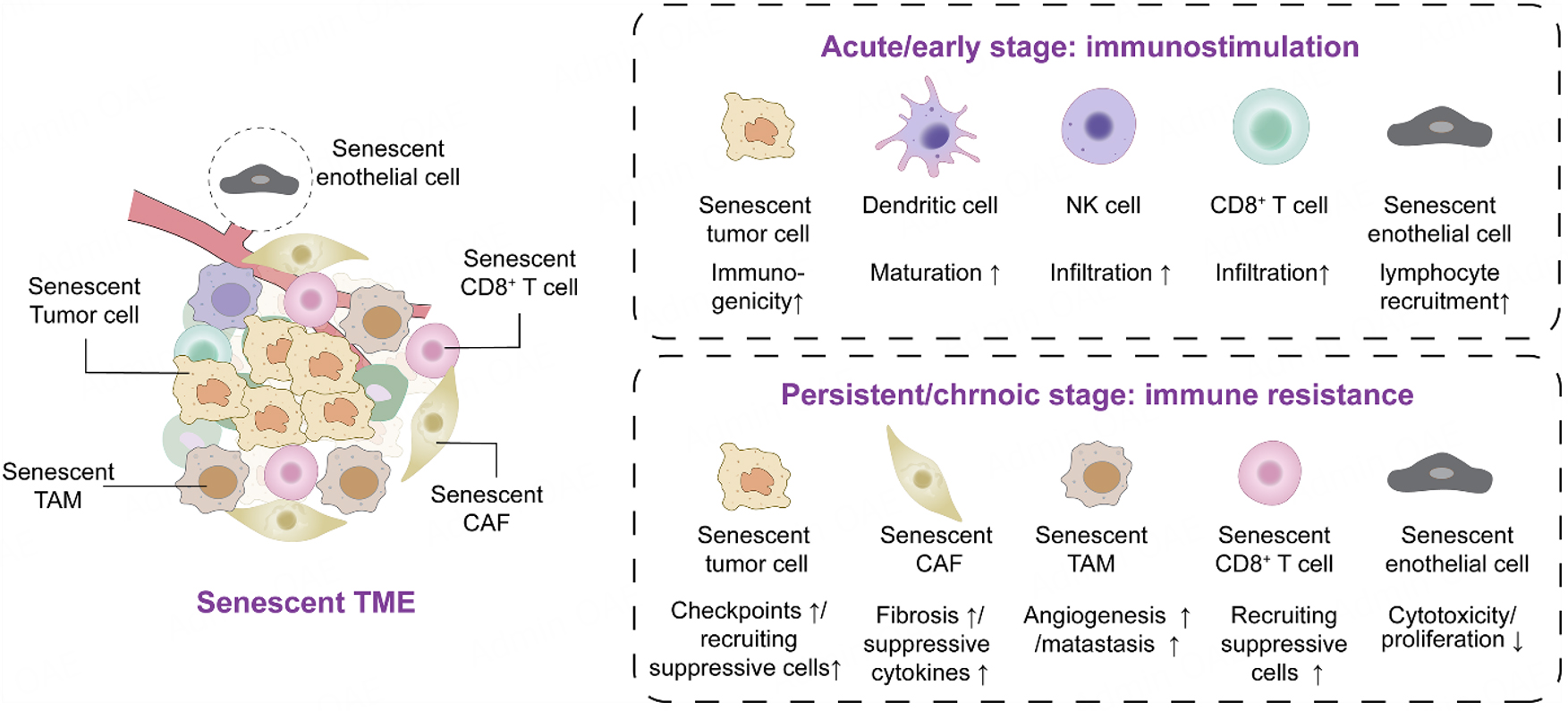
Figure 2. Stage-dependent crosstalk between cellular senescence and tumor immunity. During the acute or early stage, STCs may exhibit increased immunogenicity and release immunostimulatory mediators that promote dendritic-cell maturation and the recruitment of NK cells and CD8+ T cells. Senescence-associated endothelial activation and vascular remodeling may additionally facilitate lymphocyte adhesion, transendothelial migration, and intratumoral infiltration. By contrast, persistent or chronic senescence establishes an immunosuppressive niche through immune-checkpoint upregulation, recruitment of suppressive immune populations, profibrotic and immunosuppressive SASP signaling, myeloid-cell reprogramming, and vascular dysfunction. Concurrently, senescent CD8+ T cells exhibit impaired proliferation and cytotoxicity, collectively promoting immune resistance, tumor progression, and metastatic dissemination.
Immunostimulatory functions of tumor senescence
Senescent cells can stimulate antitumor immunity via enhancing immunogenicity and recruiting cytotoxic immune cells. Senescence-inducing CDK4/6 inhibition can reprogram mammary tumors toward a more inflamed state by increasing chemokine-mediated T-cell recruitment[58].
STCs also undergo cell-intrinsic alterations that further enhance their immunogenicity. Notably, the upregulation of antigen presentation machinery, including MHC class I molecules, together with increased expression of IFN-γ receptor 1, enhances their susceptibility to immune recognition and elimination[13]. In addition, senescent cells express higher levels of stress-induced ligands, such as MICA, ULBP2, and ICAM-1, which engage activating receptors such as NKG2D on NK cells, thereby amplifying cytotoxic responses[59]. Senescent endothelial compartment can increase vascular permeability and accessibility, thereby enhancing the intratumoral penetration of chemotherapeutic agents and facilitating immune-cell infiltration into tumors[60,61]. This immunostimulatory function represents an early, protective role of senescence, in which cell-intrinsic immune signaling cooperates with SASP-mediated recruitment and activation of effector cells to establish an effective antitumor surveillance network.
Immunosuppressive consequences of persistent senescence
When STCs persist within the tumor microenvironment and evade immune clearance, their immunoregulatory function shifts from immunostimulatory to immunosuppressive. This transition is driven by multiple, interconnected mechanisms, including the recruitment and expansion of immunosuppressive cell populations, the upregulation of immune checkpoint molecules, and the establishment of a senescent niche that sustains local immune suppression.
Recruitment of immunosuppressive cells
Persistent STCs can recruit immunosuppressive cells to remodel the TME. For instance, Xiong et al. revealed that persistent STCs significantly upregulated chemokines secretion, particularly CCL2[62]. These chemokine-enriched STCs promoted the intratumoral recruitment of MDSCs via CCL2-CCR2 signaling pathways. Furthermore, the accumulated MDSCs suppressed the cytotoxic activity of CD8+ T cells and NK cells, thereby dampening antitumor immune responses[62]. Curiel et al. reported that tumor-associated cells enhanced the secretion of CCL22. These CCL22-rich cells promoted the infiltration of regulatory T cells (Tregs) through the CCL22-CCR4 axis. The recruited Tregs suppressed effector T cell responses, leading to an immunosuppressive tumor microenvironment[63]. Taken together, persistent STCs could remodel tumor microenvironment and facilitate tumor immune evasion via recruiting suppressive immune cells.
Upregulation of immune checkpoints
Multiple immune checkpoints have been reported to be upregulated on STCs. Wang et al. revealed that STCs significantly upregulated the expression of inhibitory immune checkpoint PD-L1. These PD-L1high STCs limited the activation and immune surveillance of cytotoxic T cells and diminished the efficacy of immunotherapies such as PD-1/PD-L1 blockade[64]. Similarly, Salminen et al. reported that STCs upregulated multiple inhibitory immune checkpoint ligands associated with the PD-1, TIM-3, and NKG2A pathways. This checkpoint-high phenotype impaired the activation and surveillance functions of cytotoxic T cells and NK cells, ultimately promoting immune escape[65]. In addition, SASP-associated factors can disrupt NKG2D-mediated signaling, for example by promoting the proteolytic shedding of NKG2D ligands from target cells, thereby further impairing NK-cell recognition and cytotoxicity[66]. Collectively, these mechanisms establish a local immunosuppressive environment in which STCs evade immune surveillance while simultaneously attenuating responses to immunotherapy.
Formation of a senescent and immunosuppressive niche
Beyond STCs, senescent stromal and immune cells contribute substantially to the establishment of a senescent and immunosuppressive niche. Senescent CAFs secrete SASP factors that impair CD8+ T-cell infiltration and activation, thereby limiting antitumor immunity and reducing the efficacy of immunotherapy[50]. Senescent endothelial cells alter vascular permeability and immune-cell trafficking, indirectly facilitating tumor-cell dissemination while restricting effective immune surveillance[67]. Similarly, senescent macrophages and other myeloid cells exhibit enhanced IL-6/STAT3 signaling, which promotes the expression of immunosuppressive molecules, including PD-L1 and arginase-1, as well as protumorigenic cytokines. Collectively, these alterations suppress the proliferation, activation, and cytotoxic functions of CD8+ T cells and NK cells[68].
Moreover, intratumoral CD8+ T cells can also undergo senescence, which exhibit high expression of p16, p21, CD57, and KLRG1, but low production of cytotoxic molecules, and impaired proliferative and killing capacity[69]. T cell senescence critically influences immunotherapy outcomes across multiple modalities. For immune checkpoint inhibitors (ICIs), senescent T cells are associated with suboptimal responses[70]. In chimeric antigen receptor (CAR) T-cell therapy, chronic antigen exposure and prior chemotherapy could induce T-cell senescence and decrease the quality of source T cells[71]. Cancer vaccine efficacy is also limited by T-cell senescence, with senescent T cells serving as a predictive biomarker for poor outcomes[45]. Collectively, these findings underscore the need to address T-cell senescence to optimize immunotherapy efficacy and safety.
Overall, in the senescent TME, the transition from immunostimulatory to immunosuppressive senescence appears to emerge from the interaction between temporal remodeling of the senescent phenotype and multicellular feedback within the tumor microenvironment. For example, transient inflammatory signaling may promote immune recruitment and senescence surveillance, whereas persistent signaling can sustain paracrine senescence, immune-checkpoint expression, and suppressive myeloid-cell and Treg recruitment. This framework has direct therapeutic implications. During the early immunogenic phase, treatment should preserve or enhance immune-mediated clearance rather than indiscriminately suppress the SASPs. By contrast, persistent SASP-dominant senescence may require senomorphic intervention, whereas accumulated apoptosis-resistant senescent cells may be more appropriately targeted using senolytics.
SENESCENCE-DRIVEN METASTATIC DISSEMINATION THROUGH LOCAL AND SYSTEMIC MICROENVIRONMENTAL REMODELING
Beyond suppressing local antitumor immunity, a persistent senescent niche can promote metastatic dissemination by integrating immune suppression and stromal remodeling with multiple stages of the metastatic cascade [Figure 3]. This metastatic cascade simultaneously reprograms tumor cells[72], remodels the local microenvironment[53] and conditions distant organs[73]. Senescent tumor and stromal cells release inflammatory cytokines, chemokines[74], growth factors[75], proteases[76], metabolites[77] and extracellular vesicles[78], collectively altering multiple stages of the metastatic cascade. These signals can induce epithelial plasticity, stemness[79], loss of cell adhesion[80], collective invasion[81] and escape from therapy-induced senescence in neighboring or previously arrested tumor cells[82]. Thus, the prometastatic effects of senescence are frequently non-cell-autonomous[83] and may persist even when senescent cells themselves remain proliferation-incompetent[84].
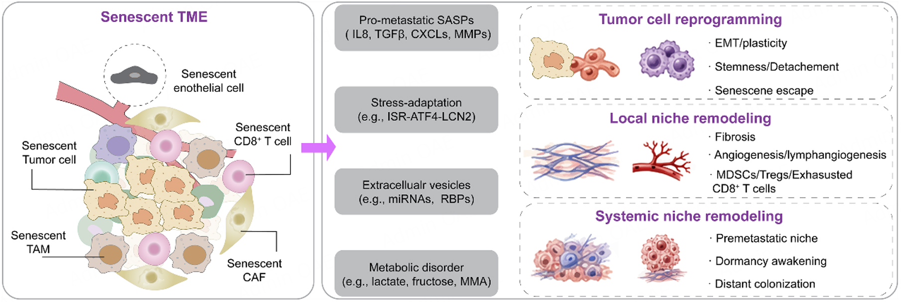
Figure 3. Senescence promotes tumor metastasis through local and systemic microenvironmental reprogramming. The senescent TME generates multiple metastasis-promoting signals. These include pro-metastatic senescence-associated secretory phenotype (SASP) factors, such as IL-8, TGF-β, CXCL chemokines and MMPs; stress-adaptation pathways, e.g., integrated stress response (ISR)-ATF4-lipocalin 2 (LCN2) signaling; extracellular vesicles, and metabolic alterations involving lactate, fructose and methylmalonic acid (MMA).
At the primary tumor site, senescent CAFs[52] and endothelial cells remodel the extracellular matrix[76], vasculature[55] and lymphatic compartments[53] while establishing an immunosuppressive niche[85]. Classical SASP factors, including IL-6, IL-8, TGF-β, CXCL chemokines[74] and matrix metalloproteinases[76], promote epithelial-mesenchymal plasticity[74], invasion[52], angiogenesis and immune-cell recruitment[55]. Recent findings have expanded this framework beyond proteinaceous SASP factors. Senescent CAF-derived lactate supports lymphatic endothelial-cell survival through lactylation-dependent metabolic reprogramming and promotes early lymph-node metastasis in pancreatic cancer[53]. Similarly, fructose present in the chemotherapy-induced senescence-associated secretome disrupts the NAD+-SIRT-SREBP axis, reduces plasma-membrane cholesterol and facilitates tumor-cell detachment and dissemination[80]. These findings identify metabolic communication as an integral component of senescence-driven metastasis[77].
Senescence also exerts systemic effects through circulating extracellular vesicles[86] and damage-associated molecules[87]. Extracellular vesicles released by senescent hepatocytes transfer prometastatic miRNAs to tumor cells and enhance dissemination across multiple cancer types[86]. Therapy-induced stromal senescence following palbociclib treatment can remodel the lung metastatic niche and increase the metastatic outgrowth of drug-resistant mammary cancer cells[88] Senescent mesenchymal stromal cells have also been identified in the premetastatic bone marrow of untreated patients with advanced breast cancer[89]. Aging-associated programs[90], including altered extracellular-matrix organization[91], methylmalonic acid accumulation[92], PDGF-C activation[93], SEMA7A-TGF-β signaling[94] and the ISR-ATF4-LCN2 signaling, converge with cellular senescence by increasing tumor-cell plasticity[95] and generating permissive metastatic niches[96]. However, organismal aging and cellular senescence should remain mechanistically distinguished, because not all age-dependent prometastatic changes arise directly from senescent cells[97].
Collectively, the metastatic outcome of senescence is determined by the cellular source, inducing stimulus, duration and efficiency of immune clearance. Acute senescence may suppress tumor growth and promote immune surveillance, whereas persistent or therapy-induced senescence converts local stress responses into a systemic prometastatic program[73]. These observations support temporally controlled senotherapeutic strategies combining senolytics or senomorphics with blockade of dominant downstream pathways.
TARGETING TUMOR SENESCENCE FOR ENHANCED IMMUNOTHERAPY
Senotherapy encompasses a broad class of therapeutic strategies that target STCs or their associated phenotypes[98]. In this review, these strategies are broadly classified into approaches that selectively eliminate STCs (senolytics), suppress or modulate the SASPs (senomorphics), or therapeutically remodel the senescent and immunosuppressive niche. Representative senotherapeutic approaches and their major therapeutic applications and molecular targets are summarized in Table 1.
Overview of senescence-targeted therapeutic strategies
| Senotherapeutics | Compound | Disease context/experimental model | Direct target or mechanism | Ref |
| Senolytic | ||||
| Small molecule | Navitoclax (ABT-263) | Senescent human fibroblast/endothelial and mouse models | Bcl-2, Bcl-xl, Bcl-w inhibitor | [100] |
| Small molecule | Venetoclax (ABT-199) | Chronic lymphocytic leukemia and acute myeloid leukemia | Selective BCL-2 inhibitor; not established as a broad-spectrum senolytic | [157] |
| Small molecule | Dasatinib plus quercetin (D+Q) | Senescent preadipocytes/endothelial cells/MEFs | Inhibition of senescent-cell anti-apoptotic pathways | [101] |
| Adoptive cell therapy | CAR T cells | Therapy-induced lung adenocarcinoma and chemically or diet-induced liver fibrosis models | uPAR | [105] |
| Small molecule | Fisetin | Radiation-induced senescent HUVECs (in vitro) | Cell-type-specific induction of apoptosis in senescent cells | [36] |
| Vaccine | SenoVax | Lung cancer (LLC model) | Senescence-associated antigens/SASP-related epitopes | [158] |
| Senomorphic | ||||
| Small molecule | Sirolimus/Rapamycin | Senescent fibroblasts; prostate tumor xenograft model | mTOR-IL1A-SASP axis | [110] |
| Small molecule | Losmapimod | Senescent-cell inflammatory hyperactivation models | p38/MAPK inhibition; reduced inflammatory SASP-associated cytokine expression | [111] |
| Small molecule | Metformin | Doxorubicin-induced senescent endothelial cells, including HUVECs | Inhibition of JNK/NF-κB signaling and reduction of SASP factors | [112] |
| Small molecule | Vitamin D3 | Irradiation-induced senescent human dermal fibroblasts; ageing-related immune models | Partial inhibition of p38/MAPK and reduction of IL-6, IL-8 and CCL2 secretion | [113] |
| Targeting the senescent and immunosuppressive niche | ||||
| Small molecule | BIRB 796 | Primary human senescent CD8+ EMRA T cells ex vivo | p38/MAPK inhibition; increased autophagy, proliferation, telomerase activity, and mitochondrial function | [126] |
| Genetic/ Senolytic depletion | Clearance of senescent macrophages | KRAS-driven lung cancer models | Elimination of senescent tumor-associated macrophages and reduction of their protumorigenic SASP | [120] |
| Small molecule | MAFP-mediated cPLA2α inhibition | Melanoma and breast cancer mouse models | Inhibition of group IVA phospholipase A2; correction of lipid-droplet accumulation and prevention of effector T-cell senescence | [128] |
| Engineering platforms for selective senolytic delivery or action | ||||
| SA-β-Gal-activated prodrug | Nav-Gal | Cisplatin-treated A549 NSCLC cells and murine lung cancer models | SA-β-Gal-dependent activation of galacto-conjugated navitoclax; reduced platelet toxicity | [135] |
| SA-β-Gal-activated prodrug | Galactose-modified duocarmycin (GMD) | Multiple senescent-cell models; preneoplastic craniopharyngioma model | GLB1-dependent activation followed by selective apoptosis of senescent cells | [136] |
| TPD | DT2216 | Drug-resistant triple-negative breast cancer and patient-derived T-ALL models | VHL-mediated BCL-XL degradation with reduced platelet toxicity | [137] |
| Drug delivery system | Navitoclax-loaded galacto-oligosaccharide-capped MSNs | Palbociclib-induced senescent triple-negative breast cancer model | SA-β-Gal-responsive release of navitoclax; reduced systemic toxicity | [145] |
| Nanovesicle | Nano-APM | Radiotherapy-treated murine tumor models | Expansion of STC-specific splenic CD8+ T cells followed by spatially restricted immune-mediated senolysis | [154] |
Senolytics: eliminating senescent cells
Current senolytic strategies can be broadly categorized into the direct pharmacological induction of STC apoptosis, sequential “one-two punch” therapy, and immune-mediated clearance of STCs [Figure 4]. Senolytics exploit intrinsic vulnerabilities of senescent cells, most notably their reliance on senescence-associated anti-apoptotic pathways (SCAPs). STCs frequently upregulate pro-survival proteins, such as BCL-XL and MCL-1, that enable their persistence despite cellular damage[99]. Pharmacological inhibition of these pathways using agents, such as navitoclax (ABT-263)[100], dasatinib plus quercetin (D+Q)[101], and fisetin[36], can selectively induce apoptosis of senescent cells. Clinical translation of senolytics remains early and has largely occurred outside oncology. D+Q has undergone a small randomized phase I pilot study in idiopathic pulmonary fibrosis[102], fisetin is being evaluated in a phase II sepsis trial[103]. These studies do not yet establish clinical clearance of STCs or improved cancer outcomes.
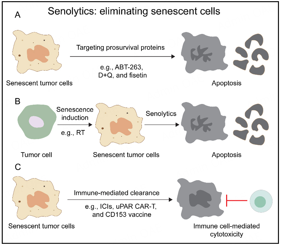
Figure 4. Senolytic strategies eliminate STCs through apoptosis induction or immune-mediated mechanisms. (A) Targeting senescence-associated anti-apoptotic pathways (SCAPs), such as BCL-XL, using agents including navitoclax (ABT-263), dasatinib plus quercetin (D+Q), and fisetin. (B) The “one-two punch” strategy, in which senescence-inducing therapies are combined with senolytics to sequentially induce and eliminate STCs. (C) Immune-mediated clearance via targeting senescence-associated antigens or surface markers using CAR-T/CAR-NK cells or vaccination approaches.
Senolytic strategies are particularly attractive when administered sequentially after senescence-inducing therapies, forming the so-called “one-two punch” approach. In this therapeutic paradigm, tumor cells are first driven into a senescent state by treatments such as chemotherapy, radiotherapy, or CDK4/6 inhibitors, and are subsequently eliminated using senolytic agents[104]. This sequential strategy can promote the clearance of residual STCs while limiting the long-term detrimental consequences of their persistence, including metastatic progression, immune evasion, and therapeutic resistance[57,104]. However, a major challenge for this sequential strategy is that the optimal interval between senescence induction and senolytic administration is unlikely to be fixed across tumor types, inducing agents, and cellular compartments. Longitudinal imaging of senescence-associated signals may therefore help identify the emergence and persistence of senescent-cell populations and guide the timing of the second therapeutic intervention.
An alternative and increasingly explored strategy involves enhancing the immune-mediated clearance of senescent cells. Given that immune surveillance represents a physiological mechanism for STC elimination, therapeutic approaches aim to potentiate this process. For instance, chimeric antigen receptor (CAR)-T or CAR-NK cells targeting senescence-associated surface antigens ( e.g., uPAR) have demonstrated selective elimination of STCs in tumor models[105]. Similarly, vaccines directed against senescence-associated antigens (e.g., CD153 or GPNMB) can induce antibody-dependent cellular cytotoxicity and facilitate immune clearance[106]. Although these approaches offer high specificity, their clinical translation remains constrained by the limited availability of robust and universal senescence-specific targets, as well as concerns regarding immune-related toxicity.
Senomorphics: reprogramming the SASP
In contrast to senolytics, senomorphic strategies aim to modulate the functional phenotype of senescent cells, particularly the production and activity of SASP components, thereby mitigating their detrimental effects on the iTME. The SASP production is governed by multiple interconnected signaling pathways, including mTOR, MAPK, cGAS-STING, JAK-STAT, and NF-κB signaling pathways[107-109]. Pharmacological targeting of these pathways using agents such as rapamycin or everolimus (mTOR inhibitors), losmapimod (p38/MAPK inhibitor), metformin, or vitamin D has been shown to suppress the production of SASPs[110-113]. Moreover, direct neutralization of individual SASP components using antibodies against IL-11, IL-6, or IL-1β has demonstrated therapeutic potential in models of aging-related diseases[114]. By attenuating chronic inflammatory profile, these interventions can partially restore immune surveillance and enhance antitumor efficacy.
Notably, SASP modulation does not necessarily require its global suppression. Emerging evidence suggests that selective reprogramming of the SASPs rather than its complete inhibition may enhance antitumor immunity[115]. For instance, certain therapeutic combinations can induce more immunostimulatory SASPs, promoting recruitment of NK cells and T cells while limiting immunosuppressive signaling[116]. These findings underscore the context-dependent nature of senomorphic interventions and suggest that preserving beneficial immune-activating signals may be critical to achieving optimal therapeutic outcomes.
In addition to targeting SASP regulatory pathways, immune checkpoint blockade represents a complementary strategy for counteracting the immunosuppressive phenotype of STCs. STCs frequently upregulate inhibitory ligands, including PD-L1, PD-L2, and HLA-E, which suppress cytotoxic immune responses and facilitate immune evasion[117]. Antibodies targeting the PD-1/PD-L1 or NKG2A/HLA-E axis can restore T-cell and NK-cell activity[118], thereby promoting the immune-mediated clearance of STCs and potentially enhancing the efficacy of immunotherapy.
Targeting the senescent and immunosuppressive niche
Given the involvement of multiple cellular compartments in senescence-mediated immune remodeling, therapeutic strategies should extend beyond targeting STCs alone to encompass the broader senescent TME. As a dominant stromal population within tumors, senescent CAFs are one of the major sources of immunosuppressive SASPs[119]. The selective elimination of senescent CAFs or suppression of their SASPs has been shown to restore immune surveillance and improve responses to chemotherapy[120]. Similarly, targeting senescent macrophages may alleviate local immune suppression, restrain early tumorigenesis, and promote the intratumoral accumulation of cytotoxic T cells. Several senolytic and senomorphic agents, including navitoclax, D+Q, and niacin, have demonstrated therapeutic potential against senescent macrophages in preclinical studies and early clinical investigations[121-124].
Senescent T cells are characterized by diminished cytotoxicity and impaired responsiveness to immunotherapy. Unlike strategies targeting senescent stromal cells, therapeutic approaches to T-cell senescence primarily focus on functional rejuvenation rather than cellular elimination. Pharmacological inhibition of PI3Kδ with leniolisib or p38 MAPK with BIRB 796 has been reported to attenuate T-cell senescence in several oncological contexts, including hematologic malignancies and solid tumors following chemotherapy[110,125,126]. Genetic or epigenetic modulation of p53- or SIRT1-associated pathways in CD8+ T cells may similarly reduce senescent features while enhancing their migratory capacity and cytotoxic function[127]. Beyond these cell-intrinsic pathways, metabolic reprogramming represents another promising therapeutic strategy. Modulation of the FOXO1-SIRT1 axis can regulate glycolytic metabolism and granzyme B production, whereas targeting group IVA phospholipase A2-mediated lipid-droplet accumulation may help preserve metabolic homeostasis[128,129]. Neuroendocrine signals mediated through β-adrenergic receptors can also influence the metabolism and function of senescent T cells, identifying catecholamine-β-adrenergic signaling as a potential therapeutic target[130].
Collectively, these findings support a paradigm shift in senotherapy from indiscriminate targeting of senescent cells toward a multi-level intervention strategy that integrates signaling pathway modulation, epigenetic reprogramming, metabolic regulation, and immune niche remodeling. Collectively, senomorphic modulation and cell-type-specific targeting of senescent stromal and immune populations provide complementary strategies for suppressing deleterious SASP signaling, restoring immune-cell function, and remodeling the senescent iTME [Figure 5].
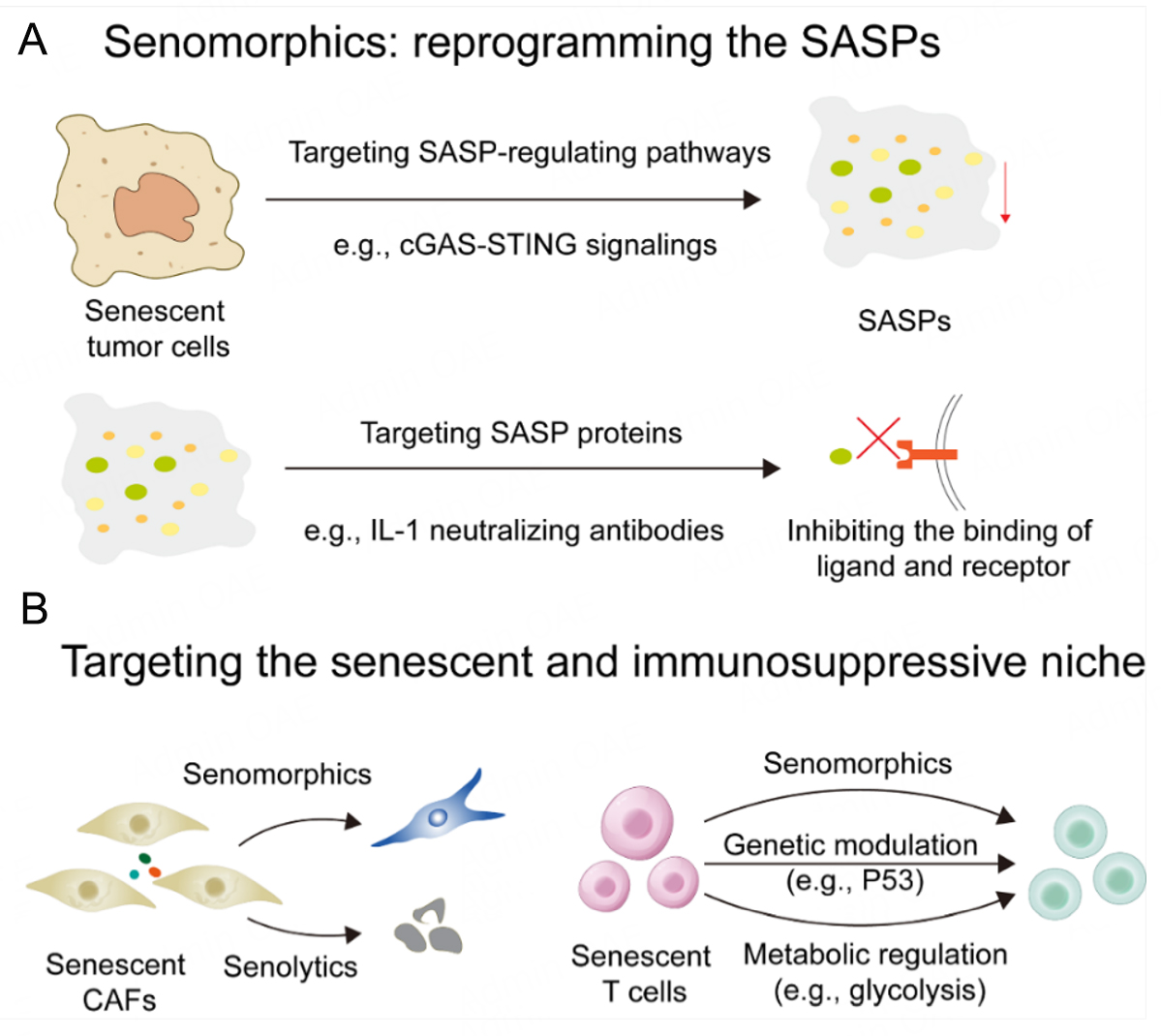
Figure 5. Reprogramming senescent cell function and the senescent TME to reverse immune evasion. (A) Senomorphic strategies suppress the deleterious effects of STCs by targeting key SASP-regulating pathways, including cGAS-STING, JAK-STAT, and NF-κB signaling, or by using neutralizing antibodies against specific SASP components. (B) Targeting senescent stromal and immune populations, including senescent CAFs and T cells, using senolytic or senomorphic approaches to remodel the senescent TME.
ADVANCED SENOTHERAPEUTIC STRATEGIES FOR TUMOR SENESCENCE MODULATION
Tumor senescence is characterized by marked heterogeneity, reflected in the lack of highly specific biomarkers and its occurrence across diverse cell types and pathological contexts[131,132]. Although markers such as SA-β-Gal, p16INK4a and p21WAF1/Cip1 are commonly used in clinical settings, their combined application in single tissue samples often yields false-positive or false-negative results[133]. This insufficient specificity poses substantial challenges for tumor senotherapy due to the narrow therapeutic window of free senolytic agents.
Prodrug strategy for tumor senotherapy
To address these challenges, prodrug strategies have emerged as a promising solution. Prodrugs are inactive compounds designed to respond to specific tumor microenvironment stimuli, such as pH, redox conditions, enzymes, or hypoxia[134]. The integration of conventional senolytic agents with prodrug strategies holds promise for achieving precise senescence intervention. For instance, González-Gualda et al. developed Nav-Gal, a galactose-conjugated navitoclax prodrug activated by SA-β-Gal in senescent cells. In non-small cell lung cancer (NSCLC) models, Nav-Gal combined with cisplatin reduced tumor burden more effectively than monotherapy, while exhibiting reduced navitoclax-related platelet toxicity[135]. Similarly, Guerrero et al. designed a galactose-modified duocarmycin (GMD) prodrug that triggers senescent cell apoptosis via SA-β-Gal. In a craniopharyngioma mouse model, GMD selectively eliminated β-catenin-accumulating senescent clusters without affecting normal pituitary cells, demonstrating its potential for targeted elimination of preneoplastic senescent cells[136]. However, the repertoire of prodrug activation strategies that enable specific responsiveness to senescent cells remains limited, warranting further development of novel approaches tailored to the unique features of the senescent microenvironment.
Targeted protein degradation for tumor senotherapy
Targeted protein degradation (TPD) technologies, such as proteolysis-targeting chimeras (PROTACs), enable selective elimination of disease-relevant proteins, offering potential advantages including reduced off-target toxicity and sustained therapeutic effects[137]. PROTACs are bivalent small molecules consisting of a ligand that recognizes the target protein linked to an E3 ubiquitin ligase, thereby facilitating selective protein degradation[138,139]. Recent studies have leveraged PROTAC technology to transform navitoclax into a platelet-sparing targeted chimera, addressing navitoclax-induced thrombocytopenia while enhancing senolytic activity[137,140]. A VHL PROTAC DT2216 targeting BCL-XL could induce STC apoptosis while minimizing platelet toxicity due to the low dependence of platelets on VHL-mediated degradation[137]. DT2216 has demonstrated excellent antitumor performance in drug-resistant triple-negative breast cancer and patient-derived T-ALL models[137], underscoring the high therapeutic potential of TPD in tumor senotherapy. Beyond PROTAC-based approaches, expanding the application of TPD technologies, including molecular glue, lysosome-targeting chimeras (LYTACs), and antibody-based degraders, holds great promise for developing next-generation senescence-targeting therapies.
Drug delivery system for tumor senotherapy
Drug delivery systems (DDS) have attracted considerable interest in senotherapy, offering the ability to actively target senescent cells or specific subsets thereof, prolong drug half-life, and minimize off-target distribution[141-143]. Mesoporous silica nanoparticles (MSNs) possess favorable chemical properties, thermal stability, and biocompatibility, promoting their application in senotherapy[144]. Several studies have developed MSN-based formulations that exploit elevated SA-β-Gal activity in senescent cells for targeted delivery of navitoclax[145-149]. These systems, capped with galacto-oligosaccharide derivatives, enable selective release within senescent cells, thereby reducing navitoclax-related thrombocytopenia while enhancing senolytic efficacy[145,150]. In preclinical models, such formulations have demonstrated improved tumor growth inhibition and reduced metastasis when combined with senescence-inducing therapies (e.g., palbociclib or doxorubicin), while mitigating off-target toxicities including endothelial dysfunction and cardiotoxicity[150,151].
The SASP produced by STCs exerts pleiotropic effects, including immune cell recruitment and enhanced vascular permeability, which can be harnessed for drug delivery[152,153]. Leveraging these properties, cell membrane coating technology has emerged as an innovative approach enabling immune evasion, prolonged circulation, and targeted delivery. Pan et al. demonstrated that STCs exhibit enhanced antigen-presenting capacity and engineered their membranes into nanovesicles (nano-APM) to selectively prime splenic CD8+ T cells. Sequential combination of nano-APM with RT achieved robust antitumor efficacy, as evidenced by significantly delayed tumor growth and prolonged survival[154]. Yang et al. engineered STC membranes into biomimetic nanovaccines. Such formulations enhance dendritic cell internalization and lymph node targeting, promoting antitumor immune responses. In murine melanoma models, these nanovaccines combined with ICIs significantly suppressed tumor growth[155]. A complementary approach utilizes senescent red blood cell membranes fused with tumor cell membrane-associated antigens. These nanoformulations accumulate in the spleen, activate antigen-presenting cells, and synergize with anti-PD-L1 therapy to achieve durable antitumor effects[156]. Venetoclax is a selective BCL-2 inhibitor with established activity in hematologic malignancies, although its role as a broad-spectrum senolytic remains unproven[157]. Senescent-cell vaccination has also reduced tumor growth in several preclinical solid-tumor models[158].
Collectively, current advanced senotherapies show promise in senescence intervention. Integrating prodrug strategies, TPD technologies, and advanced DDS is anticipated to pave the way for next-generation senotherapies. The convergence of these approaches may ultimately enable safe, effective, and broadly applicable senotherapeutic interventions.
PERSPECTIVES
Tumor senotherapy has emerged as a conceptual and therapeutic framework that extends beyond conventional cytotoxic paradigms centered on apoptosis or necrosis. By targeting a fundamental and plastic cellular state, senescence, this strategy offers a unique opportunity to reprogram the tumor immune ecosystem rather than merely eliminate malignant cells. The dual and temporally dynamic roles of senescent cells in antitumor immunity, ranging from immune activation to immune suppression, position senotherapy as a potentially powerful approach to overcome immune resistance and augment the efficacy of existing immunotherapies.
Despite this promise, the biological heterogeneity of senescence remains a major barrier to clinical translation. Foremost among these is the heterogeneity of senescence, which spans differences in cell of origin, inducing stimuli, temporal evolution, and tissue microenvironment[159]. Senescent phenotypes cannot be captured by a single marker or pathway. This complexity complicates both mechanistic interpretation and therapeutic targeting. In particular, the lack of robust and specific biomarkers for senescent cells remains a critical bottleneck, limiting patient stratification, real-time monitoring, and accurate intervention. Moreover, the molecular determinants governing the transition of senescence from an immunostimulatory to an immunosuppressive state need to be clarified. Whether distinct forms of senescence, such as oncogene-induced, therapy-induced, or microenvironment-driven senescence, exert fundamentally different immunological functions across diverse cellular compartments requires systematic investigation. Addressing these questions will necessitate integrative approaches combining single-cell and spatial omics, lineage tracing, and high-throughput functional screening (e.g., CRISPR-based perturbation platforms) to construct a high-resolution, context-aware atlas of senescence.
This heterogeneity also underscores the need for specific and clinically feasible methods to monitor tumor senescence in vivo[31]. In experimental animals, genetic reporter systems, such as p16INK4a-luciferase mice, permit longitudinal visualization of senescence-associated signaling[160] and have revealed substantial tissue- and context-dependent variation in the kinetics of senescence induction[9]. Activatable probes targeting SA-β-gal have further enabled non-invasive detection using near-infrared fluorescence[161], photoacoustic tomography[162], positron emission tomography[163], and magnetic resonance imaging. Representative probes have visualized therapy-induced senescence[161], monitored ABT263-mediated senolysis[164], and provided spatial[162] or whole-body assessment of senescent-cell burden. The early clinical evaluation of [18F] FPyGal PET further supports the translational potential of senescence imaging. These advances may enable a transition from empirically fixed treatment schedules to imaging-guided, adaptive senotherapy[26]. However, current probes generally detect individual features, such as p16INK4a activation, SA-β-gal activity, or DPP4 activity[165], none of which is specific to senescent cells. Future multiparametric imaging strategies that integrate cell-cycle arrest, lysosomal activity, SASP-associated inflammation, immune remodeling, and proliferation or apoptosis will therefore be required to distinguish early immunostimulatory senescence from persistent SASP-dominant senescence[166] and to personalize the timing and intensity of sequential senotherapy[26].
The future of senotherapy will likely depend on precision and combinatorial strategies. The emerging “one-two-punch” paradigm, inducing senescence followed by selective clearance, represents a compelling blueprint, but its clinical implementation requires careful optimization of timing, dosing, and patient selection. In this context, advanced nanoplatforms may enable the spatiotemporally controlled co-delivery or sequential release of senescence inducers, senolytics, and immunomodulators, thereby improving therapeutic efficacy while limiting systemic toxicity. Functionalization with ligands targeting senescence-associated surface molecules, such as uPAR or DPP4, may further enhance cellular specificity, whereas integration with activatable prodrugs or targeted protein-degradation technologies could provide additional layers of conditional control. Ultimately, combining multiparametric senescence monitoring with programmable delivery systems may enable precise modulation of senescent states in vivo, shifting tumor senotherapy from the indiscriminate elimination of senescent cells toward adaptive, stage-specific, and patient-tailored intervention.
CONCLUSION
Cellular senescence in cancer is a heterogeneous and temporally dynamic state that arises in malignant, stromal, vascular, and immune compartments. Although acute senescence may enhance tumor immunogenicity and immune-mediated clearance, the persistence of senescent cells promotes chronic SASP signaling, immune suppression, microenvironmental remodeling, therapeutic resistance, and metastatic dissemination. Therefore, effective senotherapy should not rely on the indiscriminate elimination of all senescent cells, but should instead account for the cellular source, inducing stimulus, temporal stage, and immunological context of senescence. Integrating senolytics, senomorphics, immune modulation, and precision delivery may provide a rational strategy for overcoming senescence-mediated immune resistance and improving the durability of cancer immunotherapy.
DECLARATION
Authors’ contributions
Conceived and designed this review: Chen S, Su J
Conducted the literature search and drafted the full text of the manuscript: Chen S
Prepared all tables and figures in the manuscript: Su J
Participated in partial literature collection and sorting, and assisted in the revision of the preliminary manuscript: Jin A, Liu C, Zhang P
Contributed to the overall study design, and critically revised the manuscript for key intellectual content: Pan J
Provided important guidance on the review design and content organization: Lai Y, Xu Z, Yu H
All authors reviewed and approved the final version of the manuscript.
Availability of data and materials
Not applicable.
AI and AI-assisted tools statement
Not applicable.
Financial support and sponsorship
This work was supported by the New Organizational Model Initiative (2025ZD0552300 to Yu H), National Natural Science Foundation of China (32571707 to Lai Y, U22A20328 and W2412035 to Yu H, 82606071 to Pan J), Science and Technology Commission of Shanghai Municipality (23ZR1475000 and 20430711800 to Yu H), the Strategic Priority Research Program of the Chinese Academy of Sciences (XDB1060000 to Yu H). and “Super Postdoc” Incentive Plan of Shanghai (2025451 to Pan J).
Conflict of interest
Yu H is an Associate Editor of Journal of Cancer Metastasis and Treatment. Xu Z is an Editorial Board Member of the Journal of Cancer Metastasis and Treatment. They were not involved in any steps of editorial processing, notably including reviewers' selection, manuscript handling, or decision-making, while the other authors have declared that they have no conflicts of interest.
Ethical approval and consent to participate
Not applicable.
Consent for publication
Not applicable.
Copyright
© The Author(s) 2026.
REFERENCES
1. Topalian SL, Forde PM, Emens LA, Yarchoan M, Smith KN, Pardoll DM. Neoadjuvant immune checkpoint blockade: a window of opportunity to advance cancer immunotherapy. Cancer Cell. 2023;41:1551-66.
2. Wang X, Jing J. Cancer immunotherapy in combination with radiotherapy and/or chemotherapy: mechanisms and clinical therapy. MedComm. 2025;6:e70346.
3. Zhou M, Wang J, Pan J, et al. Nanovesicles loaded with a TGF-β receptor 1 inhibitor overcome immune resistance to potentiate cancer immunotherapy. Nat Commun. 2023;14:3593.
4. Yan W, Lang T, Yuan W, Yin Q, Li Y. Nanosized drug delivery systems modulate the immunosuppressive microenvironment to improve cancer immunotherapy. Acta Pharmacol Sin. 2022;43:3045-54.
5. Xiao Z, Todd L, Huang L, et al. Desmoplastic stroma restricts T cell extravasation and mediates immune exclusion and immunosuppression in solid tumors. Nat Commun. 2023;14:5110.
6. Dong Y, Li J, Dai Y, et al. Redox-responsive metal-organic framework nanocapsules enhance tumor chemo-immunotherapy by modulating tumor metabolic reprogramming. Materials Today Bio. 2025;31:101487.
7. Jiang M, Yang Z, Dai J, et al. Intratumor microbiome: selective colonization in the tumor microenvironment and a vital regulator of tumor biology. MedComm. 2023;4:e376.
8. Yang M, Tao L, Jiang Y, et al. Perineural invasion reprograms the immune microenvironment through cholinergic signaling in pancreatic ductal adenocarcinoma. Cancer Res. 2020;80:1991-2003.
9. Faget DV, Ren Q, Stewart SA. Unmasking senescence: context-dependent effects of SASP in cancer. Nat Rev Cancer. 2019;19:439-53.
10. Kumari R, Jat P. Mechanisms of cellular senescence: cell cycle arrest and senescence associated secretory phenotype. Front Cell Dev Biol. 2021;9:645593.
11. Victorelli S, Passos JF. Telomeres and cell senescence - size matters not. EBioMedicine. 2017;21:14-20.
12. Li M, You L, Xue J, Lu Y. Ionizing radiation-induced cellular senescence in normal, non-transformed cells and the involved DNA damage response: a mini review. Front Pharmacol. 2018;9:522.
13. Marin I, Boix O, Garcia-Garijo A, et al. Cellular senescence is immunogenic and promotes antitumor immunity. Cancer Discov. 2023;13:410-31.
14. Marin I, Serrano M, Pietrocola F. Cellular senescence enhances adaptive anticancer immunosurveillance. OncoImmunology. 2022;12:2154115.
15. Liu Y, Yu Y, Chen Y, Zhuang J, Sun C. Tumor immunosenescence driven by chronic inflammation: mechanisms, microenvironment remodeling and therapeutic strategies. Aging Dis. 2025;17:1347-70.
16. Hayflick L, Moorhead P. The serial cultivation of human diploid cell strains. Exp Cell Res. 1961;25:585-621.
17. Yamauchi S, Takahashi A. Cellular senescence: mechanisms and relevance to cancer and aging. J Biochem. 2025;177:163-9.
19. Shang D, Sun D, Shi C, et al. Activation of epidermal growth factor receptor signaling mediates cellular senescence induced by certain pro-inflammatory cytokines. Aging Cell. 2020;19:e13145.
20. Michaloglou C, Vredeveld LCW, Soengas MS, et al. BRAFE600-associated senescence-like cell cycle arrest of human naevi. Nature. 2005;436:720-4.
21. Chicas A, Wang X, Zhang C, et al. Dissecting the unique role of the retinoblastoma tumor suppressor during cellular senescence. Cancer Cell. 2010;17:376-87.
22. Acosta JC, Banito A, Wuestefeld T, et al. A complex secretory program orchestrated by the inflammasome controls paracrine senescence. Nat Cell Biol. 2013;15:978-90.
23. Mchugh D, Durán I, Gil J. Senescence as a therapeutic target in cancer and age-related diseases. Nat Rev Drug Discov. 2024;24:57-71.
24. Tabasso A, Jones D, Jones G, Macip S. Radiotherapy-induced senescence and its effects on responses to treatment. Clin Oncol. 2019;31:283-9.
25. Frippiat C, Chen QM, Zdanov S, Magalhaes J, Remacle J, Toussaint O. Subcytotoxic H2O2 stress triggers a release of transforming growth factor-β1, which induces biomarkers of cellular senescence of human diploid fibroblasts. J Biol Chem. 2001;276:2531-7.
26. Wang L, Lankhorst L, Bernards R. Exploiting senescence for the treatment of cancer. Nat Rev Cancer. 2022;22:340-55.
27. Hou AJ, Chen LC, Chen YY. Navigating CAR-T cells through the solid-tumour microenvironment. Nat Rev Drug Discov. 2021;20:531-50.
28. Nagarsheth N, Wicha MS, Zou W. Chemokines in the cancer microenvironment and their relevance in cancer immunotherapy. Nat Rev Immunol. 2017;17:559-72.
29. Belle JI, Sen D, Baer JM, et al. Senescence defines a distinct subset of myofibroblasts that orchestrates immunosuppression in pancreatic cancer. Cancer Discov. 2024;14:1324-55.
30. Takeuchi S, Takahashi A, Motoi N, et al. Intrinsic cooperation between p16INK4a and p21Waf1/Cip1 in the onset of cellular senescence and tumor suppression in vivo. Cancer Res. 2010;70:9381-90.
31. Ogrodnik M, Carlos Acosta J, Adams PD, et al. Guidelines for minimal information on cellular senescence experimentation in vivo. Cell. 2024;187:4150-75.
32. Corpet A, Stucki M. Chromatin maintenance and dynamics in senescence: a spotlight on SAHF formation and the epigenome of senescent cells. Chromosoma. 2014;123:423-36.
33. Vizioli MG, Liu T, Miller KN, et al. Mitochondria-to-nucleus retrograde signaling drives formation of cytoplasmic chromatin and inflammation in senescence. Genes Dev. 2020;34:428-45.
34. Xiang W, Wang S, Pan Y, Cong X, Lu J, Zhang Y. Cytoplasmic chromatin fragments: divergent roles in senescence and cancer. Ageing Cancer Res Treat. 2024;3:202523.
35. Rodier F, Coppé J, Patil CK, et al. Persistent DNA damage signalling triggers senescence-associated inflammatory cytokine secretion. Nat Cell Biol. 2009;11:973-9.
36. Zhu Y, Doornebal EJ, Pirtskhalava T, et al. New agents that target senescent cells: the flavone, fisetin, and the BCL-XL inhibitors, A1331852 and A1155463. Aging. 2017;9:955-63.
37. Dörr JR, Yu Y, Milanovic M, et al. Synthetic lethal metabolic targeting of cellular senescence in cancer therapy. Nature. 2013;501:421-5.
38. Wiley CD, Campisi J. The metabolic roots of senescence: mechanisms and opportunities for intervention. Nat Metab. 2021;3:1290-301.
39. Kuilman T, Michaloglou C, Vredeveld LC, et al. Oncogene-induced senescence relayed by an interleukin-dependent inflammatory network. Cell. 2008;133:1019-31.
40. Parrinello S, Coppe J, Krtolica A, Campisi J. Stromal-epithelial interactions in aging and cancer: senescent fibroblasts alter epithelial cell differentiation. J Cell Sci. 2005;118:485-96.
41. Meng Q, Chen C, Yang N, et al. Surfaceome analysis of extracellular vesicles from senescent cells uncovers uptake repressor DPP4. Proc Natl Acad Sci USA. 2023;120:e2219801120.
42. Terlecki-Zaniewicz L, Lämmermann I, Latreille J, et al. Small extracellular vesicles and their miRNA cargo are anti-apoptotic members of the senescence-associated secretory phenotype. Aging. 2018;10:1103-32.
43. Elder SS, Emmerson E. Senescent cells and macrophages: key players for regeneration? Open Biol. 2020;10:200309.
44. Reynolds LE, Maallin S, Haston S, Martinez-Barbera JP, Hodivala-Dilke KM, Pedrosa AR. Effects of senescence on the tumour microenvironment and response to therapy. FEBS J. 2023;291:2306-19.
45. Zhang J, He T, Xue L, Guo H. Senescent T cells: a potential biomarker and target for cancer therapy. EBioMedicine. 2021;68:103409.
46. Huang M, Wang Y, Fang L, et al. T cell senescence: a new perspective on immunotherapy in lung cancer. Front Immunol. 2024;15:1338680.
47. Janelle V, Neault M, Lebel M, et al. p16INK4a regulates cellular senescence in PD-1-expressing human T cells. Front Immunol. 2021;12:698565.
48. Wada H, Otsuka R, Germeraad WTV, Murata T, Kondo T, Seino K. Tumor cell-induced macrophage senescence plays a pivotal role in tumor initiation followed by stable growth in immunocompetent condition. J Immunother Cancer. 2023;11:e006677.
49. Shen W, Huang Y, Yang X, et al. Senescent macrophages in tumor: phenotypes, roles, and interventions. Cell Death Dis. 2025;16:677.
50. Assouline B, Kahn R, Hodali L, et al. Senescent cancer-associated fibroblasts in pancreatic adenocarcinoma restrict CD8+ T cell activation and limit responsiveness to immunotherapy in mice. Nat Commun. 2024;15:6162.
51. Wang B, Han J, Elisseeff JH, Demaria M. The senescence-associated secretory phenotype and its physiological and pathological implications. Nat Rev Mol Cell Biol. 2024;25:958-78.
52. Wang T, Notta F, Navab R, et al. Senescent carcinoma-associated fibroblasts upregulate IL8 to enhance prometastatic phenotypes. Mol Cancer Res. 2017;15:3-14.
53. Zhou T, Yan J, Mao G, et al. Senescent cancer-associated fibroblasts drive early-stage lymph node metastasis in pancreatic cancer through lactate-mediated metabolic-epigenetic rewiring. Cancer Discov. 2026;16:1550-72.
54. Bloom SI, Islam MT, Lesniewski LA, Donato AJ. Mechanisms and consequences of endothelial cell senescence. Nat Rev Cardiol. 2022;20:38-51.
55. Wang D, Xiao F, Feng Z, et al. Sunitinib facilitates metastatic breast cancer spreading by inducing endothelial cell senescence. Breast Cancer Res. 2020;22:103.
56. Hwang HJ, Lee Y, Kang D, et al. Endothelial cells under therapy-induced senescence secrete CXCL11, which increases aggressiveness of breast cancer cells. Cancer Lett. 2020;490:100-10.
57. Liu D, Wang J, Wei Y, et al. Senescent cancer cells in immune surveillance and evasion: mechanisms and therapeutic implications. J Leukocyte Biol. 2025;117:qiaf125.
58. Uzhachenko RV, Bharti V, Ouyang Z, et al. Metabolic modulation by CDK4/6 inhibitor promotes chemokine-mediated recruitment of T cells into mammary tumors. Cell Rep. 2021;35:108944.
59. Sagiv A, Burton DGA, Moshayev Z, et al. NKG2D ligands mediate immunosurveillance of senescent cells. Aging. 2016;8:328-44.
60. Ruscetti M, Morris JP, Mezzadra R, et al. Senescence-induced vascular remodeling creates therapeutic vulnerabilities in pancreas cancer. Cell. 2020;181:424-41.e21.
61. Yin K, Patten D, Gough S, et al. Senescence-induced endothelial phenotypes underpin immune-mediated senescence surveillance. Genes Dev. 2022;36:533-49.
62. Xiong J, Dong L, Lv Q, et al. Targeting senescence-associated secretory phenotypes to remodel the tumour microenvironment and modulate tumour outcomes. Clin Transl Med. 2024;14:e1772.
63. Curiel TJ, Coukos G, Zou L, et al. Specific recruitment of regulatory T cells in ovarian carcinoma fosters immune privilege and predicts reduced survival. Nat Med. 2004;10:942-9.
64. Wang T, Johmura Y, Suzuki N, et al. Blocking PD-L1-PD-1 improves senescence surveillance and ageing phenotypes. Nature. 2022;611:358-64.
65. Salminen A. Inhibitory immune checkpoints suppress the surveillance of senescent cells promoting their accumulation with aging and in age-related diseases. Biogerontology. 2024;25:749-73.
66. Chitadze G, Bhat J, Lettau M, Janssen O, Kabelitz D. Generation of soluble NKG2D ligands: proteolytic cleavage, exosome secretion and functional implications. Scand J Immunol. 2013;78:120-9.
67. Krouwer VJD, Hekking LHP, Langelaar-Makkinje M, Regan-Klapisz E, Post J. Endothelial cell senescence is associated with disrupted cell-cell junctions and increased monolayer permeability. Vasc Cell. 2012;4:12.
68. Tengesdal IW, Dinarello A, Powers NE, et al. Tumor NLRP3-derived IL-1β drives the IL-6/STAT3 axis resulting in sustained MDSC-mediated immunosuppression. Front Immunol. 2021;12:661323.
69. Franzin R, Stasi A, Castellano G, Gesualdo L. Methods for characterization of senescent circulating and tumor-infiltrating T-cells: an overview from multicolor flow cytometry to single-cell RNA sequencing. Methods Mol Biol. 2021;2325:79-95.
70. Wang P, Zhang Y, Cai Q, et al. Optimal combination of immune checkpoint and senescence molecule predicts adverse outcomes in patients with acute myeloid leukemia. Ann Med. 2023;55:2201507.
71. Liu W, Stachura P, Xu HC, et al. Senescent tumor CD8+ T cells: mechanisms of induction and challenges to immunotherapy. Cancers. 2020;12:2828.
72. Milanovic M, Fan DNY, Belenki D, et al. Senescence-associated reprogramming promotes cancer stemness. Nature. 2017;553:96-100.
73. He D, Wu Q, Tian P, et al. Chemotherapy awakens dormant cancer cells in lung by inducing neutrophil extracellular traps. Cancer Cell. 2025;43:1622-36.e7.
74. Coppé J, Patil CK, Rodier F, et al. Senescence-associated secretory phenotypes reveal cell-nonautonomous functions of oncogenic RAS and the p53 tumor suppressor. PLoS Biol. 2008;6:e301.
75. Wang N, Fang Y, Hou Y, et al. Senescent cells promote breast cancer cells motility by secreting GM-CSF and bFGF that activate the JNK signaling pathway. Cell Commun Signal. 2024;22:478.
76. Guccini I, Revandkar A, D'ambrosio M, et al. Senescence reprogramming by TIMP1 deficiency promotes prostate cancer metastasis. Cancer Cell. 2021;39:68-82.e9.
77. Capparelli C, Guido C, Whitaker-Menezes D, et al. Autophagy and senescence in cancer-associated fibroblasts metabolically supports tumor growth and metastasis, via glycolysis and ketone production. Cell Cycle. 2014;11:2285-302.
78. Zhang D, Zhang J, Xu H, et al. Therapy-induced senescent tumor cell-derived extracellular vesicles promote colorectal cancer progression through SERPINE1-mediated NF-κB p65 nuclear translocation. Mol Cancer. 2024;23:70.
79. Ritschka B, Storer M, Mas A, et al. The senescence-associated secretory phenotype induces cellular plasticity and tissue regeneration. Genes Dev. 2017;31:172-83.
80. Cole AR, Buj R, Uboveja A, et al. The chemotherapy-induced senescence-associated secretome promotes cell detachment and metastatic dissemination through metabolic reprogramming. Nat Aging. 2026;6:1647-66.
81. Kim YH, Choi YW, Lee J, Soh EY, Kim J, Park TJ. Senescent tumor cells lead the collective invasion in thyroid cancer. Nat Commun. 2017;8:15208.
82. Yang L, Fang J, Chen J. Tumor cell senescence response produces aggressive variants. Cell Death Discov. 2017;3:17049.
83. Krtolica A, Parrinello S, Lockett S, Desprez P, Campisi J. Senescent fibroblasts promote epithelial cell growth and tumorigenesis: a link between cancer and aging. Proc Natl Acad Sci USA. 2001;98:12072-7.
84. Angelini PD, Fluck MFZ, Pedersen K, et al. Constitutive HER2 signaling promotes breast cancer metastasis through cellular senescence. Cancer Res. 2013;73:450-8.
85. Choi YW, Kim YH, Oh SY, et al. Senescent tumor cells build a cytokine shield in colorectal cancer. Adv Sci. 2021;8:2002497.
86. Li H, Zhao R, Wan L, et al. Extracellular vesicles derived from senescent hepatocytes drive pan-cancer metastasis in aging. Nat Aging. 2026;6:1042-63.
87. Lai P, Liu L, Bancaro N, et al. Mitochondrial DNA released by senescent tumor cells enhances PMN-MDSC-driven immunosuppression through the cGAS-STING pathway. Immunity. 2025;58:811-25.e7.
88. Gallanis G, Sharif G, Schmidt M, et al. Stromal senescence following treatment with the CDK4/6 inhibitor palbociclib alters the lung metastatic niche and increases metastasis of drug-resistant mammary cancer cells. Cancers. 2023;15:1908.
89. Ra Borzone F, Bel Giorello M, Marcelo Martinez L, et al. Senescent mesenchymal stem/stromal cells in pre-metastatic bone marrow of untreated advanced breast cancer patients. Oncol Res. 2023;31:361-74.
90. Fane ME, Chhabra Y, Alicea GM, et al. Stromal changes in the aged lung induce an emergence from melanoma dormancy. Nature. 2022;606:396-405.
91. Kaur A, Ecker BL, Douglass SM, et al. Remodeling of the collagen matrix in aging skin promotes melanoma metastasis and affects immune cell motility. Cancer Discov. 2019;9:64-81.
92. Gomes AP, Ilter D, Low V, et al. Age-induced accumulation of methylmalonic acid promotes tumour progression. Nature. 2020;585:283-7.
93. Turrell FK, Orha R, Guppy NJ, et al. Age-associated microenvironmental changes highlight the role of PDGF-C in ER+ breast cancer metastatic relapse. Nat Cancer. 2023;4:468-84.
94. Kines KT, Fairchild HR, Elder AM, et al. Aging-induced semaphorin 7a promotes TGF-β1-mediated cell plasticity and breast tumor metastases. Cell Rep. 2025;44:116042.
95. Patel AAH, Dzanan JJ, Ali KX, et al. Ageing promotes metastasis via activation of the integrated stress response. Nature. 2026;652:1339-48.
96. Ecker BL, Kaur A, Douglass SM, et al. Age-related changes in HAPLN1 increase lymphatic permeability and affect routes of melanoma metastasis. Cancer Discov. 2019;9:82-95.
97. Marino-Bravante GE, Carey AE, Hüser L, et al. Age-dependent loss of HAPLN1 erodes vascular integrity via indirect upregulation of endothelial ICAM1 in melanoma. Nat Aging. 2024;4:350-63.
98. Qi X, Jiang L, Cao J. Senotherapies: a novel strategy for synergistic anti-tumor therapy. Drug Discov Today. 2022;27:103365.
99. Troiani M, Colucci M, D’ambrosio M, et al. Single-cell transcriptomics identifies Mcl-1 as a target for senolytic therapy in cancer. Nat Commun. 2022;13:2177.
100. Zhu Y, Tchkonia T, Fuhrmann-Stroissnigg H, et al. Identification of a novel senolytic agent, navitoclax, targeting the Bcl-2 family of anti-apoptotic factors. Aging Cell. 2016;15:428-35.
101. Zhu Y, Tchkonia T, Pirtskhalava T, et al. The Achilles’ heel of senescent cells: from transcriptome to senolytic drugs. Aging Cell. 2015;14:644-58.
102. Nambiar A, Kellogg D, Justice J, et al. Senolytics dasatinib and quercetin in idiopathic pulmonary fibrosis: results of a phase I, single-blind, single-center, randomized, placebo-controlled pilot trial on feasibility and tolerability. eBioMedicine. 2023;90:104481.
103. Silva M, Wacker DA, Driver BE, et al. Senolytics to slow progression of sepsis (stop-sepsis) in elderly patients: study protocol for a multicenter, randomized, adaptive allocation clinical trial. Trials. 2024;25:698.
105. Amor C, Feucht J, Leibold J, et al. Senolytic CAR T cells reverse senescence-associated pathologies. Nature. 2020;583:127-32.
106. Yoshida S, Nakagami H, Hayashi H, et al. The CD153 vaccine is a senotherapeutic option for preventing the accumulation of senescent T cells in mice. Nat Commun. 2020;11:2482.
107. Ohtani N. The roles and mechanisms of senescence-associated secretory phenotype (SASP): can it be controlled by senolysis? Inflamm Regener. 2022;42:11.
108. Li H, Cai R, Zhou Y, Jiang Y, Tan S. cGAS-STING signaling in brain aging and neurodegeneration: molecular links and therapeutic perspectives. J Neuroinflamm. 2025;22:235.
109. Korotchkina LG, Leontieva OV, Bukreeva EI, Demidenko ZN, Gudkov AV, Blagosklonny MV. The choice between p53-induced senescence and quiescence is determined in part by the mTOR pathway. Aging. 2010;2:344-52.
110. Laberge R, Sun Y, Orjalo AV, et al. MTOR regulates the pro-tumorigenic senescence-associated secretory phenotype by promoting IL1A translation. Nat Cell Biol. 2015;17:1049-61.
111. Budamagunta V, Manohar-Sindhu S, Yang Y, et al. Senescence-associated hyper-activation to inflammatory stimuli in vitro. Aging. 2021;13:19088-107.
112. Abdelgawad IY, Agostinucci K, Sadaf B, Grant MKO, Zordoky BN. Metformin mitigates SASP secretion and LPS-triggered hyper-inflammation in doxorubicin-induced senescent endothelial cells. Front Aging. 2023;4:1170434.
113. Sayegh S, Fantecelle CH, Laphanuwat P, et al. Vitamin3 inhibits p38 MAPK and senescence-associated inflammatory mediator secretion by senescent fibroblasts that impacts immune responses during ageing. Aging Cell. 2024;23:e14093.
114. Wan M, Gray-Gaillard EF, Elisseeff JH. Cellular senescence in musculoskeletal homeostasis, diseases, and regeneration. Bone Res. 2021;9:41.
115. Dong Z, Luo Y, Yuan Z, Tian Y, Jin T, Xu F. Cellular senescence and SASP in tumor progression and therapeutic opportunities. Mol Cancer. 2024;23:181.
116. Lee CH, Minh Nguyen T, Lee Y, et al. Therapy-induced senescence (TIS) and SASP: the p53-mediated interplay in cancer progression and treatment. Int J Mol Sci. 2025;27:357.
117. Šmahel M. PD-1/PD-L1 blockade therapy for tumors with downregulated MHC class I expression. Int J Mol Sci. 2017;18:1331.
118. Li Y, Li Z, Tang Y, et al. Unlocking the therapeutic potential of the NKG2A-HLA-E immune checkpoint pathway in T cells and NK cells for cancer immunotherapy. J Immunother Cancer. 2024;12:e009934.
119. Shi H, Xiao M, Li Y, et al. The role of senescence, its therapeutic relevance and clinical implications in the tumor microenvironment. Theranostics. 2025;15:8675-703.
120. Haston S, Gonzalez-Gualda E, Morsli S, et al. Clearance of senescent macrophages ameliorates tumorigenesis in KRAS-driven lung cancer. Cancer Cell. 2023;41:1242-60.e6.
121. Lee S, Yu Y, Trimpert J, et al. Virus-induced senescence is a driver and therapeutic target in COVID-19. Nature. 2021;599:283-9.
122. Zhang Y, Tang L, Lu J, Xu L, Cheng B, Xiong J. ABT-263 enhanced bacterial phagocytosis of macrophages in aged mouse through Beclin-1-dependent autophagy. BMC Geriatr. 2021;21:225.
123. Saccon TD, Nagpal R, Yadav H, et al. Senolytic combination of dasatinib and quercetin alleviates intestinal senescence and inflammation and modulates the gut microbiome in aged mice. J Gerontol. 2021;76:1895-905.
124. Rawji KS, Young AM, Ghosh T, et al. Niacin-mediated rejuvenation of macrophage/microglia enhances remyelination of the aging central nervous system. Acta Neuropathol. 2020;139:893-909.
125. He D, Wu H, Xiang J, et al. Gut stem cell aging is driven by mTORC1 via a p38 MAPK-p53 pathway. Nat Commun. 2020;11:37.
126. Henson SM, Lanna A, Riddell NE, et al. p38 signaling inhibits mTORC1-independent autophagy in senescent human CD8+ T cells. J Clin Investig. 2014;124:4004-16.
127. Beck J, Turnquist C, Horikawa I, Harris C. Targeting cellular senescence in cancer and aging: roles of p53 and its isoforms. Carcinogenesis. 2020;41:1017-29.
128. Liu X, Hartman CL, Li L, et al. Reprogramming lipid metabolism prevents effector T cell senescence and enhances tumor immunotherapy. Sci Transl Med. 2021;13:eaaz6314.
129. Jeng MY, Hull PA, Fei M, et al. Metabolic reprogramming of human CD8+ memory T cells through loss of SIRT1. J Exp Med. 2018;215:51-62.
130. Globig A, Zhao S, Roginsky J, et al. The β1-adrenergic receptor links sympathetic nerves to T cell exhaustion. Nature. 2023;622:383-92.
131. Sharpless NE, Sherr CJ. Forging a signature of in vivo senescence. Nat Rev Cancer. 2015;15:397-408.
132. González-Gualda E, Baker AG, Fruk L, Muñoz-Espín D. A guide to assessing cellular senescence in vitro and in vivo. FEBS J. 2020;288:56-80.
133. Kohli J, Wang B, Brandenburg SM, et al. Algorithmic assessment of cellular senescence in experimental and clinical specimens. Nat Protoc. 2021;16:2471-98.
134. Handa M, Singh A, Flora SJS, Shukla R. Stimuli-responsive polymeric nanosystems for therapeutic applications. Curr Pharm Des. 2022;28:910-21.
135. González-Gualda E, Pàez-Ribes M, Lozano-Torres B, et al. Galacto-conjugation of Navitoclax as an efficient strategy to increase senolytic specificity and reduce platelet toxicity. Aging Cell. 2020;19:e13142.
136. Guerrero A, Guiho R, Herranz N, et al. Galactose-modified duocarmycin prodrugs as senolytics. Aging Cell. 2020;19:e13133.
137. Khan S, Zhang X, Lv D, et al. A selective BCL-XL PROTAC degrader achieves safe and potent antitumor activity. Nat Med. 2019;25:1938-47.
138. Toure M, Crews CM. Small-molecule PROTACS: new approaches to protein degradation. Angew Chem Int Ed. 2016;55:1966-73.
139. Inuzuka H, Liu J, Wei W, Rezaeian A. PROTAC technology for the treatment of Alzheimer’s disease: advances and perspectives. Acta Mater Med. 2022;1:24-41.
140. He Y, Zhang X, Chang J, et al. Using proteolysis-targeting chimera technology to reduce navitoclax platelet toxicity and improve its senolytic activity. Nat Commun. 2020;11:1996.
141. Xu X, Zhang J, Wang T, et al. Emerging non-antibody-drug conjugates (non-ADCs) therapeutics of toxins for cancer treatment. Acta Pharm Sin B. 2024;14:1542-59.
142. Xia W, Tao Z, Zhu B, et al. Targeted delivery of drugs and genes using polymer nanocarriers for cancer therapy. Int J Mol Sci. 2021;22:9118.
143. Yao Q, Kou L, Tu Y, Zhu L. MMP-responsive ‘smart’ drug delivery and tumor targeting. Trends Pharmacol Sci. 2018;39:766-81.
144. Bharti C, Gulati N, Nagaich U, Pal A. Mesoporous silica nanoparticles in target drug delivery system: a review. Int J Pharma Investig. 2015;5:124.
145. Galiana I, Lozano-Torres B, Sancho M, et al. Preclinical antitumor efficacy of senescence-inducing chemotherapy combined with a nanoSenolytic. J Controlled Release. 2020;323:624-34.
146. Muñoz-Espín D, Rovira M, Galiana I, et al. A versatile drug delivery system targeting senescent cells. EMBO Mol Med. 2018;10:EMMM201809355.
147. Shi Y, Zhang Y, Zhang Y, et al. Advances in nanotherapy for targeting senescent cells. Int J Nanomed. 2024;19:8797-813.
148. Jafari S, Derakhshankhah H, Alaei L, Fattahi A, Varnamkhasti BS, Saboury AA. Mesoporous silica nanoparticles for therapeutic/diagnostic applications. Biomed Pharmacother. 2019;109:1100-11.
149. Parshad B, Baker AG, Ahmed I, Estepa-Fernández A, Muñoz-Espín D, Fruk L. Improved therapeutic efficiency of senescent cell-specific, galactose-functionalized micelle nanocarriers. Small. 2024;21:2405732.
150. Estepa-Fernández A, Alfonso M, Morellá-Aucejo Á, et al. Senolysis reduces senescence in veins and cancer cell migration. Adv Ther. 2021;4:2100149.
151. Lérida-Viso A, Estepa-Fernández A, Morellá-Aucejo Á, et al. Pharmacological senolysis reduces doxorubicin-induced cardiotoxicity and improves cardiac function in mice. Pharmacol Res. 2022;183:106356.
152. Watanabe S, Kawamoto S, Ohtani N, Hara E. Impact of senescence-associated secretory phenotype and its potential as a therapeutic target for senescence-associated diseases. Cancer Sci. 2017;108:563-9.
153. Ruhland MK, Loza AJ, Capietto A, et al. Stromal senescence establishes an immunosuppressive microenvironment that drives tumorigenesis. Nat Commun. 2016;7:11762.
154. Pan J, Zhang S, Xu Y, et al. A senescent tumor cell-derived nanovesicle directly primes splenic T cells to potentiate cancer radiotherapy. Cell Rep Med. 2026;7:102709.
155. Yang C, Chen Y, Liu J, et al. Leveraging senescent cancer cell membrane to potentiate cancer immunotherapy through biomimetic nanovaccine. Adv Sci. 2024;11:2400630.
156. Han X, Shen S, Fan Q, et al. Red blood cell-derived nanoerythrosome for antigen delivery with enhanced cancer immunotherapy. Sci Adv. 2019;5:eaaw6870.
157. Itchaki G, Brown JR. The potential of venetoclax (ABT-199) in chronic lymphocytic leukemia. Ther Adv Hematol. 2016;7:270-87.
158. Ichim TE, Lopes G, Reznik R, et al. Reduction of solid tumors by senescent cell immunization. J Transl Med. 2025;23:1365.
159. Kirschner K, Rattanavirotkul N, Quince MF, Chandra T. Functional heterogeneity in senescence. Biochem Soc Trans. 2020;48:765-73.
160. Burd CE, Sorrentino JA, Clark KS, et al. Monitoring tumorigenesis and senescence in vivo with a p16INK4a-luciferase model. Cell. 2013;152:340-51.
161. Liu J, Ma X, Cui C, et al. Noninvasive NIR imaging of senescence via in situ labeling. J Med Chem. 2021;64:17969-78.
162. Wu F, Liu J, Tao M, Wang M, Ren X, Hai Z. β-galactosidase-activatable fluorescent and photoacoustic imaging of tumor senescence. Anal Chem. 2023;95:10481-5.
163. Xiang X, Dong C, Zhou L, et al. Novel PET imaging probe for quantitative detection of senescence in vivo. J Med Chem. 2024;67:5924-34.
164. Cen P, Cui C, Huang J, et al. Cellular senescence imaging and senolysis monitoring in cancer therapy based on a β-galactosidase-activated aggregation-induced emission luminogen. Acta Biomater. 2024;179:340-53.
165. Tanaka H, Sugawara S, Tanaka Y, et al. Dipeptidylpeptidase-4-targeted activatable fluorescent probes visualize senescent cells. Cancer Sci. 2024;115:2762-73.
Cite This Article
 Open Access
How to Cite
Chen S, Su J, Jin A, Liu C, Zhang P, Xu Z, Pan J, Lai Y, Yu H. Targeting senescence-mediated immune resistance for enhanced tumor immunotherapy. J Cancer Metastasis Treat. 2026;12:15. https://dx.doi.org/10.20517/2394-4722.2026.26
Download Citation
If you have the appropriate software installed, you can download article citation data to the citation manager of your choice. Simply select your manager software from the list below and click on download.
Export Citation File
Type of Import
Tips on Downloading Citation
Citation Manager File Format
Type of Import
Direct Import: When the Direct Import option is selected (the default state), a dialogue box will give you the option to Save or Open the downloaded citation data. Choosing Open will either launch your citation manager or give you a choice of applications with which to use the metadata. The Save option saves the file locally for later use.
Indirect Import: When the Indirect Import option is selected, the metadata is displayed and may be copied and pasted as needed.
About This Article
Copyright

Data & Comments
Data
0










Comments
Comments must be written in English. Spam, offensive content, impersonation, and private information will not be permitted. If any comment is reported and identified as inappropriate content by OAE staff, the comment will be removed without notice. If you have any queries or need any help, please contact us at [email protected].