

Evidence and mechanisms linking obesity to cardiovascular disease
 0
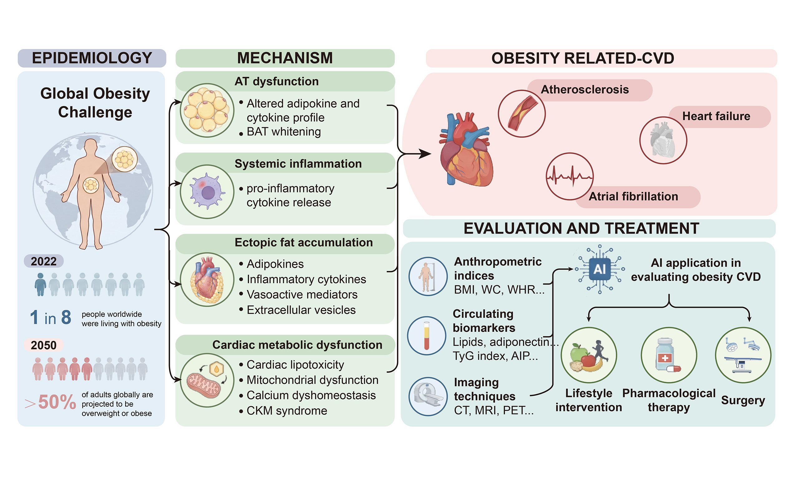
Graphical Abstract
Abstract
Obesity has emerged as a global public health crisis, with its prevalence increasing dramatically over recent decades. Accumulating evidence identifies obesity as a major independent risk factor for the development and progression of cardiovascular disease (CVD). This review comprehensively summarizes current evidence on the association between obesity and phenotype-specific CVD, and discusses the underlying pathophysiological mechanisms, including adipose tissue dysfunction, systemic inflammation, ectopic fat accumulation, and cardiac metabolic dysfunction. We also examine key indicators for assessing obesity-related cardiovascular risk and discuss emerging applications of artificial intelligence in risk prediction and personalized management. Finally, we summarize current evidence-based therapeutic approaches to provide guidance for clinicians, researchers, and public health policymakers.
Keywords


 Download PDF
INTRODUCTION
Obesity is a chronic metabolic disorder characterized by excessive or abnormal accumulation of adipose tissue (AT), which is associated with an increased risk of various metabolic complications and chronic diseases. According to current World Health Organization (WHO) guidelines, overweight and obesity in adults are defined according to body mass index (BMI), with BMI ≥ 25 kg/m2 classified as overweight and BMI ≥ 30 kg/m2 classified as obesity[1]. Driven by factors including increased caloric intake and reduced physical activity, the prevalence of obesity has risen steadily in recent decades[2]. By 2022, one in eight people worldwide were living with obesity[1], with projections indicating that by 2050, more than half of adults worldwide will be overweight or obese[3]. Obesity therefore represents an escalating global public health concern. Although BMI remains the most widely used measure for obesity classification because of its simplicity and accessibility, recent studies have suggested that BMI may not fully capture body fat distribution or obesity-related health risks. Accordingly, alternative anthropometric measures, including the body roundness index (BRI), waist circumference (WC), and waist-to-hip ratio (WHR), have attracted increasing attention for the assessment of obesity and obesity-related risk[4-6].
The association between obesity and cardiovascular disease (CVD) is well established[7-9]. More than two-thirds of deaths attributable to high BMI are due to CVD[7], and obesity is associated with substantially increased risks of ischemic heart disease, heart failure (HF), and stroke[10]. Elucidating the complex interplay between obesity and CVD, together with developing targeted preventive and therapeutic strategies, is therefore essential to reducing the global burden of CVD.
This review synthesizes current evidence on obesity-related CVD across four key domains: (1) epidemiological associations between obesity and CVD; (2) the pathophysiological mechanisms underlying obesity-related CVD; (3) conventional and artificial intelligence (AI)-based approaches to cardiovascular risk assessment in obesity; and (4) current and emerging therapeutic strategies for obesity-related CVD.
EPIDEMIOLOGICAL EVIDENCE FOR OBESITY-RELATED CVD
A robust dose-response association between adiposity and CVD risk has been well established in large-scale cohorts[11-13]. In a prospective cohort study of 360,855 participants from the China Kadoorie Biobank, early-adulthood BMI showed a monotonic dose-response association with incident CVD. Compared with individuals with an early-adulthood BMI of 20.5-22.4 kg/m2, those with a BMI ≥ 30 kg/m2 had a 58% higher risk of incident CVD (HR = 1.58, 95%CI: 1.42-1.75)[12]. Evidence from an umbrella review and meta-analysis of more than 500 cohort studies further supported the association between adiposity and CVD risk. Across cardiovascular outcomes, each 5 kg/m2 increment in BMI was associated with a 10%-49% increase in cardiovascular event risk[10]. Notably, the association between elevated BMI and CVD risk may be stronger in East and Southeast Asian populations than in Western populations. In a pooled analysis of 97 prospective cohorts involving approximately 1.8 million participants, each 5 kg/m2 increase in BMI was associated with a 40% higher risk of coronary heart disease (CHD) and a 29% higher risk of stroke in Asian cohorts, compared with 24% and 14%, respectively, in Western cohorts[14]. These epidemiological findings are summarized in Table 1.
Epidemiological evidence for obesity-related CVDs
| CVD outcome | Reference | Study design/population | Adiposity exposure | Main finding |
| Overall CVD | [12] | Prospective cohort; 360,855 participants, China Kadoorie Biobank | Early-adulthood BMI ≥ 30 kg/m2 vs. 20.5-22.4 kg/m2 | Incident CVD: HR 1.58 (95%CI: 1.42-1.75) |
| [10] | Umbrella review/meta-analysis; > 500 cohort studies | Each 5 kg/m2 increase in BMI | CVD risk increased by approximately 10%-49% across cardiovascular outcomes | |
| CHD | [14] | Pooled analysis of 97 prospective cohorts; ~1.8 million participants | Each 5 kg/m2 increase in BMI | HR 1.27 (95%CI: 1.23-1.31) |
| [15] | Meta-analysis of 95 cohorts; > 1.2 million participants | Obesity vs. normal weight | Women: HR 1.61 (95%CI: 1.42-1.82); Men: HR 1.60 (95%CI: 1.43-1.79) | |
| [16] | Nurses’ Health Study and Health Professionals Follow-up Study | BMI > 30 kg/m2 | Women: RR 2.16 (95%CI: 1.81-2.58); Men: RR 1.81 (95%CI: 1.48-2.22) | |
| [17] | Global Burden of Disease analysis, 2023 | High BMI | ~0.94 million CHD deaths attributable to high BMI (10.6% of all CHD deaths); 24.56 million DALYs | |
| VTE | [18] | Meta-analysis of 21 case-control and cohort studies | Obesity vs. non-obesity | OR 2.33 (95%CI: 1.68-3.24) |
| [19] | Mendelian randomization; INVENT consortium | Each genetically predicted 1-SD increase in BMI | OR 1.59 (95%CI: 1.30-1.93) | |
| [20] | Tromsø Study | Abdominal obesity | HR 2.03 (95%CI: 1.49-2.75) | |
| HF | [24] | Prospective cohort; Framingham Heart Study, 5,881 participants; mean follow-up 14 years | BMI ≥ 30 kg/m2 vs. normal weight | Women: HR 2.12 (95%CI: 1.51-2.97); Men: HR 1.90 (95%CI: 1.30-2.79) |
| Each 1 kg/m2 increase in BMI | HF risk increased by 7% in women and 5% in men | |||
| [25] | Pooled analysis of three prospective cohorts; 51,451 participants | Overweight vs. normal weight | HFpEF: HR 1.38 (95%CI: 1.18-1.61); HFrEF: HR 1.03 (95%CI: 0.87-1.22) | |
| [26] | Pooled analysis of four community-based cohorts; 22,681 participants | Each 1-SD increase in BMI | HFpEF: HR 1.34 (95%CI: 1.24-1.45); HFrEF: HR 1.18 (95%CI: 1.10-1.27) | |
| AF | [27] | Framingham Heart Study | Each 1 kg/m2 increase in BMI | HR 1.04 (95%CI: 1.01-1.07) in both men and women |
| [28] | Meta-analysis of 16 observational studies | Obesity vs. non-obesity | RR 1.49 (95%CI: 1.36-1.64) | |
| [29] | Mendelian randomization | Each genetically predicted 1-SD increase in BMI | OR 1.425 (95%CI: 1.346-1.509) |
CHD is among the most extensively studied obesity-related cardiovascular conditions. Large-scale prospective studies have consistently demonstrated a positive association between excess adiposity and CHD risk. In a pooled analysis, each 5 kg/m2 increase in BMI was associated with a 27% higher risk of CHD
Obesity is also an important risk factor for thrombotic disorders, particularly venous thromboembolism (VTE). A meta-analysis of 21 case-control and cohort studies showed that obesity was associated with more than a twofold higher risk of VTE than non-obese individuals (OR = 2.33, 95%CI: 1.68-3.24)[18]. Mendelian randomization analysis further provided genetic evidence supporting a causal relationship between obesity and VTE. In the INVENT consortium, each genetically predicted 1-standard-deviation increase in BMI was associated with a 59% higher risk of VTE (OR = 1.59, 95%CI: 1.30-1.93)[19]. Notably, some studies have suggested that abdominal adiposity may be particularly relevant to thrombotic risk[20,21]. In the Tromsø Study, abdominal obesity was the only component of the metabolic syndrome that remained significantly associated with VTE after multivariable adjustment (HR = 2.03, 95%CI: 1.49-2.75), suggesting that fat distribution may provide additional information beyond overall adiposity in assessing VTE risk[20].
HF represents another major cardiovascular complication associated with obesity. Based on left ventricular ejection fraction (LVEF), HF is generally classified into three phenotypes: heart failure with reduced ejection fraction (HFrEF; LVEF < 40%), HF with mildly reduced ejection fraction (HFmrEF; LVEF 40%-49%), and heart failure with preserved ejection fraction (HFpEF; LVEF ≥ 50%)[22,23]. Long-term follow-up data from the Framingham Heart Study provide compelling evidence of the association between obesity and HF. Among 5,881 participants followed for a mean of 14 years, obesity was associated with an approximately twofold higher risk of HF than normal weight, with hazard ratios of 2.12 (95%CI: 1.51-2.97) in women and 1.90 (95%CI: 1.30-2.79) in men. Moreover, each 1 kg/m2 increase in BMI was associated with a 7% and 5% increase in HF risk in women and men, respectively, supporting a graded relationship between increasing adiposity and HF incidence[24]. Notably, higher BMI appears to be more consistently and more strongly associated with the development of HFpEF than with HFrEF. In a pooled analysis of 51,451 participants from three prospective cohorts, overweight was associated with a 38% higher risk of HFpEF (HR = 1.38, 95%CI: 1.18-1.61) compared with normal weight, whereas no significant increase in HFrEF risk was observed (HR = 1.03, 95%CI: 0.87-1.22)[25]. Similarly, another pooled analysis of 22,681 participants from four community-based cohorts showed that each 1-SD increase in BMI was associated with a 34% higher risk of incident HFpEF (HR = 1.34, 95%CI: 1.24-1.45), compared with an 18% increase in HFrEF risk (HR = 1.18, 95%CI: 1.10-1.27), with a significant difference between HF subtypes[26].
Atrial fibrillation (AF) is the most common obesity-related arrhythmia. In the Framingham Heart Study, obesity was identified as an independent risk factor for incident AF, with each 1 kg/m2 increase in BMI associated with a 4% higher risk of AF in both men and women (HR = 1.04, 95%CI: 1.01-1.07)[27]. Consistently, a meta-analysis of 16 observational studies showed that obesity was associated with a 49% higher risk of AF (RR = 1.49, 95%CI: 1.36-1.64)[28]. More recently, Mendelian randomization analysis provided genetic support for a causal relationship between BMI and AF. A genetically predicted 1-standard-deviation increase in BMI was associated with a 42.5% higher risk of AF (OR = 1.425, 95%CI: 1.346-1.509)[29].
However, the so-called "obesity paradox" has challenged the conventional interpretation of the obesity-CVD relationship. This phenomenon refers to observations that individuals who are overweight or mildly obese and have established cardiovascular or other chronic diseases may exhibit better clinical outcomes than those with normal BMI[30]. Several methodological explanations have been proposed to account for the obesity paradox, with three major contributors receiving particular attention: reverse causation, selection bias, and the limitations of BMI as a surrogate measure of adiposity[31-33].
Reverse causation may contribute to the obesity paradox because underlying chronic diseases can cause unintentional weight loss and reduced adiposity, making individuals with lower BMI appear to have a higher risk of adverse outcomes compared with those with higher BMI. In an NHANES analysis of individuals with CVD, overweight or obesity initially appeared protective against mortality (HR = 0.89, 95%CI: 0.78-1.01); however, after accounting for reverse causation, the association was reversed (HR = 1.16, 95%CI: 0.95-1.41), with higher mortality observed among patients with overweight or obesity (HR = 1.51, 95%CI: 1.07-2.15)[31]. Mendelian randomization analyses provide additional support because they are less susceptible to reverse causation than conventional observational studies. Results showed that each genetically predicted 1 kg/m2 increase in BMI was associated with a 10% higher risk of cardiovascular mortality (HR = 1.10, 95%CI: 1.01-1.19) and a 12% higher risk of CHD mortality (HR = 1.12, 95%CI: 1.00-1.25), providing little support for a true protective effect of excess adiposity[34]. Collectively, these findings suggest that the apparent obesity paradox may largely reflect reverse causation arising from disease-related weight loss rather than a genuine survival benefit of obesity.
Selection bias may also contribute to the obesity paradox because studies restricted to patients with established disease may preferentially include individuals in whom low BMI reflects greater disease severity or frailty, thereby making higher BMI appear protective. Banack and Kaufman demonstrated through simulation analyses that selection bias alone could generate an apparent protective effect of obesity among individuals with CVD[35]. Using causal models and simulation, Sperrin et al. further demonstrated that conditioning on disease status can introduce collider bias and generate an apparent inverse association between obesity and mortality[36]. However, their analyses also indicated that collider bias alone is unlikely to fully explain the obesity paradox because the magnitude of bias was generally small relative to the underlying causal associations, suggesting that other factors may also contribute[36].
A more fundamental explanation for the obesity paradox may lie in the limitations of BMI as a measure of adiposity. By design, BMI is a simplified, population-level metric that cannot distinguish between fat mass and lean mass, nor capture the well-established prognostic impact of anatomical body fat distribution. Several studies have suggested that the apparent protective effect of obesity observed in patients with CVD may partly result from misclassification caused by the use of BMI alone[37,38]. Butt et al. compared BMI with alternative anthropometric measures, including waist-to-height ratio (WHtR), in 8,399 patients with HF enrolled in the PARADIGM-HF trial[39]. In unadjusted analyses, patients with BMI ≥ 25 kg/m2 appeared to have a lower risk of mortality, consistent with the conventional obesity paradox. However, after comprehensive adjustment for established prognostic factors, including N-terminal pro-B-type natriuretic peptide (NT-proBNP), this apparent survival advantage disappeared. Importantly, WHtR, which better reflects central adiposity and is less influenced by muscle mass, skeletal weight, and fluid retention, showed no obesity paradox; instead, higher WHtR was associated with a greater risk of adverse outcomes, including HF hospitalization (adjusted HR for the highest vs. lowest quintile: 1.39, 95%CI: 1.06-1.81)[39]. These findings suggest that the apparent protective effect of obesity observed with BMI may largely reflect the limitations of BMI in accurately characterizing adiposity rather than a true biological benefit of excess adiposity.
MECHANISMS UNDERLYING OBESITY-RELATED CVD
Obesity promotes CVD through a complex network of interconnected metabolic, inflammatory, and tissue-specific alterations. These processes can be integrated into four major mechanistic domains: (1) AT dysfunction and altered endocrine/metabolic signaling; (2) systemic inflammatory activation; (3) ectopic fat accumulation; and (4) cardiac metabolic dysfunction. Rather than acting independently, these mechanisms are interconnected through systemic and local pathways. AT dysfunction represents an important upstream disturbance, characterized by altered adipokine secretion and increased lipid availability. At the systemic level, elevated free fatty acids (FFAs) and dysregulated adipokine signaling promote macrophage activation and pro-inflammatory cytokine release, sustaining chronic low-grade inflammation. In parallel, excess lipid availability contributes to ectopic fat expansion and pathological remodeling. Ectopic fat depots, particularly epicardial and perivascular AT, further amplify cardiovascular injury through paracrine signaling. These systemic and local disturbances converge on cardiovascular tissues. In cardiomyocytes, excessive lipid accumulation and inflammatory stress disrupt mitochondrial function, cellular quality control, and Ca2+ homeostasis, thereby promoting cardiac metabolic dysfunction. Importantly, the interactions among these pathological mechanisms are not unidirectional. Systemic inflammation can further aggravate AT dysfunction, while cardiovascular injury may reinforce inflammatory activation and alter adipose metabolism, thereby establishing self-reinforcing feedback loops that amplify cardiovascular injury.
The relative contributions of these pathological mechanisms vary across cardiovascular phenotypes. In atherosclerosis, vascular inflammation, endothelial dysfunction, and macrophage lipid accumulation play central roles. In HF, myocardial metabolic dysfunction, inflammatory injury, and hemodynamic stress interact to promote adverse remodeling, with adiposity-related myocardial stiffness and microvascular dysfunction being particularly relevant to HFpEF. In AF, local epicardial fat-atrium crosstalk and systemic inflammation contribute to atrial fibrosis and electrical remodeling. Thus, different cardiovascular phenotypes may arise from distinct patterns of interaction among these shared mechanisms.
In the following sections, we first discuss the four mechanistic domains and then examine how their differential contributions are associated with atherosclerosis, HF, and AF. The major molecular pathways are summarized in Table 2 and Figures 1-4.
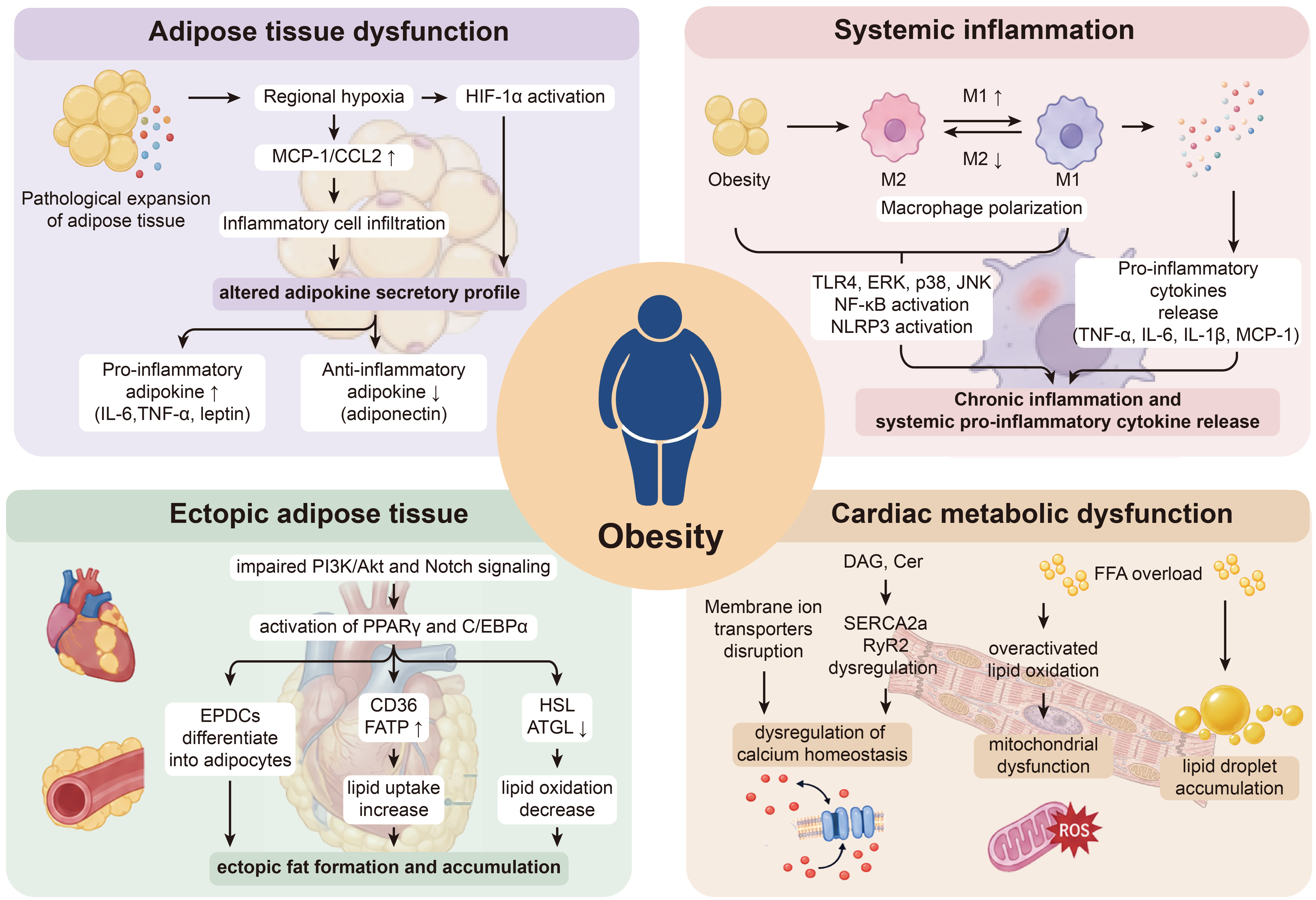
Figure 1. Molecular mechanisms linking obesity to CVD. Obesity promotes cardiovascular injury through four interconnected mechanisms: adipose tissue dysfunction, systemic inflammation, ectopic adipose tissue accumulation, and cardiac metabolic dysfunction. Pathological expansion of adipose tissue induces regional hypoxia and HIF-1α activation, accompanied by increased MCP-1/CCL2 expression and inflammatory cell infiltration, ultimately shifting the adipokine secretory profile toward increased pro-inflammatory adipokines, including IL-6, TNF-α, and leptin, and reduced anti-inflammatory adiponectin. Obesity also promotes macrophage polarization toward a pro-inflammatory M1 phenotype and suppresses the M2 phenotype. Activation of TLR4-ERK/p38/JNK, NF-κB, and NLRP3 signaling enhances the release of pro-inflammatory cytokines, including TNF-α, IL-6, IL-1β, and MCP-1, thereby sustaining chronic systemic inflammation. In parallel, impaired PI3K/Akt and Notch signaling activates PPARγ- and C/EBPα-dependent adipogenic programs, promotes the differentiation of epicardial progenitor-derived cells into adipocytes, increases CD36/FATP-mediated lipid uptake, and reduces HSL/ATGL-mediated lipid mobilization, collectively facilitating ectopic fat formation and accumulation. In cardiomyocytes, DAG and Cer disrupt membrane ion transport and SERCA2a/RyR2-mediated Ca2+ handling, whereas FFA overload promotes excessive lipid oxidation, mitochondrial dysfunction, ROS generation, and lipid droplet accumulation. Together, these processes contribute to obesity-associated cardiovascular dysfunction and remodeling. HIF-1α: Hypoxia-inducible factor-1α; MCP-1/CCL2: monocyte chemoattractant protein-1/C-C motif chemokine ligand 2; IL: interleukin; TNF-α: tumor necrosis factor-α; TLR4: Toll-like receptor 4; ERK: extracellular signal-regulated kinase; JNK: c-Jun N-terminal kinase; NF-κB: nuclear factor κB; NLRP3: NOD-like receptor family pyrin domain-containing 3; PI3K/Akt: phosphoinositide 3-kinase/protein kinase B; PPARγ: peroxisome proliferator-activated receptor γ; C/EBPα: CCAAT/enhancer-binding protein α; EPDCs: epicardium-derived progenitor cells; FATP: fatty acid transport protein; HSL: hormone-sensitive lipase; ATGL: adipose triglyceride lipase; DAG: diacylglycerol; Cer: ceramide; FFA: free fatty acid; SERCA2a: sarco/endoplasmic reticulum Ca2+-ATPase 2a; RyR2: ryanodine receptor 2; ROS: reactive oxygen species.
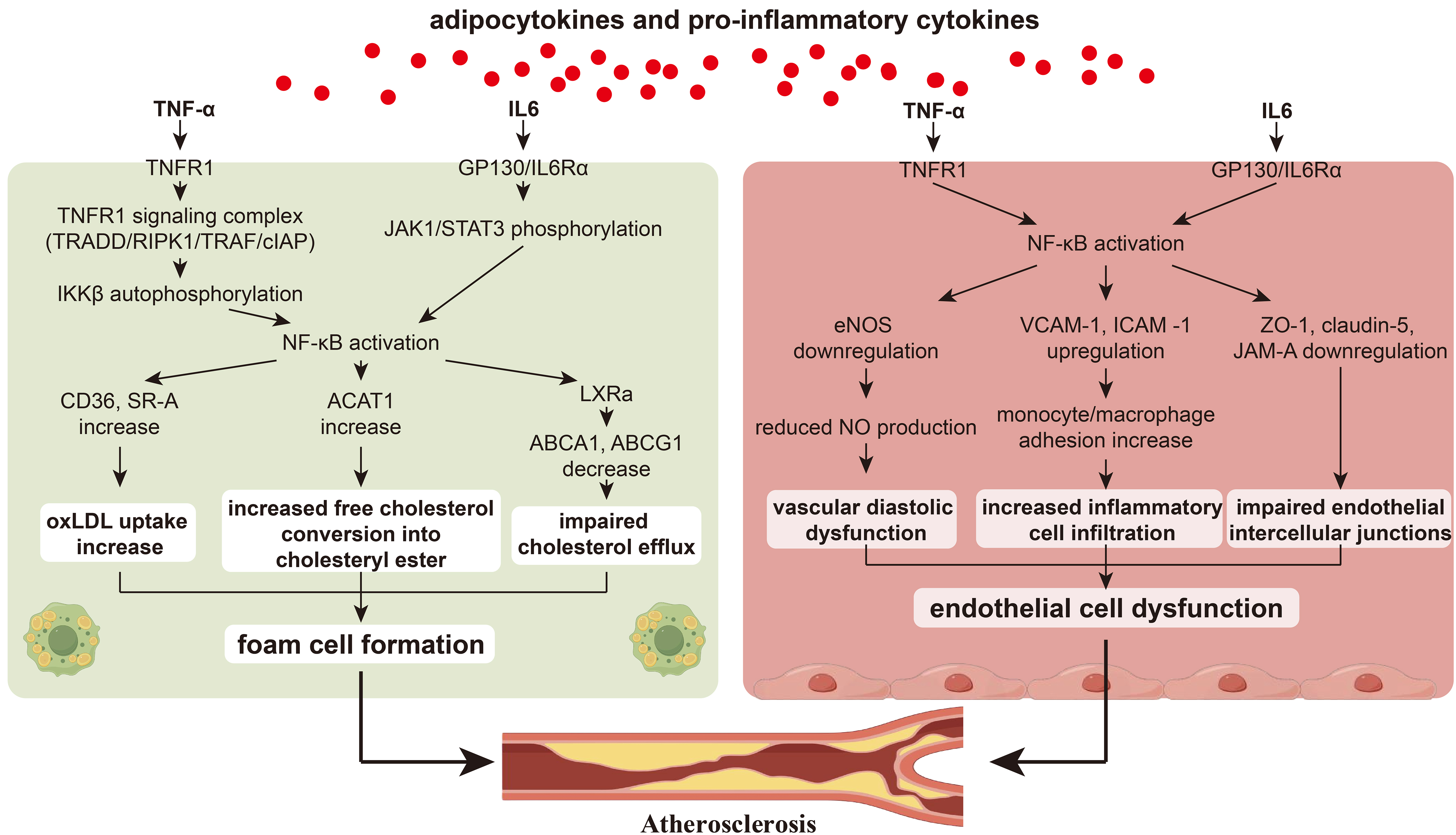
Figure 2. Mechanisms of adipocytokine- and pro-inflammatory cytokine-mediated atherosclerosis. Adipocytokines and pro-inflammatory cytokines promote atherosclerosis by enhancing macrophage lipid accumulation and impairing endothelial function. In macrophages, TNF-α binds to TNFR1 and activates the TNFR1 signaling complex, including TRADD, RIPK1, TRAFs, and cIAPs, thereby inducing IKKβ activation and subsequent NF-κB signaling. IL-6 signaling through the IL-6Rα/GP130 complex activates the JAK1/STAT3 pathway and further augments NF-κB activity. Activated NF-κB upregulates CD36 and SR-A expression, thereby increasing ox-LDL uptake, while simultaneously enhancing ACAT1 expression to promote the esterification of free cholesterol into cholesteryl esters. In parallel, suppression of LXRα-dependent ABCA1 and ABCG1 expression impairs cholesterol efflux. Together, these alterations promote intracellular cholesterol accumulation and foam-cell formation. In endothelial cells, TNF-α/TNFR1 and IL-6/IL-6Rα/GP130 signaling activate NF-κB, resulting in eNOS downregulation and reduced nitric oxide bioavailability, increased VCAM-1 and ICAM-1 expression with enhanced monocyte adhesion and transmigration, and reduced expression of endothelial junctional proteins, including ZO-1, claudin-5, and JAM-A. Collectively, these changes impair endothelium-dependent vasodilation, increase inflammatory cell recruitment and endothelial permeability, and ultimately promote endothelial dysfunction and atherosclerotic plaque development. TNF-α: Tumor necrosis factor-α; TNFR1: tumor necrosis factor receptor 1; TRADD: TNFR1-associated death domain protein; RIPK1: receptor-interacting serine/threonine-protein kinase 1; TRAF: TNF receptor-associated factor; cIAP: cellular inhibitor of apoptosis protein; IKKβ: inhibitor of nuclear factor κB kinase β; NF-κB: nuclear factor κB; IL-6: interleukin-6; IL-6Rα: interleukin-6 receptor α; GP130: glycoprotein 130; JAK1: Janus kinase 1; STAT3: signal transducer and activator of transcription 3; CD36: cluster of differentiation 36; SR-A: scavenger receptor A; ox-LDL: oxidized low-density lipoprotein; ACAT1: acyl-CoA:cholesterol acyltransferase 1; LXRα: liver X receptor α; ABCA1/ABCG1: ATP-binding cassette transporters A1/G1; eNOS: endothelial nitric oxide synthase; NO: nitric oxide; VCAM-1: vascular cell adhesion molecule-1; ICAM-1: intercellular adhesion molecule-1; ZO-1: zonula occludens-1; JAM-A: junctional adhesion molecule A.
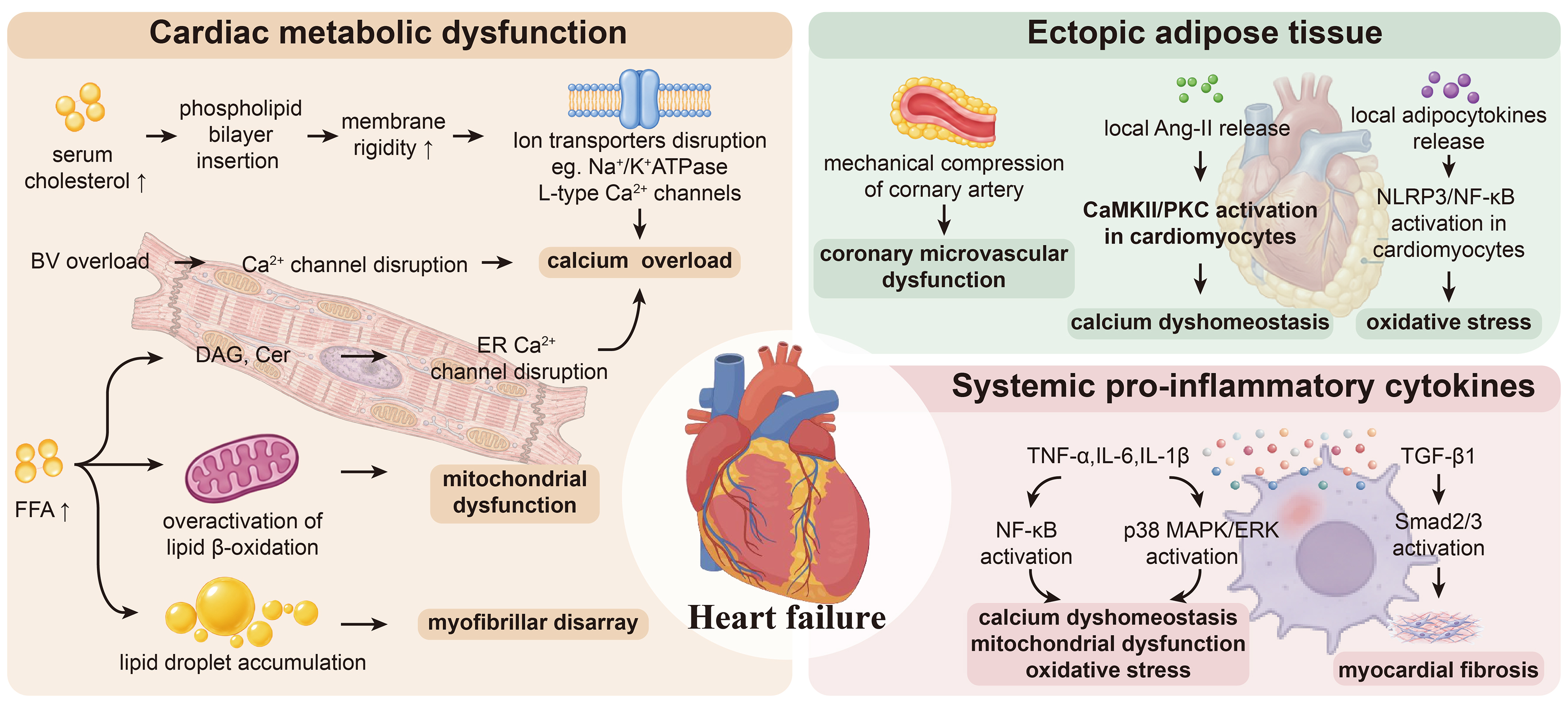
Figure 3. Mechanisms of obesity-associated HF. Obesity promotes heart failure through interconnected metabolic dysfunction and ectopic adipose tissue-mediated mechanisms. In cardiomyocytes, elevated circulating cholesterol alters membrane lipid composition and increases membrane rigidity, thereby impairing membrane transporters and ion channels, including Na+/K+-ATPase and L-type Ca2+ channels, and disrupting intracellular ionic homeostasis. Volume overload and abnormal sarcoplasmic/endoplasmic reticulum Ca2+ handling further aggravate Ca2+ dyshomeostasis. Excess free fatty acids promote lipid droplet accumulation and the generation of lipotoxic intermediates, including DAGs and Cer, while excessive fatty acid β-oxidation imposes mitochondrial metabolic stress, resulting in impaired ATP production, increased ROS generation, and myofibrillar disruption. Ectopic adipose tissue, particularly EAT, further contributes to cardiac dysfunction through mechanical compression of the coronary circulation and the local release of Ang-II and pro-inflammatory adipocytokines. These mediators activate NLRP3/NF-κB signaling, enhance oxidative stress, impair SERCA2a-dependent Ca2+ handling, and promote coronary microvascular dysfunction. In parallel, pro-inflammatory cytokines, including TNF-α, IL-6, and IL-1β, activate NF-κB and p38 MAPK/ERK signaling, thereby exacerbating mitochondrial dysfunction, lipid accumulation, oxidative stress, and cardiomyocyte injury. TGF-β1-mediated Smad2/3 signaling further promotes the expression of collagen-associated genes, including COL1A1 and COL3A1, leading to extracellular matrix deposition, myocardial fibrosis, increased ventricular stiffness, and ultimately heart failure. FFA: Free fatty acids; DAGs: diacylglycerols; Cer: ceramide; ATP: adenosine triphosphate; ROS: reactive oxygen species; Ang-II: angiotensin II; NLRP3: NOD-like receptor family pyrin domain-containing 3; NF-κB: nuclear factor κB; SERCA2a: sarco/endoplasmic reticulum Ca2+-ATPase 2a; TNF-α: tumor necrosis factor-α; IL: interleukin; MAPK: mitogen-activated protein kinase; ERK: extracellular signal-regulated kinase; TGF-β1: transforming growth factor-β1; COL1A1: collagen type I alpha 1 chain; COL3A1: collagen type III alpha 1 chain.
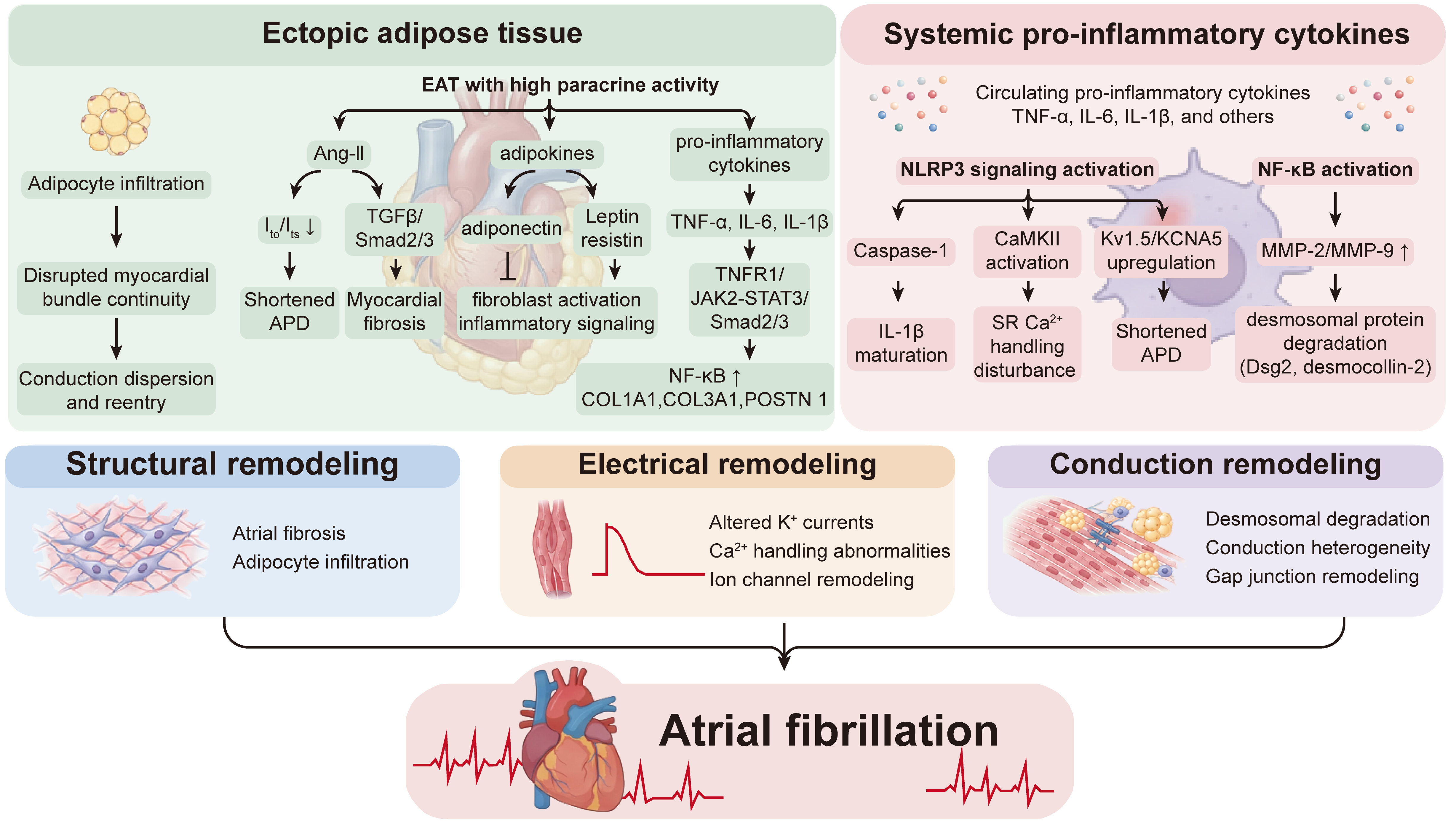
Figure 4. Mechanisms of obesity-related atrial fibrillation. Ectopic adipose tissue and systemic pro-inflammatory cytokines promote atrial fibrillation through coordinated structural, electrical, and conduction remodeling. Adipocyte infiltration disrupts the continuity of atrial myocardial bundles, thereby increasing conduction heterogeneity and susceptibility to reentry. EAT with enhanced paracrine activity further contributes to atrial remodeling through the local release of Ang-II, adipokines, and pro-inflammatory cytokines. Ang-II reduces cardiomyocyte K+ currents, thereby shortening action potential duration, while simultaneously activating TGF-β/Smad2/3 signaling in cardiac fibroblasts to promote fibrotic remodeling. In parallel, EAT-derived TNF-α, IL-6, IL-1β, leptin, and TGF-β activate TNFR1-, JAK2/STAT3-, NF-κB-, and TGFβR/Smad2/3-dependent signaling pathways, enhancing inflammatory responses and upregulating fibrosis-associated genes, including COL1A1, COL3A1, and POSTN. Systemic pro-inflammatory cytokines further activate the NLRP3/caspase-1/IL-1β axis and CaMKII signaling, resulting in abnormal sarcoplasmic reticulum Ca2+ handling and electrical instability. Concurrent upregulation of Kv1.5/KCNA5-mediated IKur contributes to action potential shortening. In addition, NF-κB-dependent induction of MMP-2 and MMP-9 promotes degradation of desmosomal proteins, including Dsg2 and Dsc2, thereby impairing intercellular coupling and increasing conduction heterogeneity and effective refractory period dispersion. Collectively, these structural, electrical, and conduction abnormalities establish a pro-arrhythmic substrate that facilitates the initiation and maintenance of atrial fibrillation. EAT: Epicardial adipose tissue; Ang-II: angiotensin II; APD: action potential duration; ERP: effective refractory period; TNF-α: tumor necrosis factor-α; IL: interleukin; TNFR1: tumor necrosis factor receptor 1; JAK2: Janus kinase 2; STAT3: signal transducer and activator of transcription 3; TGF-β: transforming growth factor-β; TGFβR: transforming growth factor-β receptor; NF-κB: nuclear factor κB; COL1A1: collagen type I alpha 1 chain; COL3A1: collagen type III alpha 1 chain; POSTN: periostin; NLRP3: NOD-like receptor family pyrin domain-containing 3; CaMKII: Ca2+/calmodulin-dependent protein kinase II; IKur: ultra-rapid delayed rectifier potassium current; Kv1.5: voltage-gated potassium channel Kv1.5; KCNA5: potassium voltage-gated channel subfamily A member 5; MMP: matrix metalloproteinase; Dsg2: desmoglein-2; Dsc2: desmocollin-2.
Mechanisms underlying obesity-related CVD
| Mechanistic category | Major molecular pathways/mediators | Major cellular consequences | Predominant CVD phenotype(s) |
| AT dysfunction and altered endocrine/metabolic signaling | Adipose hypoxia → HIF-1α activation → Leptin, IL-6, MIF, and PAI-1 increase; adiponectin decrease[44,47-50] | Pro-inflammatory adipokine imbalance, impaired metabolic homeostasis, insulin resistance | Shared upstream contribution to obesity-associated CVD |
| Hypoxia → MCP-1/CCL2 → Monocyte recruitment/M1-like macrophage activation[51-59] | Adipocyte-macrophage inflammatory crosstalk and increased TNF-α, IL-1β, IL-6 secretion | Shared upstream contribution to obesity-associated CVD | |
| FFA/SFA → TLR4/NF-κB → TNF-α, IL-6[56,61-63] | Sustained adipose inflammation and systemic inflammatory signaling | Shared upstream contribution to obesity-associated CVD | |
| BAT whitening → UCP1 downregulation; loss of FGF21, NRG4, 12,13-diHOME; TNF-α/IL-6/MCP-1/leptin increase; adiponectin decrease[64-74] | Reduced thermogenesis, impaired glucose/lipid utilization, systemic insulin resistance and inflammation | Shared upstream contribution to obesity-associated CVD | |
| Systemic inflammation | SFAs → PKC/MAPK signaling and enhanced inflammatory responses; SFAs → NLRP3 inflammasome activation → IL-1β release[77,78] | Macrophage pro-inflammatory activation and increased cytokine release | Shared upstream contribution to obesity-associated CVD |
| Palmitate/Cer → CD11b and CD36 upregulation; DAG → PKCδ/NF-κB/p38 MAPK; Cer → NLRP3/caspase-1/IL-1β[79-81] | Enhanced macrophage lipid uptake and sustained inflammatory activation | Atherosclerosis; HF; AF | |
| NF-κB → CD36/SR-A increase, ACAT1 increase; LXRα-ABCA1/ABCG1 decrease[136-140] | Increased ox-LDL uptake and cholesterol esterification; impaired cholesterol efflux; foam-cell formation | Atherosclerosis | |
| NF-κB → eNOS/NO decrease; VCAM-1/ICAM-1 increase; ZO-1/claudin-5/JAM-A decrease[141-147] | Impaired vasodilation, monocyte adhesion/transmigration, increased endothelial permeability | Atherosclerosis | |
| Ectopic fat accumulation | Altered PKC/JNK/IKK-NF-κB and PI3K/Akt-GSK-3β/Notch signaling→ Activation of PPARγ/C/EBPα[84,86-88] | Adipogenic differentiation and expansion/remodeling of EAT | HF; AF |
| CD36/FATP-mediated lipid uptake increase; ATGL/HSL-dependent lipid mobilization decrease[89,90] | Ectopic triglyceride accumulation and adipocyte hypertrophy | HF; AF | |
| EAT-derived Ang-II and adipocytokines[93,94,97,157,158] | Local inflammation, mitochondrial/metabolic dysfunction, fibrosis and myocardial remodeling | HF; AF | |
| EAT-derived EVs carrying miR-1-3p and miR-133a-3p → Reduced Kcnj2/Kcnj12 expression[95,96] | Pro-arrhythmic signaling and impaired electrical conduction | AF | |
| EAT-conditioned medium → Cardiomyocyte miR-208a upregulation[97] | Impaired mitochondrial fatty acid oxidation and contractile function | HF | |
| Cardiac metabolic dysfunction | Excess FFA → DAG/Cer/acylcarnitines → PKC activation / Akt inhibition; long-chain acylcarnitine-induced Ca2+ dysregulation[101-105] | Metabolic inflexibility, reduced ATP production, increased ROS and impaired contractility | HF |
| Lipid overload → TFEB/LAMP-2A-associated autophagic impairment; Lipid overload → Altered DRP1- and PINK1/Parkin-dependent mitophagy[106-108] | Impaired organelle quality control, accumulation of dysfunctional mitochondria and energetic impairment | HF | |
| Lipotoxic stress → DRP1 acetylation → Excessive mitochondrial fission[111] | Mitochondrial fragmentation | HF | |
| Mitochondrial ROS → RyR2 oxidation; SERCA2a-mediated Ca2+ reuptake decrease; MCU-CaMKII activation[115-117] | SR Ca2+ leak, mitochondrial Ca2+ overload and excitation-contraction dysfunction | HF | |
| Volume expansion/mechanical stretch → Mechanosensitive signaling and MAPK activation[120,121] | Cardiomyocyte hypertrophy, extracellular matrix deposition and maladaptive remodeling | HF |
Molecular mechanisms linking obesity to CVD
AT dysfunction and altered endocrine/metabolic signaling
AT is not merely an energy-storage depot but a highly active endocrine and immunometabolic tissue that regulates systemic metabolic homeostasis, immune responses, vascular function, and cardiovascular physiology[40,41]. Accordingly, pathological remodeling of AT represents an important upstream component of obesity-associated cardiovascular injury.
As obesity progresses, excessive adipocyte hypertrophy and hyperplasia drive pathological expansion of AT. When tissue growth exceeds the capacity of compensatory angiogenesis, inadequate vascularization results in local hypoxia, which represents a key initiating event in AT dysfunction[42-44]. Hypoxia stabilizes and activates hypoxia-inducible factor-1α (HIF-1α), a master transcriptional regulator of cellular adaptation to reduced oxygen availability[45,46]. Sustained activation of HIF-1α profoundly reshapes the adipokine secretory profile, shifting it toward a pro-inflammatory and metabolically unfavorable state. Hypoxic adipocytes exhibit increased expression and secretion of inflammation-associated adipokines and cytokines, including leptin, interleukin-6 (IL-6), macrophage migration inhibitory factor, and plasminogen activator inhibitor-1, whereas the production of adiponectin, a potent insulin-sensitizing and anti-inflammatory adipokine, is markedly suppressed[44,47,48]. Mechanistically, HIF-1α can directly promote leptin transcription through a hypoxia-responsive element in the leptin promoter and suppress adiponectin expression through modulation of the SOCS3-STAT3 signaling axis[49,50].
Beyond the direct effects of HIF-1α on adipocyte function, hypoxia also promotes the recruitment and activation of immune cells within AT. Hypoxic stress enhances the expression of chemokines, particularly monocyte chemoattractant protein-1 (MCP-1/CCL2), as well as endothelial adhesion molecules, thereby facilitating the recruitment of circulating monocytes into AT[51-53]. Within the hypoxic and inflammatory microenvironment, these recruited monocytes differentiate into pro-inflammatory M1-like macrophages[54]. Activated macrophages subsequently release tumor necrosis factor-α (TNF-α), interleukin-1β (IL-1β), IL-6, and other inflammatory mediators, establishing reciprocal signaling between adipocytes and immune cells through NF-κB, mitogen-activated protein kinase (MAPK), and STAT3 pathways[54-59]. Persistent activation of this inflammatory circuit disrupts adipose tissue endocrine and metabolic homeostasis, driving a pro-inflammatory adipokine imbalance that exacerbates local insulin resistance (IR) and systemic inflammation, and ultimately contributes to cardiovascular injury[60].
Elevated circulating FFAs provide an additional inflammatory stimulus that reinforces obesity-associated AT dysfunction. Excess FFAs, particularly saturated fatty acids (SFAs), can directly activate TLR4-dependent signaling in adipocytes and immune cells, resulting in activation of NF-κB and other downstream inflammatory pathways[61,62]. This, in turn, enhances the production of pro-inflammatory mediators such as IL-6 and TNF-α, thereby reinforcing adipocyte-immune cell crosstalk and perpetuating local adipose tissue inflammation[56,63].
Importantly, obesity-associated adipose tissue dysfunction is not restricted to white adipose tissue. Brown adipose tissue (BAT) also undergoes profound phenotypic and metabolic remodeling during obesity. BAT whitening refers to the pathological transition of thermogenic brown adipocytes toward a white adipocyte-like phenotype, characterized by enlarged lipid droplets, reduced mitochondrial content and function, and diminished expression of thermogenic proteins such as uncoupling protein 1 (UCP1)[64]. This process is frequently observed in obesity and is accompanied by impaired thermogenesis, reduced energy expenditure, and altered lipid and glucose metabolism[65,66]. BAT whitening not only compromises the metabolic protective functions of BAT but also promotes lipid accumulation, systemic metabolic dysfunction, and inflammation, thereby contributing to the development of obesity-associated CVD[64,65,67].
BAT whitening consequently reduces energy expenditure and the utilization of circulating glucose and fatty acids, thereby promoting dyslipidemia, hyperglycemia, and systemic IR[68,69]. In addition, loss of BAT-derived protective mediators, including fibroblast growth factor 21 (FGF21), neuregulin-4 (NRG4), and 12,13-diHOME, may diminish the metabolic and cardiovascular protection normally conferred by functional BAT[70-73]. At the same time, whitened BAT increasingly exhibits a dysfunctional secretory profile characterized by increased TNF-α, IL-6, MCP-1/CCL2, and leptin and reduced adiponectin[67,74]. Thus, dysfunction of both white and brown adipose tissue constitutes an upstream endocrine and metabolic disturbance that promotes systemic inflammatory and metabolic stress and facilitates subsequent cardiovascular injury.
Systemic inflammation and macrophage-derived pro-inflammatory cytokine release
The second mechanistic domain involves the propagation of obesity-associated metabolic stress beyond adipose tissue into a state of chronic low-grade systemic inflammation. Sustained activation and metabolic reprogramming of innate immune cells, particularly macrophages, provide an important link between adipose tissue dysfunction and cardiovascular injury[75,76].
At the systemic level, elevated circulating FFAs can activate inflammatory responses in macrophages through lipid metabolism-dependent signaling. Schwartz et al. demonstrated that SFA pretreatment markedly enhanced lipopolysaccharide-induced IL-6 and IL-8 production in human monocytes, whereas inhibition of PKC/MAPK signaling attenuated these inflammatory responses[77]. In addition, L'Homme et al. showed that SFAs, including palmitate and stearate, promoted NLRP3 inflammasome activation and IL-1β release in human monocytes/macrophages, whereas unsaturated fatty acids exhibited distinct effects[78]. Chronic activation of these signaling pathways promotes macrophage pro-inflammatory reprogramming, thereby contributing to obesity-associated chronic inflammation.
In parallel, obesity-associated lipotoxic metabolites, including ceramides (Cer) and diacylglycerols (DAGs), impose substantial metabolic stress on macrophages. It is reported that palmitate increased CD11b and CD36 expression and oxidized low-density lipoprotein (LDL) uptake in macrophages, and these effects could be attenuated by inhibition of Cer synthesis but not by reactive oxygen species (ROS) scavenging[79]. Furthermore, obesity also increased DAG accumulation in mouse peritoneal macrophages, accompanied by enhanced PKCδ, NF-κB, and p38 MAPK signaling activation. Pharmacological inhibition of adipose triglyceride lipase attenuated DAG accumulation and the associated inflammatory signaling changes[80]. Moreover, Cer accumulation can promote NLRP3 inflammasome activation in macrophages, facilitating caspase-1-dependent maturation and release of IL-1β[81].
Collectively, these alterations sustain a macrophage-centered pro-inflammatory state that drives chronic systemic inflammation, ultimately contributing to obesity-related CVD.
Ectopic fat accumulation in the cardiovascular system
Ectopic fat broadly refers to the abnormal lipid deposition within or around organs and tissues not primarily specialized for lipid storage, including the liver, pancreas, skeletal muscle, heart, and vasculature[82]. Of particular relevance to CVD are adipose depots located in close anatomical proximity to cardiovascular structures, including epicardial adipose tissue (EAT), situated between the myocardium and the visceral pericardium; pericardial adipose tissue (PAT), located external to the parietal pericardium; and perivascular adipose tissue (PVAT), which surrounds large- and medium-sized blood vessels such as the aorta, coronary arteries, and carotid arteries[83].
Ectopic fat undergoes progressive expansion and pathological remodeling, partly driven by the adipogenic conversion of resident progenitor cell populations, for example, epicardial mesothelial progenitor cells (EMPCs)[84,85]. Sustained exposure to insulin, elevated FFAs, and pro-inflammatory mediators perturb protein kinase C (PKC), JNK, inhibitor of nuclear factor-κB kinase (IKK)/NF-κB, and PI3K/Akt signaling. These alterations promote glycogen synthase kinase-3β (GSK-3β) activation and suppress Notch signaling, thereby promoting the adipogenic differentiation of progenitor cells[86,87]. Concomitantly, activation of canonical adipogenic transcriptional programs, particularly peroxisome proliferator-activated receptor γ (PPARγ) and CCAAT/enhancer-binding protein α (C/EBPα), promotes terminal differentiation into mature adipocytes, thereby contributing to the expansion and remodeling of the epicardial fat depot[84,88]. Once formed, EAT expands further through increased CD36/fatty acid transport protein (FATP)-mediated fatty acid uptake and reduced adipose triglyceride lipase (ATGL)/hormone-sensitive lipase (HSL)-dependent lipolysis and mitochondrial fatty acid oxidation, leading to triglyceride accumulation and adipocyte hypertrophy[89,90].
Pathologically remodeled cardiac ectopic fat exhibits a highly active secretory phenotype, releasing adipokines, inflammatory cytokines, vasoactive mediators, and extracellular vesicles (EVs) that can directly influence the adjacent coronary vasculature and myocardium[85,91,92]. Unlike conventional white AT, cardiac ectopic fat exhibits active sympathetic and local renin-angiotensin signaling, with enhanced catecholaminergic activity and local production of vasoactive mediators such as angiotensin II (Ang-II), which contribute to its neurohumoral effects on the heart[93,94]. EAT-derived EVs, particularly exosomes carrying regulatory microRNAs (miRNAs), constitute an additional pathway of local communication. Shaihov-Teper et al. demonstrated that EAT-derived EVs from patients with AF exhibited pro-inflammatory, pro-fibrotic, and pro-arrhythmic properties, with AF-derived EVs inducing sustained reentrant activity in induced pluripotent stem cell-derived cardiomyocytes[95]. Further mechanistic studies identified miR-1-3p and miR-133a-3p in EAT-derived EVs and demonstrated that their overexpression in neonatal rat ventricular cardiomyocytes reduced Kcnj2 and/or Kcnj12 expression and slowed electrical conduction[96].In contrast, Blumensatt et al. demonstrated that conditioned medium from EAT induced cardiomyocyte miR-208a expression, impairing mitochondrial fatty acid oxidation and contractile function[97]. However, this study did not establish whether EVs were involved in this process or whether miR-208a was directly transferred from EAT to cardiomyocytes. By delivering regulatory molecules to cardiomyocytes and other cells, EAT may influence mitochondrial function, substrate metabolism, inflammatory signaling, and fibrotic responses.
Cardiac metabolic dysfunction
Under physiological conditions, the adult heart utilizes multiple substrates to meet its energetic demands, with fatty acid oxidation accounting for approximately 60%-70% of myocardial ATP production[98].In obesity, elevated circulating FFAs, IR, and dyslipidemia shift myocardial metabolism toward excessive fatty acid uptake and utilization[99,100]. When fatty acid supply exceeds myocardial oxidative capacity, excessive fatty acids and lipotoxic intermediates initiate metabolic and organelle stress responses that disrupt intracellular signaling, cellular quality control, and energetic homeostasis, ultimately driving cardiomyocyte dysfunction and injury.
Firstly, excessive fatty acids and lipotoxic intermediates could perturb key signaling pathways in cardiomyocytes. For example, DAG and ceramide interfere with myocardial insulin signaling through PKC activation and PP2A-dependent Akt dephosphorylation, respectively, thereby impairing glucose utilization and metabolic flexibility[101,102]. Palmitate can additionally activate cAMP-Epac1 signaling through palmitoylation of soluble adenylyl cyclase; activated Epac1 subsequently interacts with Ca2+/calmodulin-dependent protein kinase II (CaMKII) and phosphorylates other key enzymes[103]. Long-chain acylcarnitines can also directly perturb cardiomyocyte Ca2+ signaling. Palmitoylcarnitine has been shown to increase Ca2+ sparks[104], while oleoylcarnitine was reported to enhance RyR2-mediated spontaneous Ca2+ release[105].
Secondly, chronic lipid overload also disrupts organelle quality-control systems. In diet-induced obesity, lipotoxicity reduces the lysosomal regulator TFEB together with LAMP-2A, Hsc70, Hsp90, and cathepsin-B activity, resulting in impaired autophagic flux[106]. Mitophagy is also dynamically remodeled rather than uniformly suppressed. Using cardiac-specific Mito-Keima mice, Tong et al. showed that high-fat feeding initially increased DRP1-dependent mitophagy; cardiomyocyte-specific Drp1 deletion abolished these adaptive responses and aggravated cardiac dysfunction[107]. Prolonged high fat feeding has been associated with reduced PINK1/Parkin-dependent mitophagy, and pharmacological restoration of mitophagy with urolithin A improved mitochondrial integrity, diastolic function, and cardiac remodeling in obese mice[108]. In contrast to mitophagy, the role of lipophagy in obesity-associated myocardial dysfunction remains less clearly defined. Tong et al. found no detectable evidence of substantial lipid droplet-associated autophagy in high-fat diet-fed mouse hearts, suggesting limited activation of conventional macrolipophagy under these conditions[109]. In contrast, Ke et al. demonstrated that microlipophagy mediated by the Rab7-RILP axis facilitates lipid droplet degradation in diabetic cardiomyopathy. Mechanistically, Rab7 phosphorylation at Tyr183 promotes RILP recruitment, while pharmacological activation of Rab7 enhanced lipid droplet clearance and improved cardiac function in diabetic mice[110]. These findings suggest that different forms of lipophagy may operate under distinct conditions, although their relative contributions to lipid clearance remain to be clarified.
Lipid overload additionally perturbs mitochondrial dynamics. In high-fat-fed mice, palmitate promoted DRP1 acetylation at Lys642 and activation; preventing this modification attenuated mitochondrial fission, contractile dysfunction, and cardiomyocyte death[111], supporting a pathogenic role for excessive fission under lipotoxic stress. However, Drp1-dependent fission also serves an adaptive function by segregating damaged mitochondrial components for subsequent mitophagic removal[107]. These findings suggest that obesity-related mitochondrial injury may arise when excessive fission becomes uncoupled from mitophagic clearance, resulting in the accumulation of dysfunctional mitochondria and impaired myocardial energetics.
In parallel, excessive fatty acids and lipotoxic intermediates can provoke endoplasmic reticulum stress and unfolded protein response. Palmitate increases membrane fatty acid saturation and activates IRE1- and PERK-dependent unfolded protein response, accompanied by increased GRP78 expression and ER-stress-mediated apoptosis[112,113]. PERK-ATF4 signaling pathway was also engaged in palmitate-induced ER stress[114].
These signaling and organelle abnormalities converge on energetic failure, oxidative stress, and Ca2+ dyshomeostasis, which are tightly interconnected. Impaired mitochondrial ATP production compromises energy-dependent Ca2+ handling, particularly sarcoplasmic reticulum Ca2+-ATPase 2a (SERCA2a)-mediated Ca2+ reuptake, whereas excessive ROS can enhance ryanodine receptor 2 (RyR2)-dependent sarcoplasmic-reticulum (SR) Ca2+ leak[115,116]. The resulting cytosolic and mitochondrial Ca2+ overload further impairs ATP production and promotes additional ROS generation[117], creating a reciprocal loop.
Lipid stress may also engage cell-death programs. For example, high-fat-diet-induced cardiac injury has been associated with increased lipid peroxidation and ferroptotic signaling[118]. Moreover, dyslipidemia may alter cardiomyocyte membrane lipid composition and cholesterol content, thereby affecting membrane fluidity and the function of ion channels and transporters involved in electrical activity and Ca2+ handling[119].
Obesity also imposes chronic hemodynamic stress on the myocardium. Expansion of circulating blood volume and increased cardiac output elevate ventricular preload and expose cardiomyocytes to sustained mechanical stretch[120]. Activation of mechanosensitive ion channels and stress-responsive signaling pathways, including MAPK cascades, subsequently promotes cardiomyocyte hypertrophy and maladaptive remodeling and, through interactions with non-myocyte cardiac cells, contributes to extracellular matrix deposition and fibrosis[121].
Cardiovascular-kidney-metabolic (CKM) syndrome has recently been proposed to describe the interconnected pathophysiological relationships among metabolic dysfunction, chronic kidney disease, and CVD[122]. Obesity represents a major driver of CKM syndrome. In addition to IR and chronic inflammation, obesity-associated AT dysfunction simultaneously contributes to renal hyperfiltration, sodium retention, and progressive kidney injury[123,124]. The resulting volume expansion, hypertension, neurohormonal activation, and persistent metabolic disturbances further increase myocardial workload and promote cardiomyocyte hypertrophy, fibrosis, and ventricular dysfunction[125-127]. Conversely, declining cardiovascular and renal function can aggravate systemic metabolic dysregulation, establishing a multidirectional feed-forward network that accelerates the progression of obesity-associated CVD[128].
Emerging evidence suggests that gut microbiota dysbiosis represents an additional mechanism linking obesity to myocardial injury. In diet-induced obesity, alterations in microbial composition and metabolites can perturb myocardial metabolism and enhance oxidative and inflammatory stress. For example, the gut microbiota-derived metabolite trimethyl-5-aminovaleric acid aggravates cardiac hypertrophy and dysfunction in high-fat diet-fed mice by disrupting carnitine metabolism, thereby suppressing myocardial fatty acid oxidation and promoting lipid accumulation and mitochondrial dysfunction[129]. In another HFD-induced obesity model, gut dysbiosis was associated with increased left ventricular mitochondrial ROS, pathological hypertrophy, fibrosis, and cardiac dysfunction, whereas fecal microbiota transplantation from metabolically protected donors reduced myocardial ROS and preserved cardiac structure and function[130]. Together, these findings support a gut-heart axis through which obesity-associated microbial dysbiosis and altered microbial metabolites may amplify myocardial metabolic stress and remodeling.
Phenotype-specific pathogenic mechanisms of obesity-related CVD
The four mechanistic domains described above do not correspond one-to-one with individual CVDs. Instead, specific CVD phenotypes emerge from different combinations and relative contributions of these shared mechanisms. The following subsections focus on atherosclerosis, HF, and AF as representative obesity-associated CVD phenotypes and illustrate how the four upstream mechanistic domains converge into disease-specific cellular and molecular pathways.
Atherosclerosis
AT-derived adipocytokines and macrophage-derived pro-inflammatory cytokines are increasingly recognized as central drivers of obesity-associated vascular injury and atherosclerosis[131]. Among these mediators, TNF-α and IL-6 can directly disrupt vascular homeostasis by activating inflammatory signaling in vascular cells. TNF-α activates TNF receptor 1 (TNFR1)-dependent signaling, promoting the assembly of a signaling complex containing TNF receptor-associated death domain protein (TRADD), receptor-interacting serine/threonine-protein kinase 1 (RIPK1), TNF receptor-associated factors (TRAFs), and cellular inhibitors of apoptosis proteins (cIAPs)[132]. This complex subsequently activates IκB kinase β (IKKβ) and nuclear factor-κB (NF-κB) signaling[133,134]. In parallel, IL-6 binds to the IL-6 receptor α (IL-6Rα)/glycoprotein 130 (GP130) complex and activates the Janus kinase 1 (JAK1)/signal transducer and activator of transcription 3 (STAT3) pathway[135].
In macrophages within the vascular intima, which are the principal precursors of lipid-laden foam cells, TNF-α induced NF-κB activation promotes the upregulation of scavenger receptors, including CD36 and scavenger receptor A (SR-A), thereby facilitating oxidized low-density lipoprotein (ox-LDL) uptake[136,137]. Simultaneously, NF-κB signaling increases acyl-CoA cholesterol acyltransferase 1 (ACAT1) activity, enhancing the esterification of free cholesterol into cholesteryl esters[138]. Conversely, inflammatory signaling suppresses liver X receptor α (LXRα) and its downstream cholesterol efflux transporters, ATP-binding cassette transporter A1 (ABCA1) and ATP-binding cassette transporter G1 (ABCG1), thereby impairing reverse cholesterol transport[139,140]. The resulting imbalance between lipid influx, intracellular cholesterol esterification, and cholesterol efflux drives excessive cholesteryl ester accumulation and foam-cell formation, a hallmark event in the initiation and progression of atherosclerotic lesions.
In endothelial cells, TNF-α/TNFR1 signaling and IL-6-dependent inflammatory pathways promote a pro-inflammatory endothelial phenotype[141,142]. NF-κB activation downregulates endothelial nitric oxide synthase (eNOS) expression and nitric oxide bioavailability, thereby impairing endothelium-dependent vasodilation[143]. Moreover, increased expression of vascular cell adhesion molecule-1 (VCAM-1) and intercellular adhesion molecule-1 (ICAM-1) enhances monocyte adhesion and transendothelial migration, facilitating macrophage accumulation within the vascular intima[144,145]. Inflammatory signaling also disrupts endothelial barrier integrity by reducing junctional proteins, including zonula occludens-1 (ZO-1), claudin-5, and junctional adhesion molecule A (JAM-A), thereby increasing vascular permeability[146,147].
Together, these alterations promote endothelial dysfunction, vascular inflammation, lipid deposition, and ultimately the progression of obesity-associated atherosclerosis [Figure 2].
HF
Obesity promotes the development of HF through multiple interconnected mechanisms, including direct cardiomyocyte injury, ectopic adipose tissue-heart crosstalk, coronary microvascular dysfunction, chronic inflammation, and fibrotic remodeling [Figure 3].
At the cardiomyocyte level, obesity disrupts myocardial metabolism, mitochondrial energetics, and Ca2+ homeostasis, ultimately impairing contractile function. Excessive fatty acid supply promotes accumulation of lipid droplets and lipotoxic intermediates, including DAG and Cer[148]. Chen et al. demonstrated that PAK3 promoted myocardial lipid accumulation and oxidative stress through activation of the mTOR-S6K1-SREBP1c pathway, whereas pharmacological inhibition of S6K1 or SREBP1c attenuated cardiac lipotoxicity and dysfunction in obese mice[149]. Lipid overload also disrupts mitochondrial homeostasis. Palmitate treatment increased Drp1 acetylation at Lys642, promoting excessive mitochondrial fission and cardiomyocyte injury, whereas preventing this modification attenuated mitochondrial fragmentation and contractile dysfunction[111]. Furthermore, excessive accumulation of long-chain acylcarnitines can disturb sarcoplasmic reticulum Ca2+ release and mitochondrial Ca2+ handling, leading to impaired mitochondrial energetics and cardiomyocyte injury in experimental models[104]. Obesity-associated dyslipidemia may further perturb cardiomyocyte membrane composition by promoting cholesterol accumulation within cellular membranes, thereby increasing membrane rigidity, and perturbing the function of membrane transporters and ion channels, including Na+/K+-ATPase and L-type Ca2+ channels[150-152]. Together, these metabolic, mitochondrial, and ionic disturbances impair cardiomyocyte contractility and promote HF development.
Ectopic adipose tissue, particularly expanded EAT, may serve as an important mechanistic link between obesity and HF. Owing to its anatomical proximity to the myocardium and coronary circulation, excessive EAT can influence cardiac function through both mechanical and paracrine/vasocrine effects. Excessive EAT mechanically compresses coronary arteries and impairs microvascular perfusion[153,154]. Moreover, EAT-derived vasoactive and inflammatory mediators, including Ang-II and adipocytokines, can act directly on adjacent myocardium and coronary vessels[97,154]. Local Ang-II signaling may activate Ca2+/CaMKII and PKC pathways in cardiomyocytes, disrupt SERCA2a-dependent SR Ca2+ handling, and thereby aggravate Ca2+ dyshomeostasis[155,156]. Meanwhile, pro-inflammatory adipocytokines released from dysfunctional EAT may amplify NLRP3 inflammasome- and redox-sensitive inflammatory signaling within the EAT myocardium axis, thereby promoting cardiomyocyte oxidative stress, coronary endothelial dysfunction, and myocardial inflammatory injury[157,158].
Systemic inflammatory cytokines further exacerbate obesity-associated myocardial damage. Pro-inflammatory mediators such as TNF-α, IL-6, and IL-1β activate NF-κB, p38 MAPK, and ERK signaling pathways in cardiomyocytes[159]. Activation of these pathways exacerbates mitochondrial dysfunction, oxidative stress, lipid accumulation, and Ca2+ dyshomeostasis, thereby establishing a self-reinforcing inflammatory-metabolic feedback loop that promotes progressive myocardial dysfunction[160,161]. In addition, persistent inflammation drives maladaptive extracellular matrix remodeling. Through Smad2/3-dependent transcriptional activation, TGF-β1 upregulates collagen-associated genes, including COL1A1 and COL3A1, thereby promoting collagen deposition, interstitial fibrosis, myocardial stiffness, and impaired ventricular compliance[162].
The contribution of obesity differs substantially between HFpEF and HFrEF. The association is particularly strong for HFpEF, in which obesity, especially visceral and epicardial adiposity, is increasingly recognized as a major pathophysiological driver rather than merely a coexisting comorbidity[163,164]. Excess adiposity promotes inflammation, microvascular dysfunction, volume expansion, and adverse cardiac remodeling, leading to increased myocardial stiffness, elevated filling pressures, and impaired diastolic function, which are hallmark features of HFpEF[165,166]. Consistent with this association, an analysis of 626 patients showed that increasing obesity severity was associated with progressively greater cardiac remodeling, higher right- and left-sided filling pressures, worse functional capacity, and an increased risk of subsequent HF events[167]. In contrast, the relationship between obesity and HFrEF is more heterogeneous and is often mediated by obesity-associated cardiovascular disorders, including hypertension, diabetes, and coronary artery disease, which increase the susceptibility to myocardial ischemia and injury[168,169]. An analysis of 6,340 participants from the Framingham Heart Study showed that higher BMI preferentially predicted incident HFpEF, whereas hypertension and pre-existing CVD were more closely associated with the development of HFrEF[170]. Similarly, a prospective cohort study reported that obesity-related hypertension and diabetes predicted HFrEF[171]. In addition, chronic lipid overload, IR, mitochondrial dysfunction, oxidative stress, Ca2+ dyshomeostasis, and sustained activation of the sympathetic nervous system and renin-angiotensin-aldosterone system may directly impair cardiomyocyte viability and contractile function[150,172]. Collectively, although metabolic, inflammatory, neurohormonal, and hemodynamic abnormalities are shared across obesity-related HF phenotypes, obesity appears to have a particularly direct role in HFpEF, whereas the development of HFrEF more commonly reflects the combined effects of obesity-associated CVD and cumulative myocardial injury.
AF
Obesity is one of the most important modifiable risk factors for the development and progression of AF, with epidemiological evidence linking adiposity to increased AF incidence, recurrence after catheter ablation, and progression from paroxysmal to persistent AF[173-175].
EAT, particularly peri-atrial EAT, represents an important local mediator linking obesity to atrial remodeling. Excessive EAT may exert mechanical and structural effects through adipocyte infiltration into the atrial myocardium and separation of myocardial fiber bundles, thereby disrupting cardiomyocyte continuity and cell-to-cell electrical coupling[176]. This structural discontinuity increases local conduction dispersion and creates regions of slow conduction or conduction block, thereby providing an anatomical substrate for reentrant excitation and increasing susceptibility to AF[176,177]. In addition, EAT acts as a metabolically active paracrine organ that releases multiple bioactive mediators. EAT-derived Ang-II can promote both electrical and structural remodeling by altering atrial ion-channel activity and Ca2+ handling and by activating profibrotic TGF-β/Smad signaling[85,178]. EAT also secretes a broad range of adipokines, including adiponectin, leptin, and resistin. Leptin and resistin have been implicated in fibroblast activation, inflammatory signaling, and atrial remodeling[179,180], whereas adiponectin generally exerts anti-inflammatory and antifibrotic effects[181], although its association with AF in clinical studies appears more complex. Thus, an imbalance between protective and deleterious adipokines may contribute to the arrhythmogenic effects of EAT. Furthermore, dysfunctional EAT releases pro-inflammatory mediators, including TNF-α, IL-6, and IL-1β, which act locally on adjacent atrial cardiomyocytes and fibroblasts. These mediators enhance oxidative stress, inflammatory signaling, and extracellular matrix remodeling, thereby reinforcing atrial fibrosis and electrical instability[95,182]. Collectively, the mechanical effects and altered secretory profile of expanded EAT promote atrial structural discontinuity, fibrosis, Ca2+ dysregulation, and electrophysiological heterogeneity.
Obesity-induced chronic low-grade systemic inflammation further promotes AF development. Increased circulating levels of TNF-α, IL-6, and IL-1β activate inflammatory pathways in cardiomyocytes, including NF-κB-, JAK2/STAT3-, and inflammasome-dependent pathways[183]. In particular, activation of the NLRP3 inflammasome-caspase-1 axis promotes the maturation of IL-1β, which can activate Ca2+/CaMKII and disturb SR Ca2+ handling[184,185]. NLRP3 inflammasome signaling may also enhance the ultra-rapid delayed rectifier potassium current (IKur) by upregulating Kv1.5/KCNA5, resulting in APD shortening and increased atrial electrical instability[184,186]. Inflammatory signaling also disrupts intercellular adhesion structures. Through NF-κB-dependent upregulation of matrix metalloproteinase-2 (MMP-2) and matrix metalloproteinase-9 (MMP-9), inflammatory signaling promotes proteolytic degradation of desmosomal cadherins, including desmoglein-2 (Dsg2) and desmocollin-2[187,188]. Loss of desmosomal integrity weakens mechanical coupling between cardiomyocytes and impairs coordinated electrical propagation[189]. As a result, heterogeneous conduction and greater dispersion of the effective refractory period develop, creating a pro-reentrant electrophysiological environment that sustains AF.
Collectively, obesity-associated AF arises from the interaction between local EAT-mediated effects and systemic inflammatory activation. These mechanisms jointly promote progressive atrial structural and electrical remodeling, providing the substrate and triggers required for AF initiation and maintenance [Figure 4].
EVALUATION OF OBESITY-RELATED CVD
The evaluation of obesity-related CVD employs a comprehensive, multidimensional assessment framework integrating anthropometric parameters, circulating biomarkers, composite metabolic indices and advanced imaging techniques [Table 3].
Summary of anthropometric, metabolic, and imaging indices for cardiovascular disease risk assessment
| Category | Index/technique | Definition or measurement | Representative association with CVD | Clinical value/advantages | Limitations |
| Anthropometric indices | BMI | Each 5 kg/m2 increase: CHD, HR 1.27 (95%CI: 1.23-1.31); hypertension, RR 1.49 (95%CI: 1.40-1.60)[10,14] | Simple, inexpensive, and extensively validated[190] | Cannot distinguish fat from lean mass or characterize fat distribution[4,193] | |
| WC | Central obesity: ≥ 90 cm in men and ≥ 80 cm in women in Asian populations; ≥ 102 and ≥ 88 cm, respectively, in non-Asian populations[199] | Each 1-cm increase: CVD, RR 1.02 (95%CI: 1.01-1.03)[200] | Simple measure of abdominal and visceral adiposity | Sensitive to measurement protocol; ethnicity-specific thresholds | |
| WHR | Cutoff value: ≥ 0.90 in men and ≥ 0.85 in women[201] | Each 0.01-unit increase: CVD, RR 1.05 (95%CI: 1.04-1.07); elevated WHR: MI, OR 1.98 (95%CI: 1.75-2.24)[200,202] | Reflects abdominal relative to gluteofemoral fat distribution | Population-dependent interpretation | |
| WHtR | Mean boundary value ≈ 0.50[197] | Each 5% relative increase: ischemic CVD, HR 1.23 (95%CI: 1.20-1.25); MI, HR 1.24 (1.21-1.27); ischemic stroke, HR 1.22 (1.18-1.26)[203] | Adjusts WC for body size; good discrimination across sex and ethnic groups | Measurement and population differences should be considered | |
| BRI | Each 1-unit increase: higher 10-year ASCVD risk category, OR 1.58 (95%CI: 1.54-1.63); moderate- and high-stable trajectories: CVD, HR 1.22 and 1.55, respectively[6,204] | Low-cost; captures central adiposity and may improve risk reclassification | No universally accepted clinical cut-off; ethnic variability | ||
| Circulating biomarkers | Lipid profile | TG, TC, LDL-C, HDL-C | Lower HDL-C and higher TC and LDL-C associated with increased cardiovascular mortality[206] | Widely available; characterizes obesity-associated dyslipidemia | Individual lipid levels may not fully capture obesity-related AT dysfunction |
| Glucose homeostasis markers | FPG, HbA1c, fasting insulin | Prediabetic-range HbA1c associated with approximately 21%-25% higher CVD risk; impaired fasting glucose and glucose tolerance associated with composite CVD and CHD[207] | Routinely available; assesses glycemic dysregulation and IR | Individual markers reflect different aspects of glucose homeostasis | |
| Adiponectin | - | Each unit increase: 10-year CVD risk, RR 0.64 (95%CI: 0.42-0.96)[208] | More directly reflects AT endocrine function; potentially protective cardiovascular marker | Clinical thresholds and routine use remain insufficiently established | |
| Leptin | - | Higher leptin: cardiovascular mortality in older men, HR 1.69 (95%CI: 1.06-2.70)[209] | Reflects adipose tissue expansion and endocrine dysfunction | Associations may vary according to sex, age, and adiposity phenotype | |
| TyG index | Highest vs. lowest TyG category: incident ASCVD, HR 1.61 (95%CI: 1.29-2.01); also associated with CAD and stroke[211] | Simple and practical surrogate marker of IR | Influenced by fasting glucose and TG levels; not a direct measure of IR | ||
| AIP | Each 1-unit increase: incident MACE, HR 1.453 (95%CI: 1.371-1.540)[212] | Integrates atherogenic and protective lipoprotein profiles | Clinical interpretation and thresholds require further standardization | ||
| Imaging techniques | CT | Quantification of VAT, SAT, EAT, VAT/SAT ratio, and pericoronary FAI | Each 1-SD increase in VAT: CVD, HR 1.44 (95%CI: 1.08-1.92); high FAI: MACE, adjusted HR 5.00 (95%CI: 3.30-7.58); each 1-HU increase in FAI: MACE risk +24%[215,218] | High spatial resolution; directly quantifies regional and ectopic fat | Ionizing radiation, relatively high cost; less suitable for population screening |
| MRI/CMR | Fat-sensitive MRI/1H-MRS for myocardial lipid; native T1 and ECV for fibrosis; T2-based techniques for edema/inflammation[220-222] | Obesity with LV hypertrophy + elevated ECV: event rate 57.1% vs. 17.7%; each 1-SD increase in EAT T1: composite outcome, adjusted HR 1.20 (95%CI: 1.02-1.41)[223,224] | No ionizing radiation; excellent soft-tissue characterization; simultaneous assessment of morphology, function, fat infiltration, and fibrosis | High cost and limited availability | |
| PET/PET-CT | 18F-FDG uptake quantified by SUV/TBR; assessment of myocardial metabolism and perfusion | Increased arterial 18F-FDG uptake independently predicted subsequent CHD events[228] | Provides molecular and metabolic information; evaluates inflammation, myocardial metabolism, and perfusion | Radiation exposure, high cost, limited availability, potential attenuation artifacts |
Anthropometric indices
BMI remains the most widely used measure for obesity screening in public health because of its simplicity, minimal cost, and extensive validation in large-scale epidemiological studies[190]. Numerous studies have demonstrated that BMI is significantly correlated with body fat percentage, skinfold thickness, and imaging-based measurements such as dual-energy X-ray absorptiometry[191,192]. Moreover, BMI exhibits substantial predictive value for obesity-related CVD risk. In a pooled analysis of 97 prospective cohorts, each
Given the limitations of BMI, anthropometric indices that more directly reflect central adiposity have been increasingly developed. The use of indices such as WC, WHR, WHtR, and BRI improves cardiovascular risk stratification beyond BMI alone[6,196,197].
WC is a direct measure of abdominal girth, typically measured at the midpoint between the lower margin of the last palpable rib and the top of the iliac crest, and serves as a simple marker of abdominal and visceral fat accumulation[198]. Commonly used WC cutoff values for central obesity are ≥ 90 cm in men and ≥ 80 cm in women in Asian populations and ≥ 102 cm in men and ≥ 88 cm in women in non-Asian populations[199]. WC has shown robust associations with cardiovascular events. A meta-analysis involving 258,114 participants demonstrated that each 1-cm increase in WC was associated with a 2% increase in CVD risk (RR = 1.02, 95%CI: 1.01-1.03)[200].
WHR, calculated as WC divided by hip circumference, reflects the relative distribution of abdominal vs. gluteofemoral adiposity. According to WHO criteria, a WHR ≥ 0.90 in men and ≥ 0.85 in women indicates increased obesity-related health risk[201]. Evidence indicates that each 0.01-unit increase in WHR was associated with a 5% increase in CVD risk (RR = 1.05, 95%CI: 1.04-1.07)[200]. A recent meta-analysis further found that an elevated WHR was associated with an approximately twofold higher risk of myocardial infarction (OR = 1.98, 95%CI: 1.75-2.24)[202].
WHtR, defined as WC divided by height, adjusts abdominal circumference for overall body size. A systematic review of 78 studies identified a mean WHtR boundary value of 0.50 across different sexes and ethnic populations and reported greater discrimination for cardiometabolic outcomes with WHtR than with WC or BMI[197]. In a prospective analysis of 468,333 UK Biobank participants followed for approximately 12 years, each 5% relative increase in WHtR was independently associated with a 23% higher risk of ischemic CVD (HR = 1.23, 95%CI: 1.20-1.25), a 24% higher risk of myocardial infarction (HR = 1.24, 95%CI: 1.21-1.27), and a 22% higher risk of ischemic stroke (HR = 1.22, 95%CI: 1.18-1.26)[203].
BRI was proposed by Thomas et al. in 2013 as a geometry-based anthropometric index designed to characterize body shape and visceral adiposity more comprehensively than BMI[5]. BRI is calculated as
Collectively, these findings suggest that anthropometric measures of central adiposity and emerging indices such as BRI can provide clinically relevant cardiovascular risk information that is not fully captured by BMI. However, ethnic variability in cutoff values, differences in measurement protocols, and the lack of standardized thresholds for newer indices such as BRI remain important considerations for their broader clinical application.
Circulating biomarkers
Circulating biomarkers are also used to evaluate obesity-related cardiovascular risk and can be broadly categorized into conventional metabolic biomarkers, adipokines, and composite indices.
Among conventional metabolic biomarkers, the lipid profile and indices of glucose homeostasis are most widely used. Triglycerides (TG), total cholesterol (TC), LDL cholesterol (LDL-C), and high-density lipoprotein cholesterol (HDL-C) characterize obesity-associated dyslipidemia[205]. Their prognostic relevance has been well established: higher TC and LDL-C levels were associated with increased cardiovascular mortality, whereas HDL-C showed an inverse association with cardiovascular mortality[206]. In parallel, fasting plasma glucose (FPG), glycated hemoglobin (HbA1c), and fasting insulin are commonly used to assess glucose homeostasis and IR. A meta-analysis involving more than 1.6 million participants showed that impaired fasting glucose, impaired glucose tolerance, and elevated HbA1c were associated with increased risks of composite CVD and CHD; elevated HbA1c within the prediabetic range was associated with approximately 21%-25% higher CVD risk[207].
Beyond conventional metabolic markers, adipokines provide more direct information on AT dysfunction. Leptin and adiponectin are the most extensively investigated. In the ATTICA prospective cohort, higher circulating adiponectin was independently associated with a lower 10-year risk of CVD, with each unit increase corresponding to an approximately 36% reduction in risk (RR = 0.64, 95%CI: 0.42-0.96)[208]. Conversely, elevated leptin may indicate adipose tissue expansion and has been associated with adverse cardiovascular outcomes. In NHANES III, higher leptin concentrations were associated with cardiovascular mortality in older men (HR = 1.69, 95%CI: 1.06-2.70), with particularly evident associations among men with obesity defined by WC or body fat percentage[209].
More recently, composite metabolic indices derived from blood parameters have emerged. The triglyceride-glucose index (TyG), calculated as
Imaging techniques
Imaging techniques, including computed tomography (CT), magnetic resonance imaging (MRI), and positron emission tomography (PET), enable direct visualization and quantitative characterization of adipose tissue, representing powerful tools for evaluating obesity-related cardiovascular risk.
CT provides high-spatial-resolution quantification of AT distribution and is particularly useful for assessing abdominal visceral adipose tissue (VAT), subcutaneous adipose tissue (SAT), and cardiovascular ectopic fat depots. CT-derived parameters, including adipose tissue area or volume, EAT volume and attenuation, and the VAT/SAT ratio, provide cardiovascular risk information beyond conventional anthropometric indices[213,214]. In the Framingham Heart Study, CT-derived VAT was independently associated with incident CVD after adjustment for conventional risk factors including BMI, with each 1-SD increase in VAT associated with a 44% higher risk of CVD (HR = 1.44, 95%CI: 1.08-1.92)[215]. Similarly, a meta-analysis involving more than 41,000 individuals demonstrated independent associations between CT-derived EAT volume and coronary stenosis, myocardial ischemia, and MACE[216]. Beyond adipose tissue quantity, pericoronary fat attenuation index (FAI) calculated from CT attenuation changes can characterize the inflammatory phenotype of pericoronary adipose tissue[217]. A recent meta-analysis found that high FAI was associated with an approximately fivefold higher risk of MACE (adjusted HR = 5.00, 95%CI: 3.30-7.58), while each 1-Hounsfield-unit increase in FAI was associated with a 24% increase in MACE risk[218].
A major advantage of MRI, particularly cardiac magnetic resonance (CMR), is the superior soft-tissue characterization and simultaneous assessment of cardiac morphology, systolic function, fat infiltration, and myocardial fibrosis[219]. Fat-sensitive MRI techniques and proton magnetic resonance spectroscopy could assess myocardial lipid accumulation[220]; native T1 mapping and extracellular volume fraction (ECV) provide quantitative measures of interstitial myocardial fibrosis[221]; and T2-based techniques are sensitive to myocardial edema and inflammatory changes[222]. These abnormalities are particularly relevant to obesity-related CVD. In a cohort of 456 patients with obesity, patients with both LV hypertrophy and elevated ECV had a substantially higher event rate than those without either abnormality (57.1% vs. 17.7%)[223]. Moreover, higher EAT T1 values were independently associated with a composite outcome of myocardial infarction, HF hospitalization, and all-cause mortality (adjusted HR per SD = 1.20, 95%CI: 1.02-1.41)[224].
PET further extends obesity-related cardiovascular assessment from anatomical and functional imaging to the molecular and metabolic level. When combined with CT or MRI, PET can quantify tissue-specific tracer uptake using parameters such as the standardized uptake value (SUV) and target-to-background ratio (TBR)[225]. Among commonly used tracers, 18F-fluorodeoxyglucose (18F-FDG) reflects glucose utilization and has been extensively applied. In obesity, increased FDG uptake in visceral and PAT may indicate adipose tissue inflammation, whereas increased arterial FDG uptake provides a surrogate measure of vascular inflammation and atherosclerotic activity[226,227]. Studies reported that increased arterial 18F-FDG uptake, quantified by TBR, independently predicted subsequent CHD events and improved risk discrimination beyond the Framingham Risk Score[228]. PET can additionally evaluate myocardial substrate metabolism and quantify myocardial perfusion, thereby offering further insight into obesity-associated coronary microvascular dysfunction and myocardial metabolic alterations[229,230].
Collectively, CT, MRI, and PET provide complementary information across different levels of obesity-related cardiovascular pathology. These imaging techniques may substantially improve phenotyping and risk stratification of obesity-related CVD.
APPLICATION OF AI IN OBESITY-RELATED CVD
Emerging applications of AI in obesity-related CVD assessment and management
AI has emerged as a powerful tool for the comprehensive evaluation of obesity-related CVD. By leveraging machine learning and deep learning algorithms, AI can analyze complex and high-dimensional data derived from electronic health records, medical imaging, electrocardiography, wearable devices, and multi-omics platforms. Its applications in obesity-related CVD can be broadly categorized into several areas, including automated image analysis, integration of multi-omics data, cardiovascular risk prediction, and personalized management [Table 4].
Application of AI in obesity-related CVD assessment and management
| Application domain | Reference | AI approach/data source | Study population/data | Key findings | Potential clinical application |
| AI-based cardiovascular imaging analysis | Commandeur et al.[231] | CNN-based automated segmentation of cardiac CT | Multicenter cardiac CT data from 850 individuals | Strong agreement with manual EAT assessment (r = 0.974); analysis time reduced to 1.57 s | Rapid EAT quantification |
| EchoNet-Measurements[232] | Deep-learning analysis of echocardiography | 877,983 echocardiographic measurements from 155,215 studies | High agreement with expert echocardiographic measurements | Automated cardiac assessment | |
| Deep-learning CMR segmentation[233] | U-Net-based automated EAT segmentation from CMR | 100 participants comprising healthy individuals and patients with obesity or diabetes who underwent 3-T cardiac cine MRI | EAT measurements comparable to interobserver variability | Automated cardiac adiposity assessment | |
| Multi-omics integration | Molecularly predicted BMI[234] | Machine learning integrating metabolomics, proteomics, genomics, gut microbiome, and other longitudinal molecular data | 1,277 individuals | Multi-omic signatures distinguished metabolically favorable and unfavorable profiles despite similar anthropometric characteristics | Molecular obesity phenotyping |
| CardiOmicScore[235] | Multitask deep learning integrating proteomic and metabolomic data | UK Biobank; 2,920 proteins and 168 metabolites | Multi-omic scores improved prediction of major CVDs | Molecular CVD risk stratification | |
| Cardiovascular risk prediction and patient stratification | OBSCORE[237] | Machine-learning integration of high-dimensional clinical variables | ~200,000 individuals with BMI > 27 kg/m2; > 2,300 candidate features | 20-variable model stratified 10-year cardiovascular mortality from ~ 0.1% to 5.7% | Cardiovascular risk prediction |
| MetsObesity classification[238] | Random forest and recursive feature elimination | 346,001 UK Biobank participants | Five-tier model stratified MACE risk from 1.4% to 8.5% and outperformed conventional obesity measures | Multidimensional risk stratification | |
| AI-ECG-derived BMI and delta-BMI[239] | Deep learning applied to 12-lead ECG | > 500,000 12-lead ECGs | Delta-BMI identified cardiometabolic risk beyond measured BMI | Detection of latent cardiometabolic risk | |
| Personalized management | Personalized nutrition program[240] | Algorithm-driven integration of postprandial responses, gut microbiome, health history, and cardiovascular risk | Multidimensional metabolic and clinical data | Personalized nutrition improved cardiometabolic parameters | Personalized lifestyle intervention |
| Prediction of response to GLP-1 receptor agonists[241] | Machine learning based on real-world clinical data | Real-world treatment data | High discrimination in predicting GLP-1 receptor agonist response | Personalized pharmacotherapy | |
| Bariatric/metabolic surgery prediction models[242,243] | AI-based prediction of longitudinal treatment response | Clinical and longitudinal surgical data | Predicted long-term weight and treatment outcomes | Personalized bariatric surgery | |
| Continuous digital monitoring[244] | AI analysis of wearable-device and digital-health data | Longitudinal physiological data from wearable and digital platforms | Enabled dynamic cardiometabolic risk assessment | Continuous risk monitoring |
AI-based cardiovascular imaging analysis represents one of the most direct applications of AI in the evaluation of obesity-related CVD. Conventional image interpretation frequently requires manual delineation and measurement, which can be time-consuming and subject to interobserver variability. AI, particularly deep-learning-based image analysis, can automate image recognition, segmentation, and quantitative measurement. Commandeur et al. developed a convolutional neural network for fully automated EAT segmentation using multicenter cardiac CT data from 850 individuals. The algorithm showed a strong correlation with expert manual measurements (r = 0.974) and reduced the average analysis time from approximately 15 min for manual assessment to only 1.57 s[231]. AI can also automate the assessment of cardiac structure and function on echocardiography. Sahashi et al. developed EchoNet-Measurements, a deep-learning system trained using 877,983 echocardiographic measurements from 155,215 studies[232]. The model demonstrated high agreement with expert measurements across internal and external validation cohorts, and importantly, its performance remained consistent across patient characteristics, including obesity status[232]. Similar approaches have been extended to CMR. In a study including healthy individuals and patients with obesity or diabetes, deep-learning-based U-Net models were used to automatically segment EAT from standard four-chamber cine CMR images. The automated measurements showed performance comparable to interobserver variability and enabled rapid estimation of EAT burden without labor-intensive manual contouring[233]. Collectively, AI can transform qualitative or semiquantitative cardiovascular image interpretation into rapid, reproducible quantitative assessment, providing a practical foundation for more precise evaluation of obesity-related cardiac structural, functional, and adipose-tissue abnormalities.
AI also provides a powerful framework for integrating multi-omics data to achieve a more comprehensive evaluation of obesity-related CVD. Through dimensionality reduction, feature selection, unsupervised clustering, and nonlinear representation learning, AI can identify cross-omics molecular signatures that may be difficult to detect using conventional analytical approaches. Watanabe et al. integrated longitudinal metabolomic, proteomic, genomic, gut microbiome, and other molecular data from 1,277 individuals and used machine-learning models to derive multi-omic signatures of BMI[234]. The resulting molecularly predicted BMI revealed substantial discordance with conventional BMI, distinguishing individuals with relatively favorable or unfavorable metabolic profiles despite similar anthropometric characteristics[234]. Notably, these molecular signatures were also responsive to lifestyle intervention, suggesting that multi-omics profiling can capture dynamic metabolic alterations that are not adequately represented by BMI alone[234]. More importantly, this approach extends from molecular phenotyping to cardiovascular risk prediction. Luo et al. recently developed CardiOmicScore, an AI-based framework using UK Biobank data to characterize cardiovascular risk from large-scale proteomic and metabolomic profiles[235]. Multitask deep-learning models analyzed 2,920 proteins and 168 metabolites to generate disease-specific molecular risk scores for six major CVDs, including coronary artery disease, HF, stroke, and AF. Proteomic and metabolomic signatures provided complementary predictive information and improved cardiovascular risk prediction when incorporated with genetic and conventional clinical risk factors[235]. Collectively, these studies demonstrate that AI enables multi-omics research to move beyond the analysis of individual biomarkers toward an integrated representation of the molecular networks underlying obesity-related cardiovascular risk. Such approaches may therefore bridge the gap between conventional obesity phenotyping and the complex biological mechanisms leading to CVD.
Cardiovascular risk prediction and patient stratification represent one of the most promising applications of AI in the evaluation of obesity-related CVD. Conventional risk assessment approaches fail to fully capture the complex interactions among adiposity, metabolic dysfunction, inflammation, comorbidities, and other determinants of cardiovascular risk. By integrating high-dimensional clinical information, including demographic characteristics, anthropometric measurements, biomarkers, imaging results, comorbidities, medication use, and potentially omics-derived features, machine-learning algorithms can identify nonlinear relationships that may not be readily detected using conventional statistical approaches[236]. Several recent studies illustrate this potential. Demircan et al. developed OBSCORE, a machine-learning-based risk prediction framework derived from approximately 200,000 individuals with a BMI > 27 kg/m2[237]. From more than 2,300 candidate features, the model identified a parsimonious set of 20 clinically informative variables to predict 18 obesity-related complications. OBSCORE distinguished markedly different levels of cardiovascular mortality risk, with the 10-year incidence of cardiovascular death ranging from approximately 0.1% in the lowest-risk group to 5.7% in the highest-risk group, demonstrating that individuals with similar degrees of excess weight can have substantially different long-term cardiovascular risks[237]. Similarly, Iqbal et al. developed the MetsObesity classification system using data from 346,001 UK Biobank participants[238]. Random forest and recursive feature-elimination algorithms were used to select cardiovascular risk predictors, resulting in a five-tier risk classification. Over a median follow-up of 15 years, the incidence of MACE increased progressively from 1.4% in class 1 to 8.5% in class 5, and the model provided better prediction than BMI, WC, body-fat percentage, or conventional metabolic-syndrome definitions[238].
The ultimate goal of applying AI to obesity-related CVD is to translate multidimensional patient information into personalized prevention and treatment strategies. By integrating clinical characteristics, imaging-derived phenotypes, longitudinal physiological data, and molecular signatures, AI can enable individualized decision-making and optimize therapeutic strategies. One important application of AI-based personalized management is the optimization of lifestyle and weight-loss interventions. For example, an algorithm-driven personalized nutrition program integrating postprandial metabolic responses, gut microbiome profiles, health history, and cardiovascular risk was shown to improve cardiometabolic parameters compared with conventional dietary advice[240]. AI may further facilitate personalized pharmacotherapy by predicting heterogeneous responses to anti-obesity medications. Machine-learning models based on real-world data have demonstrated high discrimination in predicting weight outcomes following GLP-1 receptor agonist initiation, suggesting a potential role for AI in identifying patients most likely to benefit from specific pharmacological therapies[241]. In metabolic and bariatric surgery, AI-based models can provide patient-specific predictions of long-term weight trajectories and treatment response, potentially informing the selection of surgical procedures and the intensity of postoperative follow-up[242,243]. AI-driven continuous monitoring represents another important component of personalized cardiovascular management. By analyzing data from wearable devices and digital health platforms, AI algorithms can detect early changes in cardiometabolic status and provide dynamic risk assessment[244,245].
Translational challenges and clinical considerations
Despite these promising advances, several important challenges remain before AI-based approaches can be applied to clinical practice.
First, the reliability and generalizability of AI models depend heavily on the quality and representativeness of their development datasets. Models trained in specific demographic cohorts may exhibit reduced performance when applied to populations with different ethnic backgrounds, and cardiometabolic risk profiles. Liu et al. developed neural network models for 10-year cardiovascular risk prediction using UK Biobank data and externally validated them in a Chinese cohort. Although the models maintained good discrimination in both populations (C-indices > 0.720), the predicted risks substantially underestimated the observed cardiovascular risk in the Chinese cohort by approximately 60%[246]. More broadly, differences in clinical settings, imaging acquisition systems, and data quality may further compromise model reliability. Gulati et al. conducted 158 independent external validations of 104 cardiovascular prediction models and found that median discrimination decreased from 0.76 during model development to 0.64 in external validation[247]. Nevertheless, encouraging evidence of model generalizability has also emerged. Demircan et al. externally validated OBSCORE in individuals of South Asian ancestry using a lower BMI inclusion threshold (≥ 22 kg/m2), achieving a C-index of 0.78 for incident type 2 diabetes[237].
Second, biological interpretability remains an important consideration. For example, Pastika et al. demonstrated that the greater predictive value of AI-ECG-derived delta-BMI was associated with metabolic and proteomic signatures, including adiponectin, glycine, and valine. However, the selected metabolomic and proteomic markers explained only 4.6% and 9.8% of delta-BMI variability, respectively[239]. Although limited mechanistic interpretability does not necessarily invalidate a prediction model, insufficient understanding of physiological determinants of AI models may complicate the identification of confounding factors and assessment of pathophysiological processes. Recent studies have attempted to bridge this interpretability gap. Sau et al. developed an AI-ECG risk estimation platform and employed genome-wide association analyses to identify relationships between AI-predicted cardiovascular risk and genetic loci associated with cardiac electrophysiology, myocardial structure, and metabolism[248]. It is worth noting that mechanistic validation should be completed particularly when AI-derived models are intended to inform cardiovascular risk stratification or treatment decisions.
Perhaps most importantly, improved predictive performance does not necessarily translate into meaningful clinical benefit. Whether these improvements are sufficient to alter clinical decisions or improve patient outcomes remains uncertain. For instance, in the MARS-ED randomized controlled trial involving
TREATMENT
Established guideline-recommended therapeutic strategies
Among the current therapeutic strategies for obesity-related CVD, weight reduction represents a fundamental approach supported by established clinical guideline recommendations. The main weight-management interventions include lifestyle interventions, pharmacological therapy, and metabolic and bariatric surgery [Table 5][252,253].
Effects of weight-management interventions on cardiovascular risk factors and clinical outcomes
| Intervention category | Intervention | Population | Effect on cardiovascular outcomes | |
| Lifestyle intervention | Dietary modification | Mediterranean diet | Individuals at high cardiovascular risk | Lower incidence of major cardiovascular events compared with the control diet[257] |
| Low-fat diet | 21,699 individuals with obesity | All-cause mortality: RR = 0.82 (95%CI: 0.71-0.95)[256] | ||
| Physical activity | Aerobic exercise | 796 adults with obesity | SBP: -3.4 mmHg; DBP: -2.8 mmHg[264] | |
| Resistance exercise | Adults with overweight or obesity | Improved body composition and cardiometabolic risk factors[266] | ||
| HIIT | Individuals undergoing HIIT vs. moderate-intensity continuous training | Improved vascular function, including flow-mediated dilation[268] | ||
| Pharmacological therapy | Orlistat | 36,876 patients with obesity | MACE: HR = 0.74 (95%CI: 0.66-0.83); MI: HR = 0.77 (0.66-0.88); new-onset HF: HR = 0.79 (0.67-0.94)[298] | |
| Liraglutide (GLP-1 RA) | Patients receiving liraglutide | MACE: HR = 0.87 (95%CI: 0.78-0.97); cardiovascular mortality: HR = 0.78 (0.66-0.93)[278] | ||
| Semaglutide (GLP-1 RA) | 17,604 adults with established CVD and BMI ≥27 kg/m2 without diabetes | Cardiovascular death, nonfatal MI, or nonfatal stroke: HR = 0.80 (95%CI: 0.72-0.90)[280] | ||
| Tirzepatide (dual GIP/GLP-1 RA) | 731 patients with obesity and HFpEF | Cardiovascular death or worsening HF: HR = 0.62 (95%CI: 0.41-0.95)[282] | ||
| Surgery | Metabolic and bariatric surgery | Individuals with obesity | Cardiovascular mortality: HR = 0.70 (95%CI: 0.57-0.85); all-cause mortality: HR = 0.77 (0.68-0.87)[285] | |
| Patients with obesity and T2D | Major cardiovascular outcomes: adjusted HR = 0.61 (95%CI: 0.55-0.69); all-cause mortality: HR = 0.59 (0.48-0.72)[286] | |||
Lifestyle intervention represents the foundation and first-line strategy for obesity management, which mainly includes dietary modification and physical activity[253]. Current dietary strategies generally emphasize sustained energy restriction within healthy dietary patterns, such as the Mediterranean and Dietary Approaches to Stop Hypertension diets, which are characterized by greater consumption of fruits, vegetables, whole grains, legumes, and lean protein sources, together with reduced intake of saturated and trans fats, added sugars, and sodium[254]. Animal studies indicate that switching mice with obesity and HF from a high-fat to a low-fat diet can delay cardiac remodeling and improve diastolic function[255]. Consistent with these findings, Ma et al. reported that low-fat dietary interventions were associated with an 18% reduction in all-cause mortality (RR = 0.82, 95%CI: 0.71-0.95)[256]. In the PREDIMED study, a Mediterranean diet supplemented with extra-virgin olive oil or nuts was associated with a lower incidence of major cardiovascular events among individuals at high cardiovascular risk compared with a control diet[257].
Regular physical exercise represents another cornerstone of lifestyle intervention for obesity and exerts cardiovascular benefits through reductions in adiposity as well as direct improvements in vascular, metabolic, and myocardial function[258,259]. Different exercise types are discussed separately as they impose distinct acute hemodynamic demands on the cardiovascular system. Aerobic exercise, characterized by sustained rhythmic contraction of large muscle groups, markedly increases venous return, stroke volume, and cardiac output, predominantly imposing a sustained volume load on the ventricles with a comparatively modest pressure load[260,261]. Repeated increases in vascular shear stress also enhance endothelial nitric oxide bioavailability, contributing to improved endothelial function and lower resting blood pressure[262]. In the STRRIDE AT/RT trial, aerobic training significantly reduced visceral and hepatic fat and improved IR in sedentary adults with overweight or obesity, with greater reductions in visceral adiposity than resistance training[263]. Moreover, a meta-analysis involving 796 adults with obesity showed that aerobic exercise reduced systolic and diastolic blood pressure by approximately 3.4 and 2.8 mmHg, respectively[264]. Resistance exercise is characterized by brief, repetitive muscular contractions that mechanically compress the peripheral vasculature and increase sympathetic activation. These responses produce transient elevations in arterial pressure and ventricular afterload, resulting in a predominantly pressure-loading pattern and greater short-term systolic ventricular wall stress[265]. In the STRRIDE AT/RT trial, resistance training produced greater increases in lean body mass than aerobic training, whereas combined aerobic and resistance training provided benefits across both fat reduction and lean-mass preservation[266]. Long-term resistance training has also been associated with modest reductions in ambulatory blood pressure, further supporting its complementary role in cardiovascular risk management[267]. High-intensity interval training (HIIT) combines repeated short bouts of vigorous exercise with periods of active or passive recovery and therefore produces an intermittent but more intense hemodynamic stimulus, representing a time-efficient alternative to moderate-intensity continuous training. A systematic review found that HIIT improved several indices of vascular function, including flow-mediated dilation, compared with moderate-intensity continuous training[268]. However, evidence that HIIT is consistently superior for fat loss remains mixed, and its higher peak cardiovascular demand warrants individualized prescription, particularly in patients with established CVD[269,270]. Overall, aerobic exercise, resistance training, and HIIT exert cardiovascular protection through distinct but complementary hemodynamic and metabolic mechanisms.
Pharmacological therapy represents an important adjunct to lifestyle intervention in adults with obesity, particularly in individuals with obesity-related complications such as hypertension, dyslipidemia, type 2 diabetes, or CVD[271,272]. Historically, orlistat was one of the earliest pharmacotherapies approved for long-term obesity management. It inhibits gastric and pancreatic lipases, thereby reducing the hydrolysis of dietary triglycerides and subsequent intestinal fat absorption[273]. The weight-loss effect of orlistat is relatively modest: in the 4-year XENDOS trial, participants receiving orlistat in addition to lifestyle intervention lost an average of 5.8 kg, compared with 3.0 kg with lifestyle intervention alone[274]. The introduction of glucagon-like peptide-1 receptor agonists (GLP-1 RAs) marked a major advance in pharmacological obesity treatment. GLP-1 RAs enhance satiety and reduce energy intake through central appetite-regulatory pathways, including activation of hypothalamic POMC/CART neurons and inhibition of orexigenic NPY/AgRP signaling, while also slowing gastric emptying and improving glucose homeostasis[275,276]. Liraglutide was the first GLP-1 RA widely used for chronic weight management. In the SCALE Obesity and Prediabetes trial, liraglutide produced a mean weight loss of 8.4 kg at 56 weeks compared with 2.8 kg with placebo, with 63.2% of treated participants achieving at least 5% weight loss[277]. Moreover, liraglutide reduced the incidence of major cardiovascular events by 13% (HR 0.87, 95%CI: 0.78-0.97) and cardiovascular mortality by 22% (HR 0.78, 95%CI: 0.66-0.93)[278]. The subsequent development of the longer-acting GLP-1 RA semaglutide substantially increased the magnitude of weight loss. In the STEP 1 trial, semaglutide combined with lifestyle intervention resulted in a mean body-weight reduction of 14.9% at 68 weeks compared with 2.4% with placebo[279]. Furthermore, among 17,604 participants with established CVD, semaglutide reduced the incidence of cardiovascular death, nonfatal myocardial infarction, or nonfatal stroke by 20% compared with placebo (HR 0.80, 95%CI: 0.72-0.90)[280]. More recently, tirzepatide, a dual glucose-dependent insulinotropic polypeptide (GIP) and GLP-1 receptor agonist, has further expanded the efficacy of incretin-based treatment. In the SURMOUNT-1 trial, mean weight reduction reached 15.0%, 19.5%, and 20.9% across increasing dose groups at 72 weeks, compared with 3.1% with placebo[281]. In the SUMMIT trial involving 731 patients with obesity and HFpEF, tirzepatide reduced the composite risk of cardiovascular death or worsening HF by 38% (HR 0.62, 95%CI: 0.41-0.95), while also improving health status and reducing body weight[282]. Collectively, pharmacological management of obesity has evolved from agents producing modest weight loss and improvements in cardiovascular risk factors toward incretin-based therapies capable of achieving substantial and sustained weight reduction together with demonstrable cardiovascular benefits. Drug selection should therefore consider not only weight-loss efficacy but also the patient's cardiovascular phenotype, metabolic comorbidities, treatment tolerability, and the strength of evidence for cardiovascular outcome improvement.
For individuals with severe obesity, particularly those with obesity-related metabolic or cardiovascular complications, metabolic and bariatric surgery (MBS) represents one of the most effective interventions for achieving substantial and sustained weight loss. According to the 2022 ASMBS/IFSO guidelines, MBS is recommended for individuals with a BMI ≥ 35 kg/m2 regardless of the presence or severity of obesity-related comorbidities, and should also be considered in individuals with a BMI of 30-34.9 kg/m2 who fail to achieve substantial weight loss or improvement in obesity-related complications with nonsurgical treatment; it is additionally recommended for patients with type 2 diabetes and a BMI ≥ 30 kg/m2[283]. The most commonly performed procedures, including sleeve gastrectomy and Roux-en-Y gastric bypass, exert their effects through complex physiological mechanisms beyond gastric restriction, including altered gut-hormone signaling involving GLP-1, peptide YY, and ghrelin, as well as changes in gut microbiota and gut-brain communication[284]. Beyond weight reduction, bariatric surgery is associated with substantial cardiovascular benefits. Long-term evidence from the Swedish Obese Subjects study demonstrated that bariatric surgery was associated with lower all-cause mortality (HR = 0.77, 95%CI: 0.68-0.87) and cardiovascular mortality (HR = 0.70, 95%CI: 0.57-0.85) compared with usual obesity care, together with an approximately 3-year increase in adjusted median life expectancy[285]. Similar cardiovascular benefits have been observed in high-risk populations. Among patients with obesity and type 2 diabetes, MBS was associated with a substantially lower incidence of major cardiovascular outcomes than nonsurgical management (adjusted
Emerging mechanism-targeted therapeutic strategies
Beyond the established guideline-recommended interventions for weight management, increasing understanding of the molecular mechanisms linking obesity to CVD has accelerated mechanism-targeted therapeutic development. However, the clinical translation of these approaches remains at a relatively early stage.
Some agents targeting relevant signaling pathways have already been incorporated into clinical practice for other cardiovascular indications, although their efficacy specifically in obesity-related CVD remains uncertain. Among these, anti-inflammatory therapies have achieved notable clinical progress. Low-dose colchicine, which partly modulates NLRP3 inflammasome activation, has been recommended in the 2024 ESC guidelines for patients with atherosclerotic coronary artery disease[289]. In the LoDoCo2 trial, colchicine significantly reduced the risk of major cardiovascular events (HR = 0.69, 95%CI: 0.57-0.83)[290]. Similarly, IL-1 signaling inhibitors, including anakinra and rilonacept, have received guideline recommendations for patients with recurrent pericarditis[291]. In the RHAPSODY trial, rilonacept markedly reduced the risk of pericarditis recurrence compared with placebo (HR = 0.04, 95%CI: 0.01-0.18)[292]. Nevertheless, whether these therapeutic benefits extend to obesity-related cardiovascular injury remains to be established.
Several novel agents targeting pathogenic pathways have advanced to early-phase clinical trials involving individuals with obesity. For example, pegozafermin, a long-acting analog of protective adipokine FGF21, reduced triglyceride levels by 57.3%, compared with 11.9% in the placebo group, in a phase II trial involving patients with severe hypertriglyceridemia[293]. Ninerafaxstat, which partially inhibits mitochondrial 3-ketoacyl-CoA thiolase to suppress myocardial fatty acid oxidation and to promote glucose oxidation, was evaluated in the phase IIa IMPROVE-DiCE trial. In patients with obesity, type 2 diabetes, and HFpEF, ninerafaxstat treatment was associated with improved myocardial energetics and exercise capacity[294]. Similarly, pirfenidone, an antifibrotic agent that modulates TGF-β-related signaling, significantly reduced CMR-derived myocardial extracellular volume in patients with HFpEF in the phase II PIROUETTE trial[295]. Collectively, these findings highlight the therapeutic potential of targeting metabolic and fibrotic pathways in obesity-related CVD, although these approaches remain investigational and have not yet been recommended by clinical guidelines for this indication.
Other potential therapeutic strategies targeting obesity-induced myocardial lipotoxicity and mitochondrial dysfunction remain largely at the preclinical stage. For example, myriocin, an inhibitor of serine palmitoyltransferase that suppresses ceramide synthesis, reduced myocardial ceramide levels and improved glucose utilization in diet-induced obese mice[296]. Similarly, urolithin A enhanced PINK1/Parkin-dependent mitophagy, thereby alleviating mitochondrial dysfunction, improving diastolic function, and attenuating cardiac remodeling in obese mice. Mitochondrial dynamics also represent a potential therapeutic target[108]. As mentioned above, experimental inhibition of lipid-induced DRP1 acetylation attenuated excessive mitochondrial fission and cardiomyocyte injury, whereas complete DRP1 deletion impaired adaptive mitophagy and aggravated cardiac dysfunction under high-fat feeding[107,111]. Collectively, these preclinical findings support the therapeutic potential of targeting myocardial lipotoxicity and mitochondrial dysfunction, while highlighting the need to selectively modulate pathological signaling without disrupting essential adaptive mechanisms.
Taken together, these advances highlight the growing opportunities for mechanism-guided treatment of obesity-related CVD. The key challenge now lies in determining which molecular targets are pharmacologically actionable and whether their modulation translates into meaningful cardiovascular benefits in clinical trials. Such evidence will be essential for advancing these therapeutic strategies toward clinical practice and informing future guideline recommendations.
CONCLUSION
The close interplay between obesity and CVD represents a major global public health challenge. The pathogenesis of obesity-related CVD is multifactorial and involves AT dysfunction, systemic inflammation, ectopic fat deposition, cardiac metabolic dysfunction, and other interrelated mechanisms. These alterations collectively drive adverse structural remodeling and functional impairment of the cardiovascular system, ultimately contributing to the development of atherosclerosis, HF, arrhythmias, and other CVDs. Notably, many of these obesity-associated molecular and cellular abnormalities overlap with established mechanisms of cardiovascular ageing, suggesting that obesity may accelerate biological ageing of the cardiovascular system and thereby contribute to premature cardiovascular dysfunction[297].
Addressing obesity-related CVD will require coordinated efforts spanning basic research, clinical translation, and public health. Mechanistic studies should further identify the key molecular regulators and cellular switches that determine why only a subset of individuals with obesity develop cardiovascular complications. At the same time, greater emphasis is also needed on the discovery and validation of biomarkers, including circulating inflammatory mediators, imaging-derived phenotypes, and genomic or multi-omics signatures, that enable earlier and more accurate cardiovascular risk prediction. Another major priority is to translate emerging molecular targets and mechanistic insights into safe and effective therapeutic strategies. At the population level, innovative policies and long-term management strategies are also needed to prevent weight regain and sustain cardiovascular benefits after successful weight loss. Overall, research on obesity-related CVD is progressively shifting from describing broad associations to resolving biological heterogeneity, and from uniform treatment approaches to risk-adapted and precision-based preventive and therapeutic strategies.
DECLARATIONS
Authors' contributions
Designed and revised this work: Guo D
Performed the literature search and data analysis and drafted the manuscript: Liu Y, Cao Y, Qu G
Prepared tables and figures: Hu L, Li Y
Critically revised the work: Liu Y, Hu L
All authors approved the final manuscript.
Availability of data and materials
Not applicable.
AI and AI-assisted tools statement
During the preparation of this manuscript, the authors used ChatGPT-5.5 (OpenAI, released April 23, 2026) solely for language editing and improving readability. The authors reviewed and verified all AI-assisted revisions and take full responsibility for the accuracy, integrity, and final content of the manuscript. ChatGPT was not used for study design, data collection or analysis, interpretation of results, or the formulation of scientific conclusions.
Financial support and sponsorship
This work was supported by a grant from the National Natural Science Foundation of China (grant no. 82500337) to Guo D and the Xinfei Project from The Fourth Military Medical University to Guo D.
Conflicts of interest
All authors declared that there are no conflicts of interest.
Ethical approval and consent to participate
Not applicable.
Consent for publication
Not applicable.
Copyright
© The Author(s) 2026.
REFERENCES
1. World Health Organization. Obesity and overweight Geneva; 2025. Available from: https://www.who.int/news-room/fact-sheets/detail/obesity-and-overweight [Last accessed on 30 Oct 2026].
2. Moschonis G, Trakman GL. Overweight and obesity: the interplay of eating habits and physical activity. Nutrients 2023;15:2896.
3. Ng M, Gakidou E, Lo J, et al. Global, regional, and national prevalence of adult overweight and obesity, 1990-2021, with forecasts to 2050: a forecasting study for the Global Burden of Disease Study 2021. Lancet 2025;405:813-38.
4. Rubino F, Cummings DE, Eckel RH, et al. Definition and diagnostic criteria of clinical obesity. Lancet Diabetes Endocrinol 2025;13:221-62.
5. Thomas DM, Bredlau C, Bosy-Westphal A, et al. Relationships between body roundness with body fat and visceral adipose tissue emerging from a new geometrical model. Obesity 2013;21:2264-71.
6. Wang CL, Hsu CY, Chu WM, Lin CY. Body roundness index outperforms body mass index in predicting 10-year atherosclerotic cardiovascular disease risk: evidence from a health screening cohort in Taiwan. Prev Med Rep 2025;60:103314.
7. Afshin A, Forouzanfar MH, Reitsma MB, et al. Health effects of overweight and obesity in 195 countries over 25 years. N Engl J Med 2017;377:13-27.
8. Valenzuela PL, Carrera-Bastos P, Castillo-García A, Lieberman DE, Santos-Lozano A, Lucia A. Obesity and the risk of cardiometabolic diseases. Nat Rev Cardiol 2023;20:475-94.
9. Di Angelantonio E, Bhupathiraju SN, Wormser D, et al. Body-mass index and all-cause mortality: individual-participant-data meta-analysis of 239 prospective studies in four continents. Lancet 2016;388:776-86.
10. Kim MS, Kim WJ, Khera AV, et al. Association between adiposity and cardiovascular outcomes: an umbrella review and meta-analysis of observational and mendelian randomization studies. Eur Heart J 2021;42:3388-403.
11. Larsson SC, Bäck M, Rees JMB, Mason AM, Burgess S. Body mass index and body composition in relation to 14 cardiovascular conditions in UK biobank: a mendelian randomization study. Eur Heart J 2019;41:221-6.
12. Chen Y, Yu W, Lv J, et al. Early adulthood BMI and cardiovascular disease: a prospective cohort study from the China kadoorie biobank. Lancet Public Health 2024;9:e1005-13.
13. Jia X, Hu C, Xu Y, et al. Revisiting obesity thresholds for cardiovascular disease and mortality risk in Chinese adults: age- and gender-specific insights. Cell Rep Med 2025;6:102309.
14. Lu Y, Hajifathalian K, Ezzati M, Woodward M, Rimm EB, Danaei G. Metabolic mediators of the effects of body-mass index, overweight, and obesity on coronary heart disease and stroke: a pooled analysis of 97 prospective cohorts with 1·8 million participants. Lancet 2013;383:970-83.
15. Mongraw-Chaffin ML, Peters SAE, Huxley RR, Woodward M. The sex-specific association between BMI and coronary heart disease: a systematic review and meta-analysis of 95 cohorts with 1·2 million participants. Lancet Diabetes Endocrinol 2015;3:437-49.
16. Flint AJ, Rexrode KM, Hu FB, et al. Body mass index, waist circumference, and risk of coronary heart disease: A prospective study among men and women. Obes Res Clin Pract 2010;4:e171-81.
17. Sun M, Shi Y, Li N, et al. The global burden of cardiovascular diseases attributable to high body mass index, 1990-2023. Front Cardiovasc Med 2026;13:1689708.
18. Ageno W, Becattini C, Brighton T, Selby R, Kamphuisen PW. Cardiovascular risk factors and venous thromboembolism: a meta-analysis. Circulation 2008;117:93-102.
19. Lindström S, Germain M, Crous-Bou M, et al. Assessing the causal relationship between obesity and venous thromboembolism through a mendelian randomization study. Hum Genet 2017;136:897-902.
20. Borch KH, Brækkan SK, Mathiesen EB, et al. Abdominal obesity is essential for the risk of venous thromboembolism in the metabolic syndrome: the tromsø study. J Thromb Haemost 2009;7:739-45.
21. Borch KH, Brækkan SK, Mathiesen EB, et al. Anthropometric measures of obesity and risk of venous thromboembolism: the Tromso study. Arterioscler Thromb Vasc Biol 2010;30:121-7.
22. Heidenreich PA, Bozkurt B, Aguilar D, et al. 2022 AHA/ACC/HFSA guideline for the management of heart failure: a report of the American College of Cardiology/American Heart Association Joint Committee on clinical practice guidelines. Circulation 2022;145:e895-1032.
23. McDonagh TA, Metra M, Adamo M, et al. 2021 ESC guidelines for the diagnosis and treatment of acute and chronic heart failure. Eur Heart J 2021;42:3599-726.
24. Kenchaiah S, Evans JC, Levy D, et al. Obesity and the risk of heart failure. N Engl J Med 2002;347:305-13.
25. Pandey A, LaMonte M, Klein L, et al. Relationship between physical activity, body mass index, and risk of heart failure. J Am Coll Cardiol 2017;69:1129-42.
26. Savji N, Meijers WC, Bartz TM, et al. The association of obesity and cardiometabolic traits with incident HFpEF and HFrEF. JACC Heart Fail 2018;6:701-9.
27. Wang TJ, Parise H, Levy D, et al. Obesity and the risk of new-onset atrial fibrillation. JAMA 2004;292:2471-7.
28. Wanahita N, Messerli FH, Bangalore S, Gami AS, Somers VK, Steinberg JS. Atrial fibrillation and obesity-results of a meta-analysis. Am Heart J 2008;155:310-5.
29. Ma M, Zhi H, Yang S, Yu EYW, Wang L. Body mass index and the risk of atrial fibrillation: A mendelian randomization study. Nutrients 2022;14:1878.
30. Gruberg L, Weissman NJ, Waksman R, et al. The impact of obesity on the short-term andlong-term outcomes after percutaneous coronary intervention: the obesity paradox? J Am Coll Cardiol 2002;39:578-84.
31. Stokes A, Preston SH. Smoking and reverse causation create an obesity paradox in cardiovascular disease. Obesity 2015;23:2485-90.
32. Preston SH, Stokes A. Obesity paradox: conditioning on disease enhances biases in estimating the mortality risks of obesity. Epidemiology 2014;25:454-61.
33. Banack HR, Kaufman JS. The obesity paradox: understanding the effect of obesity on mortality among individuals with cardiovascular disease. Prev Med 2014;62:96-102.
34. Wade KH, Carslake D, Sattar N, Davey Smith G, Timpson NJ. BMI and mortality in UK biobank: revised estimates using mendelian randomization. Obesity 2018;26:1796-806.
35. Banack HR, Kaufman JS. Does selection bias explain the obesity paradox among individuals with cardiovascular disease? Ann Epidemiol 2015;25:342-9.
36. Sperrin M, Candlish J, Badrick E, Renehan A, Buchan I. Collider bias is only a partial explanation for the obesity paradox. Epidemiology 2016;27:525-30.
38. Elagizi A, Kachur S, Lavie CJ, et al. An overview and update on obesity and the obesity paradox in cardiovascular diseases. Prog Cardiovasc Dis 2018;61:142-50.
39. Butt JH, Petrie MC, Jhund PS, et al. Anthropometric measures and adverse outcomes in heart failure with reduced ejection fraction: revisiting the obesity paradox. Eur Heart J 2023;44:1136-53.
40. Hauner H. Secretory factors from human adipose tissue and their functional role. Proc Nutr Soc 2005;64:163-9.
41. Scheja L, Heeren J. The endocrine function of adipose tissues in health and cardiometabolic disease. Nat Rev Endocrinol 2019;15:507-24.
42. Crewe C, An YA, Scherer PE. The ominous triad of adipose tissue dysfunction: inflammation, fibrosis, and impaired angiogenesis. J Clin Invest 2017;127:74-82.
43. Rutkowski JM, Stern JH, Scherer PE. The cell biology of fat expansion. J Cell Biol 2015;208:501-12.
44. Hosogai N, Fukuhara A, Oshima K, et al. Adipose tissue hypoxia in obesity and its impact on adipocytokine dysregulation. Diabetes 2007;56:901-11.
45. Ivan M, Kondo K, Yang H, et al. HIFα targeted for vhl-mediated destruction by proline hydroxylation: implications for O2 sensing. Science 2001;292:464-8.
47. Wang B, Wood IS, Trayhurn P. Dysregulation of the expression and secretion of inflammation-related adipokines by hypoxia in human adipocytes. Pflugers Arch 2007;455:479-92.
48. Trayhurn P. Hypoxia and adipose tissue function and dysfunction in obesity. Physiol Rev 2013;93:1-21.
49. Grosfeld A, André J, Hauguel-de Mouzon S, Berra E, Pouysségur J, Guerre-Millo M. Hypoxia-inducible factor 1 transactivates the human leptin gene promoter. J Biol Chem 2002;277:42953-7.
50. Jiang C, Kim JH, Li F, et al. Hypoxia-inducible factor 1α regulates a SOCS3-STAT3-Adiponectin signal transduction pathway in adipocytes. J Biol Chem 2013;288:3844-57.
51. Lee YS, Kim JW, Osborne O, et al. Increased adipocyte O2 consumption triggers HIF-1α, causing inflammation and insulin resistance in obesity. Cell 2014;157:1339-52.
52. Yu J, Shi L, Wang H, et al. Conditioned medium from hypoxia-treated adipocytes renders muscle cells insulin resistant. Eur J Cell Biol 2011;90:1000-15.
53. Liang X, Arullampalam P, Yang Z, Ming XF. Hypoxia enhances endothelial intercellular adhesion molecule 1 protein level through upregulation of arginase type II and mitochondrial oxidative stress. Front Physiol 2019;10:1003.
54. Fujisaka S, Usui I, Ikutani M, et al. Adipose tissue hypoxia induces inflammatory M1 polarity of macrophages in an HIF-1α-dependent and HIF-1α-independent manner in obese mice. Diabetologia 2013;56:1403-12.
55. Lumeng CN, Deyoung SM, Saltiel AR. Macrophages block insulin action in adipocytes by altering expression of signaling and glucose transport proteins. Am J Physiol Endocrinol Metab 2007;292:E166-74.
56. Suganami T, Nishida J, Ogawa Y. A paracrine loop between adipocytes and Macrophages aggravates inflammatory changes: role of free fatty acids and tumor necrosis factor alpha. Arterioscler Thromb Vasc Biol 2005;25:2062-8.
57. Suganami T, Tanimoto-Koyama K, Nishida J, et al. Role of the toll-like receptor 4/NF-κB pathway in saturated fatty acid-induced inflammatory changes in the interaction between adipocytes and Macrophages. Arterioscler Thromb Vasc Biol 2007;27:84-91.
58. Gao D, Bing C. Macrophage‐induced expression and release of matrix metalloproteinase 1 and 3 by human preadipocytes is mediated by IL‐1β via activation of MAPK signaling. J Cell Physiol 2011;226:2869-80.
59. Luan D, Dadpey B, Zaid J, et al. Adipocyte-Secreted IL-6 sensitizes Macrophages to IL-4 signaling. Diabetes 2022;72:367-74.
60. Ouchi N, Parker JL, Lugus JJ, Walsh K. Adipokines in inflammation and metabolic disease. Nat Rev Immunol 2011;11:85-97.
61. Huang S, Rutkowsky JM, Snodgrass RG, et al. Saturated fatty acids activate tlr-mediated proinflammatory signaling pathways. J Lipid Res 2012;53:2002-13.
62. Pal D, Dasgupta S, Kundu R, et al. Fetuin-A acts as an endogenous ligand of TLR4 to promote lipid-induced insulin resistance. Nat Med 2012;18:1279-85.
63. Shi H, Kokoeva MV, Inouye K, Tzameli I, Yin H, Flier JS. TLR4 links innate immunity and fatty acid-induced insulin resistance. J Clin Invest 2006;116:3015-25.
64. Shimizu I, Aprahamian T, Kikuchi R, et al. Vascular rarefaction mediates whitening of brown fat in obesity. J Clin Invest 2014;124:2099-112.
65. Rangel-Azevedo C, Santana-Oliveira DA, Miranda CS, Martins FF, Mandarim-de-Lacerda CA, Souza-Mello V. Progressive brown adipocyte dysfunction: whitening and impaired nonshivering thermogenesis as long-term obesity complications. J Nutr Biochem 2022;105:109002.
66. Ziqubu K, Dludla PV, Mthembu SXH, et al. An insight into brown/beige adipose tissue whitening, a metabolic complication of obesity with the multifactorial origin. Front Endocrinol 2023;14:1114767.
67. Kotzbeck P, Giordano A, Mondini E, et al. Brown adipose tissue whitening leads to brown adipocyte death and adipose tissue inflammation. J Lipid Res 2018;59:784-94.
68. Ouellet V, Labbé SM, Blondin DP, et al. Brown adipose tissue oxidative metabolism contributes to energy expenditure during acute cold exposure in humans. J Clin Invest 2012;122:545-52.
69. Bartelt A, Bruns OT, Reimer R, et al. Brown adipose tissue activity controls triglyceride clearance. Nat Med 2011;17:200-5.
70. Ruan CC, Kong LR, Chen XH, et al. A2A receptor activation attenuates hypertensive cardiac remodeling via promoting brown adipose tissue-derived FGF21. Cell Metab 2018;28:476-89.e5.
71. Wang GX, Zhao XY, Meng ZX, et al. The brown fat-enriched secreted factor Nrg4 preserves metabolic homeostasis through attenuation of hepatic lipogenesis. Nat Med 2014;20:1436-43.
72. Pinckard KM, Shettigar VK, Wright KR, et al. A novel endocrine role for the bat-released lipokine 12,13-diHOME to mediate cardiac function. Circulation 2021;143:145-59.
73. Villarroya F, Cereijo R, Villarroya J, Giralt M. Brown adipose tissue as a secretory organ. Nat Rev Endocrinol 2016;13:26-35.
74. Alcalá M, Calderon-Dominguez M, Bustos E, et al. Increased inflammation, oxidative stress and mitochondrial respiration in brown adipose tissue from obese mice. Sci Rep 2017;7:16082.
75. Lumeng CN, Saltiel AR. Inflammatory links between obesity and metabolic disease. J Clin Invest 2011;121:2111-7.
76. Sprenkle NT, Mills EL. Metabolite and nutrient regulation of macrophages in obesity and metabolic disease. Nat Rev Immunol 2026;26:613-26.
77. Schwartz EA, Zhang WY, Karnik SK, et al. Nutrient modification of the innate immune response: a novel mechanism by which saturated fatty acids greatly amplify monocyte inflammation. Arterioscler Thromb Vasc Biol 2010;30:802-8.
78. L'homme L, Esser N, Riva L, et al. Unsaturated fatty acids prevent activation of NLRP3 inflammasome in human monocytes/macrophages. J Lipid Res 2013;54:2998-3008.
79. Gao D, Pararasa C, Dunston CR, Bailey CJ, Griffiths HR. Palmitate promotes monocyte atherogenicity via de novo ceramide synthesis. Free Radic Biol Med 2012;53:796-806.
80. Getiye Y, Rice TA, Phillips BD, Carrillo DF, He G. Dysregulated lipolysis and lipophagy in lipid droplets of macrophages from high fat diet‐fed obese mice. J Cell Mol Med 2022;26:4825-36.
81. Vandanmagsar B, Youm YH, Ravussin A, et al. The NLRP3 inflammasome instigates obesity-induced inflammation and insulin resistance. Nat Med 2011;17:179-88.
82. Shulman GI. Ectopic fat in insulin resistance, dyslipidemia, and cardiometabolic disease. N Engl J Med 2014;371:1131-41.
83. Britton KA, Fox CS. Ectopic fat depots and cardiovascular disease. Circulation 2011;124:e837-41.
84. Yamaguchi Y, Cavallero S, Patterson M, et al. Adipogenesis and epicardial adipose tissue: a novel fate of the epicardium induced by mesenchymal transformation and PPARγ activation. Proc Natl Acad Sci USA 2015;112:2070-5.
85. Iacobellis G. Epicardial adipose tissue in contemporary cardiology. Nat Rev Cardiol 2022;19:593-606.
86. Liu B, Wang D, Xiong T, et al. Inhibition of notch signaling promotes the differentiation of epicardial progenitor cells into adipocytes. Stem Cells Int 2021;2021:1-2.
87. Tang QQ, Grønborg M, Huang H, et al. Sequential phosphorylation of CCAAT enhancer-binding protein β by MAPK and glycogen synthase kinase 3β is required for adipogenesis. Proc Natl Acad Sci USA 2005;102:9766-71.
88. Qureshi R, Kindo M, Arora H, Boulberdaa M, Steenman M, Nebigil CG. Prokineticin receptor-1-dependent paracrine and autocrine pathways control cardiac tcf21+ fibroblast progenitor cell transformation into adipocytes and vascular cells. Sci Rep 2017;7:12804.
89. Jaffer I, Riederer M, Shah P, et al. Expression of fat mobilizing genes in human epicardial adipose tissue. Atherosclerosis 2012;220:122-7.
90. Burgeiro A, Fuhrmann A, Cherian S, et al. Glucose uptake and lipid metabolism are impaired in epicardial adipose tissue from heart failure patients with or without diabetes. Am J Physiol Endocrinol Metab 2016;310:E550-64.
91. Ballasy NN, Jadli AS, Edalat P, et al. Potential role of epicardial adipose tissue in coronary artery endothelial cell dysfunction in type 2 diabetes. FASEB J 2021;35:e21878.
92. Leo S, Tremoli E, Ferroni L, Zavan B. Role of epicardial adipose tissue secretome on cardiovascular diseases. Biomedicines 2023;11:1653.
93. Parisi V, Rengo G, Perrone-Filardi P, et al. Increased epicardial adipose tissue volume correlates with cardiac sympathetic denervation in patients with heart failure. Circ Res 2016;118:1244-53.
94. Roubíček T, Dolinková M, Bláha J, et al. Increased angiotensinogen production in epicardial adipose tissue during cardiac surgery: possible role in a postoperative insulin resistance. Physiol Res 2008:911-7.
95. Shaihov-Teper O, Ram E, Ballan N, et al. Extracellular vesicles from epicardial fat facilitate atrial fibrillation. Circulation 2021;143:2475-93.
96. Ernault AC, de Winter R, Fabrizi B, et al. MicroRNAs in extracellular vesicles released from epicardial adipose tissue promote arrhythmogenic conduction slowing. Heart Rhythm O2 2023;4:805-14.
97. Blumensatt M, Fahlbusch P, Hilgers R, et al. Secretory products from epicardial adipose tissue from patients with type 2 diabetes impair mitochondrial β-oxidation in cardiomyocytes via activation of the cardiac renin-angiotensin system and induction of miR-208a. Basic Res Cardiol 2016;112:2.
98. Stanley WC, Recchia FA, Lopaschuk GD. Myocardial substrate metabolism in the normal and failing heart. Physiol Rev 2005;85:1093-129.
99. Ye RZ, Montastier É, Noll C, et al. Total postprandial hepatic nonesterified and dietary fatty acid uptake is increased and insufficiently curbed by adipose tissue fatty acid trapping in prediabetes with overweight. Diabetes 2022;71:1891-901.
100. Carreau AM, Noll C, Blondin DP, et al. Bariatric surgery rapidly decreases cardiac dietary fatty acid partitioning and hepatic insulin resistance through increased intra-abdominal adipose tissue storage and reduced spillover in type 2 diabetes. Diabetes 2020;69:567-77.
101. Zhang QJ, Holland WL, Wilson L, et al. Ceramide mediates vascular dysfunction in diet-induced obesity by PP2A-mediated dephosphorylation of the eNOS-Akt complex. Diabetes 2012;61:1848-59.
102. Zhang L, Ussher JR, Oka T, Cadete VJJ, Wagg C, Lopaschuk GD. Cardiac diacylglycerol accumulation in high fat-fed mice is associated with impaired insulin-stimulated glucose oxidation. Cardiovasc Res 2010;89:148-56.
103. Laudette M, Sainte-Marie Y, Cousin G, et al. Cyclic AMP-binding protein Epac1 acts as a metabolic sensor to promote cardiomyocyte lipotoxicity. Cell Death Dis 2021;12:824.
104. Berezhnov AV, Fedotova EI, Nenov MN, Kasymov VA, Pimenov OY, Dynnik VV. Dissecting cellular mechanisms of long-chain acylcarnitines-driven cardiotoxicity: disturbance of calcium homeostasis, activation of Ca2+-Dependent phospholipases, and mitochondrial energetics collapse. Int J Mol Sci 2020;21:7461.
105. Aitken-Buck HM, Krause J, van Hout I, et al. Long-chain acylcarnitine 18:1 acutely increases human atrial myocardial contractility and arrhythmia susceptibility. Am J Physiol Heart Circ Physiol 2021;321:H162-74.
106. Trivedi PC, Bartlett JJ, Perez LJ, et al. Glucolipotoxicity diminishes cardiomyocyte TFEB and inhibits lysosomal autophagy during obesity and diabetes. Biochim Biophys Acta 2016;1861:1893-910.
107. Tong M, Mukai R, Mareedu S, et al. Distinct roles of DRP1 in conventional and alternative mitophagy in obesity cardiomyopathy. Circ Res 2023;133:6-21.
108. Huang JR, Zhang MH, Chen YJ, et al. Urolithin A ameliorates obesity-induced metabolic cardiomyopathy in mice via mitophagy activation. Acta Pharmacol Sin 2022;44:321-31.
109. Tong M, Saito T, Zhai P, et al. Mitophagy is essential for maintaining cardiac function during high fat diet-induced diabetic cardiomyopathy. Circ Res 2019;124:1360-71.
110. Ke J, Pan J, Lin H, et al. Targeting Rab7‐rilp mediated microlipophagy alleviates lipid toxicity in diabetic cardiomyopathy. Adv Sci 2024;11:2401676.
111. Hu Q, Zhang H, Gutiérrez Cortés N, et al. Increased Drp1 acetylation by lipid overload induces cardiomyocyte death and heart dysfunction. Circ Res 2020;126:456-70.
112. Yamamoto T, Endo J, Kataoka M, et al. Palmitate induces cardiomyocyte death via inositol requiring enzyme-1 (IRE1)-mediated signaling independent of x-box binding protein 1 (XBP1). Biochem Biophys Res Commun 2020;526:122-7.
113. Zou L, Li X, Wu N, Jia P, Liu C, Jia D. Palmitate induces myocardial lipotoxic injury via the endoplasmic reticulum stress-mediated apoptosis pathway. Mol Med Rep 2017;16:6934-9.
114. Stevens SA, Gonzalez Aguiar MK, Toro AL, et al. PERK/ATF4-dependent expression of the stress response protein REDD1 promotes proinflammatory cytokine expression in the heart of obese mice. Am J Physiol Endocrinol Metab 2023;324:E62-72.
115. Joseph LC, Subramanyam P, Radlicz C, et al. Mitochondrial oxidative stress during cardiac lipid overload causes intracellular calcium leak and arrhythmia. Heart Rhythm 2016;13:1699-706.
116. Sánchez G, Araneda F, Peña J, et al. High-Fat-Diet-Induced obesity produces spontaneous ventricular arrhythmias and increases the activity of ryanodine receptors in mice. Int J Mol Sci 2018;19:533.
117. Joseph LC, Reyes MV, Homan EA, et al. The mitochondrial calcium uniporter promotes arrhythmias caused by high-fat diet. Sci Rep 2021;11:17808.
118. Zhu M, Peng L, Huo S, et al. STAT3 signaling promotes cardiac injury by upregulating NCOA4-mediated ferritinophagy and ferroptosis in high-fat-diet fed mice. Free Radic Biol Med 2023;201:111-25.
119. Lee AS, Xi Y, Lai CH, et al. Human electronegative low-density lipoprotein modulates cardiac repolarization via LOX-1-mediated alteration of sarcolemmal ion channels. Sci Rep 2017;7:10889.
120. Campain J, Wakeham DJ, Dias K, et al. Distinct blood volume and left ventricular adaptation to severe obesity in middle-aged adults at risk for heart failure. Eur J Heart Fail 2025;27:2480-9.
121. Bhatti SN, Li JM. Nox2 dependent redox-regulation of akt and ERK1/2 to promote left ventricular hypertrophy in dietary obesity of mice. Biochem Biophys Res Commun 2020;528:506-13.
122. Ndumele CE, Rangaswami J, Chow SL, et al. Cardiovascular-Kidney-Metabolic health: A presidential advisory from the American heart association. Circulation 2023;148:1606-35.
123. Basolo A, Salvetti G, Giannese D, et al. Obesity, hyperfiltration, and early kidney damage: a new formula for the estimation of creatinine clearance. J Clin Endocrinol Metab 2023;108:3280-6.
124. Sun Y, Ge X, Li X, et al. High-fat diet promotes renal injury by inducing oxidative stress and mitochondrial dysfunction. Cell Death Dis 2020;11:914.
125. Berger JH, Shi Y, Matsuura TR, et al. Two-hit mouse model of heart failure with preserved ejection fraction combining diet-induced obesity and renin-mediated hypertension. Sci Rep 2025;15:422.
126. Wang J, Li D, Zhang Y, Xing D, Lei Z, Jiao X. Angiotensin II type 1a receptor knockout ameliorates high-fat diet-induced cardiac dysfunction by regulating glucose and lipid metabolism. Acta Biochim Biophys Sin 2023;55:1380-92.
127. Ono H, Otaki Y, Watanabe T, et al. A novel mouse model of adenine-supplemented high-fat diet induced cardiovascular-kidney-metabolic syndrome. Hypertens Res 2026;49:2201-14.
128. Ndumele CE, Neeland IJ, Tuttle KR, et al. A synopsis of the evidence for the science and clinical management of cardiovascular-kidney-metabolic (CKM) syndrome: A scientific statement from the American heart association. Circulation 2023;148:1636-64.
129. Zhao M, Wei H, Li C, et al. Gut microbiota production of trimethyl-5-aminovaleric acid reduces fatty acid oxidation and accelerates cardiac hypertrophy. Nat Commun 2022;13:1757.
130. Petrick HL, Ogilvie LM, Brunetta HS, et al. Dietary nitrate and corresponding gut microbiota prevent cardiac dysfunction in obese mice. Diabetes 2023;72:844-56.
131. Tilg H, Ianiro G, Gasbarrini A, Adolph TE. Adipokines: masterminds of metabolic inflammation. Nat Rev Immunol 2024;25:250-65.
132. Gough P, Myles IA. Tumor necrosis factor receptors: pleiotropic signaling complexes and their differential effects. Front Immunol 2020;11:585880.
133. Dondelinger Y, Aguileta MA, Goossens V, et al. RIPK3 contributes to TNFR1-mediated RIPK1 kinase-dependent apoptosis in conditions of cIAP1/2 depletion or TAK1 kinase inhibition. Cell Death Differ 2013;20:1381-92.
134. Haas TL, Emmerich CH, Gerlach B, et al. Recruitment of the linear ubiquitin chain assembly complex stabilizes the TNF-R1 signaling complex and is required for TNF-mediated gene induction. Mol Cell 2009;36:831-44.
135. Zegeye MM, Lindkvist M, Fälker K, et al. Activation of the JAK/STAT3 and PI3K/AKT pathways are crucial for IL-6 trans-signaling-mediated pro-inflammatory response in human vascular endothelial cells. Cell Commun Signal 2018;16:55.
136. Kunjathoor VV, Febbraio M, Podrez EA, et al. Scavenger receptors class A-I/II and CD36 are the principal receptors responsible for the uptake of modified low density lipoprotein leading to lipid loading in Macrophages. J Biol Chem 2002;277:49982-8.
137. Dong N, Yang G, Liu Y, Wu K. Hydrogen sulfide inhibits early development of atherosclerosis by modulating macrophage uptake of oxidized lipoproteins. J Investig Med 2024;72:947-55.
138. Yang L, Yang JB, Chen J, et al. Enhancement of human ACAT1 gene expression to promote the macrophage-derived foam cell formation by dexamethasone. Cell Res 2004;14:315-23.
139. Spann NJ, Garmire LX, McDonald JG, et al. Regulated accumulation of desmosterol integrates Macrophage lipid metabolism and inflammatory responses. Cell 2012;151:138-52.
140. Tall AR, Yvan-Charvet L. Cholesterol, inflammation and innate immunity. Nat Rev Immunol 2015;15:104-16.
141. Xiao W, Oldham WM, Priolo C, Pandey AK, Loscalzo J. Immunometabolic endothelial phenotypes: integrating inflammation and glucose metabolism. Circ Res 2021;129:9-29.
142. Martino N, Bossardi Ramos R, Chuy D, Tomaszek L, Adam AP. SOCS3 limits TNF and endotoxin-induced endothelial dysfunction by blocking a required autocrine interleukin-6 signal in human endothelial cells. Am J Physiol Cell Physiol 2022;323:C556-69.
143. Kim TK, Jeon S, Park S, et al. 2′-5′ oligoadenylate synthetase‑like 1 (OASL1) protects against atherosclerosis by maintaining endothelial nitric oxide synthase mRNA stability. Nat Commun 2022;13:6647.
144. Yu L, Xu L, Chu H, et al. Macrophage-to-endothelial cell crosstalk by the cholesterol metabolite 27HC promotes atherosclerosis in male mice. Nat Commun 2023;14:4101.
145. Su M, Zhao W, Jiang H, et al. Endothelial IGFBP6 suppresses vascular inflammation and atherosclerosis. Nat Cardiovasc Res 2025;4:145-62.
146. Kim YM, Krantz S, Jambusaria A, et al. Mitofusin-2 stabilizes adherens junctions and suppresses endothelial inflammation via modulation of β-catenin signaling. Nat Commun 2021;12:2736.
147. Richards M, Nwadozi E, Pal S, et al. Claudin5 protects the peripheral endothelial barrier in an organ and vessel-type-specific manner. Elife 2022;11:e78517.
148. Park M, Wu D, Park T, et al. APPL1 transgenic mice are protected from high-fat diet-induced cardiac dysfunction. Am J Physiol Endocrinol Metab 2013;305:E795-804.
149. Chen X, Ruiz-Velasco A, Zou Z, et al. PAK3 exacerbates cardiac lipotoxicity via SREBP1c in obesity cardiomyopathy. Diabetes 2024;73:1805-20.
150. Dalt L, Cabodevilla AG, Goldberg IJ, Norata GD. Cardiac lipid metabolism, mitochondrial function, and heart failure. Cardiovasc Res 2023;119:1905-14.
151. Meza U, Delgado‐Ramírez M, Romero‐Méndez C, Sánchez‐Armass S, Rodríguez‐Menchaca AA. Functional marriage in plasma membrane: critical cholesterol level-optimal protein activity. Br J Pharmacol 2020;177:2456-65.
152. Poulet C, Sanchez-Alonso J, Swiatlowska P, et al. Junctophilin-2 tethers t-tubules and recruits functional l-type calcium channels to lipid rafts in adult cardiomyocytes. Cardiovasc Res 2020;117:149-61.
153. Venkateshvaran A, Faxen UL, Hage C, et al. Association of epicardial adipose tissue with proteomics, coronary flow reserve, cardiac structure and function, and quality of life in heart failure with preserved ejection fraction: insights from the PROMIS‐HFpEF study. Eur J Heart Fail 2022;24:2251-60.
154. Crum Y, Hoendermis ES, van Veldhuisen DJ, et al. Epicardial adipose tissue and pericardial constraint in heart failure with preserved ejection fraction. ESC Heart Fail 2024;11:1698-706.
155. Tadokoro T, Li H, Gan P, et al. Ablation of PKCα phosphorylation by CRISPR-Cas9 base editing rescues heart failure. Circ Res 2026;138:e326738.
156. Alves-Figueiredo H, Silva-Platas C, Estrada M, et al. Mitochondrial Ca2+ uniporter-dependent energetic dysfunction drives hypertrophy in heart failure. JACC Basic Transl Sci 2024;9:496-518.
157. Chen H, Liu L, Li M, Zhu D, Tian G. Epicardial adipose tissue-derived leptin promotes myocardial injury in metabolic syndrome rats through PKC/NADPH oxidase/ROS pathway. J Am Heart Assoc 2023;12:e029415.
158. Xia YY, Shi Y, Li Z, et al. Involvement of pyroptosis pathway in epicardial adipose tissue - myocardium axis in experimental heart failure with preserved ejection fraction. Biochem Biophys Res Commun 2022;636:62-70.
159. Liberale L, Ministrini S, Carbone F, Camici GG, Montecucco F. Cytokines as therapeutic targets for cardio- and cerebrovascular diseases. Basic Res Cardiol 2021;116:23.
160. Andreadou I, Ghigo A, Nikolaou PE, et al. Immunometabolism in heart failure. Nat Rev Cardiol 2025;22:751-72.
161. Dobrev D, Heijman J, Hiram R, Li N, Nattel S. Inflammatory signalling in atrial cardiomyocytes: a novel unifying principle in atrial fibrillation pathophysiology. Nat Rev Cardiol 2022;20:145-67.
162. Tuleta I, Hanna A, Humeres C, et al. Fibroblast-specific TGF-β signaling mediates cardiac dysfunction, fibrosis, and hypertrophy in obese diabetic mice. Cardiovasc Res 2024;120:2047-63.
163. Packer M. Do obesity and visceral adiposity promote heart failure with reduced ejection fraction? Eur Heart J 2025;47:12-21.
164. Borlaug BA, Sharma K, Shah SJ, Ho JE. Heart failure with preserved ejection fraction: JACC scientific statement. J Am Coll Cardiol 2023;81:1810-34.
165. Peikert A, Fontana M, Solomon SD, Thum T. Left ventricular hypertrophy and myocardial fibrosis in heart failure with preserved ejection fraction: mechanisms and treatment. Eur Heart J 2025;47:37-53.
166. Packer M. The adipokine hypothesis of heart failure with a preserved ejection fraction: a novel framework to explain pathogenesis and guide treatment. J Am Coll Cardiol 2025;86:1269-373.
167. Litwin SE, Komtebedde J, Seidler T, et al. Obesity in heart failure with preserved ejection fraction: insights from the REDUCE LAP-HF II trial. Eur J Heart Fail 2023;26:177-89.
168. Savarese G, Schiattarella GG, Lindberg F, et al. Heart failure and obesity: translational approaches and therapeutic perspectives. A scientific statement of the heart failure association of the ESC. Eur J Heart Fail 2025;27:1273-93.
169. Khan MS, Javaid SS, Petrie MC, Zieroth S, Anker SD, Butler J. Obesity and heart failure with reduced ejection fraction: a review. J Am Coll Cardiol 2026;87:3035-53.
170. Ho JE, Lyass A, Lee DS, et al. Predictors of new-onset heart failure: differences in preserved versus reduced ejection fraction. Circ Heart Fail 2013;6:279-86.
171. van Essen BJ, Emmens JE, Tromp J, et al. Sex-specific risk factors for new-onset heart failure: the PREVEND study at 25 years. Eur Heart J 2024;46:1528-36.
172. Rayner JJ, Abdesselam I, Pan J, Lewis AJM, Rider OJ. Obesity and heart failure: exploring the cardiometabolic axis. Cardiovasc Res 2025;121:1173-86.
173. Siddiqi HK, Vinayagamoorthy M, Gencer B, et al. Sex differences in atrial fibrillation risk: the VITAL rhythm study. JAMA Cardiol 2022;7:1027.
174. Tønnesen J, Pallisgaard J, Ruwald MH, et al. Short- and long-term risk of atrial fibrillation recurrence after first time ablation according to body mass index: a nationwide danish cohort study. Europace 2022;25:425-32.
175. Blum S, Aeschbacher S, Meyre P, et al. Incidence and predictors of atrial fibrillation progression. J Am Heart Assoc 2019;8:e012554.
176. Nalliah CJ, Bell JR, Raaijmakers AJA, et al. Epicardial adipose tissue accumulation confers atrial conduction abnormality. J Am Coll Cardiol 2020;76:1197-211.
177. Vickneson K, Gharaviri A, Vigneswaran V, et al. Peri-Atrial adipose tissue inflammation in atrial fibrillation: quantification of electrophysiological effects using electroanatomic mapping. JACC Clin Electrophysiol 2025;11:1968-79.
178. Ko WC, Hong CY, Hou SM, et al. Elevated expression of connective tissue growth factor in human atrial fibrillation and angiotensin II-treated cardiomyocytes. Circ J 2011;75:1592-600.
179. Fukui A, Takahashi N, Nakada C, et al. Role of leptin signaling in the pathogenesis of angiotensin II-mediated atrial fibrosis and fibrillation. Circ Arrhythm Electrophysiol 2013;6:402-9.
180. Singh R, Kaundal RK, Zhao B, Bouchareb R, Lebeche D. Resistin induces cardiac fibroblast-myofibroblast differentiation through JAK/STAT3 and JNK/c-jun signaling. Pharmacol Res 2021;167:105414.
181. Fujita K, Maeda N, Sonoda M, et al. Adiponectin protects against angiotensin II-induced cardiac fibrosis through activation of PPAR-α. Arterioscler Thromb Vasc Biol 2008;28:863-70.
182. Abe I, Teshima Y, Kondo H, et al. Association of fibrotic remodeling and cytokines/chemokines content in epicardial adipose tissue with atrial myocardial fibrosis in patients with atrial fibrillation. Heart Rhythm 2018;15:1717-27.
183. Kawagishi H, Nakada T, Numaga-Tomita T, et al. Cytokine receptor gp130 promotes postnatal proliferation of cardiomyocytes required for the normal functional development of the heart. Am J Physiol Heart Circ Physiol 2022;323:H103-20.
184. Scott Jr L, Fender AC, Saljic A, et al. NLRP3 inflammasome is a key driver of obesity-induced atrial arrhythmias. Cardiovasc Res 2021;117:1746-59.
185. Heijman J, Muna AP, Veleva T, et al. Atrial myocyte NLRP3/CaMKII nexus forms a substrate for postoperative atrial fibrillation. Circ Res 2020;127:1036-55.
186. Zeng L, Liu P, Rao F, et al. p300 upregulates ikur in atrial cardiomyocytes through activating NLRP3 inflammasome in hypertension. Chin Med J 2025;139:576-88.
187. Kamekura R, Nava P, Feng M, et al. Inflammation-induced desmoglein-2 ectodomain shedding compromises the mucosal barrier. Mol Biol Cell 2015;26:3165-77.
188. Chelko SP, Penna VR, Engel M, et al. NFĸB signaling drives myocardial injury via CCR2+ macrophages in a preclinical model of arrhythmogenic cardiomyopathy. J Clin Invest 2024;134:e172014.
189. Schinner C, Xu L, Franz H, et al. Defective desmosomal adhesion causes arrhythmogenic cardiomyopathy by involving an integrin-αVβ6/TGF-β signaling cascade. Circulation 2022;146:1610-26.
190. Emmerich SD, Fryar CD, Stierman B, Gu Q, Afful J, Ogden CL. Trends in obesity-related measures among US children, adolescents, and adults. JAMA 2025;333:1082.
191. Jeong SM, Lee DH, Rezende LFM, Giovannucci EL. Different correlation of body mass index with body fatness and obesity-related biomarker according to age, sex and race-ethnicity. Sci Rep 2023;13:3472.
192. Mouchti S, Orliacq J, Reeves G, Chen Z. Assessment of correlation between conventional anthropometric and imaging-derived measures of body fat composition: a systematic literature review and meta-analysis of observational studies. BMC Med Imaging 2023;23:127.
193. Coral DE, Smit F, Farzaneh A, et al. Subclassification of obesity for precision prediction of cardiometabolic diseases. Nat Med 2024;31:534-43.
194. Keating M, Saville GH, McCall L, Sainani KL, Ackerman KE. Dual-energy x-ray absorptiometry per cent fat z-score as a predictor of menstrual status in adolescent and young adult female athletes. Br J Sports Med 2026;60:362-9.
195. Ernst ME, Fravel MA, Woods RL, et al. Prevalence of obesity, excess central adiposity, and cardiovascular risk among older adults. JACC Adv 2025;4:102150.
196. Dardari ZA, Yao Z, Zhang J, et al. Risk reclassification beyond BMI by waist circumference and waist-to-hip ratio across 9 cardiovascular outcomes: results from the cross-cohort collaboration. J Am Coll Cardiol 2026;88:670-84.
197. Browning LM, Hsieh SD, Ashwell M. A systematic review of waist-to-height ratio as a screening tool for the prediction of cardiovascular disease and diabetes: 0·5 could be a suitable global boundary value. Nutr Res Rev 2010;23:247-69.
198. Zhou B, Bennett JE, Wickham AP, et al. General and abdominal adiposity and hypertension in eight world regions: a pooled analysis of 837 population-based studies with 7·5 million participants. Lancet 2024;404:851-63.
199. Diabetes Association Professional Practice Committee for Obesity. Screening, diagnosis, evaluation, and staging of obesity in adults: standards of care in overweight and obesity-2026. BMJ Open Diabetes Res Care 2026;13:e006247.
200. Koning L, Merchant AT, Pogue J, Anand SS. Waist circumference and waist-to-hip ratio as predictors of cardiovascular events: meta-regression analysis of prospective studies. Eur Heart J 2007;28:850-6.
201. World Health Organization. Waist circumference and waist-hip ratio: report of a WHO expert consultation, Geneva, 8-11 December 2008. Available from: https://www.who.int/publications/i/item/9789241501491 [Last accessed on 30 Oct 2026].
202. Zhang X, Yang L, Xiao C, Li J, Hu T, Li L. Association between waist-to-hip ratio and risk of myocardial infarction: a systematic evaluation and meta-analysis. Front Cardiovasc Med 2024;11:1438817.
203. Feng Q, Bešević J, Conroy M, et al. Waist-to-height ratio and body fat percentage as risk factors for ischemic cardiovascular disease: a prospective cohort study from UK biobank. Am J Clin Nutr 2024;119:1386-96.
204. Yang M, Liu J, Shen Q, et al. Body roundness index trajectories and the incidence of cardiovascular disease: evidence from the China health and retirement longitudinal study. J Am Heart Assoc 2024;13:e034768.
205. Huang Y, Sulek K, Stinson SE, et al. Lipid profiling identifies modifiable signatures of cardiometabolic risk in children and adolescents with obesity. Nat Med 2024;31:294-305.
206. Jung E, Kong SY, Ro YS, Ryu HH, Shin SD. Serum cholesterol levels and risk of cardiovascular death: A systematic review and a dose-response meta-analysis of prospective cohort studies. Int J Environ Res Public Health 2022;19:8272.
207. Huang Y, Cai X, Mai W, Li M, Hu Y. Association between prediabetes and risk of cardiovascular disease and all cause mortality: systematic review and meta-analysis. BMJ 2016;355:i5953.
208. Kyrou I, Tsantarlioti O, Panagiotakos DB, et al. Adiponectin circulating levels and 10-year (2002-2012) cardiovascular disease incidence: the ATTICA study. Endocrine 2017;58:542-52.
209. Batsis JA, Sahakyan KR, Singh P, Bartels SJ, Somers VK, Lopez-Jimenez F. Leptin, adiposity, and mortality: results from the national health and nutrition examination survey III, 1988 to 1994. Mayo Clin Proc 2015;90:481-91.
210. Gounden V, Devaraj S, Jialal I. The role of the triglyceride-glucose index as a biomarker of cardio-metabolic syndromes. Lipids Health Dis 2024;23:416.
211. Ding X, Wang X, Wu J, Zhang M, Cui M. Triglyceride-glucose index and the incidence of atherosclerotic cardiovascular diseases: a meta-analysis of cohort studies. Cardiovasc Diabetol 2021;20:76.
212. Zhi YW, Chen RG, Zhao JW, Zhou SX, He ZJ. Association between atherogenic index of plasma and risk of incident major adverse cardiovascular events. Int Heart J 2024;65:39-46.
213. West HW, Siddique M, Williams MC, et al. Deep-learning for epicardial adipose tissue assessment with computed tomography: implications for cardiovascular risk prediction. JACC Cardiovasc Imaging 2023;16:800-16.
214. Kammerlander AA, Lyass A, Mahoney TF, et al. Sex differences in the associations of visceral adipose tissue and cardiometabolic and cardiovascular disease risk: the framingham heart study. J Am Heart Assoc 2021;10:e019968.
215. Britton KA, Massaro JM, Murabito JM, Kreger BE, Hoffmann U, Fox CS. Body fat distribution, incident cardiovascular disease, cancer, and all-cause mortality. J Am Coll Cardiol 2013;62:921-5.
216. Mancio J, Azevedo D, Saraiva F, et al. Epicardial adipose tissue volume assessed by computed tomography and coronary artery disease: a systematic review and meta-analysis. Eur Heart J Cardiovasc Imaging 2017;19:490-7.
217. Antonopoulos AS, Sanna F, Sabharwal N, et al. Detecting human coronary inflammation by imaging perivascular fat. Sci Transl Med 2017;9:eaal2658.
218. Wang Z, Lyu C, Yang D, et al. Pericoronary fat attenuation index and major adverse cardiovascular events: a systematic review and meta-analysis. Quant Imaging Med Surg 2026;16:439-439.
219. Marco A. Cardiac magnetic resonance and ventricular arrhythmias: an indissoluble liaison. JACC Cardiovasc Imaging 2023;16:1550-1.
220. Soghomonian A, Dutour A, Kachenoura N, et al. Is increased myocardial triglyceride content associated with early changes in left ventricular function? Front Endocrinol 2023;14:1181452.
221. Gupta S, Ge Y, Singh A, Gräni C, Kwong RY. Multimodality imaging assessment of myocardial fibrosis. JACC Cardiovasc Imaging 2021;14:2457-69.
222. Drazner MH, Bozkurt B, Cooper LT, et al. 2024 ACC expert consensus decision pathway on strategies and criteria for the diagnosis and management of myocarditis: a report of the American College of Cardiology Solution Set Oversight Committee. J Am Coll Cardiol 2025;85:391-431.
223. Lertlaksameewilai P, Songsangjinda T, Kaolawanich Y, Yindeengam A, Krittayaphong R. Extracellular volume and left ventricular hypertrophy by cardiac magnetic resonance are independent predictors of cardiovascular outcome in obesity. Sci Rep 2022;12:18758.
224. Duca F, Mascherbauer K, Donà C, et al. Association of epicardial adipose tissue on magnetic resonance imaging with cardiovascular outcomes: quality over quantity? Obesity 2024;32:1670-9.
225. Sperry BW, Bateman TM, Akin EA, et al. Hot spot imaging in cardiovascular diseases: an information statement from SNMMI, ASNC, and EANM. J Nucl Med 2022:1-19.
226. Pahk K, Kim EJ, Lee YJ, Kim S, Seo HS. Characterization of glucose uptake metabolism in visceral fat by 18 F-FDG PET/CT reflects inflammatory status in metabolic syndrome. PLoS One 2020;15:e0228602.
227. Fernández-Friera L, Fuster V, López-Melgar B, et al. Vascular inflammation in subclinical atherosclerosis detected by hybrid PET/MRI. J Am Coll Cardiol 2019;73:1371-82.
228. Iwatsuka R, Matsue Y, Yonetsu T, et al. Arterial inflammation measured by 18F-FDG-PET-CT to predict coronary events in older subjects. Atherosclerosis 2018;268:49-54.
229. Schindler TH, Fearon WF, Pelletier-Galarneau M, et al. Myocardial perfusion PET for the detection and reporting of coronary microvascular dysfunction: a JACC: cardiovascular imaging expert panel statement. JACC Cardiovasc Imaging 2023;16:536-48.
230. Peterson LR, Soto PF, Herrero P, et al. Impact of gender on the myocardial metabolic response to obesity. JACC Cardiovasc Imaging 2008;1:424-33.
231. Commandeur F, Goeller M, Razipour A, et al. Fully automated CT quantification of epicardial adipose tissue by deep learning: a multicenter study. Radiol Artif Intell 2019;1:e190045.
232. Sahashi Y, Ieki H, Yuan V, et al. Artificial intelligence automation of echocardiographic measurements. J Am Coll Cardiol 2025;86:964-78.
233. Daudé P, Ancel P, Confort Gouny S, et al. Deep-Learning segmentation of epicardial adipose tissue using four-chamber cardiac magnetic resonance imaging. Diagnostics 2022;12:126.
234. Watanabe K, Wilmanski T, Diener C, et al. Multiomic signatures of body mass index identify heterogeneous health phenotypes and responses to a lifestyle intervention. Nat Med 2023;29:996-1008.
235. Luo Y, Zhang N, Yang J, et al. AI-based multiomics profiling reveals complementary omics contributions to personalized prediction of cardiovascular disease. Nat Commun 2026;17:2269.
236. Khera R, Oikonomou EK, Nadkarni GN, et al. Transforming cardiovascular care with artificial intelligence: from discovery to practice: JACC state-of-the-art review. J Am Coll Cardiol 2024;84:97-114.
237. Demircan K, Carrasco-Zanini J, Williamson A, et al. Data-driven prioritization of high-risk individuals for weight loss interventions. Nat Med 2026;32:2117-27.
238. Iqbal J, Wu HX, Wu YX, et al. MetsObesity: a novel classification system for predicting 15-year cardiovascular risk in the UK biobank population. J Adv Res 2026;88:947-57.
239. Pastika L, Sau A, Patlatzoglou K, et al. Artificial intelligence-enhanced electrocardiography derived body mass index as a predictor of future cardiometabolic disease. NPJ Digit Med 2024;7:167.
240. Bermingham KM, Linenberg I, Polidori L, et al. Effects of a personalized nutrition program on cardiometabolic health: a randomized controlled trial. Nat Med 2024;30:1888-97.
241. Abegaz TM, Frietze G. Machine learning algorithms for predicting glycemic control and weight loss outcomes in GLP-1 receptor agonist users. Front Artif Intell 2026;9:1861563.
242. Saux P, Bauvin P, Raverdy V, et al. Development and validation of an interpretable machine learning-based calculator for predicting 5-year weight trajectories after bariatric surgery: a multinational retrospective cohort SOPHIA study. Lancet Digit Health 2023;5:e692-702.
243. Panunzi S, Russo S, Pompa M, et al. Personalizing bariatric metabolic surgery: predictors of weight-loss success and risk of weight recurrence. Metabolism 2026;177:156495.
244. Petek BJ, Al-Alusi MA, Moulson N, et al. Consumer wearable health and fitness technology in cardiovascular medicine: JACC state-of-the-art review. J Am Coll Cardiol 2023;82:245-64.
245. Zhang T, Liu N, Xu J, et al. Flexible electronics for cardiovascular healthcare monitoring. Innovation 2023;4:100485.
246. Liu X, Zhang J, Liu L, Tang X, Gao P. A population-based recalibration method for updating survival neural networks models for cardiovascular risk prediction in united kingdom and China. J Clin Epidemiol 2025;185:111895.
247. Gulati G, Upshaw J, Wessler BS, et al. Generalizability of cardiovascular disease clinical prediction models: 158 independent external validations of 104 unique models. Circ Cardiovasc Qual Outcomes 2022;15:e008487.
248. Sau A, Pastika L, Sieliwonczyk E, et al. Artificial intelligence-enabled electrocardiogram for mortality and cardiovascular risk estimation: a model development and validation study. Lancet Digit Health 2024;6:e791-802.
249. van Dam PMEL, van Doorn WPTM, Sevenich L, et al. Machine learning for risk stratification in the emergency department (MARS-ED): a randomized controlled trial. Nat Commun 2025;17:242.
250. Khan SS, Greenland P, Hayman LL, et al. Criteria to assess the predictive and clinical utility of novel models, biomarkers, and tools for risk of cardiovascular disease: A scientific statement from the American heart association. Circulation 2026;153:e953-70.
251. Ying Z, Fan Y, Chen C, et al. Real-Time AI-assisted insulin titration system for glucose control in patients with type 2 diabetes: a randomized clinical trial. JAMA Netw Open 2025;8:e258910.
252. Gilbert O, Gulati M, Gluckman TJ, et al. 2025 Concise clinical guidance: an ACC expert consensus statement on medical weight management for optimization of cardiovascular health: a report of the American College of Cardiology Solution Set Oversight Committee. J Am Coll Cardiol 2025;86:536-55.
253. Busetto L, Dicker D, Frühbeck G, et al. A new framework for the diagnosis, staging and management of obesity in adults. Nat Med 2024;30:2395-9.
254. Pagidipati NJ, Taub PR, Ostfeld RJ, Kirkpatrick CF. Dietary patterns to promote cardiometabolic health. Nat Rev Cardiol 2024;22:38-46.
255. Sankaralingam S, Abo Alrob O, Zhang L, et al. Lowering body weight in obese mice with diastolic heart failure improves cardiac insulin sensitivity and function: implications for the obesity paradox. Diabetes 2014;64:1643-57.
256. Ma C, Avenell A, Bolland M, et al. Effects of weight loss interventions for adults who are obese on mortality, cardiovascular disease, and cancer: systematic review and meta-analysis. BMJ 2017;359:j4849.
257. Estruch R, Ros E, Salas-Salvadó J, et al. Primary prevention of cardiovascular disease with a mediterranean diet supplemented with extra-virgin olive oil or nuts. N Engl J Med 2018;378:e34.
258. Tucker WJ, Fegers-Wustrow I, Halle M, Haykowsky MJ, Chung EH, Kovacic JC. Exercise for primary and secondary prevention of cardiovascular disease: JACC focus Seminar 1/4. J Am Coll Cardiol 2022;80:1091-106.
259. Hastings MH, Castro C, Freeman R, et al. Intrinsic and extrinsic contributors to the cardiac benefits of exercise. JACC Basic Transl Sci 2024;9:535-52.
260. Martinez MW, Kim JH, Shah AB, et al. Exercise-Induced cardiovascular adaptations and approach to exercise and cardiovascular disease: JACC state-of-the-art review. J Am Coll Cardiol 2021;78:1453-70.
261. Mitchell JH, Haskell W, Snell P, Van Camp SP. Task force 8: classification of sports. J Am Coll Cardiol 2005;45:1364-7.
262. Valenzuela PL, Ruilope LM, Santos-Lozano A, et al. Exercise benefits in cardiovascular diseases: from mechanisms to clinical implementation. Eur Heart J 2023;44:1874-89.
263. Slentz CA, Bateman LA, Willis LH, et al. Effects of aerobic vs. resistance training on visceral and liver fat stores, liver enzymes, and insulin resistance by HOMA in overweight adults from STRRIDE AT/RT. Am J Physiol Endocrinol Metab 2011;301:E1033-9.
264. Mandal AK, Rana T, Shrestha S, et al. Effectiveness of aerobic exercise in reducing blood pressure among obese adults: systematic review and meta-analysis. J Hypertens 2025;43:1923-36.
265. Wakeham DJ, Pierce GL, Heffernan KS. Effect of acute resistance exercise and resistance exercise training on central pulsatile hemodynamics and large artery stiffness: part I. Pulse 2025;13:31-44.
266. Willis LH, Slentz CA, Bateman LA, et al. Effects of aerobic and/or resistance training on body mass and fat mass in overweight or obese adults. J Appl Physiol (1985) 2012;113:1831-7.
267. Braghieri HA, Silva GO, Farah BQ, et al. Effect of resistance exercise on ambulatory blood pressure: systematic review and meta-analysis. Int J Sports Med 2025;47:159-70.
268. K B S, Vaishali K, Kadavigere R, et al. Effects of high-intensity interval training versus moderate-intensity continuous training on vascular function among individuals with overweight and obesity-a systematic review. Int J Obes 2024;48:1517-33.
269. Poon ETC, Li HY, Little JP, Wong SHS, Ho RST. Efficacy of interval training in improving body composition and adiposity in apparently healthy adults: an umbrella review with meta-analysis. Sports Med 2024;54:2817-40.
270. Hansen D, Abreu A, Ambrosetti M, et al. Exercise intensity assessment and prescription in cardiovascular rehabilitation and beyond: why and how: a position statement from the secondary prevention and rehabilitation section of the European association of preventive cardiology. Eur J Prev Cardiol 2021;29:230-45.
271. Pedersen SD, Manjoo P, Dash S, Jain A, Pearce N, Poddar M. Pharmacotherapy for obesity management in adults: 2025 clinical practice guideline update. CMAJ 2025;197:E797-E809.
272. McGowan B, Ciudin A, Baker JL, et al. Framework for the pharmacological treatment of obesity and its complications from the European association for the study of obesity (EASO). Nat Med 2025;31:3229-32.
273. Yanovski SZ, Yanovski JA. Long-term drug treatment for obesity: a systematic and clinical review. JAMA 2014;311:74.
274. Torgerson JS, Hauptman J, Boldrin MN, Sjöström L. XENical in the prevention of diabetes in obese subjects (XENDOS) study: a randomized study of orlistat as an adjunct to lifestyle changes for the prevention of type 2 diabetes in obese patients. Diabetes Care 2004;27:155-61.
275. Secher A, Jelsing J, Baquero AF, et al. The arcuate nucleus mediates GLP-1 receptor agonist liraglutide-dependent weight loss. J Clin Invest 2014;124:4473-88.
276. Beutler LR. GLP-1 physiology and pharmacology along the gut-brain axis. J Clin Invest 2026;136:e194744.
277. Pi-Sunyer X, Astrup A, Fujioka K, et al. A randomized, controlled trial of 3.0 mg of liraglutide in weight management. N Engl J Med 2015;373:11-22.
278. Marso SP, Daniels GH, Brown-Frandsen K, et al. Liraglutide and cardiovascular outcomes in type 2 diabetes. N Engl J Med 2016;375:311-22.
279. Wilding JPH, Batterham RL, Calanna S, et al. Once-Weekly semaglutide in adults with overweight or obesity. N Engl J Med 2021;384:989-1002.
280. Lincoff AM, Brown-Frandsen K, Colhoun HM, et al. Semaglutide and cardiovascular outcomes in obesity without diabetes. N Engl J Med 2023;389:2221-32.
281. Jastreboff AM, Aronne LJ, Ahmad NN, et al. Tirzepatide once weekly for the treatment of obesity. N Engl J Med 2022;387:205-16.
282. Packer M, Zile MR, Kramer CM, et al. Tirzepatide for heart failure with preserved ejection fraction and obesity. N Engl J Med 2025;392:427-37.
283. Eisenberg D, Shikora SA, Aarts E, et al. 2022 American society for metabolic and bariatric surgery (ASMBS) and international federation for the surgery of obesity and metabolic disorders (IFSO): indications for metabolic and bariatric surgery. Surg Obes Relat Dis 2022;18:1345-56.
284. Akalestou E, Miras AD, Rutter GA, le Roux CW. Mechanisms of weight loss after obesity surgery. Endocr Rev 2021;43:19-34.
285. Carlsson LMS, Sjöholm K, Jacobson P, et al. Life expectancy after bariatric surgery in the swedish obese subjects study. N Engl J Med 2020;383:1535-43.
286. Aminian A, Zajichek A, Arterburn DE, et al. Association of metabolic surgery with major adverse cardiovascular outcomes in patients with type 2 diabetes and obesity. JAMA 2019;322:1271.
287. Bellicha A, Ciangura C, Poitou C, Portero P, Oppert JM. Effectiveness of exercise training after bariatric surgery-a systematic literature review and meta‐analysis. Obes Rev 2018;19:1544-56.
288. Marc-Hernández A, Ruiz-Tovar J, Aracil A, Guillén S, Moya-Ramón M. Effects of a high-intensity exercise program on weight regain and cardio-metabolic profile after 3 years of bariatric surgery: A randomized trial. Sci Rep 2020;10:3123.
289. Vrints C, Andreotti F, Koskinas KC, et al. 2024 ESC guidelines for the management of chronic coronary syndromes. Eur Heart J 2024;45:3415-537.
290. Nidorf SM, Fiolet ATL, Mosterd A, et al. Colchicine in patients with chronic coronary disease. N Engl J Med 2020;383:1838-47.
291. Schulz-Menger J, Collini V, Gröschel J, et al. 2025 ESC guidelines for the management of myocarditis and pericarditis. Eur Heart J 2025;46:3952-4041.
292. Klein AL, Imazio M, Cremer P, et al. Phase 3 trial of Interleukin-1 trap rilonacept in recurrent pericarditis. N Engl J Med 2021;384:31-41.
293. Bhatt DL, Bays HE, Miller M, et al. The FGF21 analog pegozafermin in severe hypertriglyceridemia: a randomized phase 2 trial. Nat Med 2023;29:1782-92.
294. Hundertmark MJ, Birkhoelzer SM, Portwood C, et al. IMPROVE-DiCE, a 2-part, open-label, phase 2a trial evaluating the safety and effectiveness of ninerafaxstat in patients with cardiometabolic syndromes. Circulation 2026;153:550-63.
295. Lewis GA, Dodd S, Clayton D, et al. Pirfenidone in heart failure with preserved ejection fraction: a randomized phase 2 trial. Nat Med 2021;27:1477-82.
296. Ussher JR, Folmes CDL, Keung W, et al. Inhibition of serine palmitoyl transferase I reduces cardiac ceramide levels and increases glycolysis rates following diet-induced insulin resistance. PLoS One 2012;7:e37703.
297. Ruperez C, Madeo F, de Cabo R, Kroemer G, Abdellatif M. Obesity accelerates cardiovascular ageing. Eur Heart J 2025;46:2161-85.
Cite This Article
 Open Access
How to Cite
Liu Y, Cao Y, Qu G, Hu L, Li Y, Guo D. Evidence and mechanisms linking obesity to cardiovascular disease. J Cardiovasc Aging. 2026;6:54. https://dx.doi.org/10.20517/jca.2026.57
Download Citation
If you have the appropriate software installed, you can download article citation data to the citation manager of your choice. Simply select your manager software from the list below and click on download.
Export Citation File
Type of Import
Tips on Downloading Citation
Citation Manager File Format
Type of Import
Direct Import: When the Direct Import option is selected (the default state), a dialogue box will give you the option to Save or Open the downloaded citation data. Choosing Open will either launch your citation manager or give you a choice of applications with which to use the metadata. The Save option saves the file locally for later use.
Indirect Import: When the Indirect Import option is selected, the metadata is displayed and may be copied and pasted as needed.
About This Article
Special Topic
Copyright

Data & Comments
Data
0










Comments
Comments must be written in English. Spam, offensive content, impersonation, and private information will not be permitted. If any comment is reported and identified as inappropriate content by OAE staff, the comment will be removed without notice. If you have any queries or need any help, please contact us at [email protected].