

Gut microbiota-derived caproic acid attenuates aging-associated arteriosclerosis
 0
0 
Graphical Abstract
Abstract
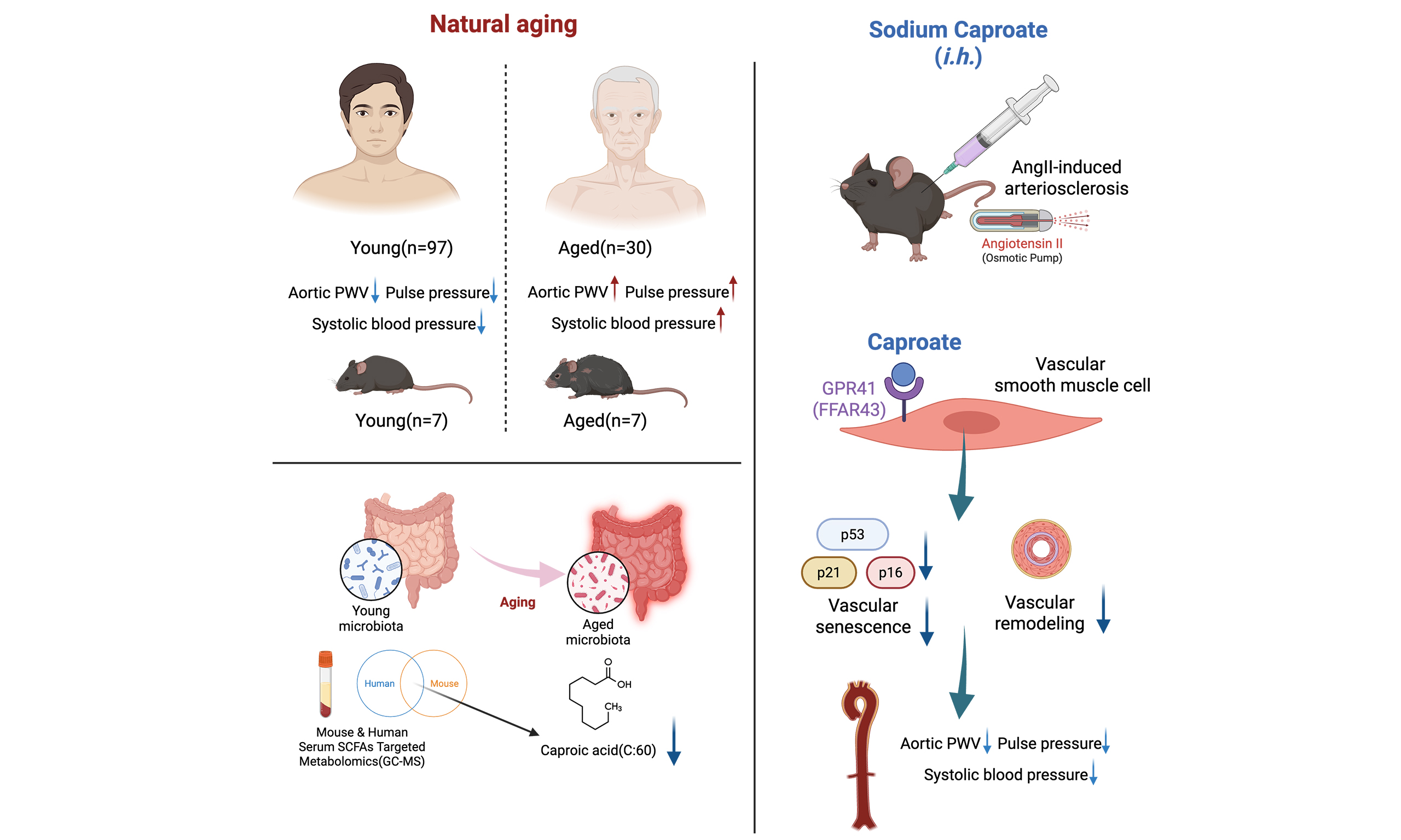
Aim: Aging is an irreversible primary risk factor for arteriosclerosis, causing high cardiovascular morbidity-mortality in the elderly via vascular remodeling and cellular senescence. Gut microbiota-derived short-chain fatty acids (SCFAs) exert cardiovascular protective effects, yet cross-species functional SCFAs counteracting age-related arteriosclerosis remain unclear. This study aimed to identify conserved SCFAs linked to vascular aging and validate caproic acid’s protective function against AngII-induced arteriosclerosis.
Methods: We assessed vascular aging phenotypes in young (8-week) and aged (24-month) mice and a human cohort (97 young < 65 years, 30 old ≥ 65 years). Serum SCFAs were quantified by targeted GC-MS. Correlation and mediation analyses evaluated relationships between caproic acid, pulse pressure (PP), and pulse wave velocity (PWV). Caproic acid protection was validated in AngII-induced arteriosclerosis mice and MOVAS cells.
Results: Circulating SCFAs declined with aging in humans and mice. Caproic acid and butyric acid were the two shared significantly down-regulated SCFAs. Serum caproic acid negatively correlated with human PP, and with both PP and aortic PWV in mice. Mediation analysis showed caproic acid directly ameliorated age-related vascular dysfunction. In AngII-treated mice, caproic acid reduced systolic blood pressure, PP and aortic PWV, mitigated aortic remodeling and elastic fiber damage, and suppressed vascular senescence protein upregulation. Gpr41 knockdown in MOVAS cells eliminated caproic acid’s anti-senescence effect, confirming GPR41-dependent protection.
Conclusion: Caproic acid acts as a conserved metabolite against age-related arteriosclerosis by inhibiting vascular cell senescence and improving hemodynamics, representing a promising therapeutic target for arteriosclerosis.
Keywords


 Download PDF
INTRODUCTION
As the core pathological basis for cardiovascular disease, arteriosclerosis represents the leading cause of death and disability among the global elderly population. It is characterized by significantly increased arterial wall stiffness, pathological structural remodeling and aberrant activation of cellular senescence pathways, which seriously threatens human health[1-3]. However, isolated systolic hypertension, elevated pulse wave velocity (PWV), and abnormal hemodynamics serve as important clinical markers of vascular aging and arteriosclerosis. These markers show a significant positive correlation with the risk of adverse cardiovascular events in the elderly population[4]. Therefore, identifying the key molecular targets of regulating vascular aging and arteriosclerosis, and exploring safe and effective interventions, have become a research hotspot in cardiovascular medicine.
As a key mediator of host-microbiome interactions, the intestinal flora and its metabolites are involved in the precise regulation of vascular homeostasis. Among them, short-chain fatty acids (SCFAs), as the core metabolites of dietary fiber fermented by gut flora, play an important role in cardiovascular protection[5,6]. SCFAs maintain vascular structure and function integrity by regulating local inflammatory response, improving endothelial relaxation, and inhibiting the abnormal proliferation and remodeling of vascular smooth muscle cells[7,8]. Previous studies indicate that the reduction of serum SCFAs levels is closely related to cardiovascular diseases, including hypertension, arteriosclerosis and myocardial ischemia. In animal models, exogenous SCFAs supplementation has been shown to significantly attenuate vascular injury and improve vascular performance[9-11]. However, the specific regulatory mechanisms and core effector molecules of SCFAs in vascular aging and age-related arteriosclerosis remain unclear. There is still a lack of systematic research on the age-related difference analysis of the SCFAs profile across species (human and mouse) and the screening of functional SCFAs, which greatly limits the clinical translational application of SCFAs as intervention targets for vascular aging and arteriosclerosis.
Building on this evidence, the study systematically characterized the hemodynamic, histomorphological, and molecular features of vascular aging in both young and aged mouse models as well as in human health cohorts. Using an AngII-induced arteriosclerosis mouse model, the study also clarified the protective effects of these SCFAs on vascular aging and arteriosclerosis in vivo, along with their underlying molecular mechanisms. Finally, this work aimed to explore the key SCFA molecules that regulate vascular aging, and provide new targets and an experimental basis for the early prevention and treatment of age-related arteriosclerosis.
MATERIAL AND METHODS
Study participants
This study included a single discovery cohort, which was recruited from the Department of Health Examination Center of the First Affiliated Hospital of Xi’an Jiaotong University. Participants were divided into two groups: the healthy young group and the healthy aged group. All enrolled individuals provided written informed consent prior to sample collection. A total of 97 young participants and 30 aged participants in the discovery cohort contributed serum samples. Immediately after collection, serum samples were snap-frozen on dry ice and stored at -80 °C until subsequent analysis. All experimental protocols involving human tissue samples and health-related data were approved by the Ethics Committee of the First Affiliated Hospital of Xi’an Jiaotong University (Approval No: XJTU1AF2020LSK-133).
Animal experiments
8-week-old and 24-month-old male C57BL/6J mice were obtained from Chengdu GemPharmatech Co. Ltd. All animal protocols were approved by the Animal Care and Use Committee of Xi’an Jiaotong University (XJTUAE2021-1492). All the mice were housed in a standard specific-pathogen-free (SPF) environment. For the interventional administration of the sodium caproate, a solution was prepared by dissolving the compound in sterile PBS. It was administered via daily subcutaneous injection at a dose of 100 mg/kg/day to mice with AngII-induced arteriosclerosis.
After 7 days of standard SPF acclimation, male C57BL/6J mice were randomly assigned to the AngII vehicle group and the AngII + sodium caproate (AngII + Na-C6) treatment group using random number sequences generated by R software (version 4.0.3). Cage positions of all mice were re-randomized every 48 h throughout the intervention period to eliminate positional confounding factors. A single-blinded design was implemented across all experimental stages: researchers performing osmotic pump implantation, daily subcutaneous injection, non-invasive blood pressure measurement and vascular ultrasound PWV scanning, as well as technicians completing histological staining and Western blot quantification, were fully masked to group allocation; all tissue and serum samples were labeled with anonymous serial numbers without group identifiers prior to testing. The sample size of each cohort was determined by referencing previously published AngII-induced vascular aging literature and our preliminary pilot observation data, without formal pre-experimental power calculation. For hemodynamic detection and histological quantitative analysis in AngII-mice, n = 5 mice per group; for hemodynamic detection and histological quantitative analysis in young mice and aged mice, n = 7 mice per group; Western blot molecular detection adopted n = 4 mice per group, and the exact number of experimental units for each test index was clearly marked in all figure legends. Predefined inclusion criteria: 8-week-old male C57BL/6J mice with body weight 24-26 g, stable food intake and normal mental status before pump surgery. Predefined exclusion criteria included intraoperative death under anesthesia and severe aortic damage during dissection that disabled section quantification. No animals were excluded from the present study, and all collected samples were included for final statistical analysis. All vascular phenotype, histology and molecular assays were performed on the same single batch of experimental animals, with all detection and quantification operations duplicated in technical replicates to guarantee data stability and reliability.
Micro-osmotic pumps implantation and AngII intervention
Osmotic pumps (Alzet model 2004) containing either normal saline or AngII (1.44 mg/kg/day) were implanted subcutaneously in 8-week-old C57BL/6J mice. Mice were anesthetized with 1% isoflurane (RWD Life Science) during implantation. Four weeks after pump implantation, the effects of drug infusion were evaluated, followed by measurements of blood pressure and PWV. Upon completion of the experiment, aortic tissue and serum samples were collected for subsequent analysis.
Serum SCFAs quantification based on GC-MS
Human and mouse serum samples were mixed with 50% acetonitrile, vortexed for 1 min, sonicated at 4 °C for 30 min, and centrifuged at 12,000 rpm at 4 °C for 15 min. Subsequently, 200 μL of the supernatant was collected, and mixed with 3-nitrophenylhydrazine (200 mM) and 1-ethyl-3-(3-dimethylaminopropyl)carbodiimide hydrochloride (120 mM; containing 6% pyridine) solutions. After vortexing for 1 min, the mixture was incubated at 40 °C for 1 h with shaking once at 5-min intervals. Following reaction completion, centrifugation was performed at 12,000 rpm and 4 °C for 15 min, and the resulting supernatant was subjected to GC-MS analysis (Agilent 7890B GC coupled to 5977B MSD, Agilent Technologies, USA). SCFAs were quantified using a standard curve (Metware, China).
Measurement of blood pressure and PWV in mice
Systolic and diastolic blood pressure (DBP) were measured in awake mice using a non-invasive tail cuff blood pressure measurement system (BP-2000, manufacturer Visitech System, Inc.). Mice were first acclimated to the tail-cuff procedure. Following acclimation, they were placed on a temperature-controlled platform (set to 37 °C) within a restraint holder. The Vevo3100 vascular ultrasound imaging system (manufacturer Fujifilm VisualSonics, Toronto, Canada) was used for PWV assessment. Before ultrasound examination, mice were anesthetized with 1% isoflurane, and then placed supine on a heating pad to maintain a constant body temperature of 37 °C. After hair removal cream was used to remove the hair in the neck and abdomen of the mice, the brightness B-mode was used to visualize the aortic arch on the transverse plane, and the resulting images were analyzed by Vevo LAB workstation.
Calculation of PWV in mice
The aortic arch PWV is calculated as the ratio of the actual distance between two designated measurement points and the time offset of the vascular waveform at the two points. For the aortic arch region, the selected measurement sites were about 1 mm in front of the left subclavian artery (lower) and the brachiocephalic trunk (upper). The time of pulse wave conduction from the upper site to the lower site is the pulse transit time (T), which is calculated as: T = T1 - T2. The distance traveled (S) between the two measurement sites was measured by B-mode ultrasound images, and the final PWV was calculated according to: PWV = S/T = S/(T1 - T2).
Histological analysis
Post animal experiment, fresh tissues were immediately fixed in 4% paraformaldehyde for ≥ 1 day. After trimming target tissues in a fume hood, samples were placed in labeled embedding frames, dehydrated with gradient alcohol, immersed in wax solution, and paraffin-embedded prior to sectioning.
Hematoxylin and eosin (H&E) staining: Tissue sections were stained with hematoxylin for 2-5 min (adjusted by staining effect), rinsed in running water for 15 min until nuclei turned blue, then eosin-stained during dehydration and mounted with neutral resin.
Masson staining: Sections were stained with Weigert’s iron hematoxylin for 10 min, followed by Ponceau magenta for 5 min. After water washing, samples were stained with phosphomolybdic acid and aniline blue for 1-2 min, and finally mounted with neutral resin. Elastic fiber staining was performed using the Elastic Fiber Stain Kit (Elastica van Gieson (EVG) Method, Catalog No. G1597, Solarbio, Beijing, China).
Media thickness was determined as the intima-media distance via the incremental distance tool in ImageJ software (calibrated step: 100 μm), with results reported as the average of at least 4 measurements per section. The degree of elastic fiber fragmentation was quantified under a light microscope (20× magnification) using a connected camera, and normalized to the media area. Collagen content was expressed as the percentage of collagen-positive area relative to the total tissue area.
Cell lines culture
MOVAS mouse vascular smooth muscle cells were purchased from Wuhan Pricella Biotechnology Co., Ltd. (Procell, CL-1167). The MOVAS cell line RRID is CVCL_0F08. STR authentication was performed and mycoplasma testing confirmed no contamination. MOVAS cells were maintained in high-glucose DMEM medium (Procell, PM150210) supplemented with 10% fetal bovine serum and 1% penicillin-streptomycin (P/S). Cells were incubated at 37 °C under a 5% CO2 humidified atmosphere and passaged upon reaching 70%-80% confluence for subsequent experiments. AngII (1 μM) and caproic acid (CA, 2.5 mM) were applied for 24 h after siRNA transfection.
siRNA transfection
Gpr41-specific siRNA (siGpr41) and negative control siRNA (siNC) were designed and synthesized by Genepharma. The detailed sequences are listed: CCAAGUUCCAAGCCGACUUTT (forward primer sequence), AAGUCGGCUUGGAACUUGGTT (reverse primer sequence). siRNA transfection was carried out with siRNA-Mate plus (Genepharma, G04026) following the manufacturer’s recommended protocols.
Western blot analysis
Total protein was extracted from mouse aortic tissues and MOVAS cell lines using radioimmunoprecipitation assay lysis buffer supplemented with protease and phosphatase inhibitor cocktail. Protein concentration was quantified via bicinchoninic acid assay. Aortic tissue lysates and cell lysates were electrophoretically separated in 10%-15% SDS-PAGE gels and subsequently transferred to polyvinylidene fluoride membranes. The membranes were blocked with 5% skim milk, and then incubated with primary antibodies overnight at 4 °C with gentle shaking. Primary antibody information and working dilutions were listed as follows: p21Waf1/Cip1 (F2C7C) rabbit monoclonal antibody (39256, 1:1,000, Cell Signaling Technology, USA), p53 (1C12) mouse monoclonal antibody (2524, 1:1,000, Cell Signaling Technology, USA), p16INK4A (F2T7H) rabbit monoclonal antibody (23200, 1:1,000, Cell Signaling Technology, USA), FFAR3 mouse monoclonal antibody (66811-1-lg, 1:500, Proteintech, China), α-Tubulin rabbit polyclonal antibody (11224-1-AP, 1:4,000, Proteintech, China), GAPDH rabbit polyclonal antibody (10494-1-AP, 1:3,000, Proteintech, China). After three rounds of Tris-buffered saline containing Tween 20 washing, membranes were incubated with corresponding horseradish peroxidase (HRP)-conjugated secondary antibodies for 1 h at room temperature: anti-rabbit IgG HRP-linked antibody (7074, 1:2,000, Cell Signaling Technology, USA) and anti-mouse IgG HRP-linked antibody (7076, 1:2,000, Cell Signaling Technology, USA). Finally, the enhanced chemiluminescence reagent Omni ECLTM (SQ201L, Epizyme Biotech Co.) was used to observe the protein bands. The grayscale density of each band was quantified by ImageJ software and normalized to the internal reference GAPDH or α-Tubulin for relative expression calculation. All unedited original Western blot gels were provided in the Supplementary Materials.
Data availability
SCFAs quantification sequencing data for all human and mouse serum samples that support the findings of this study are available from the corresponding author upon reasonable request.
Statistical analysis
Unless otherwise specified, all statistical analyses were performed using R v4.0.3 and GraphPad Prism 10.0 software. Normality of all datasets was validated via Shapiro-Wilk test and Q-Q plots, and residual distribution was checked to confirm variance homogeneity. Unpaired two-tailed Student’s t-test was applied for all two-group comparisons of mouse vascular indicators including systolic blood pressure (SBP), DBP, pulse pressure (PP), aortic medial thickness, fibrosis degree, elastic fiber fragmentation and senescence protein (p53, p21, p16) expression. As each analysis only contained two experimental groups, no multiple comparison adjustment was conducted. For cellular experiments with three groups, one-way analysis of variance (one-way ANOVA) with Tukey’s post-hoc multiple comparison correction was adopted. Cohen’s d effect sizes and 95% confidence intervals (95%CI) of mean differences were calculated and reported for all intergroup comparisons. The complete set of effect sizes, 95%CIs, and detailed statistical outputs for all analyzed parameters are provided in Supplementary Table 5. For the human and mouse SCFA datasets, we performed Bonferroni correction for multiple comparisons. For human cohort baseline and subgroup analyses presented in Supplementary Tables, two-way ANOVA was used to evaluate main effects and age-by-sex interaction effect for serum caproic acid. Stratified unpaired Student’s t-test was performed for subgroup comparisons stratified by age group or sex. Pearson correlation analysis was conducted to assess crude correlations between age and caproic acid concentration. Age-adjusted partial correlation analysis was applied to evaluate sex-stratified associations between CA and vascular parameters. Fisher’s Z-test was further implemented in R v4.0.3 to formally compare correlation coefficients between male and female subgroups. Linear regression was used to analyze correlations between serum caproic acid and vascular stiffness metrics, with regression coefficients, 95 CIs, and R2 values presented in figures. All quantitative data were expressed as mean ± standard deviation (mean ± SD). The statistical significance threshold was set at
RESULTS
Aging induces aorta vascular remodeling and cellular senescence in mice
Aging is the foremost non-modifiable risk factor for arteriosclerosis, a chronic vascular disorder characterized by progressive arterial stiffening, adverse structural remodeling, and dysregulated cellular homeostasis[12,13]. Arterial stiffness, commonly assessed by PWV, served as both a key pathological feature of arteriosclerosis and a strong predictor of cardiovascular mortality in the elderly[14]. We systematically characterized the vascular aging phenotype using young (8-week-old) and aged (24-month-old) mice, a well-established model that recapitulates major aspects of human age-related arteriosclerosis[15].
Aortic arch PWV of mice was calculated by echocardiographic imaging of the aortic arch combined with mouse electrocardiogram (ECG) [Figure 1A]. Quantitative analysis demonstrated a nearly 3-fold increase in aortic arch PWV in aged animals compared to young controls [Figure 1B], indicating a severe loss of arterial compliance, a critical mechanistic driver of arteriosclerosis[16,17]. Aged mice exhibited significantly increased body weight and aorta weight-to-body weight ratio relative to young controls
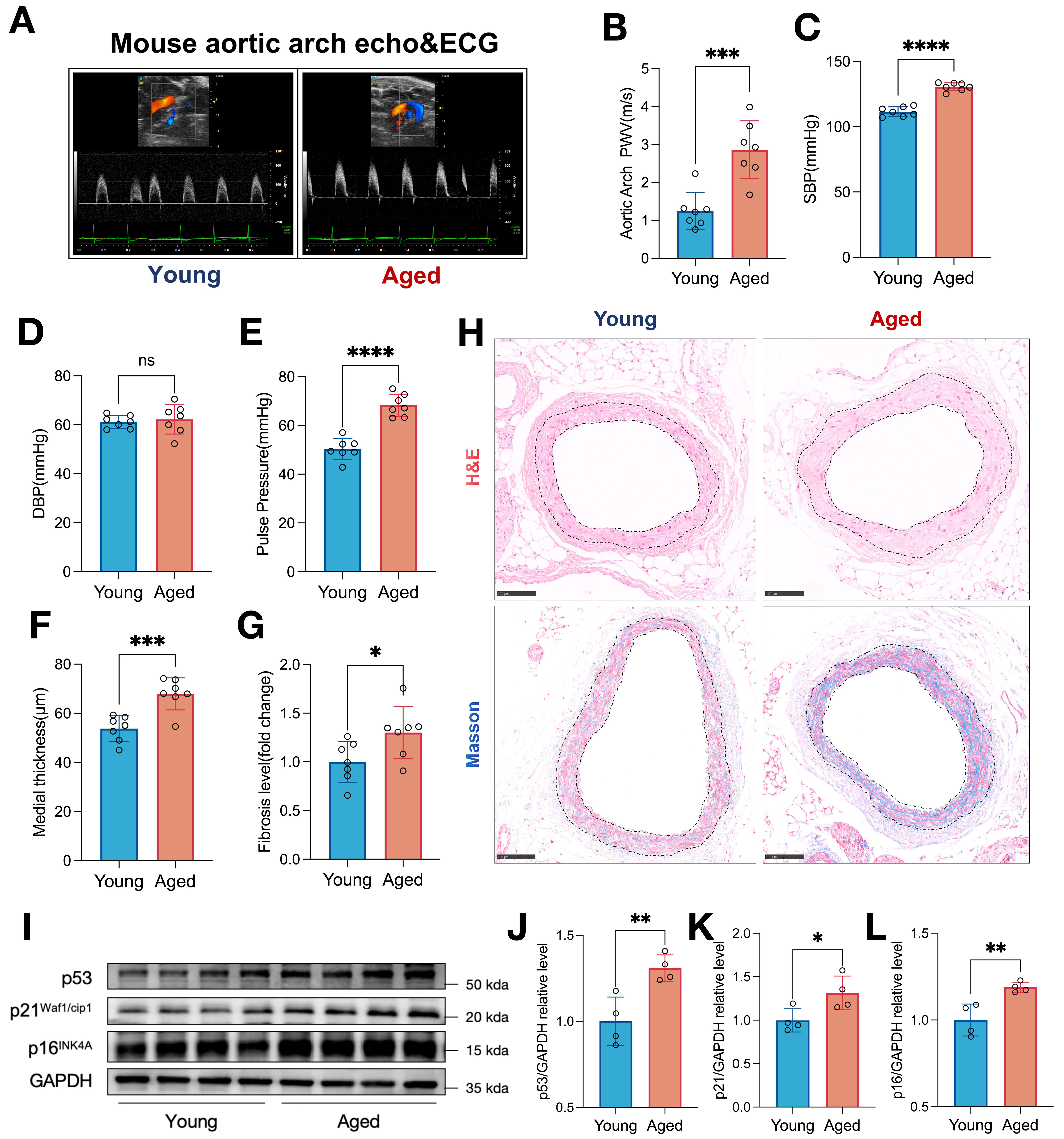
Figure 1. Aging induces aortic arch hemodynamic dysfunction, vascular remodeling, and cellular senescence in mice. (A) Representative echocardiographic images of the aortic arch and simultaneous ECG recordings in young and aged mice. (B) Quantification of aortic arch PWV in young (n = 7) and aged (n = 7) mice calculated by the measurement from echocardiogram and ECG. (C and D) SBP and DBP measured in young (n = 7) and aged (n = 7) mice. (E) Pulse pressure calculated from SBP and DBP in young (n = 7) and aged (n = 7) mice. (F) Aortic medial thickness quantified from histological sections in young (n = 7) and aged (n = 7) mice. (G) Fibrosis level (fold change relative to young mice) in the thoracic aorta of young (n = 7) and aged (n = 7) mice. (H) Representative H&E and Masson’s trichrome staining of thoracic aorta sections from young and aged mice. Scale bar, 100 μm. (I) Representative Western blot images showing the expression of senescence markers p53, p21Waf1/cip1, and p16INK4A in aortic tissues from young and aged mice. GAPDH was used as the loading control. (J-L) Densitometric quantification of (J) p53, (K) p21Waf1/cip1, and (L) p16INK4A protein levels, normalized to GAPDH, in young (n = 4) and aged (n = 4) mice. Data are presented as mean ± SD. *P < 0.05, **P < 0.01, ***P < 0.001, ****P < 0.0001 vs. young group (unpaired two-tailed Student’s t-test).
Histological analysis further confirmed pathological vascular remodeling in the aged aortas. H&E staining demonstrated significant thickening of the aortic medial layer [Figure 1F], a structural change associated with advanced arteriosclerosis[17]. Additionally, Masson’s staining revealed excessive collagen deposition and expansion of the fibrotic area [Figure 1G], with representative histological micrographs provided in
Aging is associated with altered serum SCFAs concentrations
Emerging evidence has implicated gut microbiota-derived SCFAs as key regulators of age-related cardiovascular disease[21,22]. Multiple studies demonstrate that reduced SCFAs bioavailability is linked to accelerated Cardiovascular aging. To identify age-associated alterations, we performed targeted metabolomic analysis of serum SCFAs in both young (8-week-old, n = 7) and aged (24-month-old, n = 7) mice, as well as in a human cohort of young (< 65 years, n = 97) and aged (≥ 65 years, n = 30) individuals [Figure 2A]. We compared the baseline characteristics between the two groups in this cohort and found no differences in sex and body mass index [Supplementary Table 1].

Figure 2. Aging is associated with altered SCFA profiles in both mouse and human serum. (A) Flowchart of the study design. Young (8-week-old, n = 7) and aged (24-month-old, n = 7) C57BL/6J mice, as well as young human subjects (< 65 years old, n = 97) and aged human subjects (≥ 65 years old, n = 30), were enrolled. Serum samples were collected for targeted SCFA metabolomics analysis via GC/MS. (B) Quantification of individual SCFA concentrations in serum from young (blue) and aged (red) mice (n = 7 per group). (C) Quantification of individual SCFA concentrations in serum from young (cyan) and aged (purple) human subjects (young, n = 97; aged, n = 30). (D) Heatmap of differentially expressed SCFAs in mouse serum, with rows representing individual SCFAs and columns representing individual samples, clustered by group (young, blue; aged, red; n = 7 per group). The color scale indicates z-score-normalized SCFA concentrations. (E) Heatmap of differentially expressed SCFAs in human serum, with rows representing individual SCFAs and columns representing individual samples, clustered by group (young, cyan; aged, purple; young, n = 97; aged,
Quantitative analysis of serum SCFAs profiles revealed a consistent pattern of age-related downregulation across multiple SCFAs species in both mice and humans [Figure 2B and C]. In mice, aged animals exhibited significant reductions in the concentrations of butyric acid, CA, isovaleric acid and glutaric acid relative to young controls [Figure 2B]. In the human cohort, aged individuals displayed significant decreases in butyric acid, valeric acid and CA, alongside a trend toward reduced butyric acid levels
Notably, CA and butyric acid emerged as the only two SCFAs that were significantly downregulated with aging in both species, representing a shared, conserved age-related metabolic alteration. To validate the age-related decline in CA, we performed linear regression analysis in the full human cohort, which demonstrated a significant inverse correlation between serum caproic acid concentration and chronological age (Supplementary Figure 2; R2 = 0.1562, P = 0.0028), confirming that caproic acid levels progressively decrease with advancing age in humans. We conducted sex-stratified subgroup analyses for the human cohort consisting of both male and female participants. We performed multiple statistical tests covering sex-specific baseline CA concentrations, two-way ANOVA to test the age-by-sex interaction, as well as sex-stratified partial correlation analyses evaluating the relationships between CA and vascular parameters. Overall, these analyses indicated no remarkable sex-divergent correlations between circulating CA and vascular phenotypes in this cohort [Supplementary Tables 2-4].
These findings identify CA as a key candidate SCFA linked to vascular aging, so we selected CA for further functional investigation.
CA is an age-associated SCFA linked to vascular aging and arteriosclerosis
Building on our metabolomic findings, Venn diagram analysis of age-associated differentially expressed SCFAs confirmed that CA and butyric acid were the only two SCFAs significantly downregulated with aging in both mice and humans, with CA exhibiting the most consistent reduction across species [Figure 3A]. Quantitative analysis verified that serum caproic acid concentrations were significantly lower in aged mice [Figure 3B] and aged human subjects [Figure 3C] relative to their young counterparts. To further explore the clinical relevance of CA, we performed correlation analyses with key vascular aging metrics. In the full human cohort (n = 127), serum caproic acid concentration was inversely correlated with pulse pressure (Figure 3D; R2 = 0.1451, P < 0.0001). In mice, CA levels were similarly inversely correlated with both pulse pressure [Figure 3E] and aortic arch PWV [Figure 3F], the gold-standard preclinical measure of arterial stiffness. Consistent with these findings, linear regression analysis confirmed a significant positive correlation between aortic arch PWV and pulse pressure in aged mice
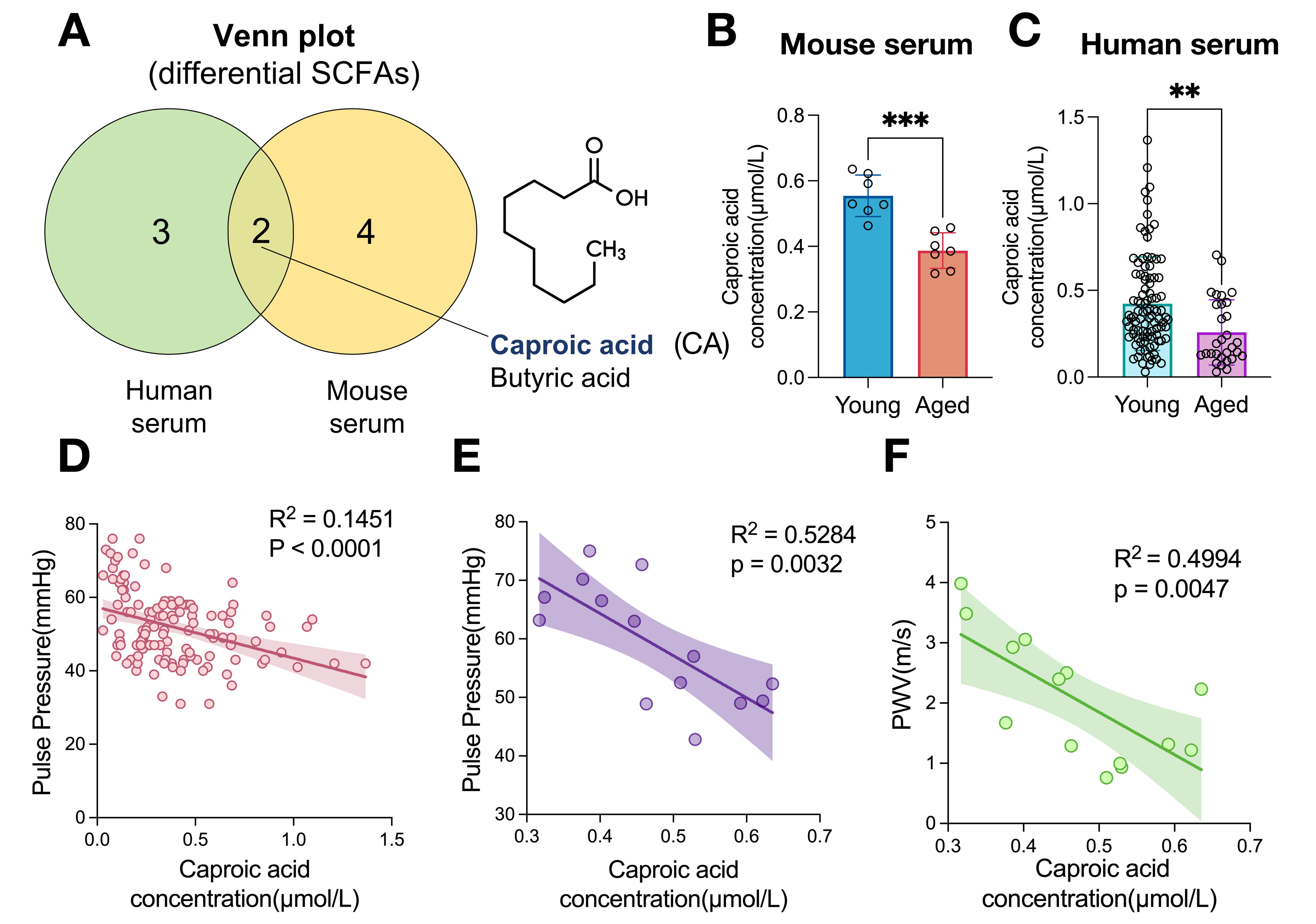
Figure 3. Caproic acid is an age-associated SCFA linked to vascular aging and arteriosclerosis in mice and humans. (A) Venn diagram of overlapping age-related differential SCFAs in human and mouse serum, with the chemical structure of caproic acid shown. (B) Serum caproic acid concentrations in young (n = 7) and aged (n = 7) mice. (C) Serum caproic acid concentrations in young (n = 97) and aged (n = 30) human subjects. (D) Correlation between serum caproic acid and pulse pressure in humans (n = 127). (E) Correlation between serum caproic acid and pulse pressure in mice (n = 14). (F) Correlation between serum caproic acid and aortic arch PWV in mice (n = 14). Data are mean ± SD. **P < 0.01, ***P < 0.001 vs. young group (unpaired student’s t-test). Linear regression analyses are shown with R2 and P values; shaded areas represent 95% confidence intervals.
CA ameliorates angiotensin II-induced arteriosclerosis and vascular dysfunction in mice
To directly validate the protective role of CA against arteriosclerosis, we administered sodium caproate (Na-C6) to young mice infused with angiotensin II (AngII) [Figure 4A], a well-established preclinical model of vascular dysfunction, arterial stiffening, and pathological vascular remodeling[20,25-27]. Functional hemodynamic analysis revealed that Na-C6 treatment partially ameliorated AngII-induced vascular injury: mice receiving Na-C6 exhibited a marked reduction in aortic arch PWV [Figure 4B], alongside partial reversal in SBP [Figure 4C] and pulse pressure [Figure 4D], with no significant changes in diastolic DBP [Supplementary Figure 4A]. Na-C6 treatment did not affect overall body weight

Figure 4. Caproic acid ameliorates angiotensin II-induced arteriosclerosis and vascular dysfunction in mice. (A) Schematic of the in vivo experimental design. Young mice were treated with either PBS (i.h.) or Na-C6 (i.h.) and infused with AngII via osmotic pump for 4 weeks, followed by sacrifice and tissue analysis. (B) Aortic arch PWV, (C) SBP, and (D) pulse pressure in AngII-infused mice with or without Na-C6 treatment (n = 5 per group). (E) Representative H&E, Masson’s trichrome, and EVG staining of thoracic aorta sections from AngII and AngII + Na-C6 mice. Scale bar, 100 μm. (F) Quantification of aortic medial thickness, (G) aortic fibrosis level (fold change), and (H) elastin breaks per section in the AngII and AngII + Na-C6 mice (n = 5 per group). (I) Representative Western blot images showing the expression of senescence markers p53, p21Waf1/cip1, and p16INK4A in aorta tissues. GAPDH was used as the loading control. (J-L) Densitometric quantification of (J) p53, (K) p21Waf1/cip1, and (L) p16INK4A protein levels in aorta, normalized to GAPDH (n = 4 per group). Na-C6: Sodium caproate; PWV: pulse wave velocity; SBP: systolic blood pressure. Data are presented as mean ± SD. *P < 0.05, **P < 0.01, ***P < 0.001, ****P < 0.0001 vs. AngII group (unpaired two-tailed Student’s t-test). (A) was created in BioRender. Xie, Y. (2026) https://BioRender.com/bmztl73.
CA mitigates AngII-induced senescence of MOVAS cells via the GPR41 receptor
To further dissect the upstream molecular mechanism by which CA alleviates vascular smooth muscle cell senescence, we performed siRNA-mediated GPR knockdown experiments in MOVAS cells (mouse aortic vascular smooth muscle cells). Considering that GPR43 shows no functional response to CA at physiological concentrations, we constructed siRNA-mediated Gpr41 knockdown MOVAS cells)
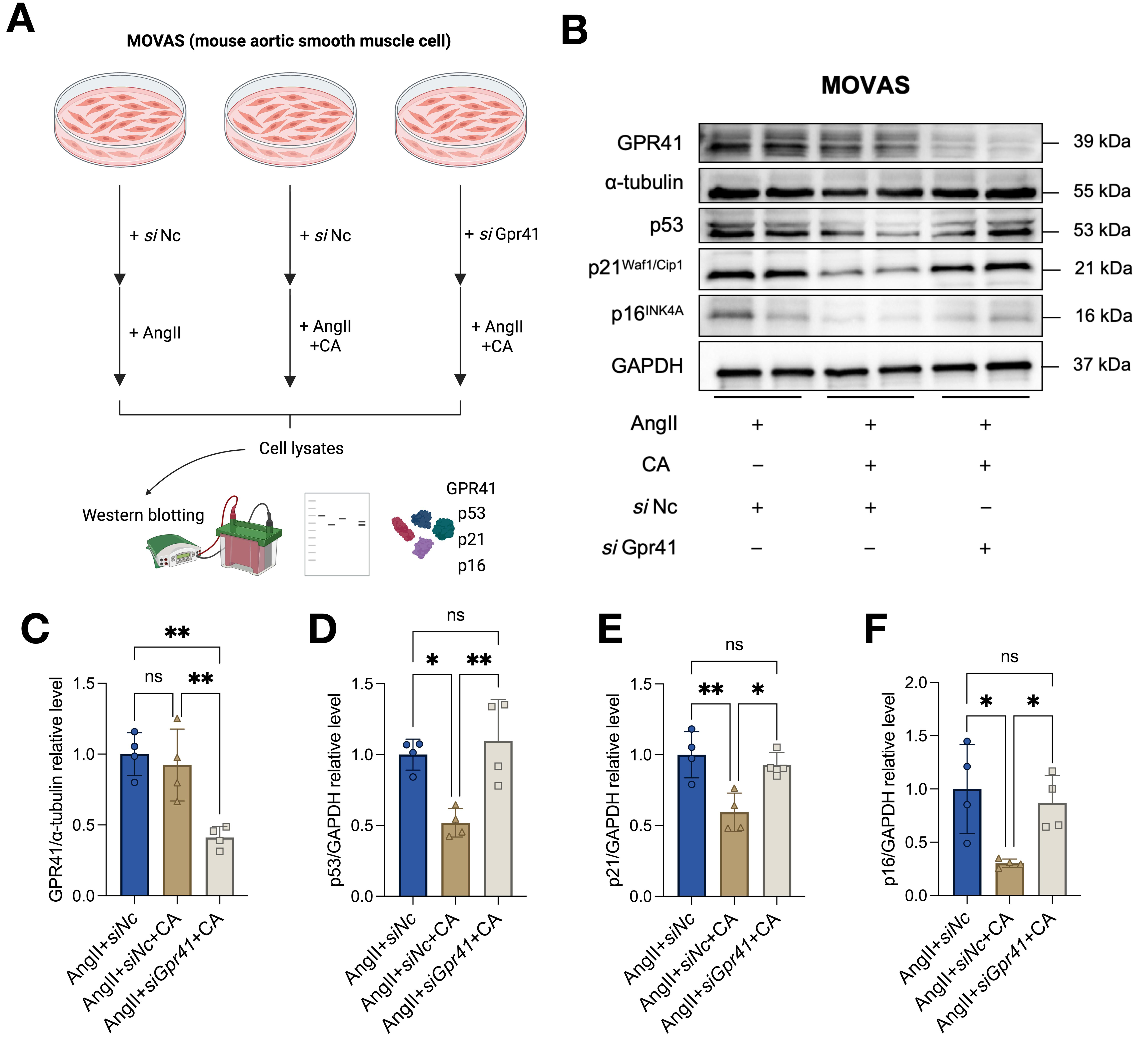
Figure 5. Caproic acid mitigates AngII-induced senescence of MOVAS cells via the GPR41 receptor. (A) Schematic workflow of Gpr41 siRNA knockdown and AngII/CA intervention in MOVAS cells. (B) Representative western blot images showing the protein expression of GPR41, p53, p21 and p16 in different treatment groups. α-tubulin and GAPDH were used as internal loading controls. (C-F) Densitometric quantitative analysis of relative protein levels of GPR41 (C), p53 (D), p21 (E) and p16 (F). n = 4 biological replicates per group. Data are presented as mean ± SD. Statistical analysis was conducted using one-way ANOVA followed by Tukey’s multiple comparison test. *P < 0.05, **P < 0.01; ns: Not significant; CA: caproic acid; AngII: angiotensin II; siNc: control siRNA; siGpr41: Gpr41-targeted siRNA. (A) was created in BioRender. Xie, Y. (2026) https://BioRender.com/yvqfe0r.
DISCUSSION
These findings not only provide new evidence for the regulation of age-related arteriosclerosis by gut microbiota metabolites but also identify potential targets for metabolic intervention, thereby advancing our understanding of the "gut-vascular axis" in the maintenance of vascular homeostasis.
The core innovation of this study is the identification of CA as a protective metabolite against age-related arteriosclerosis across both human and mouse models. This breaks from the previous research focus on SCFAs such as butyric acid and propionic acid[24,33], and clarifies a specific regulatory role for SCFAs in vascular aging. Previous studies have confirmed that gut microbiota metabolites participate in the process of cardiovascular disease by regulating vascular inflammation and endothelial function[34], but the specific effector molecules and regulatory pathways for age-related arteriosclerosis have not been clear. Through targeted metabolomic screening combined with correlation and intermediary analysis, this study confirmed that CA can play a protective role by directly regulating vascular structural remodeling and indirectly improving hemodynamics. This finding adds a new molecular node to the regulatory network of "gut-vascular axis", and explains the mechanism by which intestinal flora disturbances mediate vascular homeostasis imbalance in the process of aging[34]. Furthermore, this study confirmed that the protective effect of caproic acid depended on the inhibition of p53/p21/p16 canonical cellular senescence pathway. We further clarified the indispensable receptor mediating caproic acid’s anti-senescence effects via Gpr41 knockdown in MOVAS vascular smooth muscle cells. In vitro siRNA interference assays demonstrated that the capacity of caproic acid to suppress AngII-triggered upregulation of p53/p21/p16 senescence proteins was fully abolished after Gpr41 silencing, verifying that GPR41 acts as the essential membrane receptor transmitting caproic acid’s vascular-protective signals. Unlike GPR43 which barely responds to physiological concentrations of CA, GPR41 specifically recognizes circulating CA and initiates downstream anti-aging cascades in vascular smooth muscle[31]. This receptor-specific evidence perfects the molecular cascade linking gut-derived CA to restrained vascular senescence, laying a solid cellular foundation for targeted intervention of age-related arteriosclerosis by activating GPR41 signaling.
CA, a six-carbon gut microbial SCFAs, is primarily produced by colonic anaerobic commensal bacteria through a unique chain elongation metabolic pathway that is absent in mammalian somatic cells[35]. Multiple functional strains belonging to Lachnospiraceae and Ruminococcaceae families also catalyze the conversion of lactate and butyrate precursors into hexanoic acid under anaerobic intestinal microenvironment[35]. Notably, aging and obesity are established major drivers of intestinal barrier breakdown and gut microbiota dyshomeostasis, which disrupt the bidirectional gut-liver crosstalk and exacerbate chronic low-grade systemic inflammation in age-related metabolic syndromes[36]. Since all gut microbial SCFAs including CA are absorbed across the intestinal epithelium and transported to the liver via the portal vein before entering the peripheral vasculature, this may underlie the reduced CA-associated arteriosclerosis in elderly individuals.
The results of this study are analyzed in the context of existing research, and its clinical relevance and translational potential are of great value. In the human cohort, we found that serum caproic acid levels showed a significant negative correlation with PP and age. Given that PP is a clinically accessible marker of arteriosclerosis and is closely associated with cardiovascular adverse events in the elderly[37,38], serum caproic acid may serve as a potential biomarker for age-related arteriosclerosis, offering a new approach for early disease screening. In addition, the AngII-induced arteriosclerosis model used in this study is a classic preclinical model of hypertension-related arteriosclerosis[39], and the protective effect of CA on this model also suggests that it may have potential intervention value for patients with clinical hypertension and arteriosclerosis, pointing out the direction for subsequent clinical translational research.
Limitations and future directions
This study has several limitations, which need to be further improved in the follow-up study. First, the human cohort was a single-center, cross-sectional design with a relatively limited sample size. This design cannot establish causality between reduced CA levels and arteriosclerosis; large-scale, multicenter prospective cohorts are needed for validation. Second, only male C57BL/6J mice were used in animal experiments, and the protective effect of CA in female mice was not explored. Gender differences play an important regulatory role in the occurrence and development of vascular aging and arteriosclerosis. Whether there is a gender-specific vascular protective effect of CA still needs subsequent verification. Third, we only quantified circulating gut-derived CA in peripheral serum without accompanying fecal microbiome and fecal metabolomic testing. Although free CA in systemic circulation is predominantly produced by intestinal microbiota rather than mammalian endogenous metabolism[40], direct multi-omics evidence is still required to confirm gut dysbiosis as the root cause of decreased CA in patients with arteriosclerosis. The exploratory mouse phenotypic assay was performed with a small sample size of 5 mice per group. Despite the near-perfect statistical power derived from the remarkably large effect size (Cohen’s d = 2.53), limited sample volume may introduce sampling fluctuation and restrict result extrapolation. We plan to conduct definitive follow-up validation experiments with an adequately expanded sample cohort in future work to strengthen the reliability and repeatability of our in vivo findings.
These findings offer several insights for both basic and clinical research. Mechanistically, future studies could focus on the “gut microbiota- CA-vascular cell senescence” axis to delineate its specific molecular targets and signaling pathways, and to identify key bacterial strains that regulate CA production. In addition, cross-species and multi-cohort validation studies can be carried out to clarify the specificity and sensitivity of CA as a biomarker of vascular aging. At the same time, the serum caproic acid level can be incorporated into the risk assessment system of arteriosclerosis, and the accuracy of early disease screening can be improved by combining it with traditional markers. In addition, this study also suggests that the clinical regulation of intestinal flora CA synthesis by adjusting the dietary structure (such as increasing the intake of fermentable dietary fiber) can provide a novel non-pharmacological strategy for preventing and managing age-related arteriosclerosis.
DECLARATIONS
Acknowledgments
The Graphical Abstract was created with BioRender.com (Created in BioRender. Xie, Y. (2026) https://BioRender.com/64bzazz). We gratefully acknowledge the support and assistance provided by the staff members of the Laboratory Animal Center (LAC) at Xi’an Jiaotong University.
Authors’ contributions
Authors who contributed equally to this study: Xie Y, Pang J
Methodology, writing - original draft, review and editing, investigation: Xie Y
Methodology, investigation: Hua Y, Pang J
Conceptualization, methodology: Wu H
Investigation, funding acquisition: Ding N
Resources, investigation: Pang J
Writing - review and editing: Meng Z, Qian R
Availability of data and materials
All data presented in this research can be obtained from the corresponding author upon reasonable request.
AI and AI-assisted tools statement
Not applicable.
Financial support and sponsorship
This study was supported by the National Key R&D Program of China (2024YFA1307004, 2021YFA1301200, 2021YFA1301201, 2021YFA0805400) and the National Natural Science Foundation of China (82425004, 82430019, 82370458).
Conflicts of interest
All authors declared that there are no conflicts of interest.
Ethical approval and consent to participate
Research involving human serum and human blood pressure data was performed in accordance with the Declaration and approved by the Ethics Committee of the First Affiliated Hospital of Xi’an Jiaotong University (Approval No: XJTU1AF2020LSK-133). All animal protocols were approved by the Institutional Animal Care and Use Committee (IACUC) of the School of Medicine, Xi'an Jiaotong University (Approval No: XJTUAE2021-1492). All enrolled individuals provided written informed consent prior to sample collection.
Consent for publication
Not applicable.
Copyright
© The Author(s) 2026.
Supplementary Materials
REFERENCES
2. Minamino T, Yoshida T, Tateno K, et al. Ras induces vascular smooth muscle cell senescence and inflammation in human atherosclerosis. Circulation. 2003;108:2264-9.
4. Laurent S, Boutouyrie P, Asmar R, et al. Aortic stiffness is an independent predictor of all-cause and cardiovascular mortality in hypertensive patients. Hypertension. 2001;37:1236-41.
5. Perler BK, Friedman ES, Wu GD. The role of the gut microbiota in the relationship between diet and human health. Annu Rev Physiol. 2023;85:449-68.
6. Canfora EE, Jocken JW, Blaak EE. Short-chain fatty acids in control of body weight and insulin sensitivity. Nat Rev Endocrinol. 2015;11:577-91.
7. Yang F, Chen H, Gao Y, et al. Gut microbiota-derived short-chain fatty acids and hypertension: Mechanism and treatment. Biomed Pharmacother. 2020;130:110503.
8. O’donnell JA, Zheng T, Meric G, Marques FZ. The gut microbiome and hypertension. Nat Rev Nephrol. 2023;19:153-67.
9. Liu T, Sun Z, Yang Z, Qiao X. Microbiota-derived short-chain fatty acids and modulation of host-derived peptides formation: focused on host defense peptides. Biomed Pharmacother. 2023;162:114586.
10. Hu T, Wu Q, Yao Q, Jiang K, Yu J, Tang Q. Short-chain fatty acid metabolism and multiple effects on cardiovascular diseases. Ageing Res Rev. 2022;81:101706.
11. Verhaar BJ, Wijdeveld M, Wortelboer K, et al. Effects of oral butyrate on blood pressure in patients with hypertension: a randomized, placebo-controlled trial. Hypertension. 2024;81:2124-36.
12. Lakatta EG, Levy D. Arterial and cardiac aging: major shareholders in cardiovascular disease enterprises: Part II: the aging heart in health: links to heart disease. Circulation. 2003;107:346-54.
13. Lakatta EG, Levy D. Arterial and cardiac aging: major shareholders in cardiovascular disease enterprises: Part I: aging arteries: a “set up” for vascular disease. Circulation. 2003;107:139-46.
14. Faleeva M, Ahmad S, Theofilatos K, et al. Sox9 accelerates vascular aging by regulating extracellular matrix composition and stiffness. Circ Res. 2024;134:307-24.
15. Ungvari Z, Tarantini S, Donato AJ, Galvan V, Csiszar A. Mechanisms of vascular aging. Circ Res. 2018;123:849-67.
16. Ji Y, Peng X, Chen S, Song Q, Bai J, Cai J. Evening home pulse pressure predicted cardiovascular events and mortality in older adults with hypertension: findings based on the STEP trial. Hypertens Res. 2025;48:2801-10.
17. Pucci G, Spronck B, Avolio AP, et al. Age-specific acute changes in carotid-femoral pulse wave velocity with head-up tilt. Am J Hypertens. 2020;33:1112-8.
18. Tsimploulis A, Sheriff HM, Lam PH, et al. Systolic-diastolic hypertension versus isolated systolic hypertension and incident heart failure in older adults: insights from the Cardiovascular Health Study. Int J Cardiol. 2017;235:11-6.
19. Saeedi Saravi SS, Pugin B, Constancias F, et al. Gut microbiota-dependent increase in phenylacetic acid induces endothelial cell senescence during aging. Nat Aging. 2025;5:1025-45.
20. Gan L, Liu D, Liu J, et al. CD38 deficiency alleviates Ang II-induced vascular remodeling by inhibiting small extracellular vesicle-mediated vascular smooth muscle cell senescence in mice. Sig Transduct Target Ther. 2021;6:223.
21. Liu S, He Y, Zhang Y, et al. Targeting gut microbiota in aging-related cardiovascular dysfunction: focus on the mechanisms. Gut Microbes. 2023;15:2290331.
22. Xu J, Moore BN, Pluznick JL. Short-chain fatty acid receptors and blood pressure regulation: council on hypertension mid-career award for research excellence 2021. Hypertension. 2022;79:2127-37.
23. Tang WW, Kitai T, Hazen SL. Gut microbiota in cardiovascular health and disease. Circ Res. 2017;120:1183-96.
24. Zhang D, Jian Y, Zhang Y, et al. Short-chain fatty acids in diseases. Cell Commun Signal. 2023;21:212.
25. Weiss D, Kools JJ, Taylor WR. Angiotensin II-induced hypertension accelerates the development of atherosclerosis in ApoE-deficient mice. Circulation. 2001;103:448-54.
26. Yin L, Bai J, Yu W, Liu Y, Li H, Lin Q. Blocking VCAM-1 prevents angiotensin II-induced hypertension and vascular remodeling in mice. Front Pharmacol. 2022;13:825459.
27. Trejo-Moreno C, Jiménez-Ferrer E, Castro-Martínez G, et al. Characterization of a murine model of endothelial dysfunction induced by chronic intraperitoneal administration of angiotensin II. Sci Rep. 2021;11:21193.
28. Zhang W, Zhang S, Yan P, et al. A single-cell transcriptomic landscape of primate arterial aging. Nat Commun. 2020;11:2202.
29. Pang J, Pan L, Shen W, et al. Endothelial LRRC8A delays vascular ageing in natural and accelerated ageing mouse models. Cardiovasc Res. 2025;121:2549-64.
30. Muralitharan RR, Nakai ME, Snelson M, et al. Influence of angiotensin II on the gut microbiome: modest effects in comparison to experimental factors. Cardiovasc Res. 2024;120:1155-63.
31. Kimura I, Ichimura A, Ohue-Kitano R, Igarashi M. Free fatty acid receptors in health and disease. Physiol Rev. 2020;100:171-210.
32. Brown AJ, Goldsworthy SM, Barnes AA, et al. The orphan G protein-coupled receptors GPR41 and GPR43 are activated by propionate and other short chain carboxylic acids. J Biol Chem. 2003;278:11312-9.
33. Pham QH, Bui TVA, Sim W, et al. Daily oral administration of probiotics engineered to constantly secrete short-chain fatty acids effectively prevents myocardial injury from subsequent ischaemic heart disease. Cardiovasc Res. 2024;120:1737-51.
34. Bellet MM, Curcio F, Frati L, Pariano M, Romani L, Corsi-Romanelli MM. The gut microbiome as a possible mediator in autoimmunity and cardiovascular disease: shared pathways and therapeutic implications. Am J Pathol. 2025;195:2288-302.
35. Fitzgerald BG, Govind M, Firth IJ, et al. Members of Lachnospiraceae produce valerate and caproate in response to short-chain fatty acids. Microbiome. 2025;13:201.
36. Wei G, Liu W, Shen F, et al. Polystyrene microplastics exposure increases the disruption of intestinal barrier integrity and gut microbiota homeostasis during obesity and aging. J Environ Sci. 2026;162:430-43.
37. Melgarejo JD, Thijs L, Wei D, et al. Relative and absolute risk to guide the management of pulse pressure, an age-related cardiovascular risk factor. Am J Hypertens. 2021;34:929-38.
38. Safar ME. Arterial aging—hemodynamic changes and therapeutic options. Nat Rev Cardiol. 2010;7:442-9.
39. Wang H, Martinez Yus M, Brady T, et al. Sex differences and role of lysyl oxidase-like 2 in angiotensin II-induced hypertension in mice. Am J Physiol Heart Circ Physiol. 2024;327:H642-59.
Cite This Article
 Open Access
How to Cite
Xie Y, Pang J, Wu H, Hua Y, Meng Z, Qian R, Ding N. Gut microbiota-derived caproic acid attenuates aging-associated arteriosclerosis. J Cardiovasc Aging. 2026;6:44. https://dx.doi.org/10.20517/jca.2026.41
Download Citation
If you have the appropriate software installed, you can download article citation data to the citation manager of your choice. Simply select your manager software from the list below and click on download.
Export Citation File
Type of Import
Tips on Downloading Citation
Citation Manager File Format
Type of Import
Direct Import: When the Direct Import option is selected (the default state), a dialogue box will give you the option to Save or Open the downloaded citation data. Choosing Open will either launch your citation manager or give you a choice of applications with which to use the metadata. The Save option saves the file locally for later use.
Indirect Import: When the Indirect Import option is selected, the metadata is displayed and may be copied and pasted as needed.
About This Article
Copyright

Data & Comments
Data
0











Comments
Comments must be written in English. Spam, offensive content, impersonation, and private information will not be permitted. If any comment is reported and identified as inappropriate content by OAE staff, the comment will be removed without notice. If you have any queries or need any help, please contact us at [email protected].