

fig1
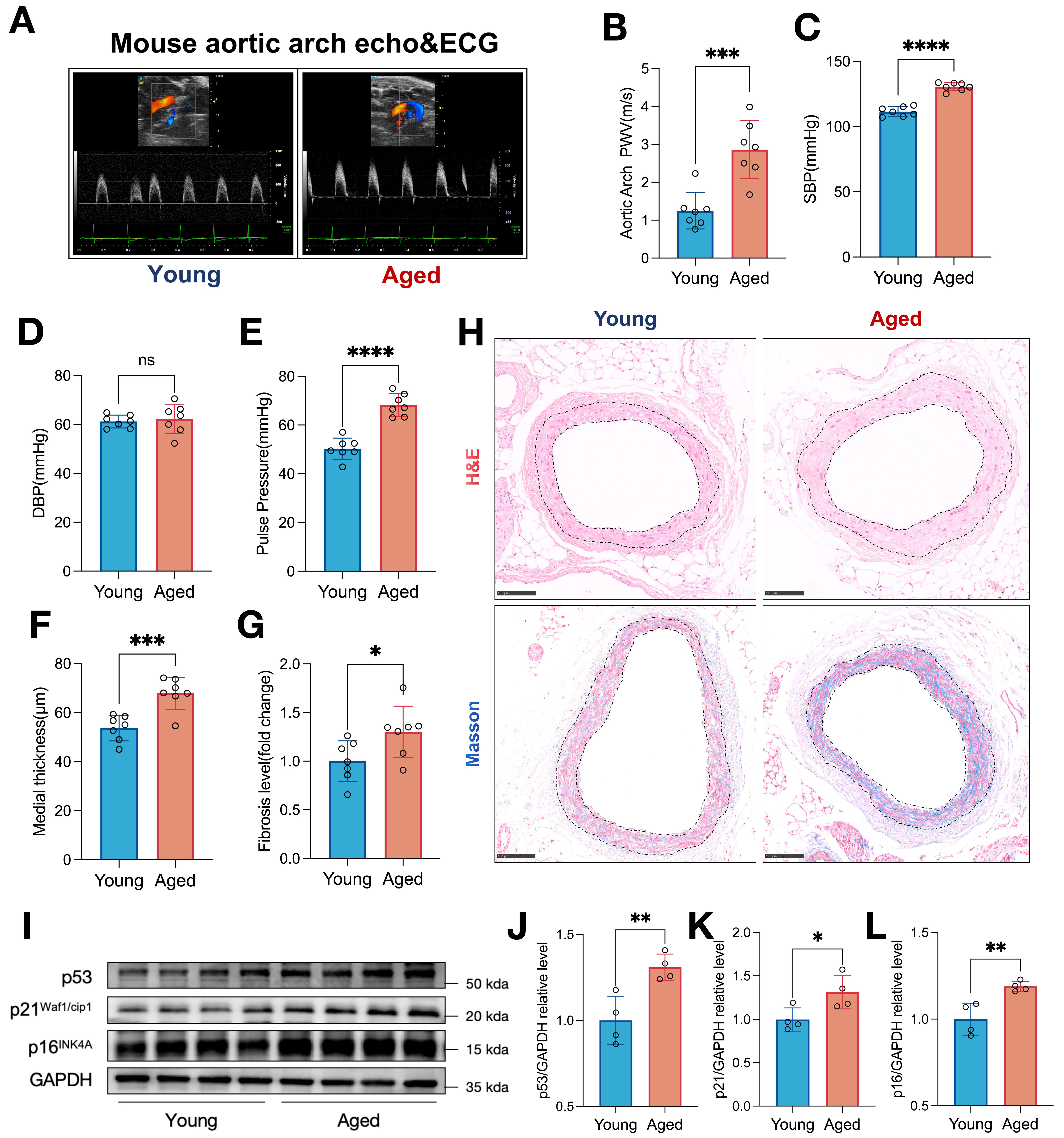
Figure 1. Aging induces aortic arch hemodynamic dysfunction, vascular remodeling, and cellular senescence in mice. (A) Representative echocardiographic images of the aortic arch and simultaneous ECG recordings in young and aged mice. (B) Quantification of aortic arch PWV in young (n = 7) and aged (n = 7) mice calculated by the measurement from echocardiogram and ECG. (C and D) SBP and DBP measured in young (n = 7) and aged (n = 7) mice. (E) Pulse pressure calculated from SBP and DBP in young (n = 7) and aged (n = 7) mice. (F) Aortic medial thickness quantified from histological sections in young (n = 7) and aged (n = 7) mice. (G) Fibrosis level (fold change relative to young mice) in the thoracic aorta of young (n = 7) and aged (n = 7) mice. (H) Representative H&E and Masson’s trichrome staining of thoracic aorta sections from young and aged mice. Scale bar, 100 μm. (I) Representative Western blot images showing the expression of senescence markers p53, p21Waf1/cip1, and p16INK4A in aortic tissues from young and aged mice. GAPDH was used as the loading control. (J-L) Densitometric quantification of (J) p53, (K) p21Waf1/cip1, and (L) p16INK4A protein levels, normalized to GAPDH, in young (n = 4) and aged (n = 4) mice. Data are presented as mean ± SD. *P < 0.05, **P < 0.01, ***P < 0.001, ****P < 0.0001 vs. young group (unpaired two-tailed Student’s t-test).




