

Intravenous immunoglobulin use and in-hospital outcomes in adult fulminant myocarditis: a two-center retrospective cohort study
 0
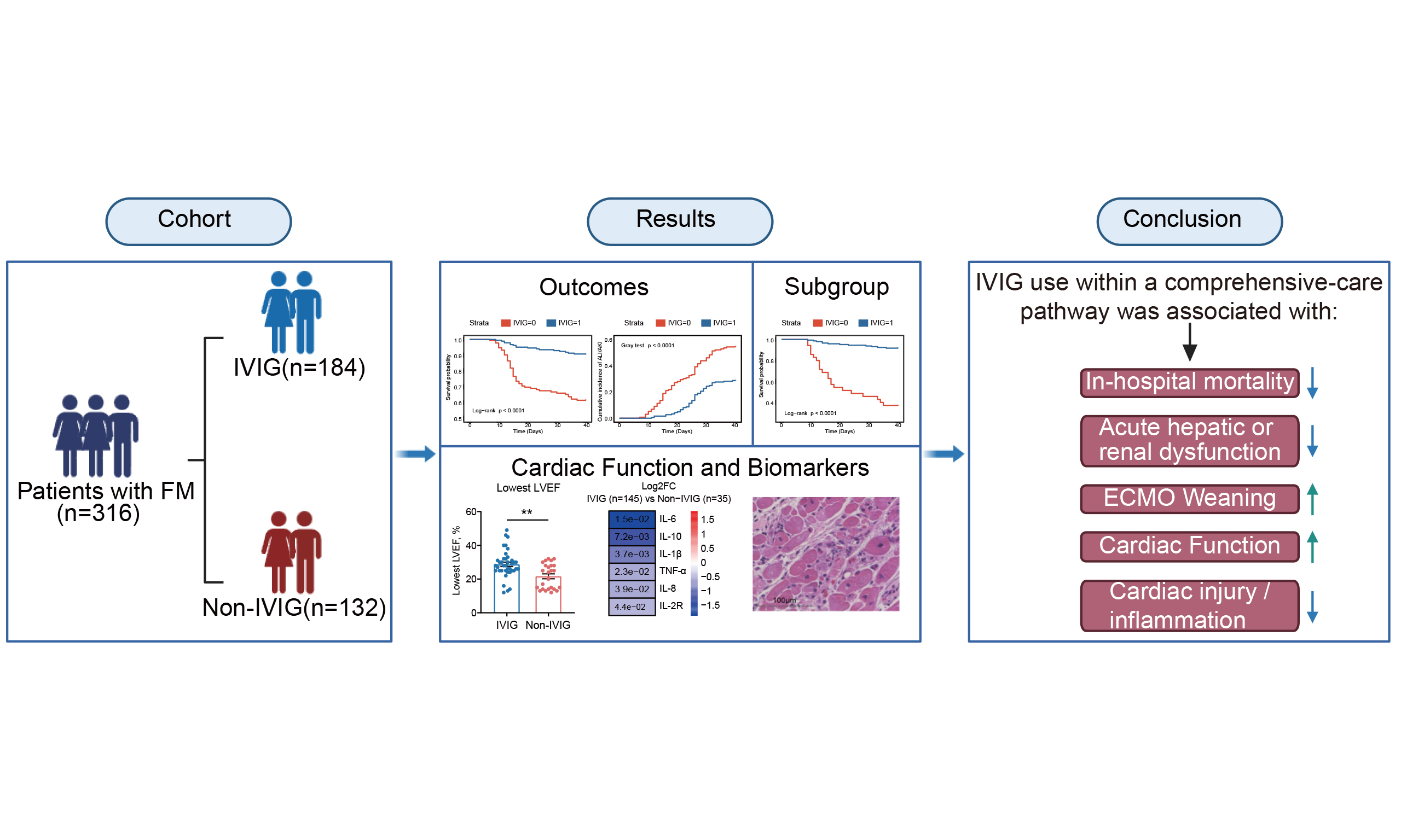
0 Graphical Abstract
Abstract
Aim: To examine the association between intravenous immunoglobulin (IVIG) use and in-hospital outcomes in adults with fulminant myocarditis (FM).
Methods: This two-center retrospective cohort study included 316 adults hospitalized with FM between January 2020 and January 2025; 184 received IVIG, and 132 did not. The primary outcome was all-cause in-hospital mortality. Secondary outcomes were a composite of acute liver injury (ALI) or Kidney Disease: Improving Global Outcomes-defined acute kidney injury (AKI) and successful extracorporeal membrane oxygenation (ECMO) weaning with survival to discharge. We evaluated associations using multivariable and time-dependent Cox models, an analysis restricted to methylprednisolone-treated patients, propensity-score overlap weighting, Aalen-Johansen cumulative-incidence analyses, and Fine-Gray competing-risk models. Dose- and timing-related analyses were exploratory.
Results: In-hospital death occurred in 17 of 184 IVIG-treated patients (9.2%) and 51 of 132 non-IVIG-treated patients (38.6%). IVIG use was associated with lower mortality across multiple models, including the multivariable Cox model (hazard ratio [HR], 0.23; 95% confidence interval [95%CI]: 0.14-0.40; P < 0.001), the time-dependent model (HR, 0.15; 95%CI: 0.08-0.29), the methylprednisolone-treated cohort (HR, 0.15; 95%CI: 0.08-0.27), and the overlap-weighted analysis (HR, 0.24; 95%CI: 0.07-0.87). ALI/AKI occurred in 53 of 184 IVIG-treated patients (28.8%) and 72 of 132 non-IVIG-treated patients (54.5%; adjusted HR, 0.43; 95%CI: 0.30-0.61; P < 0.001). Among ECMO-supported patients, successful weaning with survival to discharge occurred in 50 of 61 IVIG-treated patients and 6 of 18 non-IVIG-treated patients (subdistribution HR, 5.89; 95%CI: 2.96-11.73; P < 0.001). Exploratory dose- and timing-related estimates did not establish an optimal IVIG regimen.
Conclusions: In this two-center retrospective cohort, IVIG use within a corticosteroid-containing care pathway was associated with lower in-hospital mortality, less ALI/AKI, and a greater likelihood of successful ECMO weaning with survival. Residual and unmeasured confounding preclude causal inference; prospective multicenter validation is required.
Keywords


 Download PDF
INTRODUCTION
Fulminant myocarditis (FM) is a severe inflammatory myocardial syndrome characterized by rapid hemodynamic deterioration, ventricular arrhythmias, cardiogenic shock, and multiorgan dysfunction[1]. Contemporary diagnostic and management frameworks emphasize early recognition of high-risk clinical phenotypes, exclusion of ischemic and non-inflammatory causes of myocardial injury, and prompt referral to centers with expertise in advanced heart failure and temporary mechanical circulatory support (MCS)[2-4]. Venoarterial extracorporeal membrane oxygenation (VA-ECMO) can provide a bridge to myocardial recovery or transplantation in patients with refractory cardiogenic shock; nevertheless, mortality remains substantial even in contemporary ECMO-supported cohorts[5,6]. Accordingly, adjunctive therapies are still needed to target the inflammatory myocardial injury underlying FM rather than circulatory failure alone.
The pathobiology of myocarditis is heterogeneous and may involve direct pathogen-mediated injury, innate and adaptive immune activation, autoimmunity, endothelial dysfunction, and subsequent adverse myocardial remodeling[7,8]. Intravenous immunoglobulin (IVIG) is a pooled polyclonal IgG preparation with pleiotropic immunomodulatory effects, including modulation of activating and inhibitory Fcγ receptors, complement interception, neutralization of autoantibodies and inflammatory mediators, and regulation of innate and adaptive immune-cell responses[9,10]. However, evidence supporting IVIG in myocarditis remains inconsistent. A Cochrane review concluded that the available clinical evidence was insufficient to establish a treatment benefit in either children or adults[11]. A randomized trial in recent-onset dilated cardiomyopathy did not demonstrate an incremental improvement in ventricular function with IVIG, although the enrolled population differed substantially from patients with acute FM[12]. Conversely, pooled analyses of predominantly observational studies have suggested possible associations with lower mortality and improved ventricular recovery, but interpretation has been constrained by clinical heterogeneity, limited adjustment for treatment allocation, and variation in IVIG dosing and concomitant immunomodulatory therapy[13].
Chinese centers have developed a life-support-based comprehensive management strategy combining temporary MCS with immunomodulatory therapy in selected patients with FM. Registry data have shown favorable outcomes with this bundled treatment approach, but the relative contribution of IVIG cannot be isolated from corticosteroids, MCS, referral patterns, and other components of intensive care[14]. In particular, adult FM data remain limited regarding whether IVIG exposure is associated with outcomes after accounting for concomitant methylprednisolone and advanced organ support. The clinical relevance of cumulative IVIG exposure and timing of initiation are also uncertain, and analyses of these post-admission exposures are susceptible to treatment-selection and survivorship biases.
Therefore, we conducted a two-center retrospective cohort study of adults with FM to evaluate the association between IVIG treatment and in-hospital mortality, acute hepatic or renal dysfunction, and successful ECMO weaning with survival to discharge. To address major sources of bias inherent to the observational design, we incorporated multivariable adjustment, analyses restricted to methylprednisolone-treated patients, propensity-score overlap weighting, time-dependent exposure modeling, competing-risk analysis, and dose- and timing-related sensitivity analyses. We also examined cardiac functional recovery, circulating biomarkers, and an exploratory histopathological subset to characterize biological findings accompanying the observed clinical associations.
METHODS
Study design and population
This two-center retrospective cohort study was conducted at Tongji Hospital, Tongji Medical College, Huazhong University of Science and Technology, and the First Affiliated Hospital of Zhengzhou University. The study complied with the Declaration of Helsinki and was approved by the institutional review boards of both participating centers (Tongji Hospital approval No. TJ-IRB202604153; First Affiliated Hospital of Zhengzhou University approval No. 2025-KY-1274-001). Written informed consent was obtained from all patients or their legal representatives. The study was registered at ClinicalTrials.gov (NCT03268642).
Consecutive patients admitted with suspected FM between January 2020 and January 2025 were screened. FM was adjudicated according to the Chinese expert consensus and the American Heart Association scientific statement. Required features were an abrupt presentation with severe heart failure, cardiogenic shock, hypotension requiring vasoactive or inotropic support, or life-threatening arrhythmia; biochemical evidence of myocardial injury; and exclusion of alternative causes of acute myocardial injury or circulatory collapse. Acute coronary syndrome, sepsis-related myocardial injury, hypovolemic shock, and pre-existing structural myocardial disease were excluded using coronary angiography or coronary computed tomography angiography when feasible, serial electrocardiography and biomarker trajectories, echocardiography, cardiac magnetic resonance imaging when clinically possible, and multidisciplinary adjudication.
Of 404 patients screened, those aged < 18 years, those with an admission left-ventricular ejection fraction (LVEF) > 50%, and those with a confirmed alternative diagnosis were excluded. The final cohort comprised 316 adults, categorized according to IVIG exposure during hospitalization as IVIG-treated (n = 184) or non-IVIG-treated (n = 132) [Figure 1].
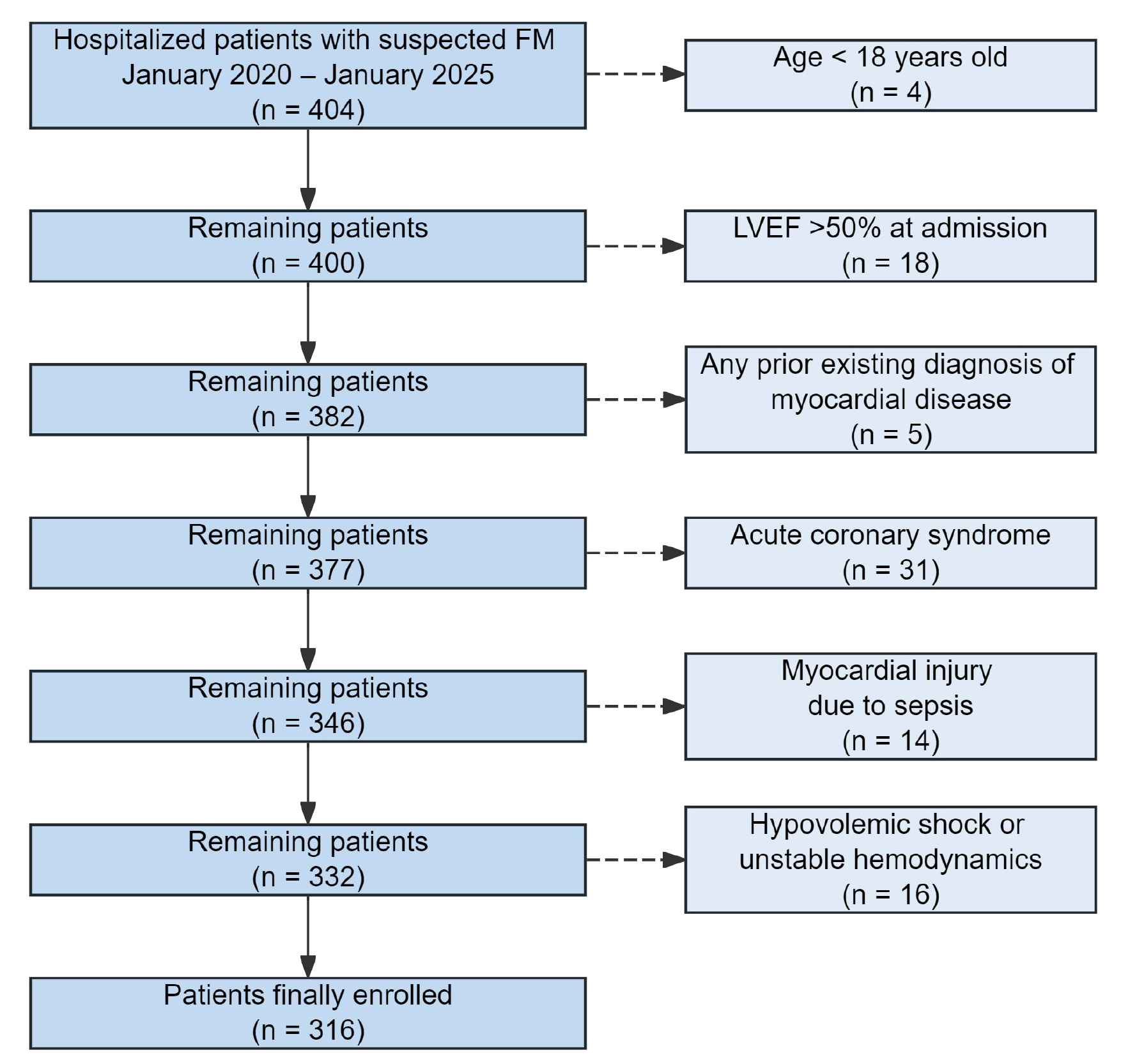
Figure 1. Flowchart of patient selection and enrollment. Patients admitted with suspected fulminant myocarditis (FM) between January 2020 and January 2025 were screened at two tertiary centers (n = 404). Sequential exclusions were age < 18 years (n = 4), admission left ventricular ejection fraction (LVEF) > 50% (n = 18), pre-existing myocardial disease (n = 5), acute coronary syndrome (n = 31), sepsis-related myocardial injury (n = 14), and hypovolemic shock or unstable hemodynamics attributable to an alternative cause (n = 16). The final cohort comprised 316 adults, including 184 IVIG-treated and 132 non-IVIG-treated patients.
Treatment protocols and exposure definition
All patients received comprehensive care according to hemodynamic status, organ perfusion, arrhythmia burden, and treating-team judgment, including vasoactive or inotropic therapy, mechanical ventilation, renal replacement therapy, and temporary MCS when clinically indicated. Intra-aortic balloon pump and peripheral VA-ECMO were used for refractory cardiogenic shock or persistent hypoperfusion despite pharmacological support.
IVIG was prescribed at the treating team's discretion, without a study-mandated dose or schedule. Both centers used the same 5% human immunoglobulin preparation (Human Immunoglobulin [pH4] for Intravenous Injection; Hualan Bio, Xinxiang, China). Actual daily doses ranged from 5 to 20 g and treatment duration from 1 to 5 days. Cumulative exposure was categorized as < 10 g (n = 65), 10 to < 20 g (n = 83), or
Methylprednisolone was administered to all 184 IVIG-treated patients and 60 of 132 non-IVIG-treated patients. Intravenous methylprednisolone was given at 200-500 mg/day for 3-5 days and subsequently tapered according to clinical response, using the same overall treatment framework at both centers. Neuraminidase inhibitors and empirical antibacterial therapy were administered when clinically indicated and were not determined by IVIG exposure. Detailed information regarding IVIG formulation, infusion procedures, weight-adjusted exposure, safety monitoring, corticosteroid tapering, and antiviral and antibacterial regimens is provided in Supplementary Table 1.
Data collection and clinical definitions
Demographic characteristics, presenting symptoms, admission vital signs, laboratory measurements, imaging findings, organ-support therapies, and in-hospital outcomes were abstracted from electronic medical records using a standardized case report form. Baseline variables included age, sex, blood pressure, cardiac troponin I (cTnI), N-terminal pro-B-type natriuretic peptide (NT-proBNP), white blood cell count, high-sensitivity C-reactive protein (hsCRP), alanine aminotransferase, aspartate aminotransferase, creatinine, circulating cytokines, and admission LVEF. Center, admission year, symptom-to-admission interval, interhospital transfer, residence, insurance/payment category, distance to the tertiary center, admission lactate, Sequential Organ Failure Assessment score, vasoactive-inotropic score within 24 h, and MCS initiated within 24 h were collected to characterize healthcare access, treatment selection, and early disease severity. Serial echocardiographic and laboratory data were also abstracted for descriptive analyses of admission, nadir, and discharge LVEF and in-hospital cardiac and inflammatory biomarker profiles.
The primary outcome was all-cause in-hospital mortality. Secondary outcomes were acute hepatic or renal dysfunction and successful ECMO weaning with survival to discharge. Acute hepatic or renal dysfunction was a composite of acute liver injury (ALI) and/or Kidney Disease: Improving Global Outcomes (KDIGO)-defined acute kidney injury (AKI). ALI was defined as persistent elevation of alanine aminotransferase or aspartate aminotransferase to > 3 times the upper limit of normal. All patients requiring continuous renal replacement therapy (CRRT) were classified as having AKI. For cumulative-incidence analyses of ALI/AKI, the first occurrence of ALI/AKI was the event of interest; in-hospital death before ALI/AKI and discharge alive without prior ALI/AKI were treated as competing events. Successful ECMO weaning with survival to discharge was defined as ECMO decannulation followed by survival to hospital discharge. For ECMO time-to-event analyses, time zero was the date of ECMO cannulation, and the event time was the date of ECMO decannulation in patients who subsequently survived to discharge. Death before qualifying for successful weaning was treated as a competing event. A patient who was decannulated but subsequently died before discharge would not be classified as having achieved the endpoint and would instead be coded as a competing death at the date of death. In the present cohort, no patient underwent heart transplantation before ECMO weaning, was transferred or discharged while receiving ECMO, or died after ECMO decannulation but before hospital discharge.
Histopathological and immunohistochemical analysis
Histopathological analyses were restricted to clinically indicated endomyocardial biopsy specimens obtained during hospitalization; tissue acquisition was not mandated by the study protocol. Specimens were eligible when sufficient myocardium was available for hematoxylin-and-eosin staining and immunohistochemical assessment and when tissue processing, staining, and image quality permitted reliable quantification. Specimens with insufficient myocardial tissue, substantial processing artifact, or inadequate staining were excluded.
Eleven patients met these criteria, including 6 IVIG-treated and 5 non-IVIG-treated patients. Formalin-fixed, paraffin-embedded sections (4 μm) were stained with hematoxylin and eosin. Immunohistochemistry was performed using antibodies against myeloperoxidase (MPO; Abcam, ab109116, clone EPR4792, 1:250), CD68 (Abcam, ab125157, 1:100), CD3 (Abcam, ab17143, clone F7.2.38, 1:20), and CD20 (Abcam, ab78237, clone EP459Y, 1:50) to identify neutrophils, macrophages, T cells, and B cells, respectively. Five non-overlapping myocardium-containing high-power fields per marker were evaluated for each specimen, excluding endocardium, thrombus, and areas with major processing artifact. Positive cells were quantified as cells/mm2 using QuPath version 0.4.4. Two pathologists, blinded to treatment allocation, independently reviewed the specimens and quantitative results.
Statistical analysis
Continuous variables are presented as median (Q1, Q3) and were compared using the Mann-Whitney U test. Categorical variables are presented as n (%) and were compared using the chi-square test or Fisher's exact test, as appropriate. Variable-level missingness is reported in Supplementary Table 2; no listed variable had
For all-cause in-hospital mortality, Kaplan-Meier curves were compared using log-rank tests, and multivariable Cox proportional-hazards models were used for adjusted analyses. Time zero was hospital admission, and patients discharged alive were censored on their discharge dates. Numbers at risk were generated from the same survival objects used to estimate the Kaplan-Meier curves. IVIG initiation was additionally modeled as a time-varying exposure to address immortal-time bias. Proportional-hazards assumptions were assessed using Schoenfeld residuals.
For ALI/AKI, cumulative incidence functions were estimated using the Aalen-Johansen method and compared using Gray's test, with in-hospital death before ALI/AKI and discharge alive without prior ALI/AKI treated as competing events. Adjusted associations were estimated using cause-specific Cox proportional-hazards models. Because methylprednisolone use differed substantially between treatment groups, a prespecified analysis was restricted to methylprednisolone-treated patients. Propensity-score overlap weighting was additionally performed in this cohort, with covariate balance assessed using absolute standardized mean differences; values < 0.10 were considered acceptable.
For successful ECMO weaning with survival to discharge, cumulative incidence functions were estimated using the Aalen-Johansen method and compared using Gray's test, with death before qualifying for successful weaning treated as a competing event. Time zero was ECMO cannulation. The Fine-Gray subdistribution hazards model was used for the primary adjusted analysis, with cause-specific Cox, accelerated failure-time, and penalized logistic models used as sensitivity analyses. Exact model-specific covariates, censoring rules, competing-event definitions, and analytic specifications are summarized in Supplementary Table 3.
Dose- and timing-related analyses were exploratory. A day-4 landmark analysis evaluated subsequent mortality according to cumulative IVIG exposure, and alternative early-treatment thresholds of 3, 4, and 5 days were examined. Clinical subgroup analyses were performed according to white blood cell count, alanine aminotransferase, aspartate aminotransferase, and creatinine. All tests were two-sided, with P < 0.05 considered statistically significant. Analyses were performed using R version 4.2.2.
RESULTS
Study population and baseline characteristics
Among 404 patients screened for suspected FM, 316 met the eligibility criteria; 184 received IVIG, and 132 did not [Figure 1]. The cohort included 149 patients from Tongji Hospital and 167 from the First Affiliated Hospital of Zhengzhou University. The median age was 38.00 years (Q1, Q3: 25.00, 53.00), and 157 patients (49.68%) were male [Table 1].
Baseline characteristics, healthcare access proxies, and admission severity indicators
| Variable | Total (n = 316) | IVIG (n = 184) | Non-IVIG (n = 132) | P value |
| Demographics, symptoms, and admission vital signs | ||||
| Age, years | 38.00 (25.00, 53.00) | 38.00 (24.00, 50.25) | 42.00 (26.75, 55.00) | 0.190 |
| Sex, male | 157 (49.68) | 84 (45.65) | 73 (55.30) | 0.091 |
| SBP, mmHg | 102.00 (92.00, 110.00) | 99.50 (92.00, 110.00) | 102.00 (93.00, 113.00) | 0.239 |
| DBP, mmHg | 67.00 (50.00, 81.00) | 65.00 (50.50, 80.50) | 70.00 (50.00, 82.00) | 0.256 |
| Fever | 120 (37.97) | 66 (35.87) | 54 (40.91) | 0.363 |
| Chest distress | 154 (48.73) | 87 (47.28) | 67 (50.76) | 0.542 |
| Chest pain | 88 (27.85) | 49 (26.63) | 39 (29.55) | 0.569 |
| Dyspnea | 30 (9.49) | 20 (10.87) | 10 (7.58) | 0.325 |
| Syncope | 29 (9.18) | 19 (10.33) | 10 (7.58) | 0.404 |
| Gastrointestinal symptoms | 56 (17.72) | 27 (14.67) | 29 (21.97) | 0.094 |
| Admission biomarkers and cardiac function | ||||
| cTnI, pg/mL | 19,675.60 (15,980.42, 39,456.00) | 20,305.33 (15,949.49, 34,664.88) | 18,928.90 (15,980.42, 47,294.74) | 0.298 |
| NT-proBNP, pg/mL | 6,738.00 (5,349.20, 18,663.00) | 6,976.00 (5,166.21, 14,996.00) | 6,736.04 (5,400.26, 22,401.29) | 0.154 |
| WBC, 109/L | 11.40 (10.17, 12.91) | 11.48 (9.50, 12.55) | 11.37 (10.29, 14.66) | 0.094 |
| hsCRP, mg/L | 36.65 (20.65, 105.40) | 40.46 (19.98, 75.55) | 34.95 (21.27, 138.60) | 0.084 |
| ALT, IU/L | 58.00 (42.00, 124.00) | 61.00 (41.75, 152.00) | 56.00 (43.75, 78.25) | 0.543 |
| AST, IU/L | 155.50 (80.08, 228.75) | 163.30 (79.80, 248.25) | 147.60 (80.95, 212.75) | 0.518 |
| Creatinine, μmol/L | 89.46 (66.00, 124.41) | 89.00 (66.00, 131.00) | 90.23 (66.92, 113.27) | 0.848 |
| IL-1β, pg/mL | 3.38 (2.17, 11.62) | 3.08 (2.17, 11.62) | 5.87 (2.00, 32.32) | 0.141 |
| IL-2R, IU/mL | 558.00 (311.00, 859.25) | 540.00 (312.00, 859.25) | 583.50 (299.50, 851.00) | 0.375 |
| IL-6, pg/mL | 18.73 (6.06, 79.15) | 14.57 (3.87, 86.12) | 19.50 (10.53, 59.41) | 0.306 |
| IL-8, pg/mL | 28.70 (11.10, 49.60) | 29.40 (11.30, 58.15) | 25.60 (8.10, 42.78) | 0.067 |
| IL-10, pg/mL | 5.00 (5.00, 10.40) | 5.00 (5.00, 9.80) | 5.50 (5.00, 10.80) | 0.163 |
| TNF-α, pg/mL | 10.90 (8.60, 14.33) | 10.90 (8.60, 14.40) | 10.90 (9.00, 13.65) | 0.714 |
| Admission LVEF, % | 42.00 (30.00, 49.00) | 40.00 (30.00, 48.25) | 48.00 (29.00, 50.00) | 0.122 |
| Center, healthcare-access proxies, and early severity indicators | ||||
| Center: First Affiliated Hospital of Zhengzhou University | 167 (52.85) | 96 (52.17) | 71 (53.79) | 0.866 |
| Admission year | 2022.00 (2021.00, 2023.25) | 2022.00 (2021.00, 2024.00) | 2022.00 (2020.75, 2023.00) | 0.002 |
| Symptom-to-admission interval, days | 3.30 (2.00, 5.53) | 3.00 (1.90, 4.72) | 4.00 (2.30, 6.93) | 0.001 |
| Interhospital transfer | 156 (49.37) | 98 (53.26) | 58 (43.94) | 0.128 |
| Rural residence | 103 (32.59) | 52 (28.26) | 51 (38.64) | 0.069 |
| Urban employee/commercial insurance | 141 (44.62) | 93 (50.54) | 48 (36.36) | 0.017 |
| Distance to tertiary center, km | 86.60 (37.87, 186.22) | 88.32 (38.70, 190.87) | 83.32 (36.72, 177.51) | 0.981 |
| Admission lactate, mmol/L | 4.28 (2.83, 6.14) | 4.55 (2.99, 6.29) | 3.99 (2.28, 5.94) | 0.043 |
| Admission SOFA score | 7.00 (4.00, 9.00) | 7.00 (4.00, 9.00) | 7.00 (4.00, 9.00) | 0.466 |
| Vasoactive-inotropic score within 24 h | 29.70 (17.58, 47.98) | 33.50 (22.48, 51.70) | 25.55 (11.53, 41.38) | < 0.001 |
| MCS initiated within 24 h | 133 (42.09) | 107 (58.15) | 26 (19.70) | < 0.001 |
| Coronary angiography or coronary CTA performed | 287 (90.82) | 174 (94.57) | 113 (85.61) | 0.012 |
| Cardiac MRI performed | 122 (38.61) | 62 (33.70) | 60 (45.45) | 0.045 |
Most admission vital signs, cardiac and inflammatory biomarkers, organ-function indices, cytokines, and admission LVEF did not differ significantly between groups. Nevertheless, treatment allocation was clearly nonrandom: IVIG-treated patients were admitted in more recent years, had a shorter symptom-to-admission interval, more often had urban employee or commercial insurance, and had higher admission lactate, 24-h vasoactive-inotropic scores, and more frequent MCS use within 24 h [Table 1]. Coronary angiography or coronary computed tomography angiography was performed in 287 patients, and cardiac magnetic resonance imaging was performed in 122, of whom 103 had Lake Louise-compatible findings. Missingness was < 5% for each listed variable [Supplementary Table 2].
In-hospital management and crude outcomes
In-hospital management differed substantially between groups [Table 2]. Methylprednisolone was administered to all 184 IVIG-treated patients and to 60 of 132 non-IVIG-treated patients. MCS was used in 145 IVIG-treated and 35 non-IVIG-treated patients, and intra-aortic balloon pump, ECMO, temporary pacing, CRRT, and vasoactive agents were generally used more frequently in the IVIG group.
In-hospital management and clinical outcomes
| Variable | Total (n = 316) | IVIG (n = 184) | Non-IVIG (n = 132) | P value |
| Methylprednisolone | 244 (77.22) | 184 (100.00) | 60 (45.45) | < 0.001 |
| Any MCS (IABP and/or ECMO) | 180 (56.96) | 145 (78.80) | 35 (26.52) | < 0.001 |
| IABP | 147 (46.52) | 121 (65.76) | 26 (19.70) | < 0.001 |
| ECMO | 79 (25.00) | 61 (33.15) | 18 (13.64) | < 0.001 |
| Temporary pacemaker | 44 (13.92) | 40 (21.74) | 4 (3.03) | < 0.001 |
| CRRT | 52 (16.46) | 44 (23.91) | 8 (6.06) | < 0.001 |
| Endotracheal intubation | 73 (23.10) | 41 (22.28) | 32 (24.24) | 0.785 |
| Norepinephrine | 216 (68.35) | 135 (73.37) | 81 (61.36) | 0.032 |
| Dopamine or dobutamine | 194 (61.39) | 124 (67.39) | 70 (53.03) | 0.014 |
| Neuraminidase inhibitor | 232 (73.42) | 147 (79.89) | 85 (64.39) | 0.003 |
| Empirical antibacterial therapy | 197 (62.34) | 115 (62.50) | 82 (62.12) | 1.000 |
| ALI/AKI | 125 (39.56) | 53 (28.80) | 72 (54.55) | < 0.001 |
| In-hospital death | 68 (21.52) | 17 (9.24) | 51 (38.64) | < 0.001 |
In-hospital death occurred in 17 IVIG-treated patients (9.24%) and 51 non-IVIG-treated patients (38.64%). The ALI/AKI composite occurred in 53 IVIG-treated patients (28.80%) and 72 non-IVIG-treated patients (54.55%) [Table 2].
Association between IVIG use and in-hospital mortality
The Kaplan-Meier curves separated early and remained divergent through discharge [Figure 2A]. In the primary multivariable Cox model, IVIG use was associated with a lower hazard of in-hospital death (hazard ratio [HR], 0.23; 95% confidence interval [95%CI]: 0.14-0.40; P < 0.001). Modeling IVIG initiation as a time-varying exposure yielded a similar association (HR, 0.15; 95%CI: 0.08-0.29; P < 0.001) [Table 3].
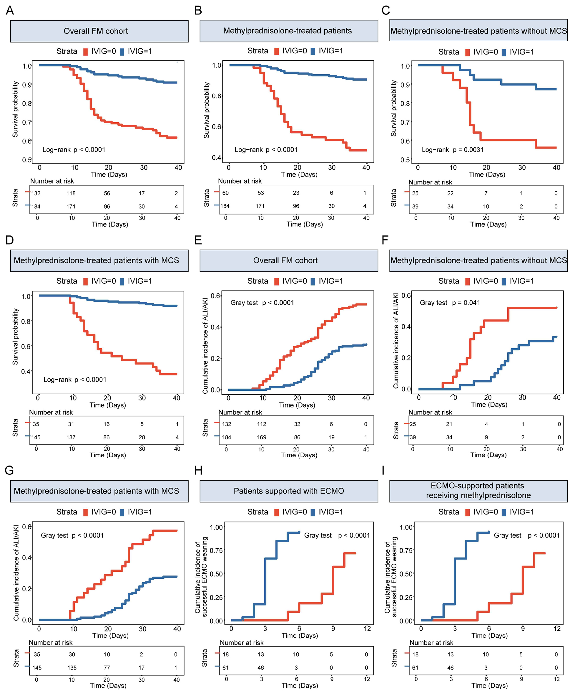
Figure 2. Survival, ALI/AKI, and ECMO weaning curves according to IVIG use. (A) Kaplan-Meier survival curves in the overall FM cohort (n = 316). (B) Kaplan-Meier survival curves in methylprednisolone-treated patients (n = 244). (C) Kaplan-Meier survival curves in methylprednisolone-treated patients without mechanical circulatory support (MCS; n = 64). (D) Kaplan-Meier survival curves in methylprednisolone-treated patients with MCS (n = 180). (E) Aalen-Johansen cumulative incidence of ALI and/or AKI in the overall cohort. (F) Aalen-Johansen cumulative incidence of ALI/AKI in methylprednisolone-treated patients without MCS. (G) Aalen-Johansen cumulative incidence of ALI/AKI in methylprednisolone-treated patients with MCS. (H) Aalen-Johansen cumulative incidence of successful ECMO weaning with survival to discharge in all ECMO-supported patients (n = 79). (I) Aalen-Johansen cumulative incidence of successful ECMO weaning with survival to discharge in ECMO-supported patients receiving methylprednisolone (n = 73). ALI/AKI was defined as persistent alanine aminotransferase or aspartate aminotransferase elevation > 3 times the upper limit of normal and/or Kidney Disease: Improving Global Outcomes (KDIGO)-defined AKI; all patients receiving continuous renal replacement therapy were classified as having AKI. For (E-G), in-hospital death before ALI/AKI and discharge alive without prior ALI/AKI were treated as competing events. For (H and I) time zero was ECMO cannulation and death before qualifying for successful ECMO weaning was treated as a competing event. Displayed P values are from log-rank tests for (A-D) and Gray's tests for (E-I). Patients discharged alive in (A-D) were censored on their actual discharge dates, and the displayed numbers at risk were generated directly from the same survival objects used to estimate the Kaplan-Meier curves.
Association of IVIG use with clinical outcomes: primary, confounding-control, and sensitivity analyses
| Outcome | Analysis population | Statistical model | Estimate (95%CI) | P value |
| In-hospital mortality | Overall cohort (N = 316; 68 deaths) | Multivariable Cox | HR 0.23 (0.14-0.40) | < 0.001 |
| In-hospital mortality | Overall cohort (N = 316; 68 deaths) | Time-dependent Cox; IVIG as a time-varying exposure | HR 0.15 (0.08-0.29) | < 0.001 |
| In-hospital mortality | Methylprednisolone-treated cohort (N = 244; 35 deaths) | Multivariable Cox | HR 0.1 (0.08-0.27) | < 0.001 |
| In-hospital mortality | Methylprednisolone-treated cohort (N = 244; 35 deaths) | Propensity-score overlap-weighted Cox | HR 0.24 (0.07-0.87) | 0.030 |
| ALI/AKI | Overall cohort (N = 316; 125 events) | Cause-specific Cox proportional-hazards model | HR 0.43 (0.30-0.61) | < 0.001 |
| Successful ECMO weaning with survival to discharge | ECMO-supported cohort (N = 79; 56 events) | Fine-Gray competing-risk model | sHR 5.89 (2.96-11.73) | < 0.001 |
| Successful ECMO weaning with survival to discharge | ECMO-supported cohort (N = 79; 56 events) | Cause-specific Cox | HR 19.30 (4.60-81.01) | < 0.001 |
| ECMO support duration | ECMO-supported cohort (N = 79) | Accelerated failure-time model | TR 0.32 (0.24-0.42) | < 0.001 |
| Successful ECMO weaning with survival to discharge | ECMO-supported cohort (N = 79; 56 events) | Penalized logistic regression; 1,000-bootstrap CI | OR 5.57 (2.45-13.10) | - |
The methylprednisolone-treated cohort comprised 184 IVIG-treated and 60 non-IVIG-treated patients, with 17 and 18 deaths, respectively. Survival curves remained separated after restriction to this cohort [Figure 2B], and IVIG use was associated with lower mortality after multivariable adjustment (HR, 0.15; 95%CI: 0.08-0.27; P < 0.001) [Table 3]. The unadjusted association was also evident among methylprednisolone-treated patients without MCS and among those receiving MCS [Figure 2C and D].
Overlap weighting substantially improved measured covariate balance in the methylprednisolone-treated cohort; every post-weighting absolute standardized mean difference was < 0.10 [Supplementary Table 4]. The overlap-weighted Cox model remained directionally consistent with the primary analysis (HR, 0.24; 95%CI: 0.07-0.87; P = 0.030) [Table 3].
Acute hepatic or renal dysfunction and ECMO weaning
The Aalen-Johansen cumulative incidence of ALI/AKI was lower among IVIG-treated patients in the overall cohort and in the methylprednisolone-treated strata with and without MCS [Figure 2E-G]. In the cause-specific Cox proportional-hazards model, IVIG use was associated with a lower hazard of ALI/AKI (HR, 0.43; 95%CI: 0.30-0.61; P < 0.001) [Table 3].
Among 79 ECMO-supported patients, 50 of 61 IVIG-treated patients (81.97%) and 6 of 18 non-IVIG-treated patients (33.33%) achieved successful weaning with survival to discharge. The cumulative incidence of successful ECMO weaning with survival to discharge was higher among IVIG-treated patients in the full ECMO cohort and after restriction to methylprednisolone-treated patients [Figure 2H and I]. In the Fine-Gray model, IVIG use was associated with a higher subdistribution rate of successful weaning with survival (sHR, 5.89; 95%CI: 2.96-11.73; P < 0.001). Cause-specific Cox, accelerated failure-time, and penalized logistic analyses yielded concordant estimates [Table 3].
Exploratory dose- and timing-related analyses
Within the methylprednisolone- and MCS-treated subgroup, survival and ALI/AKI curves differed across dose categories [Figure 3A and B]; dose-stratified time-to-weaning curves are shown for the ECMO-supported subgroup [Figure 3C]. IVIG was initiated within 4 days of admission in 105 patients and after 4 days in 79 [Supplementary Table 5]. In descriptive analyses stratified by cumulative dose, initiation by day 4 was associated with more favorable survival and ALI/AKI curves than later initiation [Figure 3D-H].
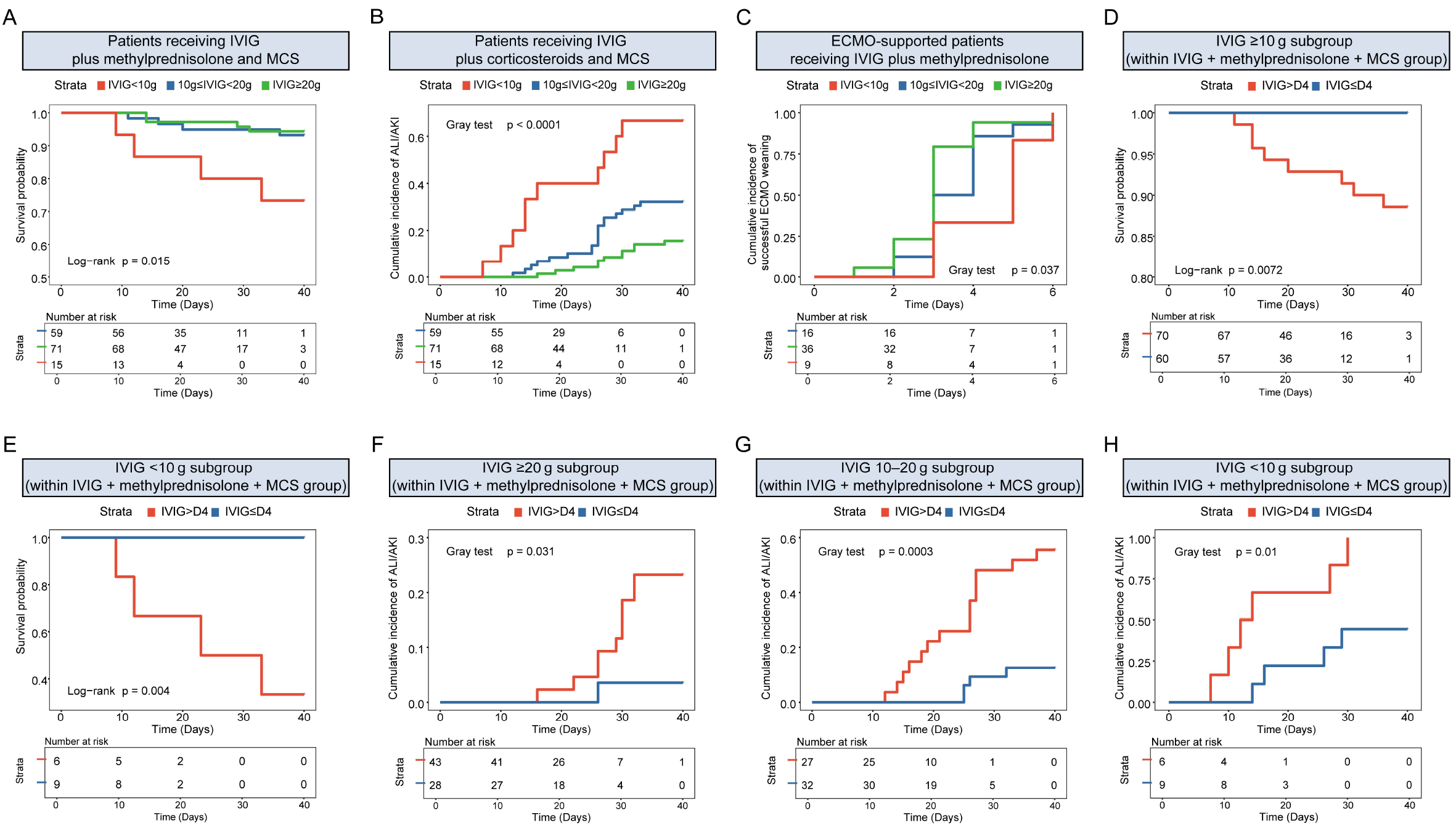
Figure 3. Exploratory dose- and timing-stratified outcomes among IVIG-treated patients. (A) Kaplan-Meier survival curves among patients who received IVIG plus methylprednisolone and MCS, stratified by cumulative IVIG dose (< 10 g, n = 15; 10 to < 20 g, n = 59; ≥ 20 g,
Associations with mortality were directionally consistent using early-treatment thresholds of 3 days
Cardiac function, biomarkers, and exploratory histopathology
Among methylprednisolone-treated patients without MCS, admission LVEF was similar between groups, whereas the lowest in-hospital and discharge LVEF values were higher in IVIG-treated patients [Figure 4A]. In-hospital cTnI, NT-proBNP, and hsCRP levels were lower with IVIG [Figure 4B], and circulating cytokines were generally lower [Figure 4C]. The same pattern was observed among methylprednisolone-treated patients receiving MCS: admission LVEF was comparable, lowest and discharge LVEF values were higher with IVIG [Figure 4D]; cardiac injury and inflammatory biomarkers were lower [Figure 4E]; and all measured cytokines were lower in the IVIG group [Figure 4F].
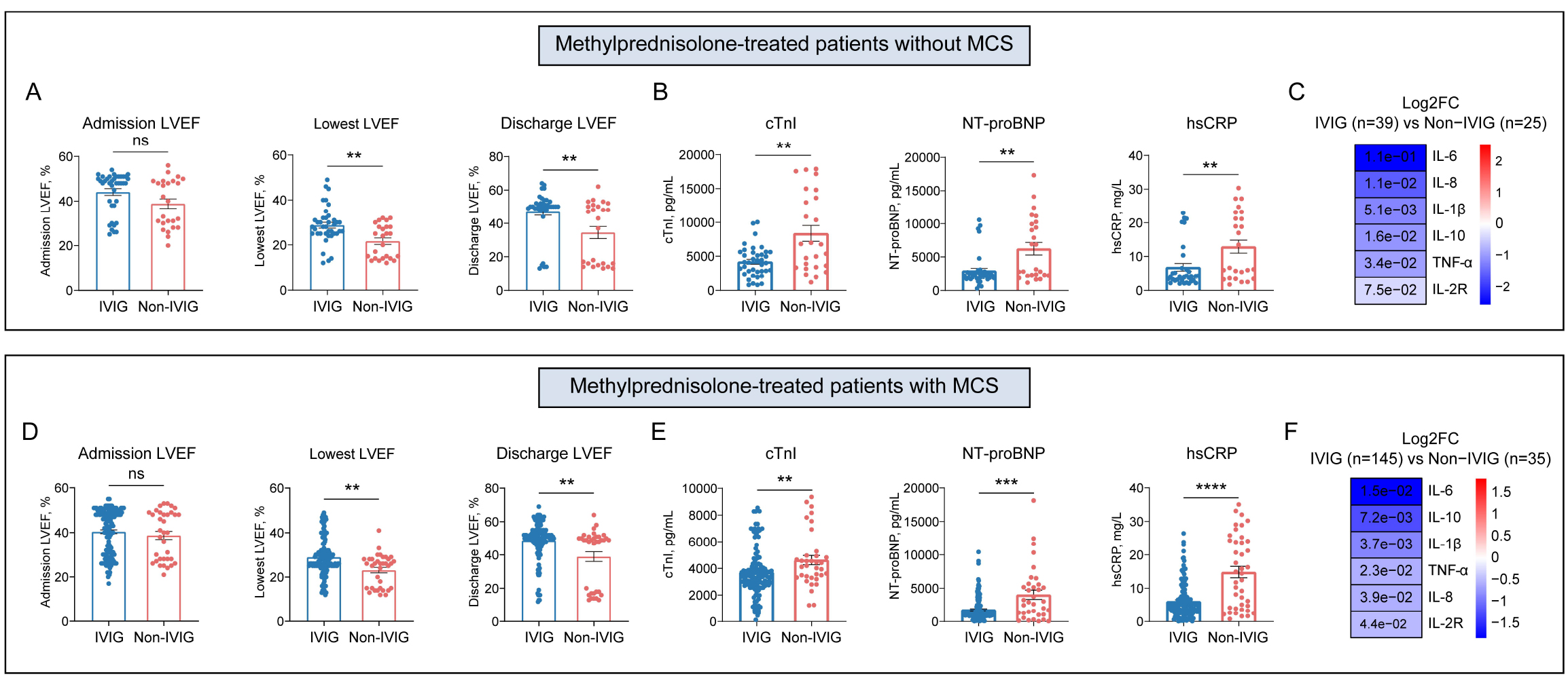
Figure 4. Cardiac function and in-hospital cardiac and inflammatory biomarker profiles according to IVIG use. (A-C) show methylprednisolone-treated patients without MCS (IVIG, n = 39; non-IVIG, n = 25). (A) admission, lowest in-hospital, and discharge LVEF. (B) in-hospital cTnI, NT-proBNP, and hsCRP. (C) heatmap of circulating cytokine differences between groups. (D-F) show methylprednisolone-treated patients receiving MCS (IVIG, n = 145; non-IVIG, n = 35). (D) admission, lowest in-hospital, and discharge LVEF. (E) in-hospital cTnI, NT-proBNP, and hsCRP. (F) heatmap of circulating cytokine differences between groups. Heatmap color represents log2 fold change for IVIG relative to non-IVIG, and the numbers within cells are P values. Dot-bar plots show individual observations with mean ± standard error of the mean. Group comparisons used the Mann-Whitney U test. **P < 0.01, ***P < 0.001, ****P < 0.0001; ns: not significant.
Clinically indicated endomyocardial biopsy specimens were available for 11 patients (IVIG, n = 6; non-IVIG, n = 5), with a median admission-to-biopsy interval of 4.80 days [Supplementary Table 6]. IVIG-treated specimens had lower histological inflammatory scores and lower densities of myeloperoxidase-positive neutrophils, CD68-positive macrophages, and CD3-positive T cells; CD20-positive B-cell density did not differ significantly [Figure 5]. This selected tissue analysis was exploratory.
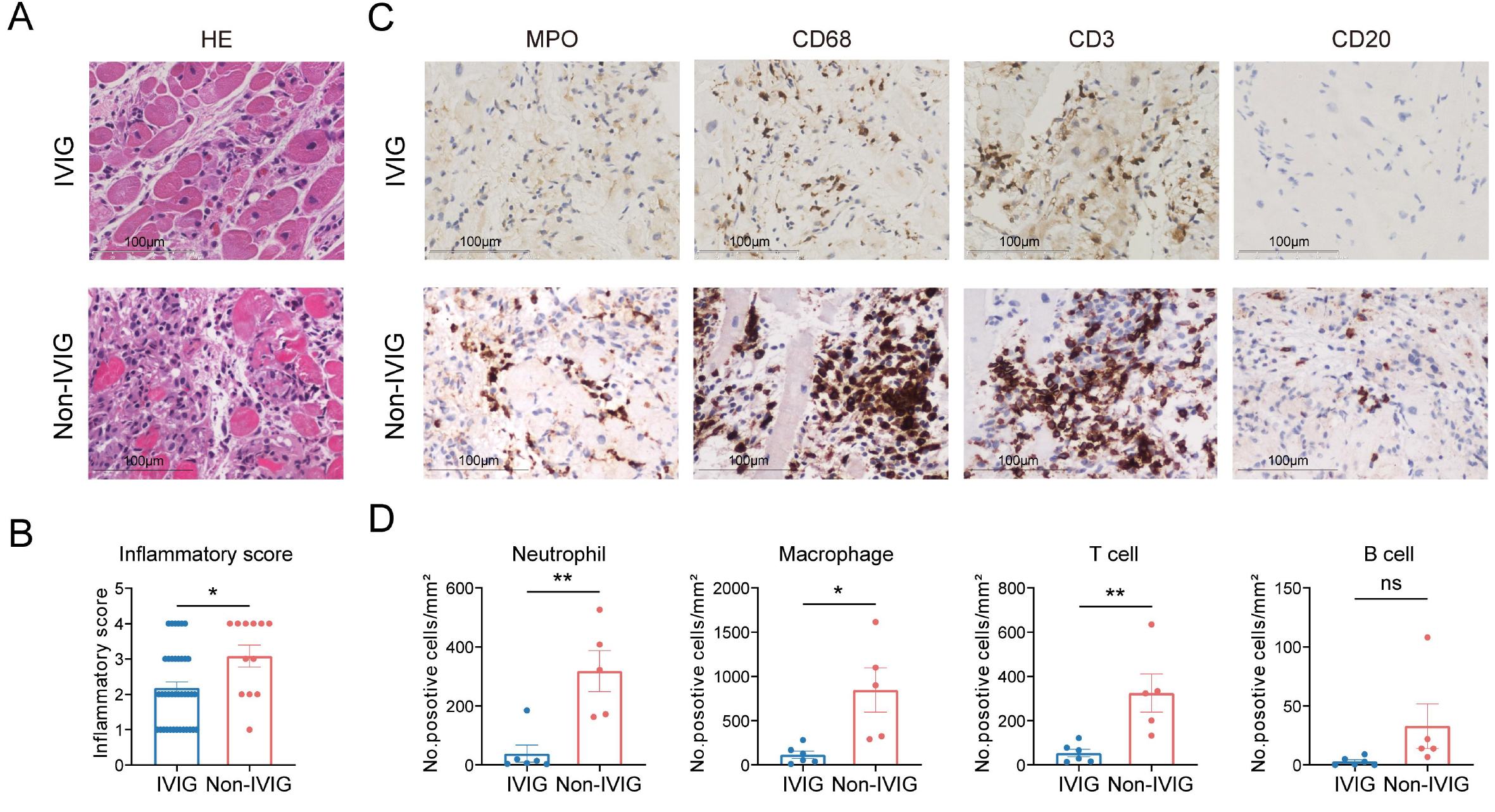
Figure 5. Exploratory histological and immunohistochemical findings in patients with available endomyocardial biopsy specimens. (A) representative hematoxylin-and-eosin-stained myocardial sections from IVIG-treated and non-IVIG-treated patients. (B) semiquantitative histological inflammatory scores. (C) representative immunohistochemical staining for myeloperoxidase (MPO), CD68, CD3, and CD20. (D) densities of MPO-positive neutrophils, CD68-positive macrophages, CD3-positive T cells, and CD20-positive B cells. Quantitative analyses included 6 IVIG-treated and 5 non-IVIG-treated patients, and each point represents one patient. Tissue acquisition was clinically indicated; the analysis was exploratory. Scale bar = 100 μm. Comparisons used the Mann-Whitney U test. *P < 0.05, **P < 0.01; ns: not significant.
Clinical subgroup analyses
Across the evaluated strata of white blood cell count, alanine aminotransferase, aspartate aminotransferase, and creatinine, all mortality HR point estimates favored IVIG [Figure 6 and Supplementary Figure 1]. Adjusted HRs ranged from 0.17 to 0.30; these subgroup estimates were exploratory and were not interpreted as evidence of effect modification.
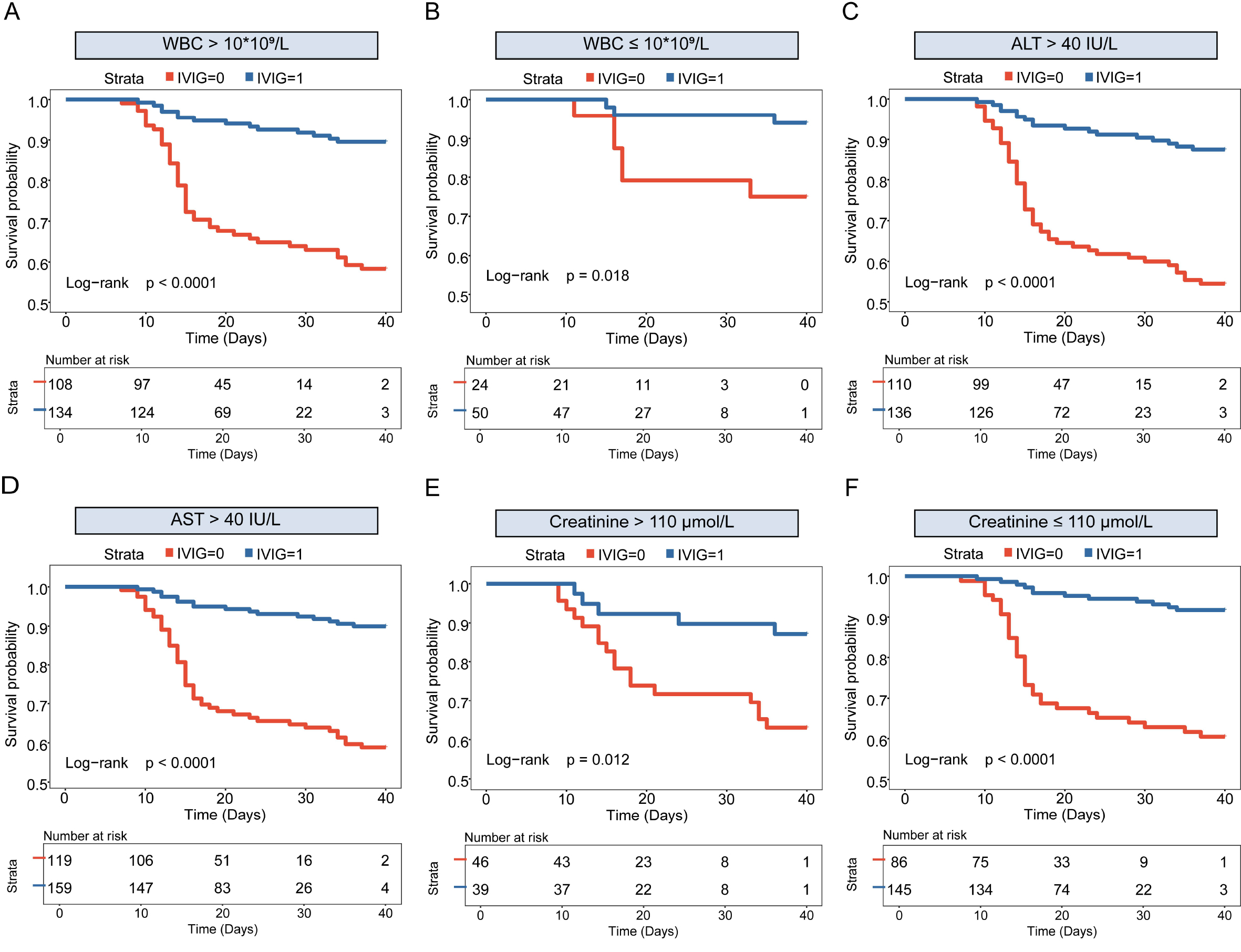
Figure 6. Kaplan-Meier survival curves within selected clinical subgroups. Kaplan-Meier curves for in-hospital survival were stratified by IVIG use within subgroups defined by admission white blood cell count (WBC), alanine aminotransferase (ALT), aspartate aminotransferase (AST), and serum creatinine. (A) WBC > 10 × 109/L. (B) WBC ≤ 10 × 109/L. (C) ALT > 40 IU/L. (D) AST > 40 IU/L. (E) creatinine >110 μmol/L. (F) creatinine ≤ 110 μmol/L. Patients discharged alive were censored on their actual dates of hospital discharge, and displayed numbers at risk were generated directly from the same survival objects used to estimate the Kaplan-Meier curves. Displayed P values are from log-rank tests.
DISCUSSION
In this two-center retrospective cohort of 316 adults with FM, IVIG treatment was associated with lower in-hospital mortality, less acute hepatic or renal dysfunction, and a higher cumulative incidence of successful ECMO weaning with survival to discharge. These associations remained directionally consistent after multivariable adjustment, restriction to methylprednisolone-treated patients, propensity-score overlap weighting, time-dependent exposure modeling, and competing-risk analyses. IVIG-treated patients also showed more favorable trajectories of LVEF, cardiac injury biomarkers, systemic inflammatory markers, and myocardial inflammatory-cell infiltration. Nevertheless, because treatment allocation was nonrandomized and IVIG was embedded within a more intensive treatment pathway, the findings should be interpreted as evidence of association rather than proof of an independent causal effect.
We found a consistent association between IVIG exposure and lower in-hospital mortality. This association remained evident after restricting the analysis to patients who received methylprednisolone, thereby comparing methylprednisolone plus IVIG with methylprednisolone without IVIG rather than contrasting two markedly different immunomodulatory strategies. The result extends an earlier adult retrospective study in which IVIG was associated with improved ventricular recovery and fewer arrhythmic events, although a mortality difference was not established[15]. Pediatric evidence has also suggested possible benefit, but differences in etiology, immune phenotype, spontaneous recovery, and treatment practice preclude direct extrapolation to adult FM[16]. Clinically, the persistence of the association across conventional and weighted models supports further evaluation of IVIG as an adjunct to corticosteroid-containing care in selected patients. However, residual confounding from referral patterns, socioeconomic position, treatment availability, and clinician selection remains plausible, and the magnitude of the observed effect should not be regarded as a treatment estimate equivalent to that obtained from a randomized trial.
Another major finding was that IVIG treatment was associated with less acute hepatic or renal dysfunction and with more favorable cardiac and inflammatory trajectories. These observations are biologically compatible with the pleiotropic actions of pooled IgG, including modulation of activating and inhibitory Fcγ-receptor signaling, complement interception, neutralization of pathogenic antibodies or inflammatory mediators, and regulation of innate and adaptive immune responses[17,18]. Such mechanisms are relevant to FM, in which CCR2-dependent monocyte recruitment contributes to myocardial inflammation[19], neutrophil activation and neutrophil extracellular trap formation can amplify cardiomyocyte injury and fibrotic signaling[20], and spatially organized interactions among endothelial cells, recruited leukocytes, fibroblasts, and stressed cardiomyocytes shape tissue damage[21]. The lower hsCRP and cytokine levels, improved cTnI and NT-proBNP trajectories, and reduced MPO-positive, CD68-positive, and CD3-positive cell densities in IVIG-treated patients are therefore mechanistically coherent. However, these findings do not demonstrate mediation: biomarker sampling was not uniform, myocardial tissue was available in only 11 selected patients, and Fcγ-receptor signaling, complement activation, viral persistence, autoantibodies, and immune-cell state transitions were not directly measured.
A further finding concerned ECMO recovery and the exploratory dose- and timing-related analyses. Among ECMO-supported patients, IVIG treatment was associated with a higher cumulative incidence of successful weaning with survival and a shorter observed duration of support. This observation is clinically relevant because early myocardial recovery is a principal determinant of outcome during VA-ECMO support in
The study has several strengths, including a relatively large adult FM cohort, detailed organ-support data, correction of the renal outcome definition so that all patients receiving continuous renal replacement therapy were classified as having AKI, and complementary analyses addressing measured confounding, immortal-time bias, and competing risks. Important limitations remain. FM was diagnosed predominantly on clinical grounds; endomyocardial biopsy remains important in selected patients with shock, malignant arrhythmias, or suspected treatment-responsive histological subtypes[24,25], whereas cardiac magnetic resonance improves noninvasive diagnostic confidence when contemporary tissue-characterization criteria are fulfilled[26-28]. Propensity-score overlap weighting improved measured covariate balance[29], and time-dependent exposure modeling reduced immortal-time bias[30], but neither approach can remove unmeasured confounding or fully emulate treatment assignment in a target trial. All IVIG-treated patients received methylprednisolone, advanced life support was more frequent in the IVIG group, no independent validation cohort was available, and long-term remodeling, transplantation, arrhythmias, functional status, and quality of life were not assessed. IVIG-related adverse events were not prospectively adjudicated; although randomized evidence has not shown a clear overall increase in thromboembolic events[31], renal and thrombotic risks remain clinically relevant in patients with shock, vascular cannulation, and multiorgan dysfunction and require attention to formulation, dose, infusion rate, and baseline kidney function[32].
In conclusion, IVIG treatment within a corticosteroid-containing comprehensive care pathway was associated with more favorable in-hospital outcomes in adults with FM, but the observational design, treatment imbalance, diagnostic heterogeneity, and absence of external validation preclude causal or practice-changing conclusions. Prospective multicenter studies should standardize diagnostic adjudication, corticosteroid exposure, IVIG formulation and weight-adjusted dose, timing from symptom onset and admission, ECMO management, and adverse-event surveillance while incorporating viral, immunological, and histological phenotyping.
DECLARATIONS
Acknowledgments
We thank our colleagues from the Division of Cardiology, Tongji Hospital, for technical assistance and stimulating discussions during this investigation.
Author contributions
Conceived the study, designed the analyses, collected and analyzed the data, and drafted and revised the manuscript: Yan Y
Assisted with study design, statistical analysis, data visualization, and manuscript revision: Li H
Provided clinical samples and case information and contributed to data interpretation: Nie J, Li P
Contributed to clinical data review: Jiang J
Contributed to study conceptualization and design, methodology, supervision, resources and funding acquisition, interpretation of the results, and critical revision of the manuscript for important intellectual content: Wang DW
Availability of data and materials
The de-identified data that support the findings of this study are available from the corresponding author upon reasonable request, subject to approval by the participating institutions and applicable data-protection requirements.
AI and AI-assisted tools statement
Not applicable.
Financial support and sponsorship
This study was supported by the National Key Research and Development Program of China (Grant No. 2022YFC3400700 to Dao Wen Wang) and by the National Natural Science Foundation of China (Grant No. 82304229) and the Henan Medical Researcher Overseas Training Program (Grant No. HNMOT2024046), both awarded to Li P.
Conflicts of interest
Wang DW is a Consulting Editor of The Journal of Cardiovascular Aging. Wang DW is also a Guest Editor of the special issue entitled “Fulminant Myocarditis: Mechanisms, Diagnostics, and Life Support Strategies” in The Journal of Cardiovascular Aging. Wang DW was not involved in any steps of editorial processing, notably including reviewers’ selection, manuscript handling, or decision-making, while the other authors have declared that they have no conflicts of interest.
Ethical approval and consent to participate
The study was approved by the institutional review boards of both participating centers. At Tongji Hospital, the study protocol was originally approved in 2021 and was subsequently continued/renewed under Approval No. TJ-IRB202604153 on April 27, 2026. At the First Affiliated Hospital of Zhengzhou University, Approval No. 2025-KY-1274-001 was issued under the umbrella retrospective clinical research project entitled “Clinical Characteristics and Prognostic Study of Heart Failure,” which encompassed patients with severe acute heart-failure syndromes, including fulminant myocarditis complicated by acute heart failure and/or cardiogenic shock, and permitted retrospective analyses of routinely administered treatments, including IVIG exposure, and associated prognostic outcomes. IVIG administration was part of routine physician-directed clinical care and was not assigned by the research protocol. Written informed consent was obtained from all patients or their legal representatives.
Consent for publication
Not applicable.
Copyright
© The Author(s) 2026.
Supplementary Materials
REFERENCES
1. Kociol RD, Cooper LT, Fang JC, et al. Recognition and initial management of fulminant myocarditis: a scientific statement from the american heart association. Circulation. 2020;141:e69-92.
2. Drazner MH, Bozkurt B, Cooper LT, et al. 2024 ACC expert consensus decision pathway on strategies and criteria for the diagnosis and management of myocarditis: a report of the American College of Cardiology Solution Set Oversight Committee. J Am Coll Cardiol. 2025;85:391-431.
3. Ammirati E, Frigerio M, Adler ED, et al. Management of acute myocarditis and chronic inflammatory cardiomyopathy: an expert consensus document. Circ Heart Fail. 2020;13:e007405.
4. Schulz-Menger J, Collini V, Gröschel J, et al. 2025 ESC guidelines for the management of myocarditis and pericarditis. Eur Heart J. 2025;46:3952-4041.
5. Nunez JI, Grandin EW, Reyes-Castro T, et al. Outcomes with peripheral venoarterial extracorporeal membrane oxygenation for suspected acute myocarditis: 10-year experience from the extracorporeal life support organization registry. Circ Heart Fail. 2023;16:e010152.
6. Schmidt M, Ponnaiah M, Huang F, et al. Temporary mechanical support in fulminant myocarditis: prognostic factors and clinical implications from the FULLMOON study. Intensive Care Med. 2026;52:240-51.
7. Tschöpe C, Ammirati E, Bozkurt B, et al. Myocarditis and inflammatory cardiomyopathy: current evidence and future directions. Nat Rev Cardiol. 2020;18:169-93.
8. Ammirati E, Cartella I, Varrenti M, et al. Acute myocarditis: 2024 state of the art. Eu Heart J Suppl. 2025;27:i56-60.
9. Schwab I, Nimmerjahn F. Intravenous immunoglobulin therapy: how does IgG modulate the immune system? Nat Rev Immunol. 2013;13:176-89.
10. Danieli MG, Antonelli E, Gammeri L, et al. Intravenous immunoglobulin as a therapy for autoimmune conditions. Autoimmun Rev. 2025;24:103710.
11. Robinson J, Hartling L, Vandermeer B, Sebastianski M, Klassen TP; Cochrane Heart Group. Intravenous immunoglobulin for presumed viral myocarditis in children and adults. Cochrane Database Syst Rev. 2020;8:CD004370.
12. Mcnamara DM, Holubkov R, Starling RC, et al. Controlled trial of intravenous immune globulin in recent-onset dilated cardiomyopathy. Circulation. 2001;103:2254-9.
13. Huang X, Sun Y, Su G, Li Y, Shuai X. Intravenous immunoglobulin therapy for acute myocarditis in children and adults: a meta-analysis. Int Heart J. 2019;60:359-65.
14. Zhou N, Zhao Y, Jiang J, et al. Impact of mechanical circulatory support and immunomodulation therapy on outcome of patients with fulminant myocarditis: Chinese registry of fulminant myocarditis. Sig Transduct Target Ther. 2021;6:350.
15. Yu D, Wang Y, Ma G, et al. Intravenous immunoglobulin in the therapy of adult acute fulminant myocarditis: a retrospective study. Exp Ther Med. 2014;7:97-102.
16. Thai TBT, Kang Y, Nguyen HS, et al. Effect of intravenous immunoglobulin and steroids in acute myocarditis in children: a systematic review and network meta-analysis. Pediatr Res. 2026:4655.
17. Nimmerjahn F, Ravetch JV. Anti-inflammatory actions of intravenous immunoglobulin. Annu Rev Immunol. 2008;26:513-33.
18. Shock A, Humphreys D, Nimmerjahn F. Dissecting the mechanism of action of intravenous immunoglobulin in human autoimmune disease: lessons from therapeutic modalities targeting Fcγ receptors. J Allergy Clin Immunol. 2020;146:492-500.
19. Leuschner F, Courties G, Dutta P, et al. Silencing of CCR2 in myocarditis. Eur Heart J. 2015;36:1478-88.
20. Wang Y, Chen J, Yu S, et al. Neutrophil CAPNS1 regulates cardiac inflammation and injury by promoting NETosis in CVB3-induced myocarditis. Free Radic Biol Med. 2026;248:500-15.
21. Mantri M, Hinchman MM, Mckellar DW, et al. Spatiotemporal transcriptomics reveals pathogenesis of viral myocarditis. Nat Cardiovasc Res. 2022;1:946-60.
22. Ueda T, Amiya E, Hatano M, et al. Prognostic factors associated with early recovery from veno‐arterial extracorporeal membrane oxygenation support in patients with fulminant myocarditis. J Am Heart Assoc. 2025;14:e039673.
23. Hsu HR, Sekhar P, Grover J, et al. Predictors of successful weaning from veno-arterial extracorporeal membrane oxygenation (V-A ECMO): a systematic review and meta-analysis. PLoS ONE. 2025;20:e0310289.
24. Cooper LT, Baughman KL, Feldman AM, et al. The role of endomyocardial biopsy in the management of cardiovascular disease: a scientific statement from the American Heart Association, the American College of Cardiology, and the European Society of Cardiology. Circulation. 2007;116:2216-33.
25. Baumeier C, Harms D, Aleshcheva G, Gross U, Escher F, Schultheiss H. Advancing precision medicine in myocarditis: current status and future perspectives in endomyocardial biopsy-based diagnostics and therapeutic approaches. J Clin Med. 2023;12:5050.
26. Ferreira VM, Schulz-Menger J, Holmvang G, et al. Cardiovascular magnetic resonance in nonischemic myocardial inflammation. J Am Coll Cardiol. 2018;72:3158-76.
27. Caobelli F, Cabrero JB, Galea N, et al. Cardiovascular magnetic resonance (CMR) and positron emission tomography (PET) imaging in the diagnosis and follow-up of patients with acute myocarditis and chronic inflammatory cardiomyopathy: a review paper with practical recommendations on behalf of the European Society of Cardiovascular Radiology (ESCR). Int J Cardiovasc Imaging. 2023;39:2221-35.
28. Caforio ALP, Kaski JP, Gimeno JR, et al. Endomyocardial biopsy: safety and prognostic utility in paediatric and adult myocarditis in the European Society of Cardiology EURObservational Research Programme Cardiomyopathy and Myocarditis Long-Term Registry. Eur Heart J. 2024;45:2548-69.
29. Thomas LE, Li F, Pencina MJ. Overlap weighting: a propensity score method that mimics attributes of a randomized clinical trial. JAMA. 2020;323:2417.
30. Hernán MA, Sauer BC, Hernández-Díaz S, Platt R, Shrier I. Specifying a target trial prevents immortal time bias and other self-inflicted injuries in observational analyses. J Clin Epidemiol. 2016;79:70-5.
31. Ammann EM, Haskins CB, Fillman KM, et al. Intravenous immune globulin and thromboembolic adverse events: a systematic review and meta‐analysis of RCTs. Am J Hematol. 2016;91:594-605.
Cite This Article
 Open Access
How to Cite
Download Citation
If you have the appropriate software installed, you can download article citation data to the citation manager of your choice. Simply select your manager software from the list below and click on download.
Export Citation File
Type of Import
Tips on Downloading Citation
Citation Manager File Format
Type of Import
Direct Import: When the Direct Import option is selected (the default state), a dialogue box will give you the option to Save or Open the downloaded citation data. Choosing Open will either launch your citation manager or give you a choice of applications with which to use the metadata. The Save option saves the file locally for later use.
Indirect Import: When the Indirect Import option is selected, the metadata is displayed and may be copied and pasted as needed.
About This Article
Special Topic
Copyright

Data & Comments
Data
0











Comments
Comments must be written in English. Spam, offensive content, impersonation, and private information will not be permitted. If any comment is reported and identified as inappropriate content by OAE staff, the comment will be removed without notice. If you have any queries or need any help, please contact us at [email protected].