



Intelligent implants: AI-integrated microchips from neural interfaces to precision cardiothoracic surgery
 0
0 
Abstract
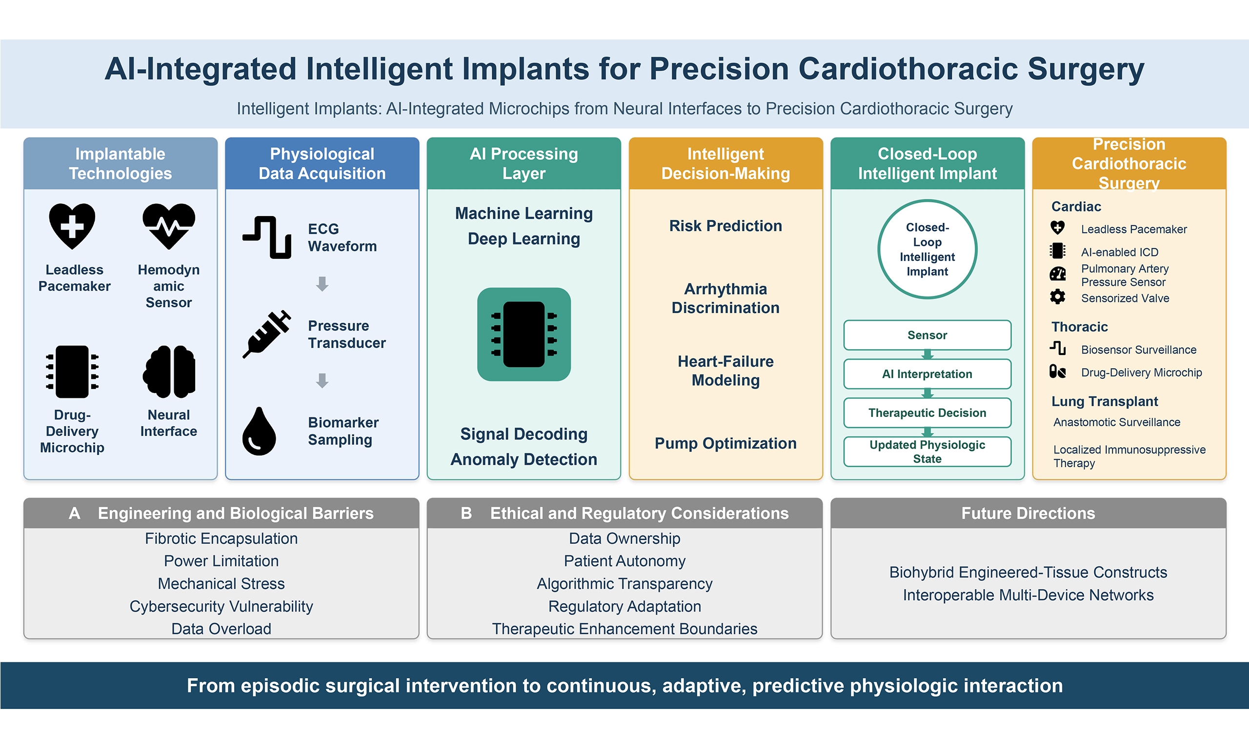
The integration of microchips into the human body has progressed from passive identification devices to intelligent, implantable systems capable of sensing, computation, stimulation, and therapeutic delivery. Advances in microelectronics, wireless power transfer, biomaterials, and artificial intelligence (AI) have enabled the development of brain-computer interfaces, implantable drug-delivery platforms, and continuous biosensing systems. Increasingly, these technologies are reshaping cardiovascular and thoracic care. In cardiothoracic surgery, microchip-enabled systems are increasingly integrated into cardiac rhythm management, hemodynamic monitoring, mechanical circulatory support, structural heart interventions, and transplant surveillance. Implantable cardiac monitors, leadless pacemakers, pressure micro-sensors, AI-driven arrhythmia detection systems, and bioelectronic interfaces for autonomic modulation illustrate the convergence of microelectronics and cardiovascular therapeutics. In thoracic oncology and lung transplantation, implantable biosensors and microfluidic platforms offer emerging potential for early detection of rejection, infection, and tumor recurrence. This review examines the technological foundations of human-integrated microchips and explores their multidisciplinary applications, with particular emphasis on cardiothoracic surgery. Engineering challenges, biocompatibility constraints, cybersecurity concerns, and ethical implications are analyzed. As AI transforms these devices from passive hardware into adaptive, closed-loop therapeutic systems, the future of cardiothoracic surgery may increasingly depend on intelligent implants capable of real-time physiologic interpretation and precision intervention.
Keywords
1. INTRODUCTION
The integration of microchips into the human body represents a major inflection point in modern medicine, enabling a shift from episodic intervention to continuous physiologic interaction[1]. Early subcutaneous radiofrequency identification devices have evolved into sophisticated implantable systems capable of neural recording, targeted stimulation, wireless communication, and real-time data processing[2]. Advances in semiconductor miniaturization, flexible electronics, microfluidics, and biocompatible materials have accelerated the development of devices that can function chronically within complex biological environments[3,4]. In parallel, artificial intelligence (AI) has emerged as a critical enabling layer, transforming implantable microchips from passive components into adaptive, closed-loop systems[5].
While much public attention has focused on neural interfaces, the cardiovascular system has been one of the earliest and most impactful domains of human-microchip integration. Contemporary cardiothoracic practice already depends heavily on implantable electronic technologies. These systems represent foundational examples of bioelectronic medicine - devices that sense physiologic signals and deliver targeted therapy within the body.
Recent advances extend far beyond traditional rhythm management. Indeed, new developments in implantable cardiovascular technologies reflect a broader trend toward device miniaturization, continuous physiologic monitoring, and increasingly sophisticated data analysis capabilities[6]. Modern systems integrate microelectronic sensors and computational algorithms that enable real-time assessment of cardiovascular function, remote disease management, and improved diagnostic and therapeutic precision across multiple domains of cardiac care[7].
AI plays a pivotal role in this evolution. For the purposes of this review, the term “artificial intelligence” is used as an umbrella term encompassing data-driven computational approaches, primarily machine learning (ML) and its deep learning (DL) subfield[8]. Where relevant, specific architectures such as convolutional neural networks (CNNs), long short-term memory (LSTM) networks, and transformer models are discussed individually according to their clinical applications.
Modern implantable systems generate high-volume, high-frequency physiologic data streams that exceed human interpretive capacity. AI algorithms enable signal decoding, anomaly detection, predictive modeling, and dynamic therapy adjustment[9]. In cardiothoracic surgery, this convergence raises the possibility of fully closed-loop systems capable of autonomously modulating pacing, adjusting ventricular assist device parameters, or titrating pharmacologic delivery based on continuous physiologic feedback. Figure 1 illustrates the evolution of intelligent implantable systems from conventional devices to AI-enabled closed-loop therapeutics.
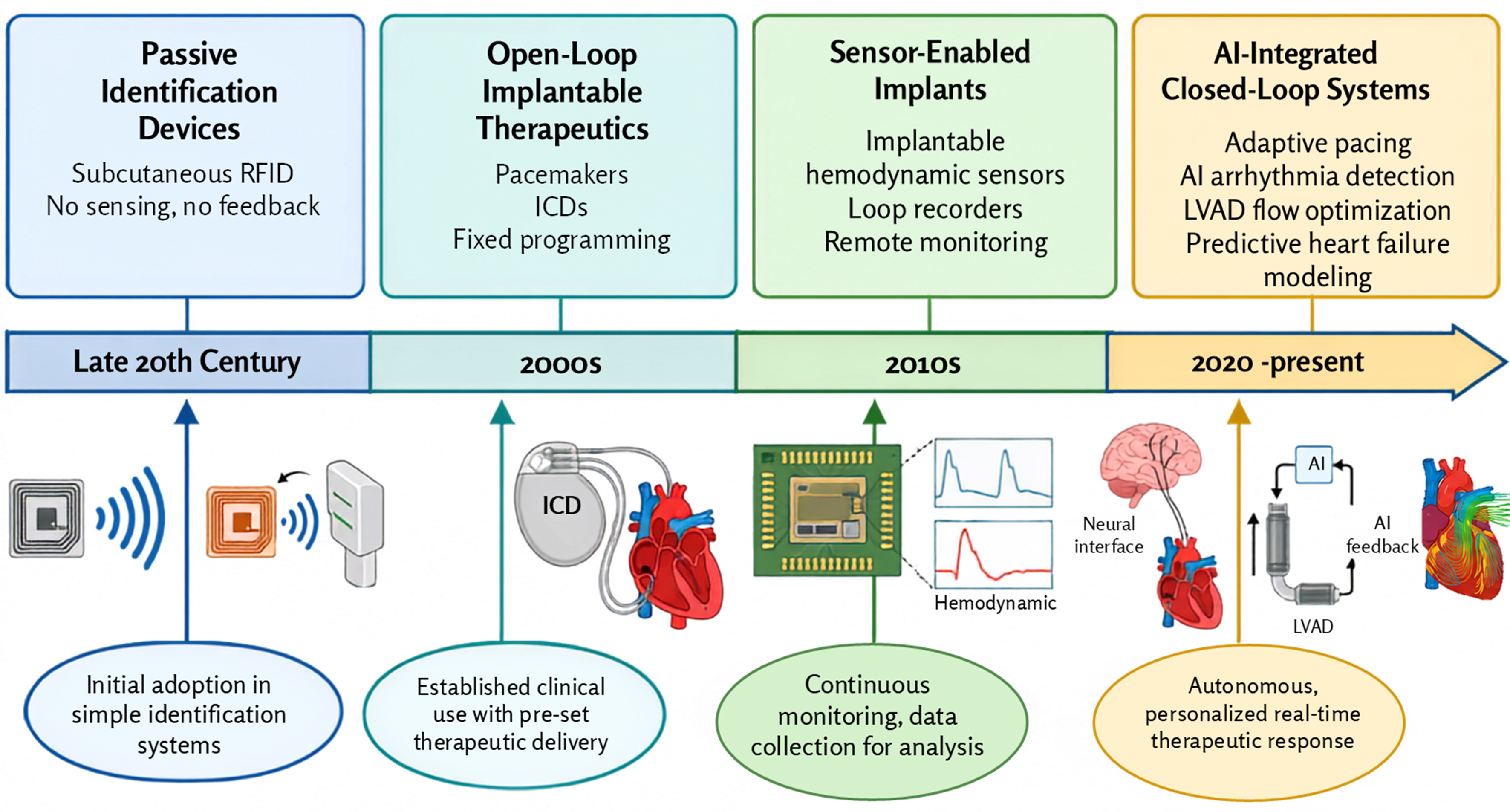
Despite these advances, substantial challenges remain. Long-term biocompatibility, device durability in high-motion cardiac environments, power constraints, cybersecurity risks, and regulatory oversight represent critical barriers[12]. Ethical considerations, including data ownership, patient autonomy, algorithmic transparency, and the boundary between therapy and enhancement, are increasingly relevant as implantable systems gain cognitive and adaptive capabilities[13].
As microchip technologies mature in the era of AI, cardiothoracic surgery stands at the intersection of bioengineering and digital intelligence. The field is uniquely positioned to shape the development of intelligent implants that not only support cardiac and thoracic function, but actively interpret and respond to physiologic signals in real time. This review examines the technological foundations of human-integrated microchips, explores their multidisciplinary applications with emphasis on cardiothoracic surgery, and outlines future directions that may redefine surgical therapeutics in the age of AI.
2. HISTORICAL EVOLUTION OF HUMAN MICROCHIP INTEGRATION
The history of human-integrated microchip technologies is closely intertwined with the development of implantable cardiac rhythm management devices, with the major technological milestones and their clinical implications summarized in Table 1. The first fully implantable cardiac pacemaker was introduced in the late 1950s and represented one of the earliest clinical applications of long-term implantable electronic devices[10]. Early pacemakers relied on relatively simple circuitry and battery systems, delivering fixed-rate electrical stimulation to maintain cardiac rhythm in patients with complete atrioventricular block or severe bradyarrhythmias.
Historical progression of implantable cardiac microelectronic systems.
| Era | Device type | Technological characteristics | Clinical impact |
| Early Era[10] | Single-chamber pacemakers | Fixed-rate pacing | Basic rhythm support |
| Intermediate[14,15] | ICD, CRT | Arrhythmia detection + resynchronization | Reduced sudden death |
| Sensor Era[16-18] | Hemodynamic sensors | Continuous pressure monitoring | Remote HF management |
| AI Era[19,20] | Adaptive closed-loop systems | Predictive modeling + auto-adjustment | Precision physiologic control |
Subsequent technological advances during the 1970s and 1980s led to the development of demand-based pacemakers capable of sensing intrinsic cardiac activity and delivering stimulation only when necessary[21]. The introduction of implantable cardioverter-defibrillators (ICDs) in the 1980s further expanded the role of implantable electronics in cardiovascular therapeutics by enabling detection and termination of life-threatening ventricular arrhythmias[14]. Cardiac resynchronization therapy subsequently emerged as a therapeutic strategy for patients with heart failure and ventricular conduction delay, demonstrating that implantable devices could actively modulate cardiac physiology to improve hemodynamic performance and clinical outcomes[15].
Throughout the late 20th and early 21st centuries, improvements in semiconductor miniaturization and battery technology facilitated the development of smaller, more durable devices capable of increasingly sophisticated sensing and computational functions. Implantable loop recorders were developed to enable long-term cardiac rhythm surveillance in patients with unexplained syncope or cryptogenic stroke[11].
More recently, a major technological transition has occurred from transvenous lead-based systems toward leadless and wireless platforms. Leadless pacemakers represent a miniaturized intracardiac device that can be delivered via catheter without the need for transvenous leads or subcutaneous generator pockets, thereby reducing infection risk and lead-related complications. Clinical studies have demonstrated high implantation success rates and favorable long-term safety profiles for leadless pacing technologies compared with conventional systems[22,23].
3. CURRENT IMPLANTABLE TECHNOLOGIES ACROSS MEDICINE
3.1. Neural and neurostimulation systems
Neurostimulation technologies represent one of the most rapidly advancing domains of implantable bioelectronics. Deep brain stimulation has become an established therapeutic intervention for neurological disorders including Parkinson disease, essential tremor, and dystonia[24]. These systems consist of intracranial electrodes connected to implantable pulse generators capable of delivering controlled electrical stimulation to targeted neural circuits. By modulating abnormal neuronal activity, deep brain stimulation can significantly improve motor symptoms and quality of life in affected patients.
Beyond traditional neurostimulation, brain-computer interfaces have emerged as a transformative technology capable of establishing bidirectional communication between neural networks and electronic systems. Implantable neural interfaces can record neuronal activity and translate neural signals into digital commands, enabling control of external devices such as prosthetic limbs or communication systems[25,26]. Conversely, electrical stimulation delivered through neural interfaces can restore sensory perception or modulate neural circuits involved in movement and cognition[27].
The relevance of neural implants to cardiothoracic medicine lies in the growing understanding of neurocardiac interactions. The autonomic nervous system plays a central role in regulating cardiac rhythm, myocardial contractility, and vascular tone. Neuromodulation strategies targeting autonomic pathways have been investigated as potential therapies for heart failure and arrhythmias[28].
These translational links become more concrete when weighed against specific, unmet needs in cardiothoracic surgical practice. Chronic post-thoracotomy and post-sternotomy pain remains common after cardiac and thoracic operations and is attributed largely to intercostal nerve injury during retraction, cannulation, or rib spreading, a problem current analgesic strategies address incompletely[29]. Closed-loop spinal cord stimulation systems, which sense evoked compound action potentials and titrate stimulation amplitude in real time, have recently been shown to operate safely alongside ICDs, illustrating a direct extension of neural-interface technology into a surgical pain problem that pharmacologic approaches alone cannot solve[30].
A second unmet need concerns neurocognitive protection during mechanical circulatory support. Acute brain injury affects up to one-third of patients supported with venoarterial ECMO, and stroke remains a major complication of left ventricular assist devices (LVADs), yet current mechanical circulatory support (MCS) control algorithms optimize pump flow and thrombosis detection rather than cerebral perfusion[31]. Continuous cerebral autoregulation monitoring, which uses near-infrared spectroscopy-derived indices to identify a patient-specific optimal blood pressure range, is a neural-interface-adjacent technology that could in principle be integrated into a closed loop with MCS controllers, adjusting flow parameters to real-time cerebral perfusion rather than systemic hemodynamics alone, a capability existing devices do not offer[31].
A third area concerns cardiac denervation after heart transplantation. Because the transplanted heart loses its afferent and efferent autonomic connections, recipients exhibit an elevated resting heart rate, a blunted chronotropic response to exercise, and loss of anginal warning of graft ischemia; sympathetic reinnervation, when it occurs, is partial, delayed by months to years, and remains incomplete in the majority of recipients[32]. Autonomic Neuromodulation strategies developed for neurocardiac disease could plausibly be adapted to accelerate or substitute for this reinnervation process, representing a concrete cardiothoracic surgical application that conventional BCI- or DBS-oriented neural interface research does not currently address.
Despite this translational potential, several limitations temper enthusiasm for neural-interface technologies in cardiothoracic applications. Electrode-tissue interfaces are subject to chronic gliosis and fibrous encapsulation, which progressively increase impedance and attenuate signal fidelity over months to years, and brain-computer interface trials report substantial variability in long-term signal stability, with some systems requiring recalibration within the first year[33,34].
It also remains unresolved how a neural interface implanted in or near the thorax would tolerate the mechanical stresses of cardiac and respiratory motion over years of use, an environment considerably more dynamic than the cranial cavity in which most existing evidence has been generated. Furthermore, established, less invasive alternatives already exist for some of the same physiological targets: catheter-based renal or pulmonary denervation achieves durable autonomic modulation without a permanently implanted neural device, and recent meta-analyses confirm meaningful and sustained blood-pressure reduction with this approach[35].
Whether an implantable neural interface can offer a clinically meaningful advantage over these established, reversible, and comparatively inexpensive catheter-based techniques, rather than simply a more complex alternative, remains an open question that the cardiothoracic surgical literature has yet to address.
3.1.1. Implantable drug delivery microchips
Implantable microchip-based drug delivery systems represent another emerging application of microelectronics in medicine. These devices typically consist of microfabricated reservoirs capable of storing pharmacologic agents and releasing them in a controlled manner through programmable electronic
Programmable drug delivery microchips have been investigated for a variety of therapeutic applications including hormone replacement therapy, oncology, and chronic disease management[38]. The ability to electronically control drug release allows physicians to adjust dosing schedules remotely and may enable personalized pharmacotherapy based on physiologic monitoring data. Such technologies may have particular relevance for transplant medicine, where precise titration of immunosuppressive therapy is essential for balancing rejection risk against drug toxicity[39].
3.2. Implantable biosensors and microfluidic platforms
Implantable biosensors represent a rapidly expanding field within biomedical engineering. These devices incorporate microelectromechanical sensors capable of detecting physiologic signals such as pressure, oxygen concentration, metabolic metabolites, or inflammatory biomarkers. Advances in microfabrication techniques have enabled the development of sensors with extremely small dimensions, allowing implantation within vascular structures, organs, or engineered tissue constructs[40,41].
Microfluidic technologies further enhance the capabilities of implantable biosensors by enabling continuous sampling and analysis of biological fluids. Particularly, microfluidic channels integrated within implantable devices can analyze biomarkers in blood, interstitial fluid, or lymphatic fluid, potentially enabling real-time monitoring of disease processes at the molecular level[42].
Such technologies have the potential to transform clinical monitoring by shifting from intermittent laboratory testing toward continuous physiologic surveillance. Indeed, early detection of biochemical changes may allow clinicians to intervene before the onset of overt clinical deterioration.
A recent computational framework combining IoT-enabled implantable biosensors with CNN classification demonstrated 97.2% accuracy in distinguishing cancer biomarker patterns (including HER2 and CEA), illustrating how AI-integrated sensing could extend beyond cardiothoracic applications into oncologic surveillance, though this proof-of-concept has not yet been validated in fabricated devices or patients[43].
As with neural interfaces, implantable drug-delivery microchips carry limitations that warrant explicit acknowledgment. Most current designs use a fixed drug reservoir and are single-use, requiring surgical removal and replacement once the reservoir is depleted, which limits their suitability for indefinite chronic therapy. Fibrotic capsule formation around the implanted device is a further, well-documented problem, progressively impeding drug diffusion into surrounding tissue and altering release kinetics in ways that are difficult to predict prospectively[44].
Refillable and antifibrotic-coated designs are under active development, but none has yet reached routine clinical use in cardiothoracic practice. Compared with conventional systemic pharmacotherapy, implantable microchips offer more precise, programmable, and localized dosing, but at the cost of a surgical implantation procedure, a finite device lifespan, and a substantially higher unit cost; for most cardiothoracic indications where oral or intravenous therapy remains effective, this trade-off has not yet been shown to justify routine adoption outside investigational settings[38,44].
Taken together, the neural interface, biosensing, and drug-delivery technologies described above represent the core technological building blocks from which cardiothoracic-specific intelligent implants are assembled. Neurostimulation platforms provide mechanisms for autonomic and pain modulation; implantable biosensors provide the continuous physiologic sensing layer needed to detect deterioration before it becomes clinically overt; and programmable drug-delivery microchips provide a localized, adjustable therapeutic effector. The remainder of this review examines how these building blocks have been combined, individually and in tandem, into the cardiac rhythm management devices, hemodynamic monitors, mechanical circulatory support systems, and structural, thoracic, and transplant applications that define the current state of intelligent implants in cardiothoracic surgery.
3.3. Cardiothoracic applications
The cardiothoracic domain represents one of the most mature and clinically impactful applications of implantable microelectronic technologies across cardiothoracic surgery. Cardiac rhythm management devices remain among the most widely used implantable electronic systems. Contemporary pacemakers and ICDs incorporate advanced sensing algorithms capable of detecting complex arrhythmias and delivering targeted therapy through pacing or defibrillation[45], as summarized in Table 2. Leadless pacemakers have further reduced device complexity by eliminating transvenous leads and generator pockets while maintaining effective pacing capability[23].
Current intelligent implant applications in cardiothoracic surgery
| Application | Implant type | Timing | AI integration | Clinical objective |
| Rhythm management | Leadless pacemaker | Long-term postoperative management | Arrhythmia discrimination | Prevent bradyarrhythmia |
| Sudden death prevention | Subcutaneous ICD | Long-term postoperative management | Shock optimization algorithms | Avoid inappropriate shocks |
| Heart failure | PA pressure sensor | Long-term postoperative management | Predictive decompensation modeling | Early intervention |
| Mechanical support | LVAD embedded sensors | Long-term postoperative management | Flow and thrombosis detection | Optimize circulatory support |
| Transplantation | Biosensor platforms | Long-term postoperative management | Rejection biomarker detection | Early graft surveillance |
| Valve deployment guidance[46,47] | Sensor-integrated delivery catheter | Intraoperative | Positional/apposition feedback | Reduce malposition and paravalvular leak |
| Graft flow assessment[48] | Transit-time flow probe | Intraoperative | Flow/PI pattern recognition | Detect inadequate anastomosis intraoperatively |
| Airway anastomosis surveillance[49,50] | Implantable tissue-oximetry probe | Early postoperative surveillance | Perfusion trend analysis | Flag impending bronchial dehiscence |
| Esophageal anastomotic leak[51] | Peri-anastomotic microdialysis probe | Early postoperative surveillance | Lactate trend pattern recognition | Early leak warning before clinical onset |
AI has increasingly been integrated into arrhythmia detection algorithms to improve diagnostic accuracy[52,53]. CNNs and LSTM networks have been among the most widely adopted deep-learning architectures for this task, extracting both morphological features (e.g., QRS width, intracardiac electrogram amplitude, and slew rate) and temporal features (e.g., RR-interval dynamics and heart-rate variability), to distinguish pathologic ventricular arrhythmias from supraventricular rhythms or benign electrical noise[54-56]. Transformer-based architectures, which apply multi-head self-attention to capture long-range dependencies across the full signal window, have recently emerged as a promising next-generation approach for long-sequence rhythm classification, with several recent studies reporting improved performance in complex arrhythmia classification[52,57].
Another important milestone in implantable microelectronics has been the emergence of remote physiologic monitoring systems. Wireless pulmonary artery pressure sensors enable continuous hemodynamic surveillance in patients with heart failure by transmitting pressure measurements from an implanted sensor directly to clinicians. In the pivotal CHAMPION trial, this pressure-guided management strategy reduced heart failure hospitalizations by 39% compared with standard care over a mean follow-up of 15 months, and subsequent large real-world post-approval registry data have confirmed a similarly substantial reduction in hospitalization rates outside the trial setting[16-18]. Continuous hemodynamic surveillance enables earlier identification of physiologic deterioration, allowing proactive therapeutic adjustments before overt clinical decompensation and hospitalization occur.
Mechanical circulatory support systems also incorporate extensive microelectronic integration. Modern LVADs rely on embedded sensors and control algorithms to regulate pump speed, maintain physiologic flow conditions, and detect abnormal operating states such as suction events or thrombosis[58,59]. These systems represent an early example of closed-loop bioelectronic therapy in cardiovascular medicine.
Emerging applications extend into structural heart interventions. Sensor-integrated prosthetic valves and grafts are being investigated as a means of monitoring hemodynamic performance and detecting early structural deterioration[60,61].
Beyond post-implant surveillance, intelligent implants are also beginning to inform the surgical act itself. Sensor-integrated delivery catheters that combine intravascular ultrasound or impedance sensing with the valve-deployment mechanism can provide real-time positional and apposition feedback during transcatheter valve implantation, complementing angiographic guidance and potentially reducing malpositioning and paravalvular leak, which remain among the most common intraprocedural complications of transcatheter valve replacement[46].
Intraoperative graft assessment represents another distinctly surgical, rather than cardiological, application. Transit-time flow measurement is used at the time of coronary artery bypass grafting to quantify mean graft flow, pulsatility index, and diastolic filling, allowing the surgeon to identify technically inadequate anastomosis or graft spasm before chest closure, when revision is still straightforward. Embedding this flow-sensing capability directly into the graft or anastomotic site, with automated pattern recognition to flag values associated with early graft failure, would extend a well-established intraoperative quality-assessment tool into a continuous, AI-supported perioperative monitoring system[48].
Thoracic surgery and transplant medicine are also entering a phase of rapid technological evolution driven by advances in implantable bioelectronics. Implantable sensing platforms capable of monitoring biochemical and physiologic signals may enable earlier identification of complications such as graft dysfunction, infection, or tumor recurrence[8,62]. In parallel, implantable microfluidic technologies are being explored for localized therapeutic delivery, potentially allowing more precise administration of immunomodulatory or anticancer agents[63,64]. Future biohybrid approaches may combine engineered tissues with integrated sensing technologies, creating constructs capable of continuously assessing tissue function and viability after implantation[65].
Two further surgical scenarios illustrate the translational gap that intelligent implants could close. First, healing of the bronchial anastomosis has long been considered the Achilles’ heel of lung transplantation: the donor bronchus loses its systemic arterial supply at the time of transplant, and airway complications, including anastomotic dehiscence, affect a substantial proportion of recipients, most often in the first weeks after surgery when bronchoscopic surveillance remains the primary monitoring tool[66]. Continuous microvascular perfusion sensing technologies developed for free-flap monitoring in reconstructive surgery, such as implantable or attached tissue-oximetry probes[50], represent a directly analogous engineering solution that has not yet been adapted to the bronchial anastomosis, where early perfusion data could flag impending ischemia before endoscopic or radiographic signs appear[66,67].
Second, esophagogastric anastomotic leak remains one of the most feared complications of esophagectomy, with delayed clinical recognition contributing substantially to morbidity and mortality. Peri-anastomotic microdialysis probes, placed on both the esophageal and gastric sides of the anastomosis to sample local tissue lactate in real time, have already been used in esophagectomy patients and can flag the metabolic signature of impaired perfusion before a leak becomes clinically or radiographically apparent[51].
Beyond implantable sensing, AI is also beginning to influence postoperative therapeutic management in lung transplantation. Although current AI applications predominantly focus on predictive analytics rather than implantable systems, recent LSTM-based models have demonstrated accurate forecasting of individualized tacrolimus dosing requirements in lung transplant recipients, illustrating how AI-guided therapeutic decision-making could ultimately be integrated with continuous implantable biosensing to enable closed-loop immunosuppressive management following lung transplantation[68].
4. AI AS AN ENABLING LAYER
AI represents a critical technological layer transforming implantable microelectronic systems from passive therapeutic devices into adaptive physiologic interfaces. Figure 2 illustrates how AI enhances signal interpretation and therapeutic decision-making within implantable cardiac devices. Implantable cardiovascular devices generate large volumes of high-frequency physiologic data, including intracardiac electrograms, hemodynamic pressure signals, device performance metrics, and patient activity patterns. Traditional rule-based algorithms embedded in early implantable devices relied on predetermined thresholds to detect arrhythmias or trigger therapy[53].
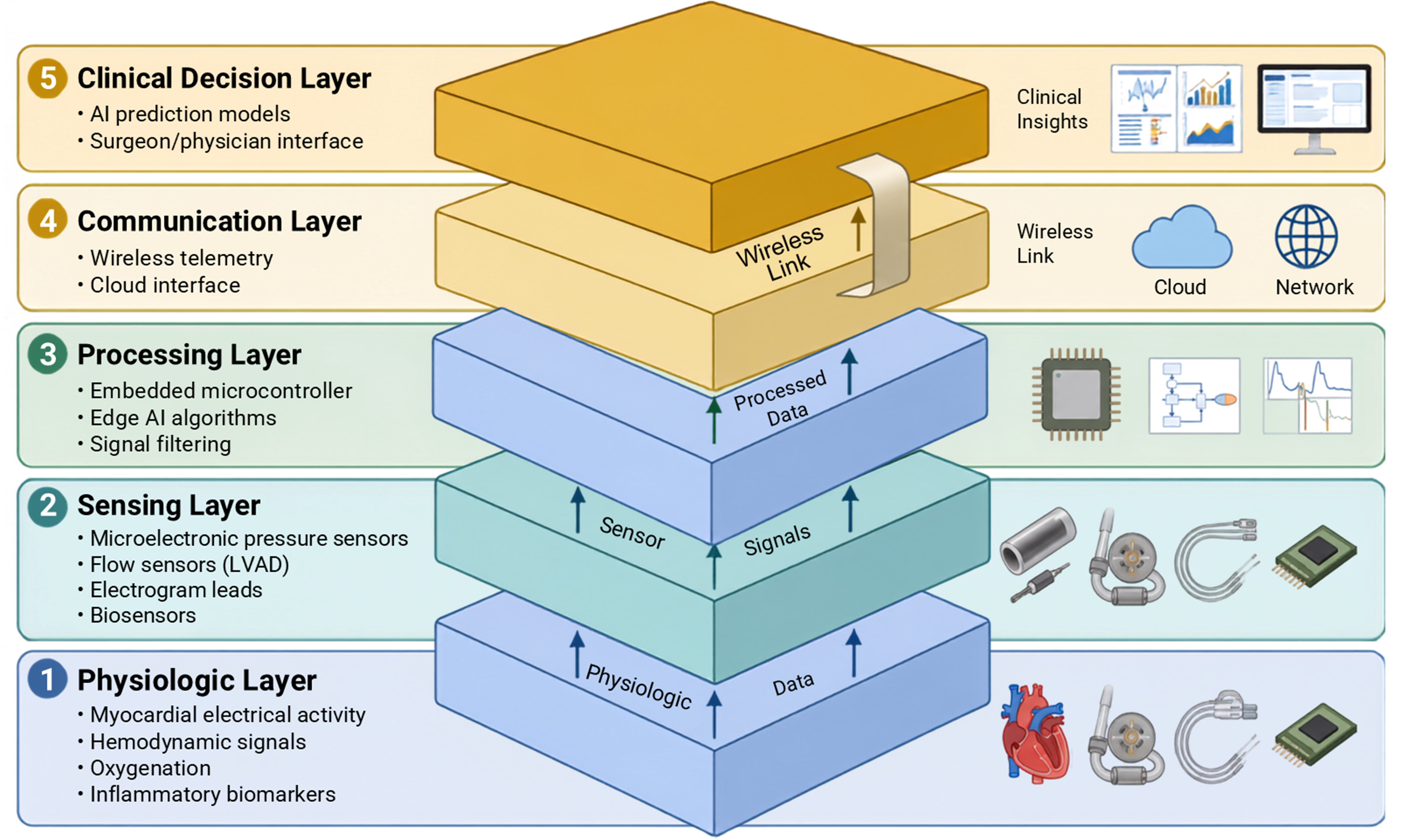
Figure 2. Architecture of an intelligent cardiothoracic implant ecosystem. AI: Artificial intelligence; LVAD: left ventricular assist device.
In contrast, contemporary ML approaches enable more sophisticated pattern recognition, predictive modeling, and dynamic therapeutic modulation based on continuously evolving physiologic signals[55]. In cardiac rhythm management, AI has been applied to improve arrhythmia discrimination algorithms within ICDs and pacemakers[52]. ML models trained on large datasets of intracardiac electrograms can distinguish ventricular tachyarrhythmias from supraventricular rhythms or electrical noise with greater accuracy than earlier rule-based systems[54]. Improved arrhythmia classification has the potential to reduce inappropriate shocks and optimize device-mediated therapy delivery, a critical consideration given the psychological and physiologic consequences of unnecessary defibrillation therapy[69].
AI also enables predictive modeling in heart failure management using data streams derived from implantable hemodynamic sensors. Continuous pulmonary artery pressure monitoring systems provide granular hemodynamic measurements that can be integrated into predictive algorithms capable of identifying early physiologic deterioration before the onset of symptomatic decompensation[70]. Clinical trials evaluating pulmonary artery pressure-guided management strategies have demonstrated reductions in heart failure hospitalization when therapy is adjusted according to implant-derived hemodynamic data[16,71].
Mechanical circulatory support systems represent another domain in which AI may enable adaptive device control. Modern LVADs incorporate multiple sensors that monitor pump speed, motor current, pressure gradients, and flow characteristics[58]. Integration of ML algorithms into device controllers may allow automated detection of pump thrombosis, suction events, or hemodynamic instability, enabling real-time adjustments in pump performance[19]. Such adaptive control systems may represent an early example of closed-loop cardiovascular bioelectronics in which physiologic sensing directly modulates device therapy.
The computational architecture supporting AI-enabled implants may be distributed across multiple layers. Edge computing strategies incorporate lightweight algorithms directly within implantable hardware to enable real-time decision making without external data transmission[72]. Alternatively, cloud-based analytic platforms can process larger datasets derived from multiple implantable devices across patient populations, allowing development of predictive models that can be updated over time and deployed across device networks[73].
Human-AI interaction remains a central consideration in the deployment of intelligent implants. While automated algorithms can process physiologic data at a scale and speed beyond human capacity, clinical oversight remains essential for interpreting algorithmic outputs and integrating them within the broader context of patient care. As implantable devices increasingly incorporate autonomous or semi-autonomous decision-making capabilities, the relationship between algorithmic control and physician supervision will become a defining feature of future bioelectronic medicine[20].
5. ENGINEERING AND BIOLOGICAL BARRIERS
Despite rapid technological progress, the development of long-term implantable microchip systems remains constrained by multiple engineering and biological challenges, which are summarized together with their underlying mechanisms and clinical consequences in Table 3. Biocompatibility represents one of the most fundamental limitations[74]. Implantable devices inevitably trigger a foreign body response characterized by inflammatory cell recruitment, fibrotic encapsulation, and tissue remodeling around the device interface. Fibrotic capsule formation can interfere with sensor performance by altering analyte diffusion or attenuating physiologic signals, thereby reducing the accuracy of implantable biosensors[75].
Engineering and biological constraints in cardiothoracic intelligent implants
| Challenge | Mechanism | Clinical consequence |
| Fibrotic encapsulation[74,75] | Foreign body response | Reduced sensor accuracy |
| Power limitation[76,77] | Battery constraints | Limited longevity |
| Mechanical stress[78,79] | Cardiac motion | Device fatigue/failure |
| Cybersecurity[80-82] | Wireless vulnerability | Malicious interference risk |
| Data overload[83,84] | High-frequency streams | Interpretability challenges |
Mitigation strategies are an active area of investigation: antifibrotic-coated devices, such as the tranilast-eluting microchannel systems described earlier[44], and zwitterionic hydrogel coatings that resist protein adsorption and cellular attachment have demonstrated promising reductions in fibrotic capsule formation in preclinical studies, with one recent study reporting over a 60% reduction in capsule thickness compared with an uncoated surface[85].
The dynamic mechanical environment of the thoracic cavity introduces additional challenges for device durability. The heart undergoes continuous cyclic motion, generating mechanical stress on implanted hardware and electrical leads. Repetitive mechanical forces can contribute to device fatigue, lead fracture, insulation degradation, and sensor drift over time[78]. Advances in flexible electronics and polymeric encapsulation materials aim to improve device resilience in high-motion biologic environments[79].
Power supply and energy transfer remain central engineering constraints in implantable microelectronics. Traditional implantable cardiac devices rely on lithium-based batteries with limited operational lifespan, necessitating periodic device replacement procedures[76]. Alternative strategies including wireless power transfer, inductive coupling, and energy harvesting from physiologic motion are under investigation as potential methods for extending device longevity[77]. Fully implantable systems, such as total artificial hearts, have demonstrated the feasibility of transcutaneous energy transfer systems capable of recharging internal batteries without transcutaneous wires, thereby reducing infection risk[86].
Cybersecurity represents an increasingly important consideration as implantable devices become connected to external monitoring networks. Modern implantable cardiac devices incorporate wireless communication systems that allow remote monitoring and device reprogramming. Although these features enhance clinical management, they also introduce potential vulnerabilities to unauthorized access or malicious interference. Theoretical studies have demonstrated that ICDs could potentially be subjected to remote reprogramming attacks capable of altering therapy parameters or inducing inappropriate shocks if adequate security protections are not implemented[80]. More recent countermeasures have moved beyond encryption alone: body-coupled communication techniques confine wireless signals to conduction through the tissue itself rather than open-air radiofrequency transmission, reducing the effective interception range from several meters to roughly a centimeter from the skin[81]. Lightweight authentication protocols tailored to the power and processing constraints of implantable hardware are being developed to secure device-reprogramming commands without exceeding a device’s limited energy budget[82].
Another critical challenge lies in interpreting the large volumes of physiologic data generated by implantable systems. Continuous monitoring devices can produce vast datasets that may overwhelm conventional clinical workflows if not appropriately filtered and interpreted[83]. AI may help address this challenge by identifying clinically meaningful patterns within large physiologic datasets, but the development of robust algorithms capable of operating reliably across diverse patient populations remains an active area of research[84].
6. ETHICAL AND REGULATORY CONSIDERATIONS
The integration of intelligent implantable microchips into human physiology raises complex ethical and regulatory considerations, including issues related to data governance, patient autonomy, algorithmic transparency, and regulatory oversight, as summarized in Table 4. Continuous physiologic monitoring generates detailed personal health data that may include information about cardiac rhythm, hemodynamic status, physical activity patterns, and other physiologic parameters. Determining ownership and governance of these data represents a major ethical challenge. Patients, healthcare providers, device manufacturers, and digital health platforms may all have potential interests in accessing and analyzing implant-derived data[87].
Ethical and regulatory considerations in AI-integrated implantable devices
| Domain | Key issue | Implication |
| Data ownership[87,88] | Continuous physiologic surveillance | Privacy concerns |
| Autonomy[89] | AI-driven therapeutic decisions | Reduced human oversight |
| Transparency[90] | Algorithm interpretability | Trust and accountability |
| Regulation[90] | Adaptive AI approval pathways | Evolving oversight models |
| Enhancement[91] | Beyond-therapeutic applications | Ethical boundary ambiguity |
This challenge is compounded by the fact that existing governance frameworks may not yet be adequate to the task: a 2025 survey of healthcare, IT, and blockchain professionals evaluating ISO, GDPR, and HIPAA compliance mechanisms found persistent dissatisfaction with their effectiveness across data encryption, access control, audit trails, and consent management, suggesting that regulatory frameworks designed for conventional electronic health records may require substantial adaptation for continuously streaming, implant-derived physiologic data[88]. Unlike a single clinical encounter, an implantable device generates a persistent data stream whose ownership, secondary use, and commercial value remain unresolved questions that current health-data law addresses only partially.
Autonomy represents another important ethical dimension in the context of closed-loop implantable systems. Devices capable of automatically adjusting therapy based on physiologic inputs introduce a degree of algorithmic control over patient physiology. While such automation may improve therapeutic precision, it also raises questions about the appropriate balance between automated device function and human decision-making authority. For example, ICDs deliver potentially life-saving but painful shocks in response to detected arrhythmias, and the criteria used by device algorithms to trigger therapy must be carefully validated and clinically justified[89].
Transparency of AI algorithms is particularly important for life-sustaining devices such as pacemakers, ventricular assist devices, and defibrillators. Clinicians must be able to understand the operational logic of algorithms that influence therapeutic decisions, particularly when these systems operate autonomously within the body. Regulatory frameworks for medical devices have historically been designed around static hardware and software configurations[90]. Adaptive ML algorithms that evolve over time pose new regulatory challenges because their behavior may change as additional data are incorporated into training models.
The distinction between therapeutic and enhancement applications also warrants ethical consideration. Implantable microelectronic technologies developed for medical treatment may potentially be adapted for human enhancement applications, including augmentation of cognitive performance, physical endurance, or sensory perception[91]. The ethical boundaries governing such applications remain the subject of ongoing debate within bioethics and regulatory policy.
7. TOWARD BIOHYBRID AND INTELLIGENT CARDIOTHORACIC SYSTEMS
Emerging developments in tissue engineering, bioelectronics, and AI are beginning to converge toward the concept of biohybrid cardiothoracic systems[92]. These platforms combine living biological tissues with embedded electronic sensing and control systems capable of monitoring physiologic function in real time. One example involves engineered myocardial patches designed to repair damaged cardiac tissue following myocardial infarction[93]. Integration of microelectronic sensors within such constructs could enable continuous monitoring of electrical activity, contractile function, and metabolic status within engineered tissues.
Similarly, tissue-engineered vascular grafts and prosthetic conduits may eventually incorporate embedded sensors capable of detecting flow dynamics, pressure gradients, or early structural degeneration[94]. Such “smart grafts” could provide early warning signals indicating graft failure, thrombosis, or infection before clinical symptoms emerge.
Neuromorphic implants represent another emerging frontier. These devices are designed to mimic neural architectures and may enable advanced forms of autonomic neuromodulation for cardiovascular disease. Modulation of vagal or sympathetic neural pathways has been explored as a therapeutic strategy for heart failure and arrhythmias, and implantable neuromorphic systems could potentially enable more precise control of autonomic cardiac regulation[95,96].
Another transformative concept involves the development of digital twins of the cardiovascular system. Digital twins are computational models that simulate the physiologic behavior of individual patients using real-time physiologic data streams. Implantable sensors could continuously feed physiologic data into patient-specific digital models, allowing predictive simulation of disease progression and therapeutic responses[97]. Such integrated systems could eventually allow clinicians to test therapeutic strategies within computational simulations before implementing them in vivo.
Distributed implant ecosystems may ultimately represent the next stage of bioelectronics medicine. Instead of relying on single implantable devices, future systems may consist of networks of interconnected micro-sensors distributed throughout the myocardium, vasculature, and thoracic organs[98]. These sensor networks could collectively monitor physiologic parameters and coordinate therapeutic responses through integrated computational platforms.
8. FUTURE RESEARCH AGENDA
The barriers outlined above - fibrotic encapsulation, power limitation, mechanical stress, cybersecurity vulnerability, and data overload on the engineering side [Table 3], and data ownership, algorithmic transparency, and adaptive regulatory pathways on the ethical side [Table 4] - define the priorities for the research agenda that follows. Rather than representing an independent set of technological aspirations, the directions below are framed explicitly as responses to these constraints.
Future research in intelligent cardiothoracic implants will likely focus on developing integrated therapeutic systems capable of continuously sensing physiologic signals, interpreting these data through advanced computational methods, and delivering adaptive therapeutic responses. Achieving this will require concurrent progress on the power-limitation and mechanical-stress constraints identified in Table 3, since closed-loop responsiveness is only clinically useful if the underlying device can sustain years of reliable operation in a moving thoracic field. Such systems aim to move beyond passive monitoring toward closed-loop platforms in which physiologic information directly informs therapeutic adjustments, enabling more responsive and individualized disease management. Figure 3 depicts the closed-loop architecture linking physiologic sensing, AI-based data interpretation, and adaptive therapeutic intervention.
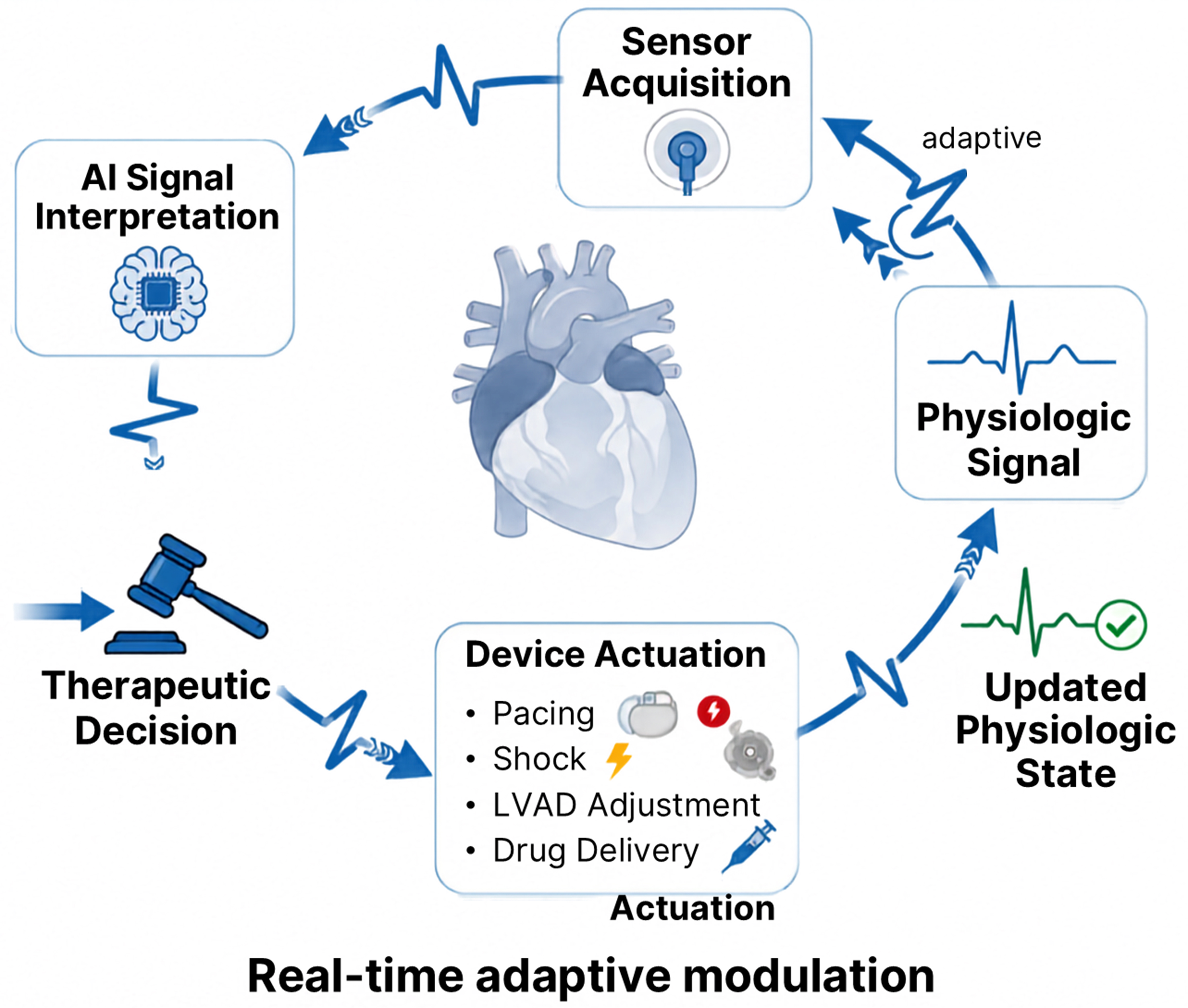
Figure 3. Closed-loop therapeutic control in AI-enhanced cardiac devices. AI: Artificial intelligence; LVAD: left ventricular assist device.
Another important research direction involves expanding the use of implantable biosensing technologies for the early detection of pathophysiologic changes within cardiovascular and thoracic systems. Progress here is contingent on mitigating fibrotic encapsulation, the mechanism identified in Table 3 as the principal driver of declining sensor accuracy over time; without durable sensor-tissue interfaces, expanded biosensing coverage will not translate into reliable long-term data. Coupling these sensing capabilities with programmable therapeutic platforms may allow dynamic adjustment of treatment strategies based on real-time biologic feedback.
Advances in sensing technologies and computational analytics may also enhance the safety and performance of implantable therapeutic devices by enabling earlier identification of device-related complications and physiologic instability[53,99]. These same analytic pipelines must be designed against the cybersecurity vulnerabilities and data-overload/interpretability challenges flagged in Table 3, since a system that improves complication detection while introducing a wireless attack surface or an uninterpretable alert burden would trade one clinical risk for another. Improved integration of physiologic monitoring with device control systems could support more precise regulation of therapeutic function and reduce the incidence of adverse events associated with long-term implantable support technologies.
As the number and complexity of implantable devices increase, the development of standardized communication frameworks will become increasingly important. Interoperability at this scale directly raises the data-ownership and regulatory questions summarized in Table 4: cross-device data sharing multiplies the number of parties with access to continuous physiologic surveillance, and current adaptive-AI approval pathways were not designed with multi-device ecosystems in mind. Interoperable systems capable of exchanging physiologic data across multiple devices and external monitoring platforms may enable coordinated networks of implantable technologies that collectively support patient monitoring and therapy, Figure 4 summarizes the future vision of biohybrid, interconnected implant networks supporting precision cardiothoracic care.
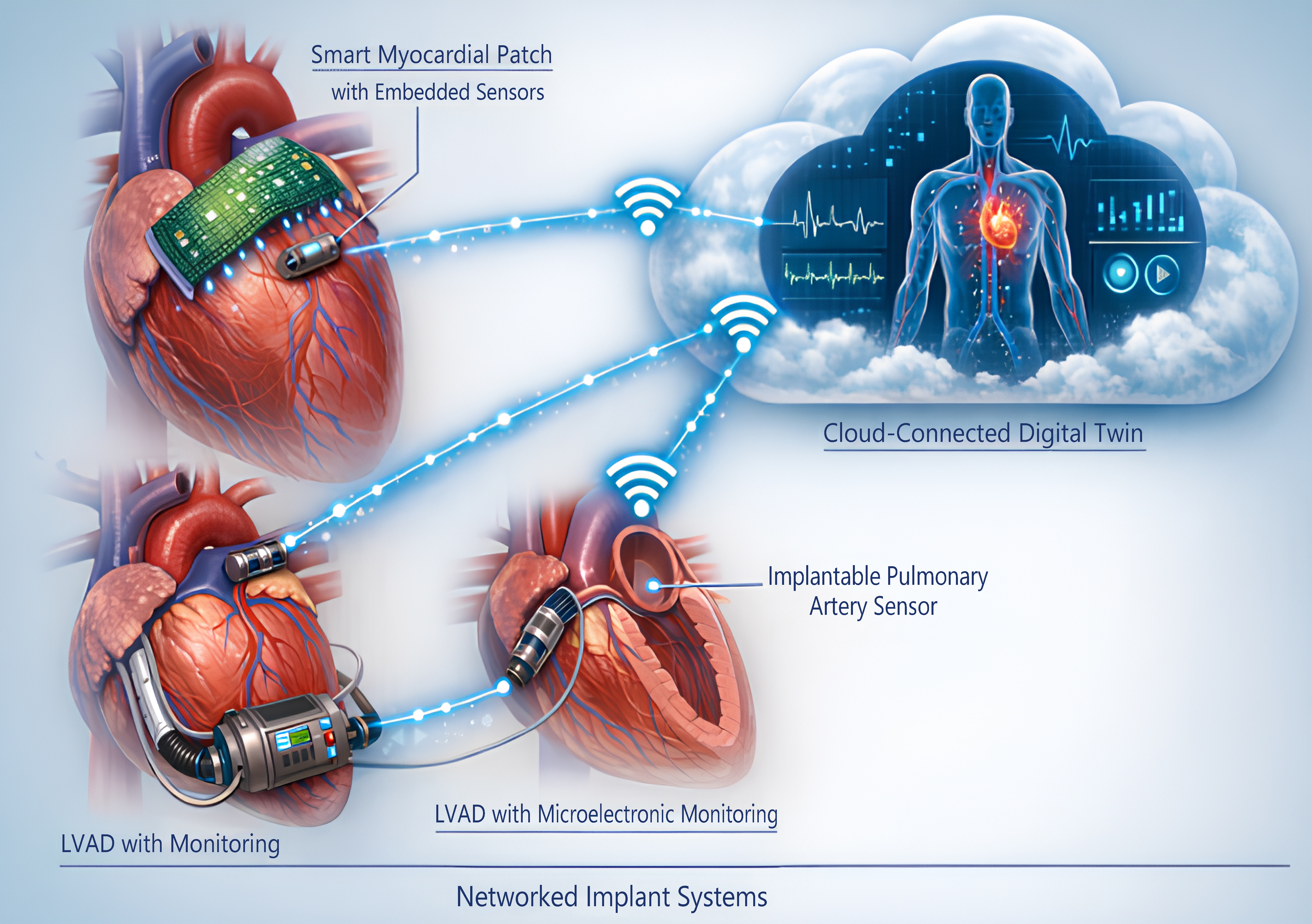
Figure 4. Future paradigm: biohybrid and distributed implant networks. LVAD: Left ventricular assist device.
9. CONCLUSION
Redefining the human-technology boundary: The integration of intelligent microelectronic implants into human physiology is redefining the relationship between medicine, technology, and the human body. In cardiothoracic surgery, implantable electronic devices have substantially advanced the management of arrhythmias, heart failure, and advanced cardiac disease. Continued advances in microelectronics, biosensing technologies, wireless communication, and AI are expanding the capabilities of these systems far beyond traditional therapeutic paradigms.
The transition from episodic surgical intervention toward continuous physiologic interaction represents a fundamental shift in the practice of cardiovascular medicine. Implantable devices are evolving from isolated hardware components into integrated physiologic interfaces capable of sensing, interpreting, and modulating biologic function in real time. AI further accelerates this transformation by enabling adaptive therapeutic systems that can respond dynamically to complex physiologic signals.
As these technologies continue to mature, the role of the cardiothoracic surgeon will extend beyond device implantation toward participation in the multidisciplinary development of intelligent bioelectronic systems. Collaboration among surgeons, engineers, computer scientists, and bioethicists will be essential for ensuring that these technologies are deployed safely, ethically, and effectively.
Ultimately, intelligent implants may redefine the boundaries between human physiology and digital technology, enabling a future in which cardiovascular care is increasingly personalized, predictive, and continuously integrated with advanced bioelectronic systems.
DECLARATIONS
Authors’ contributions
Conceptualization, methodology, validation, investigation, writing - original draft, writing - review and editing, project administration: Baudo, M.
Methodology, validation, investigation, writing - original draft, writing - review and editing, visualization: Rahouma, M.
Investigation, data curation, writing - review and editing, visualization: Abdelhemid, M.; Shenoda, D.
Investigation, literature review, writing - review and editing, assistance in preparing responses to reviewers, manuscript editing, approval of the final manuscript: Mansour, M. H.
Investigation, data curation, writing - review and editing, visualization, preparing responses to reviewers, approval of the final manuscript: Mohsen, H.
Conceptualization, methodology, validation, investigation, resources, writing - original draft, writing - review and editing, supervision, project administration: Rahouma, M.
Availability of data and materials
Not applicable.
AI and AI-assisted tools statement
During the preparation of this manuscript, Google Gemini 2.5 Flash Image (Nano Banana), released August 26, 2025, was used to assist the authors in generating and refining visual elements of Figures 1-4. Figures 1-3 were composed and finalized by the authors, with selected individual visual elements generated with assistance from Google Gemini. Figure 4 was also prepared by the authors with assistance from Google Gemini. The Graphical Abstract was created entirely by the authors using Microsoft PowerPoint, including icons from PowerPoint’s built-in icon library. No icons or graphical elements used in Figures 1-3 were obtained from third-party icon libraries or stock image platforms. The AI tool was used solely for visual/graphic assistance and did not influence the study design, data collection, analysis, interpretation, or scientific content of the work. All authors reviewed and approved the final figures and take full responsibility for their accuracy, integrity, and final content.
Financial support and sponsorship
None.
Conflict of interest
All authors declared that there are no conflicts of interest.
Ethical approval and consent to participate
Not applicable.
Consent for publication
Not applicable.
Copyright
© The Author(s) 2026.
REFERENCES
1. Kazanskiy, N. L.; Khorin, P. A.; Khonina, S. N. Biochips on the move: emerging trends in wearable and implantable lab-on-chip health monitors. Electronics 2025, 14, 3224.
2. Deshmukh, A.; Brown, L.; Barbe, M. F.; et al. Fully implantable neural recording and stimulation interfaces: peripheral nerve interface applications. J. Neurosci. Methods. 2020, 333, 108562.
3. Chandel, M.; Rosenkranz, A.; Moraru, D.; Woźniak, A.; Jastrzębska, A. M. Measuring the future - nanometrology for advanced manufacturing of miniaturized devices. Adv. Funct. Mater. 2025, 35, 2501663.
4. Corzo, D.; Tostado-Blázquez, G.; Baran, D. Flexible electronics: status, challenges and opportunities. Front. Electron. 2020, 1, 594003.
5. Zhu, B.; Shin, U.; Shoaran, M. Closed-loop neural prostheses with on-chip intelligence: a review and a low-latency machine learning model for brain state detection. IEEE. Trans. Biomed. Circuits. Syst. 2021, 15, 877-97.
6. Rav Acha, M.; Soifer, E.; Hasin, T. Cardiac implantable electronic miniaturized and micro devices. Micromachines 2020, 11, 902.
7. Lin, J.; Fu, R.; Zhong, X.; et al. Wearable sensors and devices for real-time cardiovascular disease monitoring. Cell. Rep. Phys. Sci. 2021, 2, 100541.
8. Rahouma, M.; Mohsen, H.; Mahmoud, A.; et al. Artificial intelligence (AI) applications and their impact on thoracic surgery: a narrative review. Curr. Chall. Thorac. Surg. 2025, 7, 27.
9. Yang, X.; Qi, X.; Zhou, X. Deep learning technologies for time series anomaly detection in healthcare: a review. IEEE. Access. 2023, 11, 117788-99.
10. DeForge, W. F. Cardiac pacemakers: a basic review of the history and current technology. J. Vet. Cardiol. 2019, 22, 40-50.
11. Mittal, S. Implantable loop recorders-syncope, cryptogenic stroke, atrial fibrillation. Card. Electrophysiol. Clin. 2021, 13, 439-47.
12. Zeng, Q.; Liu, H.; Zhang, Y.; et al. Materials and system design for self-decision bioelectronic systems. Adv. Mater. 2026, 38, e21164.
13. Chiurco, C.; Favaro, A.; Storti, S. F.; et al. The marriage of neurotechnologies and artificial intelligence: ethical, regulatory, and technological aspects. IEEE. Signal. Process. Mag. 2025, 42, 80-97.
14. Matchett, M.; Sears, S. F.; Hazelton, G.; Kirian, K.; Wilson, E.; Nekkanti, R. The implantable cardioverter defibrillator: its history, current psychological impact and future. Expert. Rev. Med. Devices. 2009, 6, 43-50.
15. Gorodeski, E. Z.; Magnelli-Reyes, C.; Moennich, L. A.; Grimaldi, A.; Rickard, J. Cardiac resynchronization therapy-heart failure (CRT-HF) clinic: a novel model of care. PLoS. One. 2019, 14, e0222610.
16. Abraham, W. T.; Adamson, P. B.; Bourge, R. C.; et al.; CHAMPION Trial Study Group. Wireless pulmonary artery haemodynamic monitoring in chronic heart failure: a randomised controlled trial. Lancet 2011, 377, 658-66.
17. Lander, M. M.; Aldweib, N.; Abraham, W. T. Wireless hemodynamic monitoring in patients with heart failure. Curr. Heart. Fail. Rep. 2021, 18, 12-22.
18. Shavelle, D. M.; Desai, A. S.; Abraham, W. T.; et al.; CardioMEMS Post-Approval Study Investigators. Lower rates of heart failure and all-cause hospitalizations during pulmonary artery pressure-guided therapy for ambulatory heart failure: one-year outcomes from the CardioMEMS post-approval study. Circ. Heart. Fail. 2020, 13, e006863.
19. Balcioglu, O.; Ozgocmen, C.; Ozsahin, D. U.; Yagdi, T. The role of artificial intelligence and machine learning in the prediction of right heart failure after left ventricular assist device implantation: a comprehensive review. Diagnostics 2024, 14, 380.
20. Dodda, J. M.; Kunrath, M. F.; Zhao, L. Z.; Bodaghi, M.; Yousefi, A.; Ashammakhi, N. Autonomous implants. Adv. Mater. 2025, 37, e10702.
21. Ward, C.; Henderson, S.; Metcalfe, N. H. A short history on pacemakers. Int. J. Cardiol. 2013, 169, 244-8.
22. Crossley, G. H.; Piccini, J. P.; Longacre, C.; Higuera, L.; Stromberg, K.; El-Chami, M. F. Leadless versus transvenous single-chamber ventricular pacemakers: 3 year follow-up of the Micra CED study. J. Cardiovasc. Electrophysiol. 2023, 34, 1015-23.
23. Saleem-Talib, S.; Hoevenaars, C. P. R.; Molitor, N.; et al. Leadless pacing: a comprehensive review. Eur. Heart. J. 2025, 46, 1979-90.
24. Cury, R. G.; Fraix, V.; Castrioto, A.; et al. Thalamic deep brain stimulation for tremor in Parkinson disease, essential tremor, and dystonia. Neurology 2017, 89, 1416-23.
25. Dong, Y.; Wang, S.; Huang, Q.; Berg, R. W.; Li, G.; He, J. Neural decoding for intracortical brain-computer interfaces. Cyborg. Bionic. Syst. 2023, 4, 0044.
26. Luo, S.; Rabbani, Q.; Crone, N. E. Brain-computer interface: applications to speech decoding and synthesis to augment communication. Neurotherapeutics 2022, 19, 263-73.
27. Tan, D. W.; Schiefer, M. A.; Keith, M. W.; Anderson, J. R.; Tyler, J.; Tyler, D. J. A neural interface provides long-term stable natural touch perception. Sci. Transl. Med. 2014, 6, 257ra138.
28. Stavrakis, S.; Kulkarni, K.; Singh, J. P.; Katritsis, D. G.; Armoundas, A. A. Autonomic modulation of cardiac arrhythmias: methods to assess treatment and outcomes. JACC. Clin. Electrophysiol. 2020, 6, 467-83.
29. Shelley, B.; Middleton, L.; Boyles, R.; et al. Thoracic epidural versus paravertebral blockade for reducing chronic post-thoracotomy pain (TOPIC-2): an open-label, allocation-concealed, multicentre, randomised controlled trial. medRxiv 2025.
30. Romero-Serrano, E.; Delgado Navarro, C.; Quesada Ocete, B.; De Andrés, J. A closed-loop spinal cord stimulation system in a patient with an implantable cardioverter defibrillator. Pain. Rep. 2025, 10, e1302.
31. Soulé, Z.; Wang, S.; Cao, M.; et al. Microcirculation and cerebrovascular autoregulation in patients with mechanical circulatory support devices. J. Stroke. 2026, 28, 201-17.
32. Weiner, O. J. F.; Das, M.; Clayton, R. H.; et al. Sympathetic reinnervation in cardiac transplant recipients: prevalence, time course, and association with long-term survival. J. Heart. Lung. Transplant. 2025, 44, 204-12.
33. Xu, Y.; Chen, D.; Ye, Q.; et al. Emerging neural recording and neurostimulation technologies based on brain-computer interface: a promising approach for neuropsychiatric disorders. MedComm 2026, 7, e70739.
34. Lv, S.; Xu, Z.; Mo, F.; et al. Long-term stability strategies of deep brain flexible neural interface. npj. Flex. Electron. 2025, 9, 410.
35. Salah, H. M.; Gutierrez, J. A.; Rymer, J. A.; et al. Efficacy and safety of renal denervation therapy in hypertension: a meta-analysis. Eur. Heart. J. Open. 2025, 5, oeaf026.
36. He, F.; Zhang, M.; Wang, W.; et al. Designable polymeric microparticles from droplet microfluidics for controlled drug release. Adv. Mater. Technol. 2019, 4, 1800687.
37. Villarruel Mendoza, L. A.; Scilletta, N. A.; Bellino, M. G.; Desimone, M. F.; Catalano, P. N. Recent advances in micro-electro-mechanical devices for controlled drug release applications. Front. Bioeng. Biotechnol. 2020, 8, 827.
38. Saha, A.; Khanna, A.; Nair, A.; Butola, B. S.; Sharma, N.; Sharma, N. Precision beyond pills: the era of implantable microchips in controlled drug delivery. J. Biomed. Mater. Res. B. Appl. Biomater. 2025, 113, e35645.
39. Kahan, B. D.; Keown, P.; Levy, G. A.; Johnston, A. Therapeutic drug monitoring of immunosuppressant drugs in clinical practice. Clin. Ther. 2002, 24, 330-50; discussion 329.
40. Alyami, A. M.; Kirimi, M. T.; Neale, S. L.; Mercer, J. R. Implantable biosensors for vascular diseases: directions for the next generation of active diagnostic and therapeutic medical device technologies. Biosensors 2025, 15, 147.
41. Rodrigues, D.; Barbosa, A. I.; Rebelo, R.; Kwon, I. K.; Reis, R. L.; Correlo, V. M. Skin-integrated wearable systems and implantable biosensors: a comprehensive review. Biosensors 2020, 10, 79.
42. Wang, Z.; Shah, A.; Lee, H.; Lee, C. H. Microfluidic technologies for wearable and implantable biomedical devices. Lab. Chip. 2025, 25, 4542-76.
43. KalaiSelvi, B.; Babu, R. G.; Cho, J.; Yoo, S. Gene expression profiling and predictive modeling of cancer biomarkers using machine learning and IoT-enabled biosensors. Sci. Rep. 2025, 16, 722.
44. Ji, H. B.; Hong, J. Y.; Kim, C. R.; et al. Microchannel-embedded implantable device with fibrosis suppression for prolonged controlled drug delivery. Drug. Deliv. 2022, 29, 489-98.
45. Kossaify, A. Sensing and detection functions in implantable cardioverter defibrillators: the good, the bad and the ugly. Acta. Cardiol. Sin. 2020, 36, 308-17.
46. Synetos, A.; Ktenopoulos, N.; Katsaros, O.; et al. Paravalvular leak in transcatheter aortic valve implantation: a review of current challenges and future directions. J. Cardiovasc. Dev. Dis. 2025, 12, 125.
47. Bandari, N.; Dargahi, J.; Packirisamy, M. Optical fiber array sensor for force estimation and localization in TAVI procedure: design, modeling, analysis and validation. Sensors 2021, 21, 5377.
48. Leviner, D. B.; Puskas, J. D.; Taggart, D. P. Transient time flow measurement in arterial grafts. J. Cardiothorac. Surg. 2024, 19, 224.
49. Dhillon, G. S.; Zamora, M. R.; Roos, J. E.; et al. Lung transplant airway hypoxia: a diathesis to fibrosis? Am. J. Respir. Crit. Care. Med. 2010, 182, 230-6.
50. Guo, H.; Bai, W.; Ouyang, W.; et al. Wireless implantable optical probe for continuous monitoring of oxygen saturation in flaps and organ grafts. Nat. Commun. 2022, 13, 3009.
51. Hedberg, J.; Linder, G.; Sundbom, M. Peri-anastomotic microdialysis lactate assessment after esophagectomy. Esophagus 2021, 18, 783-9.
52. Ansari, Y.; Mourad, O.; Qaraqe, K.; Serpedin, E. Deep learning for ECG arrhythmia detection and classification: an overview of progress for period 2017-2023. Front. Physiol. 2023, 14, 1246746.
53. Antoun, I.; Alkhayer, A.; Abdelrazik, A.; et al. Artificial intelligence and the future of cardiac implantable electronic devices: diagnostics, monitoring, and therapy. J. Clin. Med. 2025, 14, 8824.
54. Kolk, M. Z. H.; Deb, B.; Ruipérez-Campillo, S.; et al. Machine learning of electrophysiological signals for the prediction of ventricular arrhythmias: systematic review and examination of heterogeneity between studies. EBioMedicine 2023, 89, 104462.
55. Shusterman, V.; London, B. Personalized ECG monitoring and adaptive machine learning. J. Electrocardiol. 2024, 82, 131-5.
56. Gupta, U.; Paluru, N.; Nankani, D.; Kulkarni, K.; Awasthi, N. A comprehensive review on efficient artificial intelligence models for classification of abnormal cardiac rhythms using electrocardiograms. Heliyon 2024, 10, e26787.
57. Al-Bairmani, M. T.; Yazdchi, M.; Nasimi, F. A joint CNN-Bi-LSTM-transformer architecture with SHAP explanations for multi-label arrhythmia detection from 12-lead ECGs. Sci. Rep. 2026, 16, 11123.
58. Moscato, F.; Gross, C.; Maw, M.; et al. The left ventricular assist device as a patient monitoring system. Ann. Cardiothorac. Surg. 2021, 10, 221-32.
59. Numan, L.; Moazeni, M.; Oerlemans, M. I. F. J.; et al. Data-driven monitoring in patients on left ventricular assist device support. Expert. Rev. Med. Devices. 2022, 19, 677-85.
60. Vennemann, B.; Obrist, D.; Rösgen, T. A smartphone-enabled wireless and batteryless implantable blood flow sensor for remote monitoring of prosthetic heart valve function. PLoS. One. 2020, 15, e0227372.
61. Marcelli, E.; Bortolani, B.; Corazza, I.; Cercenelli, L. A novel sensorized heart valve prosthesis: preliminary in vitro evaluation. Sensors 2018, 18, 3905.
62. Li, Q.; Wang, W.; Yin, H.; Zou, K.; Jiao, Y.; Zhang, Y. One-dimensional implantable sensors for accurately monitoring physiological and biochemical signals. Research 2024, 7, 0507.
63. Liu, H. C.; Di Trani, N.; Conte, M.; et al. Nanofluidic delivery implant sustains localization and maximizes efficacy of intratumoral immunotherapy. Nano. Today. 2024, 56, 102258.
64. Dabbagh Moghaddam, F.; Anvar, A.; Ilkhani, E.; et al. Advances in engineering immune-tumor microenvironments on-a-chip: integrative microfluidic platforms for immunotherapy and drug discovery. Mol. Cancer. 2025, 24, 271.
65. Yong, U.; Kim, J.; Jang, J. Biohybrid-engineered tissue platforms: bridging the gap in tissue engineering. Trends. Biotechnol. 2025, 43, 3020-39.
66. Mendogni, P.; Carrinola, R.; Gherzi, L.; et al. Usefulness of autofluorescence bronchoscopy in early diagnosis of airway complications after lung transplantation. Sci. Rep. 2020, 10, 22316.
67. Jiang, H.; Han, Y.; He, G.; et al. Evaluating bronchial anastomotic stenosis dynamically by electrical impedance tomography in a lung transplant recipient: a case report. Ann. Transl. Med. 2023, 11, 222.
68. Choshi, H.; Miyoshi, K.; Tanioka, M.; et al. Long short-term memory algorithm for personalized tacrolimus dosing: a simple and effective time series forecasting approach post-lung transplantation. J. Heart. Lung. Transplant. 2025, 44, 351-61.
69. Hussein, A. A.; Wilkoff, B. L. Cardiac implantable electronic device therapy in heart failure. Circ. Res. 2019, 124, 1584-97.
70. Scheeren, T. W. L.; Ramsay, M. A. E. New developments in hemodynamic monitoring. J. Cardiothorac. Vasc. Anesth. 2019, 33, S67-72.
71. Givertz, M. M.; Stevenson, L. W.; Costanzo, M. R.; et al.; CHAMPION Trial Investigators. Pulmonary artery pressure-guided management of patients with heart failure and reduced ejection fraction. J. Am. Coll. Cardiol. 2017, 70, 1875-86.
72. Chinta, S. Edge AI for real-time decision making in IOT networks. Int. J. Innov. Res. Comput. Commun. Eng. 2024, 12, 11293-309.
73. Ponnambalath Mohanadas, H.; Manikandan, A.; Ismail, A. F.; Tucker, N.; Jaganathan, S. K. Artificial intelligence applications in medical devices for personalized health care solutions: systematic review. J. Med. Internet. Res. 2026, 28, e72410.
74. Lu, T.; Ji, S.; Jin, W.; Yang, Q.; Luo, Q.; Ren, T. L. Biocompatible and long-term monitoring strategies of wearable, ingestible and implantable biosensors: reform the next generation healthcare. Sensors 2023, 23, 2991.
75. Capuani, S.; Malgir, G.; Chua, C. Y. X.; Grattoni, A. Advanced strategies to thwart foreign body response to implantable devices. Bioeng. Transl. Med. 2022, 7, e10300.
76. Deng, J.; Sun, X.; Peng, H. Power supplies for cardiovascular implantable electronic devices. EcoMat 2023, 5, e12343.
77. Hannan, M. A.; Mutashar, S.; Samad, S. A.; Hussain, A. Energy harvesting for the implantable biomedical devices: issues and challenges. Biomed. Eng. Online. 2014, 13, 79.
78. Persons, A. K.; Ball, J. E.; Freeman, C.; et al. Fatigue testing of wearable sensing technologies: issues and opportunities. Materials 2021, 14, 4070.
79. Mariello, M.; Kim, K.; Wu, K.; Lacour, S. P.; Leterrier, Y. Recent advances in encapsulation of flexible bioelectronic implants: materials, technologies, and characterization methods. Adv. Mater. 2022, 34, e2201129.
80. Alexander, B.; Neira, V.; Campbell, D.; et al. Implantable cardioverter-defibrillator-cybersecurity. Circ. Arrhythm. Electrophysiol. 2020, 13, e008261.
81. Das, D.; Maity, S.; Chatterjee, B.; Sen, S. Enabling covert body area network using electro-quasistatic human body communication. Sci. Rep. 2019, 9, 4160.
82. Merchant, A.; Panchami, V.; Justine, S.; Eswaran, S. An efficient lightweight authentication scheme for smart healthcare in IoMT applications. Discov. Comput. 2025, 28, 261.
83. Fletcher, E.; Burns, A.; Wiering, B.; et al. Workload and workflow implications associated with the use of electronic clinical decision support tools used by health professionals in general practice: a scoping review. BMC. Prim. Care. 2023, 24, 23.
84. Rush, B.; Celi, L. A.; Stone, D. J. Applying machine learning to continuously monitored physiological data. J. Clin. Monit. Comput. 2019, 33, 887-93.
85. Peel, A.; Bennion, D.; Horne, R.; Hansen, M. R.; Guymon, C. A. Photografted zwitterionic hydrogel coating durability for reduced foreign body response to cochlear implants. ACS. Appl. Bio. Mater. 2024, 7, 3124-35.
86. Dowling, R. D.; Etoch, S. W.; Stevens, K. A.; Johnson, A. C.; Gray, L. A. Jr. Current status of the AbioCor implantable replacement heart. Ann. Thorac. Surg. 2001, 71, S147-9; discussion S183.
87. Khatiwada, P.; Yang, B.; Lin, J. C.; Blobel, B. Patient-generated health data (PGHD): understanding, requirements, challenges, and existing techniques for data security and privacy. J. Pers. Med. 2024, 14, 282.
88. Ahmed, A.; Shahzad, A.; Naseem, A.; Ali, S.; Ahmad, I. Evaluating the effectiveness of data governance frameworks in ensuring security and privacy of healthcare data: a quantitative analysis of ISO standards, GDPR, and HIPAA in blockchain technology. PLoS. One. 2025, 20, e0324285.
89. Aliot, E.; Nitzsché, R.; Ripart, A. Arrhythmia detection by dual-chamber implantable cardioverter defibrillators. a review of current algorithms. Europace 2004, 6, 273-86.
90. Tettey, F.; Parupelli, S. K.; Desai, S. A review of biomedical devices: classification, regulatory guidelines, human factors, software as a medical device, and cybersecurity. Biomed. Mater. Devices. 2024, 2, 316-41.
91. Cinel, C.; Valeriani, D.; Poli, R. Neurotechnologies for human cognitive augmentation: current state of the art and future prospects. Front. Hum. Neurosci. 2019, 13, 13.
92. Carnicer-Lombarte, A.; Malliaras, G. G.; Barone, D. G. The future of biohybrid regenerative bioelectronics. Adv. Mater. 2025, 37, e2408308.
93. Liu, T.; Hao, Y.; Zhang, Z.; et al. Advanced cardiac patches for the treatment of myocardial infarction. Circulation 2024, 149, 2002-20.
94. Gupta, P.; Mandal, B. B. Tissue‐engineered vascular grafts: emerging trends and technologies. Adv. Funct. Mater. 2021, 31, 2100027.
95. Oh, S.; Jekal, J.; Liu, J.; et al. Bioelectronic implantable devices for physiological signal recording and closed‐loop neuromodulation. Adv. Funct. Mater. 2024, 34, 2403562.
96. Paton, J. F. R.; Żera, T.; Vadigepalli, R.; Herring, N.; Paterson, D. J. Multimodal, device-based therapeutic targeting of the cardiovascular autonomic nervous system. Nat. Rev. Cardiol. 2026, 23, 255-78.
97. Yogev, D.; Goldberg, T.; Arami, A.; Tejman-Yarden, S.; Winkler, T. E.; Maoz, B. M. Current state of the art and future directions for implantable sensors in medical technology: clinical needs and engineering challenges. APL. Bioeng. 2023, 7, 031506.
98. Baghezza, R.; Bouchard, K.; Bouzouane, A.; Gouin-Vallerand, C. From offline to real-time distributed activity recognition in wireless sensor networks for healthcare: a review. Sensors 2021, 21, 2786.
Cite This Article
 Open Access
How to Cite
Download Citation
Export Citation File:
Type of Import
Tips on Downloading Citation
Citation Manager File Format
Type of Import
Direct Import: When the Direct Import option is selected (the default state), a dialogue box will give you the option to Save or Open the downloaded citation data. Choosing Open will either launch your citation manager or give you a choice of applications with which to use the metadata. The Save option saves the file locally for later use.
Indirect Import: When the Indirect Import option is selected, the metadata is displayed and may be copied and pasted as needed.
About This Article
Copyright

Data & Comments
Data
0










Comments
Comments must be written in English. Spam, offensive content, impersonation, and private information will not be permitted. If any comment is reported and identified as inappropriate content by OAE staff, the comment will be removed without notice. If you have any queries or need any help, please contact us at [email protected].