

Self-powered neural intervention via piezoelectric nanogenerators
 0
Graphical Abstract
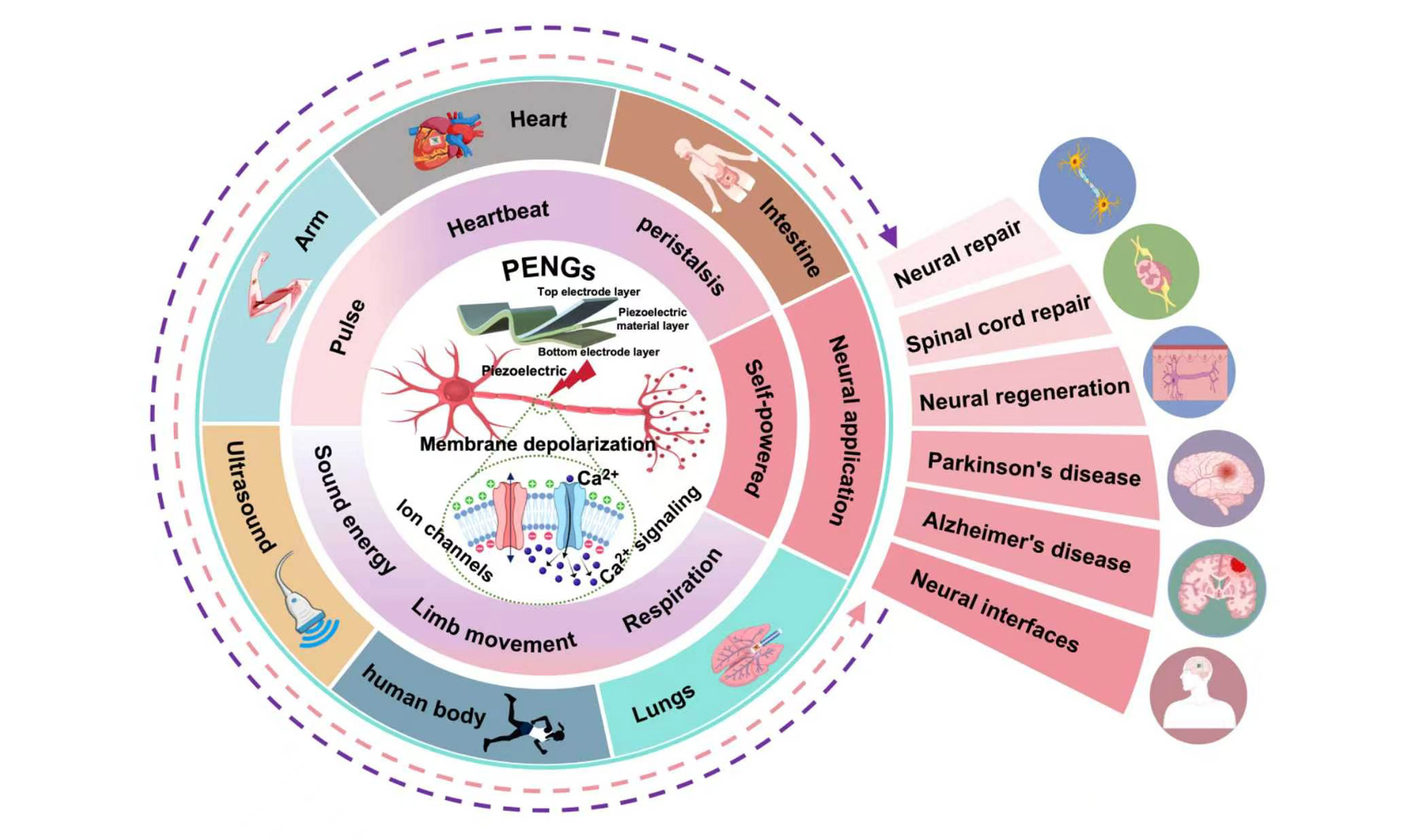
Abstract
Piezoelectric nanogenerators (PENGs) are electromechanical energy-conversion devices constructed from piezoelectric materials. They convert physiological motion, local tissue deformation, or externally applied mechanical stimuli into localized electrical signals and have attracted considerable interest in self-powered neural interventions. Despite the rapid expansion of research in this field, the systematic understanding needed to guide translational optimization remains limited. This review delineates the current research landscape by organizing the discussion around mechanical inputs, piezoelectric transduction, neural responses, and functional restoration, thereby linking fundamental principles with representative application studies. This review discusses neural-interface-compatible materials, device architectures, and powering strategies for PENGs; the underlying neurobiological mechanisms; and recent advances in their applications across the peripheral, central, and autonomic nervous systems, neurodegenerative diseases, and neural interfaces. It further examines key barriers related to the precision of deep-tissue targeting, the long-term safety of implanted devices, and the lack of standardized frameworks for clinical translation. To this end, PENGs are systematically compared with other self-powered and wirelessly powered transduction technologies, and a standardized reporting framework linking device output to the biologically effective dose is proposed, while outlining future directions in multimodal energy coupling, closed-loop feedback, intelligent energy management, and the integration of piezoelectric stimulation with genetic, immune, and metabolic reprogramming. Collectively, this review aims to advance both fundamental research and clinical translation of PENGs in neural engineering.
Keywords


 Download PDF
INTRODUCTION
Piezoelectric nanogenerators (PENGs) exploit the direct piezoelectric effect to convert dynamic mechanical energy into electrical output. Their nanoscale character arises from the micro- or nanoscale dimensions of the piezoelectric functional phases or structural units[1,2]. In 2006, Wang et al. used ZnO nanowire arrays to convert minute mechanical deformations into measurable electrical signals, advancing piezoelectric research from material-level characterization toward miniaturized devices and integrated systems[3]. Subsequent advances in flexible piezoelectric materials, electrode integration, and encapsulation technologies enabled PENGs to enter the field of implantable bioelectronics[4,5]. Between 2010 and 2019, their self-powered capability was increasingly explored for neural stimulation and regenerative modulation, with flexible piezoelectric systems conformally integrated with the heart, lungs, and diaphragm demonstrating the feasibility of harvesting biomechanical energy in vivo[6,7]. From 2020 to 2025, the development of PENG-based neural applications accelerated markedly. ZnO-nanogenerator-based biomimetic scaffolds first demonstrated self-powered motor recovery[8]. Barium zirconate titanate–barium calcium titanate/poly(vinylidene fluoride) (BZT-BCT/PVDF) films enabled ultrasound-driven sciatic nerve stimulation[9], whereas biodegradable poly(3-hydroxybutyrate-co-3-hydroxyvalerate)/poly(L-lactic acid)/potassium sodium niobate (K0.5Na0.5NbO3) (PHBV/PLLA/KNN) devices provided transcutaneous lead-free electrical stimulation of the sciatic nerve while supporting concurrent repair monitoring[10]. In parallel, molecular piezoelectric materials and piezoelectrets were successively incorporated into PENG platforms for wireless deep brain stimulation and vagus nerve stimulation (VNS)[11,12]. By 2025, programmable ultrasonic wireless implants had extended self-powered neuromodulation to personalized chronic pain management[13]. By 2026, the emergence of bioresorbable Rochelle salt/PLLA devices and piezoelectric neuromodulation of the subthalamic nucleus further indicated that PENGs were evolving beyond single-function proof-of-concept studies toward a versatile technological platform integrating flexible implantation, remote powering, biodegradable construction, and combined diagnostic and therapeutic capabilities[14,15].
Two decades of technological evolution have converged on two longstanding challenges in clinical neural engineering. First, although autologous nerve grafting remains the gold standard for peripheral nerve repair[16], its clinical use is constrained by limited donor availability, size mismatch between donor and recipient nerves, donor-site functional deficits, and the additional trauma associated with a second surgical procedure[17]. Second, commercially available nerve conduits rely predominantly on passive structural guidance and are therefore unable to reconstitute, particularly across long-segment defects, the cellular, trophic, vascular, and bioelectrical microenvironments required for sustained axonal extension and remyelination[18]. At the core of both challenges is the inability of existing approaches to provide sustained, physiologically relevant electromechanical cues at the site of injury. Owing to their distinctive electromechanical transduction capability, PENGs can address these limitations at three levels[19]. At the level of power delivery, endogenous mechanical inputs—including respiration, cardiac motion, vascular pulsation, muscle contraction, and rehabilitative movement—can directly actuate implanted devices to generate self-powered electrical pulses[20]. Exogenous mechanical fields, such as transcutaneous ultrasound, further enable battery-free, wireless, and on-demand stimulation of deep neural targets[12]. At the interface level, transduction units can be placed directly on nerve bundles, within lesion cavities, or in targeted brain regions to generate localized electric fields and charge transfer, thereby reducing the need for long interconnecting leads and bulky implanted power sources[21,22]. At the material level, aligned fibrous scaffolds, nerve conduits, and piezoelectric hydrogels can modulate the electrophysiological microenvironment at the injury site while simultaneously providing mechanical support and directional guidance for cell growth[23-25]. Bioresorbable piezoelectric materials, including PLLA, PHB, and their copolymers, can be integrated with biodegradable electrodes and encapsulation layers, allowing the device to gradually degrade after the therapeutic period and potentially reducing the risks of secondary surgery and chronic inflammation[26,27]. Together, these three complementary dimensions establish an integrated technological framework that combines localized electrical intervention, mechanical conformity to tissue, and a programmable therapeutic lifetime.
However, existing studies remain fragmented across the fields of piezoelectric materials, energy-harvesting devices, tissue engineering, and neurobiology, underscoring the urgent need for systematic integration at the system level. Accordingly, this review conceptualizes PENGs as electromechanical-biological interfaces comprising four interconnected elements: mechanical input, piezoelectric transduction, tissue loading, and neural response. It systematically reviews their operating principles, neural-interface-compatible materials, device architectures, and powering strategies; elucidates mechanisms involving membrane potential, ion channels, and Ca2+ signaling; summarizes advances in peripheral nerve regeneration, central and autonomic neuromodulation, interventions for neurodegenerative diseases, and neural interfaces; and critically evaluates key challenges related to dose calibration, deep-tissue targeting, long-term safety, device reliability, and clinical assessment. Finally, this review outlines emerging directions in multimodal energy coupling, intelligent energy management, closed-loop feedback, and integration with genetic regulation, immune modulation, and metabolic reprogramming, thereby establishing a systematic conceptual framework for the design, mechanistic validation, and clinical translation of self-powered neural intervention systems.
ENGINEERING FOUNDATIONS OF PENG-MEDIATED NEURAL INTERVENTIONS
Operating principles and system architecture of PENGs
PENGs exploit the linear electromechanical coupling of piezoelectric materials to convert dynamic mechanical inputs into electrical potential and current[28]. Under the direct piezoelectric effect, an applied force induces ionic displacement within a non-centrosymmetric crystal lattice or reorientation of molecular dipoles in an organic piezoelectric phase[29,30]. The resulting separation of positive and negative charge centers produces macroscopic polarization, generating bound charges on the material surfaces and a potential difference between the electrodes. As the deformation varies, charges redistribute through the external circuit to generate an electric current, thereby converting mechanical energy into electrical energy[31], as shown in [Figure 1A and B]. Conversely, the converse piezoelectric effect refers to the reversible mechanical deformation of a piezoelectric material induced by an applied electric field and is primarily exploited in piezoelectric actuators, acoustic transduction, and the design of electrical stimulation interfaces. Piezoelectric operating modes are defined by the corresponding piezoelectric coefficients. In the d33 mode, the applied stress is parallel to the polarization direction, making this mode suitable for through-thickness compression; in the d31 mode, the stress is perpendicular to the polarization direction and is commonly exploited in thin-film bending or in-plane stretching[32], as shown in [Figure 1C]. Under Voigt notation, d14 and d15 describe the coupling of electric displacement along the 1-direction with shear stresses in the 2-3 and 1-3 planes, respectively[33], as shown in [Figure 1D]. A PENG device system primarily comprises a piezoelectric active layer, substrate, electrodes, interconnects, and insulating encapsulation [Figure 1E][34]; For implantable PENGs, biocompatible encapsulation and tissue-conformal interfaces are additionally required
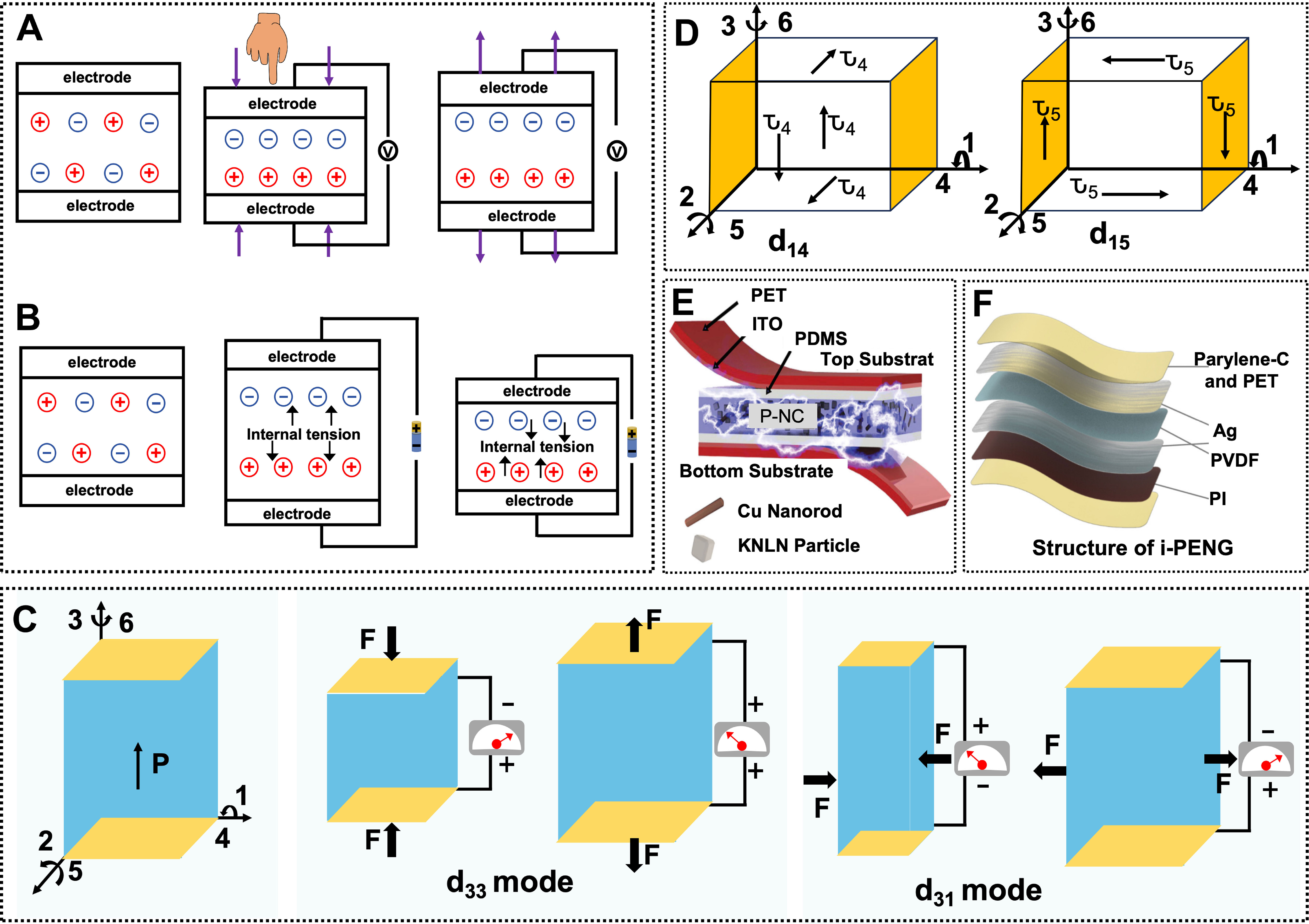
Figure 1. Working principles, operating modes, and system architecture of PENGs. (A) Direct piezoelectric effect; (B) Converse piezoelectric effect. (C) Schematic illustrations of the d31 and d33 strain modes in piezoelectric materials. (D) Schematic illustrations of the shear piezoelectric strain modes associated with d14 and d15. (E) Structural components of a PENG based on KNLN particles and Cu, adapted with permission from Ref.[34]. Copyright © 2014 Wiley-VCH. (F) Multilayer structure of an implantable piezoelectric nanogenerator (i-PENG), adapted from Ref.[35] under CC BY 4.0 license.
Piezoelectric materials for neural interfaces
Piezoelectric materials used in neural applications encompass inorganic ceramics and single crystals, conventional and biodegradable polymers, naturally piezoelectric biomaterials[39], polymer-ceramic composites, and conductive or multifunctional composites. Collectively[40], these materials can be classified into three principal categories: organic, inorganic, and composite piezoelectric systems[41]. Organic piezoelectric materials are represented primarily by PVDF, poly(vinylidene fluoride-co-trifluoroethylene) (P(VDF-TrFE)), and PLLA. Their flexibility and amenability to electrospinning into aligned fibrous architectures have enabled their use in studies of Schwann-cell adhesion, axonal extension, myelination, and the neuronal differentiation of stem cells[42-45]. Inorganic materials, including BaTiO3 (BT), ZnO, boron nitride (BN), boron nitride nanotubes (BNNTs), lead zirconate titanate (Pb(Zr,Ti)O3) (PZT), and lead-free (K, Na)NbO3-based ceramics, exhibit high piezoelectric responses and can generate localized electrical stimulation under ultrasound or mechanical perturbation[46,47]. However, mechanical mismatch with soft neural tissues, particle retention, and, in the case of lead-containing materials, heavy-metal safety concerns constrain their direct implantation. Composite systems combine PVDF-based polymers, poly(ε-caprolactone) (PCL), silk fibroin, or hydrogels with functional fillers such as BaTiO3, ZnO, and MXene. In addition to enhancing electrical output, these combinations improve flexibility[48], cell-material interactions, and processability into nerve conduits, making them particularly important for central nervous system repair[49]. Recent advances have further incorporated emerging nanomaterials, including MoS2, SbSeI, and hBN, into flexible piezoelectric systems[50].
Representative neural-interface studies include aligned PVDF-TrFE scaffolds supporting Schwann cells, neurite extension, and myelination[51]; biodegradable PLLA piezoelectric transducers[52]; PHBV-based neural scaffolds and conduits[53-54]; collagen nerve guides and collagen-mimetic piezoelectric systems[55,56]; piezoelectric silk-fibroin fibers and scaffolds[57,58]; ZnO nanogenerators for functional nerve stimulation[59]; BNNT- and hBN-based piezoelectric systems[60,61]; PZT-based neural and cochlear interfaces[62,63]; Aluminum nitride (AlN) acoustic transducers for auditory applications[64,65]; self-powered PVDF/PCL scaffolds for peripheral nerve regeneration[66]; high-performance ferroelectric perovskites[67]; and directional PHBV-based neurosupport scaffolds[68]. Table 1 summarizes representative fabrication methods, piezoelectric properties, and neural applications of these material systems.
Representative materials for piezoelectric nanogenerators in neural applications: properties and applications
| Material type | Materials | Preparation technology | Piezoelectric coefficient | Application | References |
| Organic piezoelectric | P(VDF-TrFE) | Electrospinning; Thermal annealing | d33 ≈ -40 to -20 pC N-1 | Dorsal root ganglion; Sciatic nerve; Spinal cord | [42,51] |
| PLLA | Electrospinning; Thermal treatment | d14 ≈ 6-19 pC N-1 | Rat pheochromocytoma PC12 cells | [52] | |
| PHBV | Solution casting; Electrospinning | d14 ≈ 1.2-2.0 pC N-1 | Peripheral nerve | [53,54] | |
| COL/CMP | Magnetic alignment; Self-assembly | COL: d14 ≈ 0.2-2.0 pC N-1; CMP: d35 ≤ 27 pm V-1 | Peripheral nerve | [55,56] | |
| SF | Electrospinning; Dry spinning | d33 ≈ 3.24-18.0 pm V-1 | Human neural stem cell-derived neuronal networks; Rat pheochromocytoma PC12 cells | [57,58] | |
| Inorganic piezoelectric | BTO | Hydrothermal synthesis; Solid-state synthesis | d33 ≈ 75-350 pC N-1 | Human SH-SY5Y neuroblastoma cells | [53] |
| ZnO | Hydrothermal growth; Chemical bath deposition | d33 ≈ 9-14 pC N-1 | Frog sciatic nerve and gastrocnemius neuromuscular pathway | [62] | |
| BNNT/h-BN | Chemical vapor deposition; Non-covalent glycol-chitosan coating | d31 ≈ 1-30 pC N-1 | Rat pheochromocytoma PC12 cells | [63,64] | |
| PZT | Poling and sintering; Radio-frequency magnetron sputtering | d33 ≈ 350-650 pC N-1 | Guinea pig cochlea and auditory nerve pathway; rat cerebral cortex | [65,66] | |
| KNN | Solid-state synthesis; Ceramic film preparation | d33 ≈ 100-560 pC N-1 | Mouse hippocampus; substantia nigra pars compacta; nigrostriatal dopaminergic pathway | [67] | |
| Sm-PMN-PT | Modified Bridgman growth; Array microfabrication | d33 ≤ 4,000 pC N-1 | Rat periaqueductal gray and deep-brain analgesia pathway | [68] | |
| AlN | Reactive radio-frequency magnetron sputtering; Microelectromechanical-system micromachining | Effective d33 ≈ 3.52 pm V-1 | Cochlea and auditory nerve pathway | [69,70] | |
| Polymer-based and composite piezoelectric | PHBV/CA | Electrospinning; Post-drying | Effective d33 ≈ 6.68 pm V-1 | Unspecified neural cells | [54] |
| PVDF/PCL | Solvent casting; Annealing | d33 ≈ 13 pC N-1 | Rat sciatic nerve | [66] | |
| Organic-inorganic perovskite ferroelectric | Solution crystallization | Large piezoelectric response | Deep-brain neuronal circuits | [67] | |
| PHBV/MgOl/NAC | Electrospinning; Drug incorporation | NR | Rat pheochromocytoma PC12 cells | [68] |
Device architectures
As PENGs have progressed from nanoscale material validation toward implantable and tissue-interfacing applications, neural-interface devices have diversified into aligned fibrous membranes, porous or continuous films, nerve conduits, cuff-type interfaces, piezoelectric hydrogels, multilayer composites, and systems with biodegradable encapsulation[69-70]. Early devices were predominantly based on continuous films and multilayer thin-film architectures. Zhang et al. fabricated a multiunit P(VDF-TrFE)/BaTiO3 membrane through layer-by-layer electrospinning to redistribute pulsatile mechanical loads, increasing the open-circuit voltage and short-circuit current to 3.3- and 2.1-fold those of a single-layer device, respectively[71]. For interfacing with nerve bundles, planar films were further developed into conduits, cuffs, and tubular architectures. Ma et al. established a radially compartmentalized structure comprising an outer piezoelectric membrane and an inner multichannel conductive scaffold[72]. Tai et al. rolled an aligned P(VDF-TrFE) fibrous mat into a hollow cylindrical conduit and incorporated electrodes on opposite sides, thereby supporting regeneration across sciatic nerve defects[73]. Xu et al. further integrated
Powering strategies
PENG powering strategies can be classified according to their energy source into endogenous physiological motion and exogenous mechanical fields[75]. The former harnesses cardiac motion, respiration, gastrointestinal peristalsis, and limb movement, offering the advantages of continuous energy availability and autonomous power generation[76,77]. Harnessing cyclic organ motion, Wang et al. transversely sutured an instantaneous-discharge PVDF i-PENG onto the surfaces of the left and right ventricles in pigs[78]. Cardiac motion increased the output voltage from 1.6 ± 0.4 V for a conventional device to 10.5 ± 0.5 V and charged a 100-μF capacitor to 4.53 V within 1,000 s. A flexible piezoelectric energy harvester integrated on the epicardium similarly converts cardiac contraction into voltage outputs [Figure 2A][79]. Cheng et al. deployed a PVDF film on the porcine ascending aorta, achieving a sensitivity of 14.32 mV mmHg-1 and an instantaneous power output of 40 nW. These results indicated that conformal electrode contact and integrated energy storage could compensate for the limited mechanical input associated with low-frequency cardiovascular deformation[80]. However, fixation through open-chest surgery increases implantation complexity. Li et al. used a bioadhesive to attach a PLLA/glycine core-shell nanofibrous PENG to the outer wall of the rat transverse colon. Under enema-induced peristalsis at approximately 0.2 Hz, the device generated a stable output exceeding 2 V and distinguished different states of colonic motility from the signal envelope[27], extending endogenous energy harvesting to conformal in situ coupling with soft, folded organs. Yu et al. developed a mesoporous PVDF/polydimethylsiloxane (PDMS) device that generated an open-circuit voltage of approximately 200 mV[81]. Zhang et al. reported a mesoporous piezoelectric polymer composite film with a tunable mechanical modulus, which can further harvest energy from liquid-pressure fluctuations
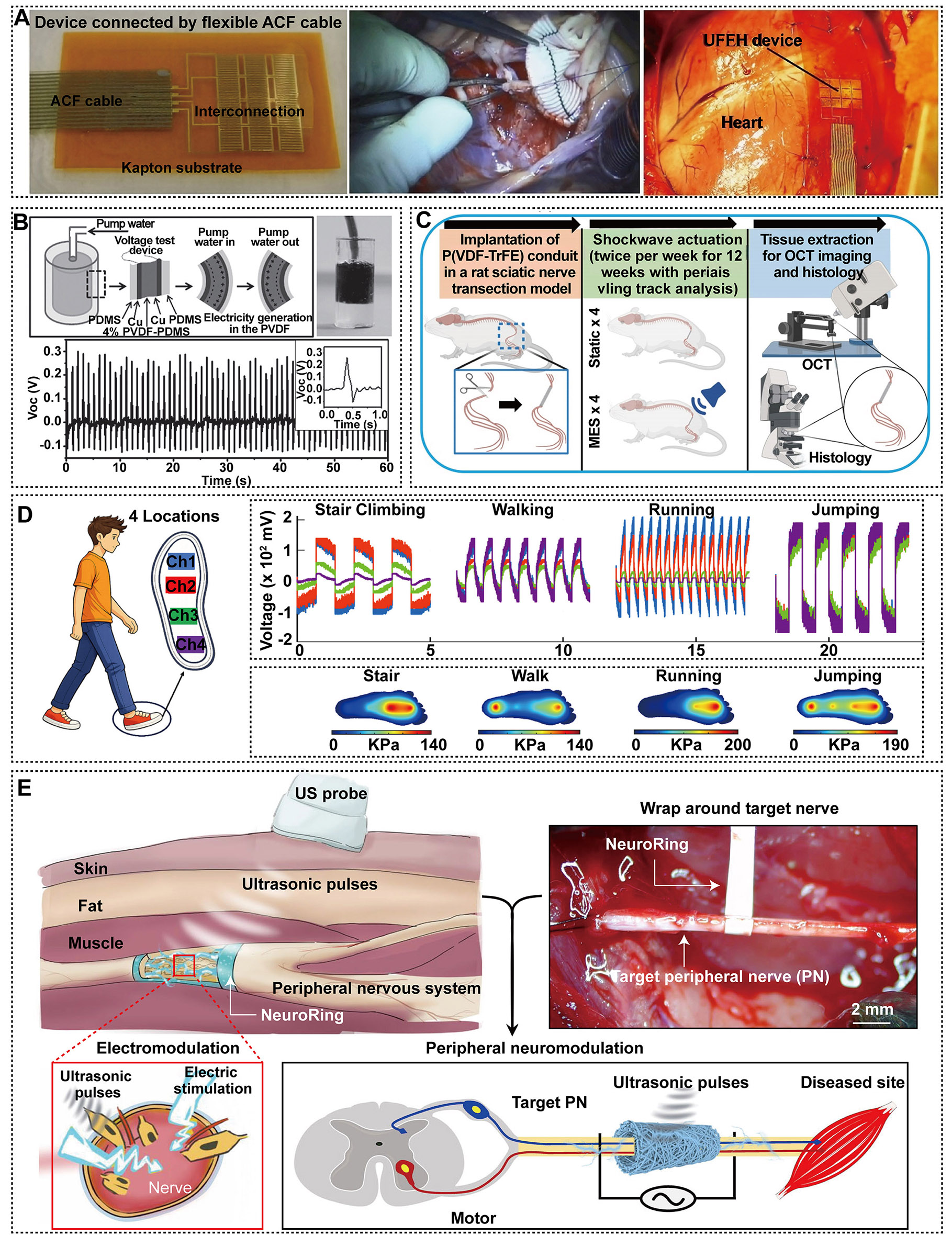
Figure 2. Representative endogenous and exogenous powering strategies for piezoelectric nanogenerator-based systems. (A) In vivo cardiac biomechanical energy harvesting: a flexible piezoelectric energy harvester integrated on the epicardium (between the left-ventricular apex and the right ventricle, and between the atrioventricular groove and the right-ventricular outflow tract) converts cardiac contraction into voltage outputs, adapted from Ref.[79] under CC BY 4.0 license. (B) Mesoporous PVDF/PDMS piezoelectric nanogenerator with a tissue-matched mechanical modulus for harvesting energy from pulsatile liquid pressure (simulated blood pressure), adapted with permission from Ref.[82]. Copyright © 2016 Wiley-VCH. (C) In vivo piezoelectric characterization of an electrospun P(VDF-TrFE) nerve conduit bridging the rat sciatic nerve and the outline of the animal experiments, Adapted from Ref.[73] under CC BY 4.0 license. (D) AI-driven gait monitoring using a P3HB/ZnO piezoelectric nanofiber smart insole: multichannel voltage outputs, plantar-pressure mapping, and gait recognition, adapted from Ref.[85]. under the CC BY 4.0 license. (E) Ultrasound-driven peripheral neuromodulation using a ferroelectret NeuroRing wrapped around the sciatic nerve, adapted from Ref.[12] under CC BY 4.0 license.
Ultrasound-driven PENGs must operate within established acoustic-exposure limits. Diagnostic-ultrasound guidance restricts the mechanical index (MI) to ≤ 1.9 and the thermal index to ≤ 6.0, with lower thresholds for ophthalmic and fetal applications, and caps the spatial-peak temporal-average intensity at
Technological positioning and advantages of PENGs
Conventional electrical stimulation relies on implantable pulse generators, transcutaneous leads, or subcutaneous batteries. Although these approaches have an established clinical track record, they remain constrained by several limitations, including lead fracture, infection, battery replacement, and the lack of synchronization with physiological states[90]. Inductive and radiofrequency power transfer eliminates the need for an implanted battery, but it relies on external coils and precise spatial alignment and may cause localized tissue heating[91]. Optogenetics and chemogenetics offer cell-type-specific neuromodulation but are constrained by the need for transgene expression and exogenous light or drug delivery. By harnessing piezoelectric electromechanical transduction, PENGs can directly convert physiological motion or externally applied ultrasound into localized electrical pulses, enabling neural stimulation without implanted batteries or transcutaneous leads. This platform can further be engineered into flexible, conformable, and biodegradable devices[92]. Compared with triboelectric nanogenerators (TENGs), magnetoelectric generators, and biofuel cells, PENGs offer distinct advantages in terms of output characteristics, tissue compatibility, and device integration. Although TENGs can generate relatively high voltages, charge leakage in the humid implantable environment can compromise their long-term stability[93]. Magnetoelectric generators and biofuel cells, in contrast, are constrained by their dependence on external magnetic fields and by limitations in power output and enzymatic stability, respectively[94,95]. Existing PENG devices can already generate electrical outputs sufficient for neural stimulation. For example, a five-layer P(VDF-TrFE)/BaTiO3 film can produce an output voltage of 84 V and a current of 1.32 μA[71], whereas a BZT-BCT/PVDF thin film can generate an output voltage of 13.5 V under ultrasound actuation[9]. Accordingly, Table 2 provides a detailed comparison of the advantages and limitations of different self-powered technologies for neural applications. Overall, PENGs integrate energy conversion and neural stimulation within a single platform, while offering the potential for wireless actuation, flexible tissue conformability, and biodegradability. Furthermore, hybrid systems that combine piezoelectric transduction with triboelectric, magnetoelectric, or biofuel-cell mechanisms may further enhance the continuity of power delivery and the stability of neural stimulation.
Comparison of PENGs with alternative self-powered and wireless powering technologies for neural applications
| Technology | Output characteristics | Key advantages | Key limitations | References |
| PENGs | Capacitive high-impedance pulses; Voc ~0.2-84 V; Isc nA-mA | Battery-free; conformable; biodegradable; intrinsically charge-limited | Output input-dependent; dose calibration required | [9,71] |
| Electromagnetic induction | Near-field power (mW-W) | Mature technology; high power | Coil alignment; tissue heating; bulky receiver | [91] |
| TENGs | High V (~kV); low I (μA); contact electrification | Low cost; high sensitivity | Charge leakage and instability in wet environments | [93] |
| Magnetoelectric generators | Charge via magnetostrictive-piezoelectric coupling | Deep-tissue access; injectable forms | External field required; loss at small dimensions | [94] |
| Biofuel cells | Low-voltage DC (μW-mW) | Continuous operation; endogenous fuel | Low power; enzyme deactivation | [95] |
NEUROBIOLOGICAL MECHANISMS OF PENG-MEDIATED ELECTRICAL STIMULATION
Membrane-potential perturbation and ion-channel gating
Neuronal membranes combine capacitive charge storage with selective ionic conductance. Mechanical deformation of a PENG generates a localized polarization potential that perturbs the resting membrane potential. Once depolarization exceeds the excitation threshold[92], voltage-gated Naᵥ channels initiate the action potential, Kv channels drive repolarization, and Cav channels mediate Ca2+ influx, thereby translating transient electrical activity into sustained cellular responses such as neuronal differentiation and neurite outgrowth[96]. Ion-channel blockade provides the most direct causal evidence for this mechanism.
Ca2+-mediated translation of neural function
Ca2+ signaling serves as the central hub through which PENGs convert transient piezoelectric stimulation into sustained neural functional responses[103]. Following membrane depolarization and the opening of voltage-gated Ca2+ channels, elevated cytosolic Ca2+ engages calmodulin (CaM)-dependent intracellular signaling[104]. The resulting signals proceed through Ca2+/calmodulin-dependent protein kinase, adenylyl cyclase/cAMP (cyclic adenosine monophosphate)/PKA (protein kinase A), and rat sarcoma GTPase/mitogen-activated protein kinase kinase/extracellular signal-regulated kinase (Ras/MEK/ERK) pathways, which converge on cAMP response element-binding protein (CREB) to encode electrical activity into cytoskeletal remodeling and gene transcription[105].
Piezoelectric output-neural response relationship
The neural effects of PENGs are not determined directly by the open-circuit voltage, but primarily by the local electric field delivered to neural tissue, charge per phase, and pulse waveform. The local electric field must induce sufficient membrane depolarization to reach the excitation threshold of the target neural population. The charge per phase determines both neural activation efficacy and the electrochemical safety limit, with typical charge levels of approximately 1-50 nC per pulse for retinal prostheses and 135-400 nC per phase for deep brain stimulation (DBS)[113,114]. Pulse duration and stimulation intensity follow the strength-duration relationship, with shorter pulses generally requiring higher stimulation currents. Stimulation frequency further modulates neuronal firing patterns and plasticity; frequencies of 1-100 Hz are generally associated with promoting neural regeneration and plasticity, whereas frequencies > 100 Hz are typically used to suppress abnormal neural activity[92,115]. Stimulation safety can be evaluated using the Shannon model, k = log10(D) + log10(Q), where k < 1.5 is generally considered within the safe range, whereas k > 2.0 indicates an increased risk of tissue damage[116]. McCreery et al. further demonstrated that neural injury is jointly governed by charge density and charge per phase, providing experimental support for the Shannon criterion[117]. Meanwhile, the charge-injection capacity and effective area of the electrode, together with tissue impedance, collectively determine the efficiency of PENG output transfer to neural tissue. The charge-injection capacities of platinum and activated iridium oxide are approximately 50-150 μC cm-2 and
Proposed standardized reporting guidelines for PENG neural stimulation studies
| Parameter | Metric (unit) | Reference range | References |
| Device output | Voc, Isc at defined load (V; nA-μA) | 0.2-84 V; nA-μA | [71,81] |
| Pulse parameters | Pulse width; frequency (μs; Hz) | 60-200 μs (DBS); 1-100 Hz; > 100 Hz | [90,114,115] |
| Electric field | Depolarization at target membrane (mV) | 20-100 mV | [90,114] |
| Delivered charge | Charge per phase (nC) | 1-50 (retinal); 135-400 (DBS) | [113,114] |
| Electrode interface | Material; area; charge-injection capacity | Pt 50-150 μC cm-2; IrOx 1-4 mC cm-2 | [114] |
| Charge density | Per phase (μC cm-2) | ≤ 30 (safe; Shannon k < 1.5); DBS: 2.3-6.7 | [116,117] |
| Functional outcome | CAP; NCV; behavior | Not yet standardized | [118] |
APPLICATIONS OF PENGS IN NEURAL INTERVENTION
Peripheral nerves
Repair of peripheral nerve defects
Peripheral nerve defects represent a major application of PENGs in neural regeneration. Repair of traumatic nerve transection, crush injury, and segmental defects requires the simultaneous restoration of gap bridging, directional axonal regeneration, and electrophysiological conduction[22,36]. The field has therefore evolved by progressively addressing three principal limitations: insufficient electrical stimulation after bridging, inadequate spatial guidance, and a lack of microenvironmental support. Pi et al. developed aligned PCL/PVDF nanofiber bundles (piezoelectric nanotracts) that, under ultrasound-driven piezoelectric stimulation, restored motor function and axonal maturity across a 15 mm rat sciatic nerve defect to levels comparable with autologous nerve grafting[119]. In parallel, Jeon et al. constructed polyethylene glycol (PEG)-modified PLLA four-channel conduits that achieved substantial reinnervation at 12 weeks after repair of an 8-mm defect, with regenerative outcomes approaching those of autologous nerve grafting [Figure 3A][38]; Huang et al. similarly combined longitudinally oriented guidance cues with a porous sheath in a compound scaffold that promoted peripheral nerve regeneration in vivo[120]. However, these therapeutic effects remained dependent on an external acoustic field. One strategy to address this limitation is to alter the mode of mechanical input. More recently, Chen et al. constructed a biodegradable PENG conduit from aligned PCL/β-glycine nanofibers [Figure 3B]. Following 15-Hz vibrational stimulation delivered using a massage gun in a 10-mm nerve-defect model, motor function and nerve conduction recovered to 99% and 96%, respectively[121]. Zhang et al. instead enhanced piezoelectric performance through structural innovation, developing a bamboo-inspired, aligned hollow bilayer conduit whose collaborative oriented architecture increased the electrical output from approximately 20 mV to 0.95 ± 0.02 V, accelerated axonal elongation and myelination by 3.1- and 1.6-fold, respectively, and promoted peripheral nerve and muscle regeneration in vivo [Figure 3C][118]. Compared with the preceding approaches, these findings suggest that structure-mediated stress concentration may provide greater performance gains than material substitution alone. However, although stress concentration can increase peak voltage, the effective charge output remains limited. More recently, Li et al. integrated piezoelectric and triboelectric energy-harvesting units into a liquid-metal/ZnO-reinforced PVDF conduit, increasing the d33 coefficient from 0.9 to 4.6 pC N-1[122]. This hybrid design enhanced effective charge generation while concurrently improving functional recovery of the sciatic nerve. As electrical stimulation performance improved, the evaluation focus shifted toward whether the generated electrical signals could act stably on cells along the direction of regeneration. To address this requirement, Zhou et al. combined PLGA/PVDF microgrooves with an reduced graphene oxide (rGO)/gelatin methacryloyl (GelMA) filling matrix to simultaneously enhance longitudinal guidance, interfacial conductivity, and cell adhesion, thereby coupling electrical stimulation with structural guidance within a single nerve conduit [Figure 3D][123]. To overcome the inability of planar microstructures to reconstruct the complex spatial organization of nerve fascicles, Qi et al. further combined digital light processing printing with directional freezing to create hierarchically aligned channels across multiple length scales, advancing interfacial design from surface patterning toward three-dimensional spatial compartmentalization[124]. After the physical and electrical design parameters had been progressively optimized, Yin et al. further integrated self-powered electrical stimulation with sustained NGF release[125]. In a 10 mm nerve-defect model, this system attenuated muscle atrophy and promoted axonal regeneration and remyelination, thereby addressing the insufficient long-term trophic support provided by material interfaces alone. This integration transformed the PENG conduit from a passive bridging scaffold with intermittent electrical stimulation into a regenerative platform that combines energy supply, spatial guidance, and biochemical modulation.
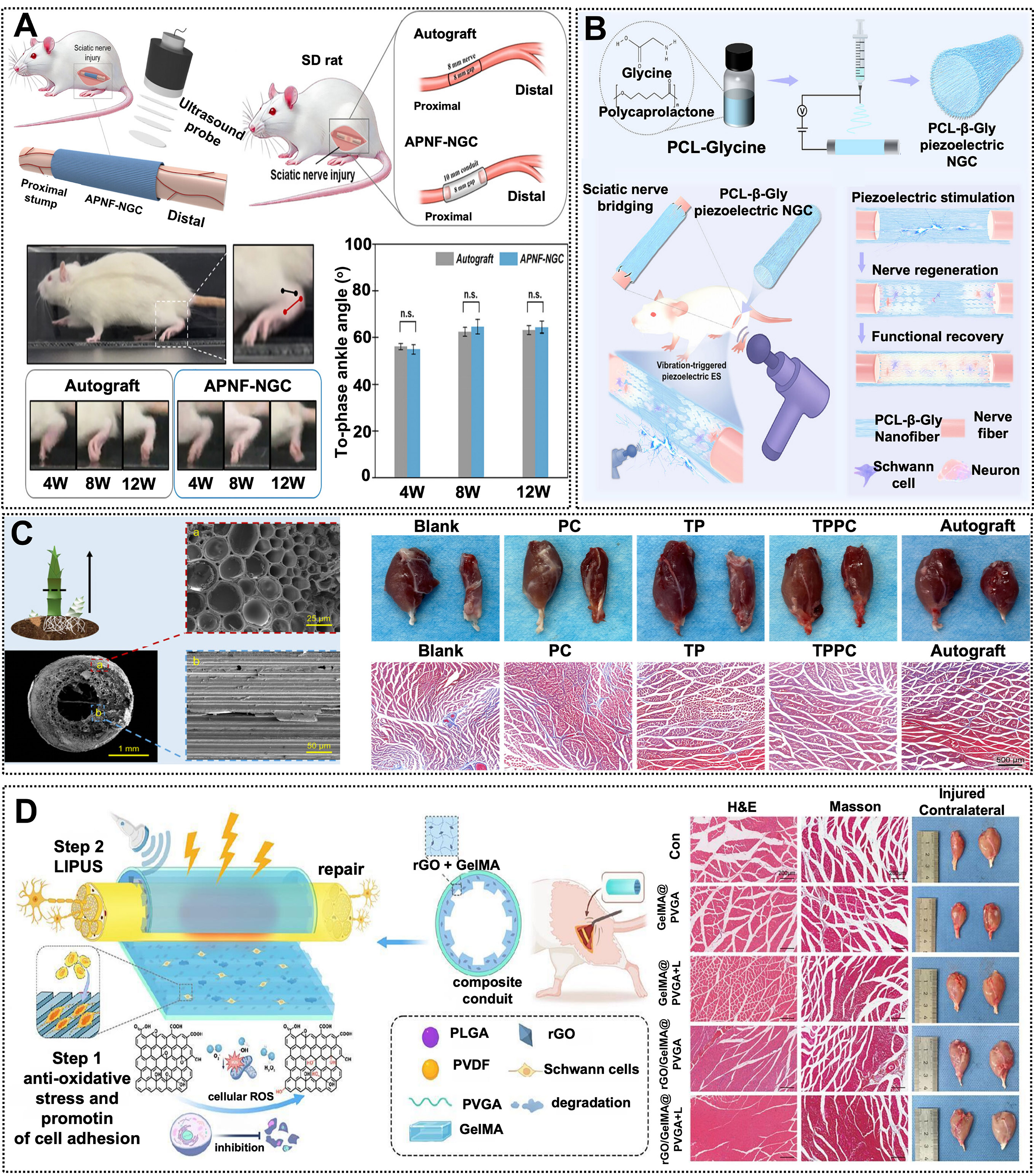
Figure 3. Representative applications of PENGs in nerve injury repair. (A) Ultrasound-driven aligned piezoelectric nanofibers nerve guidance conduit (APNF-NGC) for peripheral nerve regeneration, comparative analysis of the autograft group and the NGC group in an 8-mm sciatic nerve defect model, showing comparable outcomes, adapted from Ref.[38] under CC BY 4.0 license. (B) Biodegradable PCL-β-glycine piezoelectric nerve guidance conduit activated by low-frequency mechanical vibration for peripheral nerve regeneration, adapted with permission from Ref.[121]. Copyright © 2026 Wiley-VCH. (C) Structural characterization of the bamboo-inspired hollow bilayer conduit and its synergistic oriented architecture for piezoelectric acceleration of peripheral nerve and muscle regeneration, adapted with permission from Ref.[118]. Copyright © 2026 Wiley-VCH. (D) Schematic of the conductive-piezoelectric integrated microstructured nerve conduit (rGO/GelMA@PVGA), where piezoelectric stimulation combined with structural guidance promotes nerve and target muscle regeneration, adapted from Ref.[123] under CC BY 4.0 license.
Sensorimotor functional restoration
Sensorimotor reconstruction determines whether regenerated nerves can re-establish sensory input and motor output. Accordingly, the evaluation focus has shifted from axonal regeneration alone toward the coordinated recovery of sensory afference, motor efference, and target-muscle control. Qian et al. embedded a ZnO nanogenerator within a biomimetic scaffold and demonstrated, through electrophysiological and motor-function improvements, that piezoelectric stimulation could yield functional benefits; however, the duration of stimulation remained limited[8]. To extend the effective stimulation window, Jin et al. developed a respiration-driven triboelectric-piezoelectric biofeedback implantable electrical stimulation system for long-lasting neural modulation[126]. This hybrid nanogenerator produced 1.5 V and 0.071 μA and maintained stable operation for 14 weeks, enabling gait and neuromuscular function after repair of a 15 mm nerve defect to approach those achieved with autologous nerve grafting [Figure 4A]. The design paradigm therefore shifted from intermittent exogenous activation toward sustained endogenous powering, thereby overcoming interruptions in electrical stimulation. Nevertheless, efficient energy transfer to regenerating axons and target muscles remained dependent on the contact configuration and charge-transport properties of the conduit interface. To address this limitation, Hu et al. incorporated rGO and a microneedle architecture into a PCL/ZnO system. The rGO conductive network enhanced charge transport, whereas the microneedles improved local fixation and stimulation delivery, enabling an output voltage of 4.6 V[127]. This design extended the therapeutic effects beyond axonal regeneration to target-muscle reinnervation and protection against denervation-induced muscle atrophy. A millimeter-scale implantable magneto-mechano-electric transducer based on BTO piezoelectric ceramics enables remote wireless electrical stimulation of injured sciatic nerves [Figure 4B][128]. However, a high peak output alone does not ensure stable action-potential propagation. Lv et al. therefore developed a P(VDF-TrFE)/rGO/PCL scaffold that integrated piezoelectric stimulation, conductive signal transmission, and myelin sheath reconstruction within a single regenerative system[129]. As therapeutic efficacy improved, matching the stimulation dose to motor recovery emerged as a new challenge. Shan et al. therefore repurposed PLLA/BaTiO3 from a therapeutic material into a biodegradable sensor[28]. Its linear pressure-output relationship R2 = 0.9445 and close agreement with electromyographic signals enabled wireless monitoring of functional recovery [Figure 4C]. Because passive monitoring cannot actively adjust therapeutic intervention in response to rehabilitative activity, Tan et al. further converted movement-derived mechanical energy into a drug-release trigger, enabling on-demand delivery of aspirin/lysine and increasing the mass of denervated muscle[130]. Gu et al. developed an aligned PLLA/poly(ethylene oxide) (PEO)/Mg2Si piezoelectric fibrous scaffold whose optimized composition generated a piezoelectric current of 144.11 ± 1.55 pA and, under ultrasound activation, improved electrophysiological conduction and gait recovery while mitigating gastrocnemius muscle atrophy in a
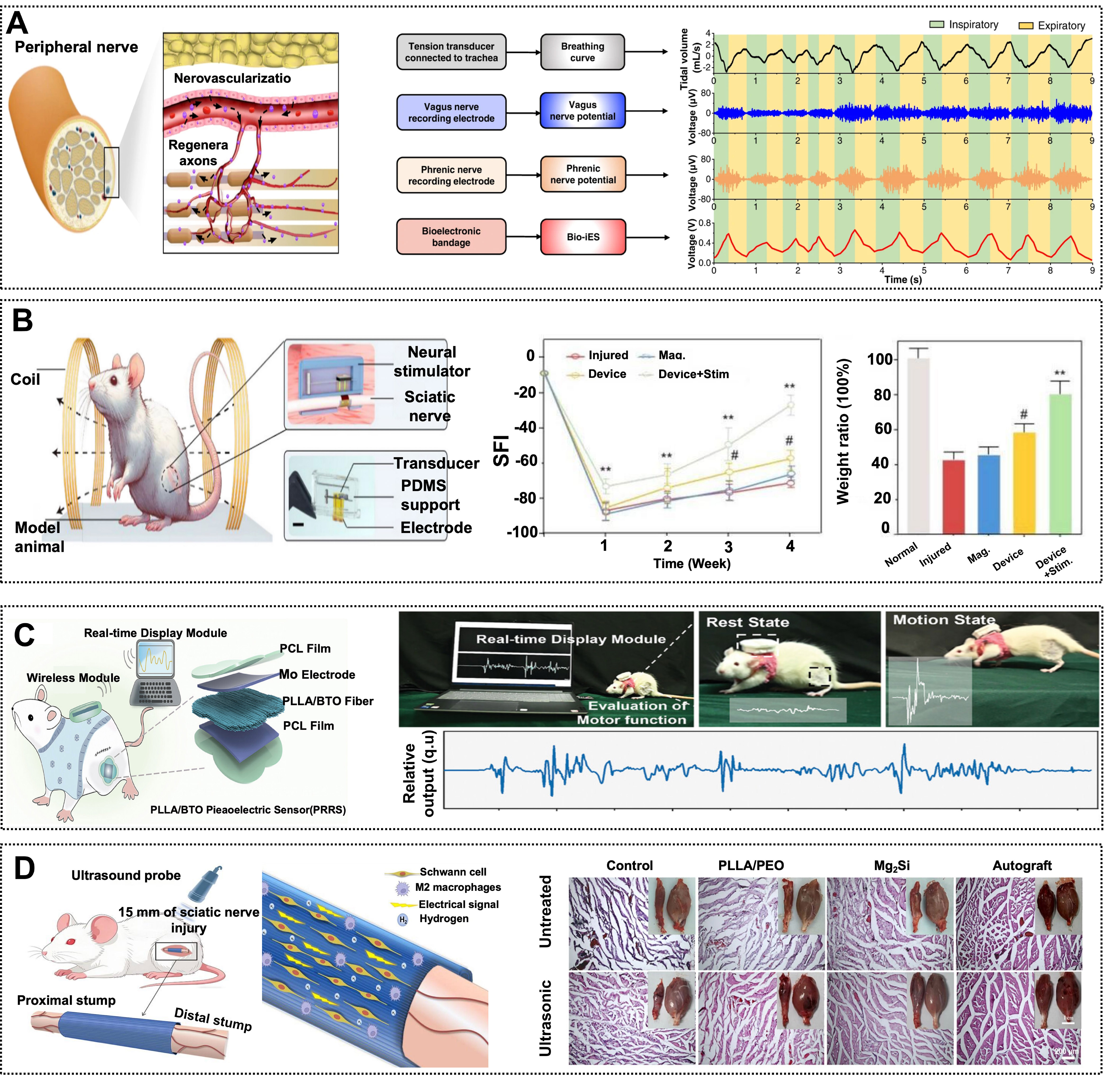
Figure 4. Representative piezoelectric and piezoelectric-hybrid strategies for sensorimotor functional restoration after peripheral nerve injury. (A) Implantable electrical stimulation (iES) system, including signal generation and its effects on neural cell and tissue regeneration, adapted from Ref.[126] under CC BY 4.0 license. (B) Wireless electrical stimulation of the injured sciatic nerve by a millimeter-scale implantable magneto-mechano-electric transducer based on BTO piezoelectric ceramics, adapted from Ref.[128] under CC BY 4.0 license. (C) Monitoring of rat motor function and output signals using a PLLA/BTO piezoelectric sensor (PBPS), adapted with permission from Ref.[28]. Copyright © 2024 Wiley-VCH. (D) Ultrasound-activated aligned PLLA/PEO/Mg2Si piezoelectric fibrous scaffold for repair of a
Central nervous system
Spinal cord injury repair
In traumatic spinal cord injury repair, PENG-based therapeutic constructs have evolved from passive bridging scaffolds into multifunctional platforms that can be remotely activated, deliver therapeutic cells, and modulate the lesion microenvironment[40]. In an early study, Lee et al. constructed a Schwann cell-loaded conduit from aligned P(VDF-TrFE) fibers. In a rat model of complete spinal cord transection, the conduit provided longitudinal bridging and promoted the ingrowth of brainstem-derived noradrenergic axons, astrocytic processes, and blood vessels into the graft, thereby establishing the feasibility of piezoelectric scaffolds for structural reconstruction of the injured spinal cord[133]. Li et al. developed a wirelessly activatable silk fibroin/BaTiO3-5 piezoelectric hydrogel that generated an output current of 124 nA, reduced lesion-cavity formation, and improved Basso-Beattie-Bresnahan (locomotor rating scale) (BBB) locomotor scores and gait performance[134]. This study marked the transition from static scaffolds to remotely activated electroactive implants. Because piezoelectric stimulation alone cannot simultaneously control post-injury excitotoxicity and myelin loss, an SFBP (silk fibroin/BaTiO3/polypyrrole)/PER (perampanel) conductive piezoelectric hydrogel incorporating PER was subsequently developed. Ultrasound activation concurrently provided electrical stimulation and pharmacological intervention, thereby further enhancing remyelination and locomotor recovery[135]. As the functional scope of these systems expanded, the research focus shifted toward recapitulating the spinal cord extracellular matrix while ensuring long-term biosafety[136]. Shi et al. incorporated KNN nanoparticles into a porous GelMA-reinforced decellularized spinal cord matrix hydrogel to construct a biomimetic piezoelectric porous decellularized spinal cord matrix gel/KNN (pDGK) system. Under ultrasound irradiation at 0.4 W cm-2, the integration of KNN with the decellularized spinal cord matrix simultaneously promoted vascularization and tissue repair while maintaining systemic organ safety over an 8-week period [Figure 5A][137]. However, acellular materials remain unable to replenish the neural and supportive cell populations lost after injury. Zhong et al. therefore co-transplanted a wireless piezoelectric hydrogel with neural stem cells and human umbilical cord-derived mesenchymal stem cells (NSCs (neural stem cells)-hUCMSCs), thereby reducing lesion-cavity formation and supporting axonal regeneration [Figure 5B][138]. Building on this strategy, Wang et al. further employed a three-dimensional system for the co-delivery of NSCs and Schwann cells, which enhanced transplanted-cell survival, synaptic density, and remyelination. These advances shifted the design paradigm from material-mediated repair toward the reconstruction of an electroactive regenerative niche[139].
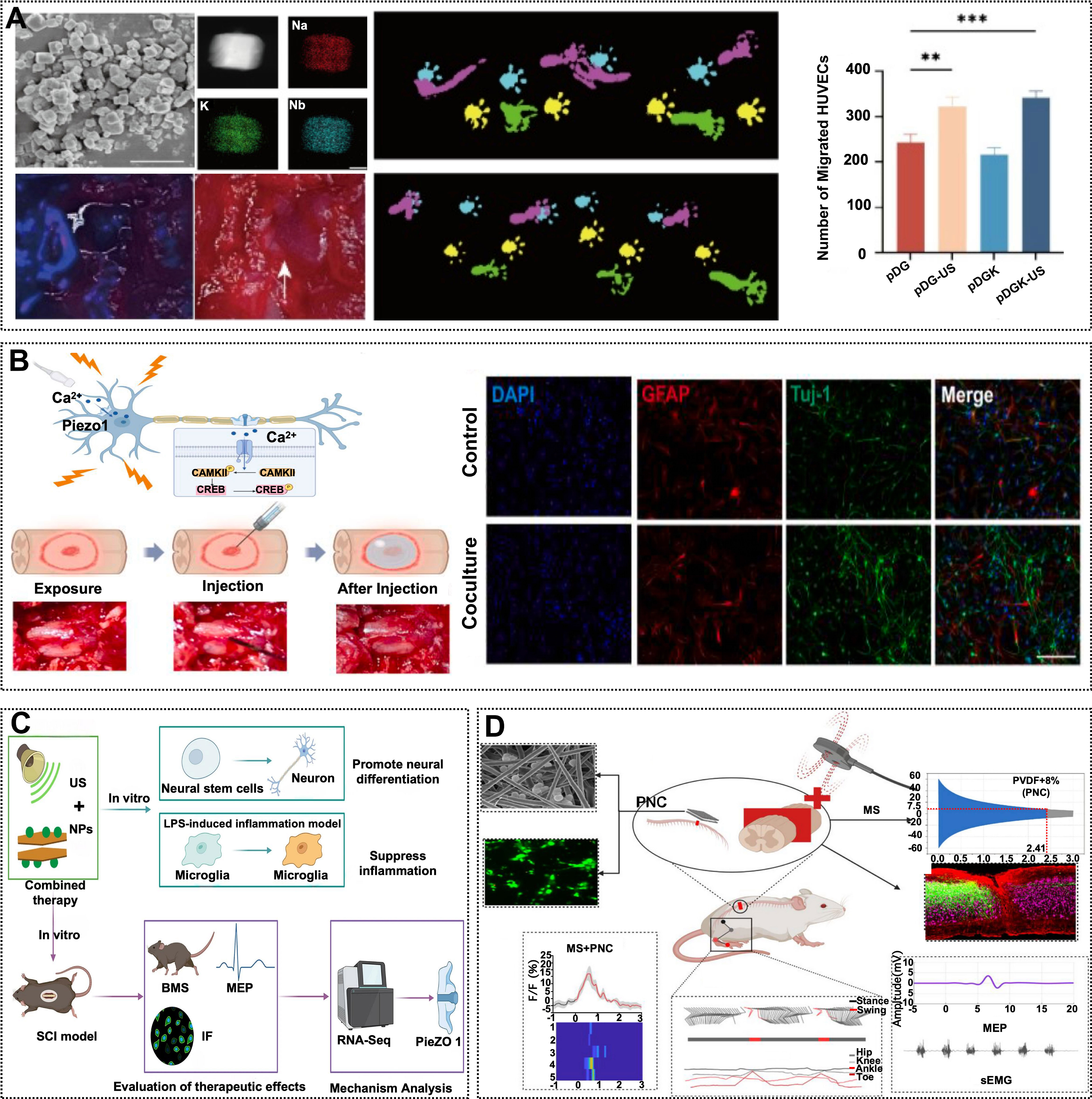
Figure 5. Representative applications of PENGs in spinal cord injury repair. (A) In vivo repair of spinal cord injury using a biomimetic piezoelectric hydrogel (decellularized spinal-cord matrix reinforced with KNN nanoparticles, pDGK), adapted from Ref.[137] under CC BY 4.0 license. (B) Ultrasound-activated wireless conductive piezoelectric hydrogel (PEDOT/polydopamine@BaTiO3, WPC) for spinal cord injury repair, and its immunofluorescence images promoting nerve regeneration, adapted from Ref.[138] under CC BY 4.0 license. (C) Ultrasound-assisted MgFe-LDH/NT3 nanoparticle therapy for neural stem cell differentiation and spinal cord injury recovery, adapted from Ref.[140] under CC BY 4.0 license. (D) BaTiO3-modified PVDF-based piezoelectric nanocapacitors (PNCs) for improving spinal cord injury outcomes and restoring neural innervation, adapted from Ref.[141] under CC BY 4.0 license. **P < 0.01, ***P < 0.001.
Functional modulation of brain regions
Piezoelectric modulation of brain regions has evolved from unidirectional power delivery toward state sensing and precise neural encoding. Accordingly, therapeutic evaluation has expanded beyond transient stimulation to encompass target-region specificity, behavioral reversibility, and long-term safety across applications involving synaptic plasticity, epilepsy, pain, and homeostatic regulation[142]. In an early study, Zhao et al. developed a body-motion-powered PZT/PVDF system that induced LTP and long-term depression in the hippocampal CA3-CA1 pathway through frequency-conditioned stimulation, demonstrating that piezoelectric energy could support bidirectional neural encoding[143]. However, stimulation timing remained externally prescribed by the operator, and the system therefore addressed energy delivery without achieving state-dependent control. To incorporate sensing and decision-making capabilities, Yang et al. integrated an enzyme/ZnO sensor into a stimulation circuit targeting the dorsomedial ventromedial hypothalamus. Hypoglycemic signals automatically triggered intervention, increasing blood glucose levels by 39% within 10 min[144]. This system provided an integrated closed-loop demonstration encompassing physiological-state detection, threshold-based decision-making, target-specific stimulation, and corrective restoration of glucose homeostasis. Within this architecture, Liang et al. combined a PZT arch-shaped transducer with a PVDF/ZnO membrane to convert respiratory motion into stimulation of the medial forebrain bundle[145], whereas Xu et al. integrated a PVDF pulse sensor with a piezoelectric generator to drive electrodes in the median preoptic nucleus[146]. Xiang et al. similarly used a PZT arch-shaped transducer and PVDF/ZnO membrane to encode carotid pulsation into periaqueductal gray stimulation[147]. Collectively, these studies extended a common piezoelectric platform to the regulation of exercise endurance, body temperature, and behavior. These studies indicate that system optimization has progressed from establishing whether sufficient power can be generated toward matching specific physiological sensing variables with the functions of targeted brain regions. However, the input signals remain derived from normal physiological activity and therefore cannot directly capture pathological neural discharges. Epilepsy provided a more stringent test of whether state-triggered control could be extended from normal physiological variables to disease-related signals. Lin et al. developed a PVDF/ZnO system that used seizure-associated body movements to trigger stimulation of the dentate gyrus-hilus circuit, reducing cumulative seizure duration by 40%-50%[148]. However, motor activity represents a delayed surrogate that occurs only after the onset of pathological electrical discharges.
Autonomic nervous system
Cardiac neuromodulation
Cardiac neuromodulation aims to convert cardiac motion, arterial pulsation, or ultrasound into electrical stimulation delivered through the cervical vagus nerve, auricular vagal branches, or intrinsic cardiac neural plexuses to regulate cardiac rhythm and preserve cardiac function. The central objective is to transform cardiac mechanical energy from a passive power source for implanted devices into neural stimulation that is matched to the underlying pathological state[6,78,151,152]. A flexible single-crystalline lead magnesium niobate–lead titanate (Pb(Mg1/3Nb2/3)O3-PbTiO3) (PMN-PT) piezoelectric energy harvester attached to the beating heart enables self-powered cardiac pacing
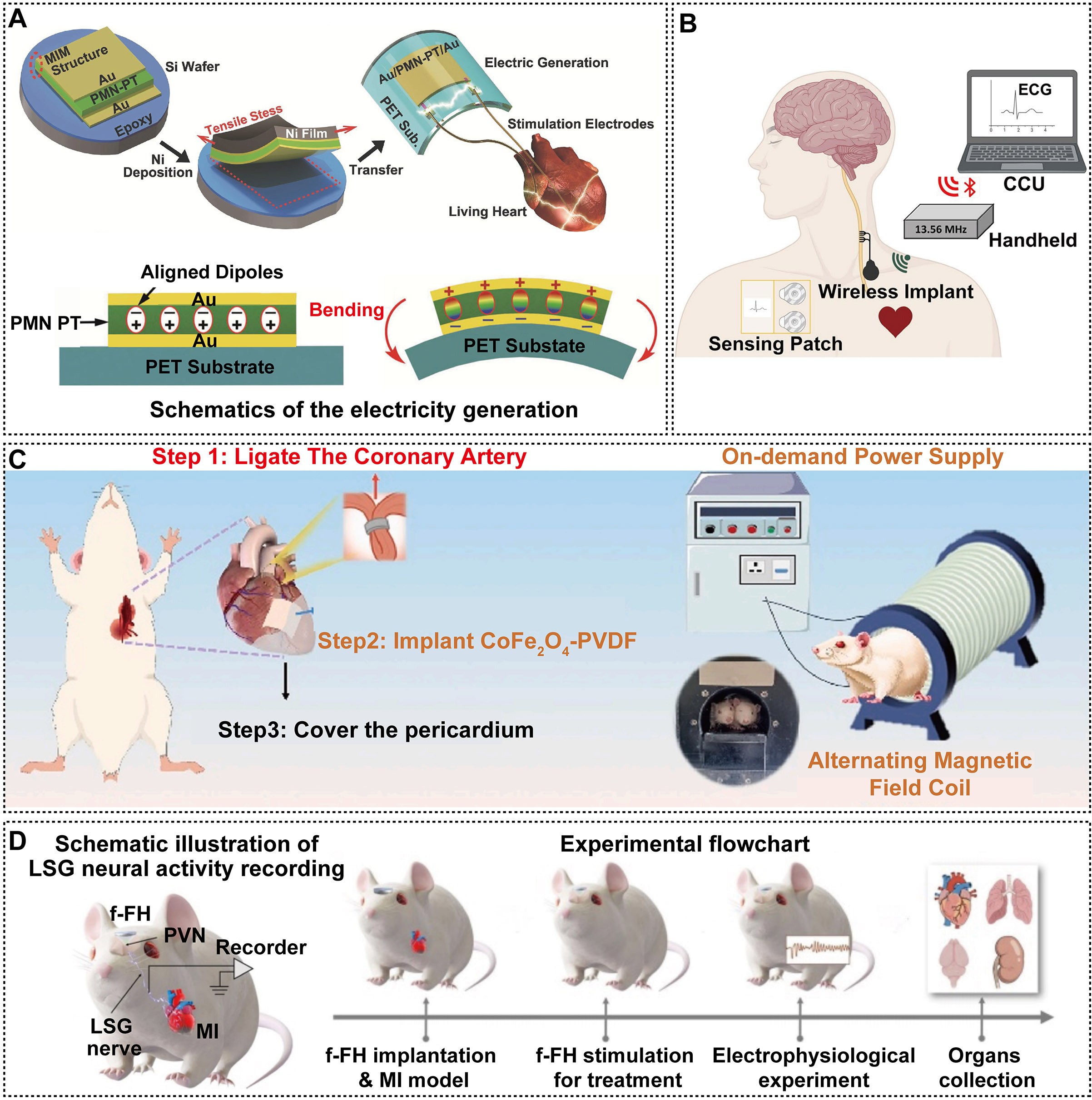
Figure 6. Representative applications of PENGs in cardiac neuromodulation. (A) Self-powered cardiac pacemaker enabled by a flexible single-crystalline PMN-PT piezoelectric energy harvester attached to the beating heart, adapted with permission from Ref.[151]. Copyright © 2014 Wiley-VCH. (B) Fully automated wireless vagus nerve stimulation (FAW-VNS) system incorporating electrocardiogram (ECG) sensing, closed-loop control, and a 13.56-MHz wirelessly powered implant, Adapted from Ref.[154] under CC BY 4.0 license. (C) Magneto-responsive piezoelectric CoFee2Oe4-PVDF system for on-demand electrical modulation of the infarcted myocardium, adapted from Ref.[155] under CC BY 4.0 license. (D) Schematic illustration of ultrasound-driven paraventricular nucleus (PVN) neuromodulation using a KNN-based ferroelectric heterojunction for myocardial infarction treatment, with left stellate ganglion (LSG) neural activity recording, adapted from Ref.[156] under CC BY 4.0 license.
Gastrointestinal neuromodulation
Gastrointestinal neuromodulation exploits continuous gastrointestinal deformation and accessible mucosal interfaces to convert mechanical energy into localized electrical stimulation or physiological-state signals for metabolic, motility, and neuroimmune interventions. Early studies first established the piezoelectric readout of gastrointestinal motility[159-163]. Li et al. constructed an adherent PENG from PLLA/glycine core-shell nanofibers, stabilizing the piezoelectric β-phase of PLLA and enabling the device to maintain a consistent electromechanical response under wet physiological conditions and deformation along the curved colonic surface[27]. Following implantation onto the transverse colon of rats, the device recorded distinct peristaltic signals during healthy, colitic, and remission states, thereby advancing gastrointestinal motility monitoring toward the identification of disease progression. Dai et al. embedded Rochelle salt within PLLA nanofibers to fabricate a biodegradable three-channel sensor with a d33 coefficient of 43.1 pC N-1[14]. When implanted on the rabbit colon, the device wirelessly resolved the amplitude, frequency, and propagation velocity of peristaltic contractions, thereby transforming motility signals into spatially resolved inputs for gastrointestinal regulation. Once physiological-state encoding had been established, piezoelectric outputs began to be translated into therapeutic stimulation. Poling et al. activated a P(VDF-TrFE) nanofibrous scaffold using low-intensity pulsed ultrasound, promoting cell proliferation in human intestinal organoids and demonstrating that gastrointestinal tissue models can respond to remotely delivered piezoelectric stimulation[164]. Building on this concept, Mac et al. conjugated capsaicin to BaTiO3 particles to target transient receptor potential vanilloid 1-positive sensory terminals in the gastric mucosa[165]. Peristaltic motion activated the particles to generate piezoelectric signals, which enhanced vagal afferent signaling of satiety and improved body-weight control and glucose-lipid metabolism in obese mice. A subsequent study further employed ultrasound-activated, gastric mucosa-adherent piezoelectric particles, extending localized piezoelectric stimulation to vagus nerve-mediated immunomodulation. In septic mice, this strategy reduced systemic inflammation and organ injury while improving survival[166]. Within non-piezoelectric gastrointestinal neuroelectronics, Srinivasan et al. further developed an integrated neuroprosthetic platform combining sensing, stimulation, and endoscopic implantation[167]. The system used multichannel inputs to drive the myenteric plexus and restore coordinated peristalsis, providing a transferable system architecture for the future integration of PENGs into closed-loop gastrointestinal neuromodulation.
Neurodegenerative diseases
Parkinson’s disease (PD)
PD is the second most common neurodegenerative disorder and is characterized by the progressive degeneration of dopaminergic neurons in the substantia nigra[168]. The resulting dopamine deficiency profoundly impairs motor function and also affects mood, cognition, gastrointestinal function, and olfaction. PENG-based research for PD has progressed sequentially through advances in acoustoelectric conversion, intracerebral delivery, circuit-specific targeting, and stimulation selectivity. The central objective is to achieve stable, precise, and sustained deep-brain neuromodulation while minimizing the burden associated with implanted hardware. Zhao et al. first fabricated carbon-coated BaTiO3 (barium titanate) (C@BT) piezoelectric nanoparticles from a BaTiO3/carbon composite to improve acoustic impedance matching with brain tissue[169]. Under 1-MHz ultrasound at
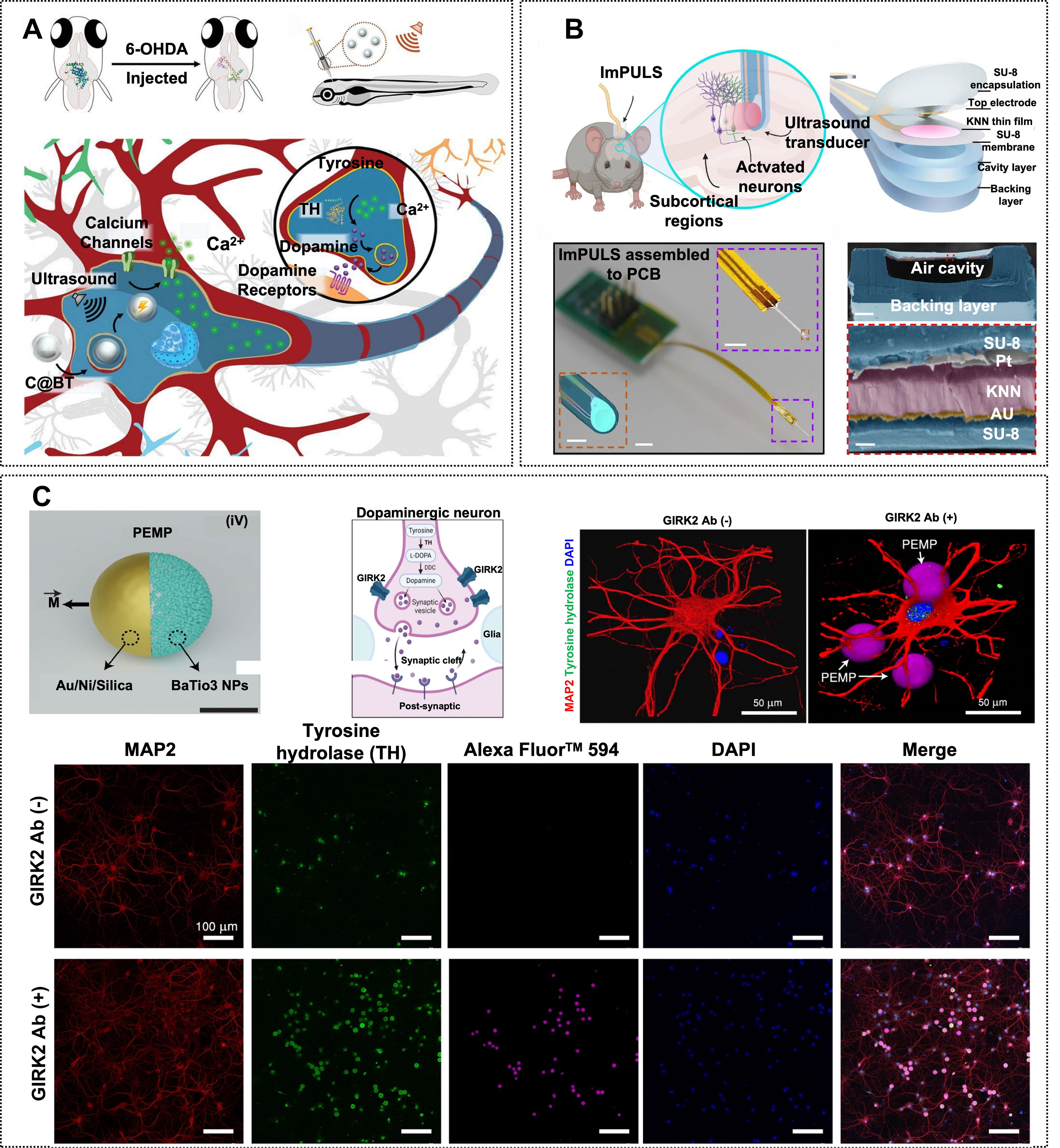
Figure 7. Representative applications of PENG-based neuromodulation for Parkinson's disease. (A) Recovery of degenerating midbrain dopaminergic neurons mediated by ultrasound-driven C@BT piezoelectric nanoparticles, adapted with permission from Ref.[169]. Copyright © 2020 Wiley-VCH. (B) Representative design and subcortical implantation of an implantable piezoelectric ultrasound stimulator (ImPULS) for wireless deep-brain activation, adapted from Ref.[170] under CC BY 4.0 license. (C) Specific targeting of dopaminergic neurons using piezoelectric magnetic Janus microparticles (PEMPs) and Immunofluorescence image of neurons, adapted from Ref.[97], under CC BY 4.0 license.
Alzheimer’s disease (AD)
AD is the most common cause of dementia and is characterized by progressive decline in cognitive and memory functions, predominantly affecting older adults[173]. Extensive evidence indicates that the abnormal accumulation of self-assembled amyloid-β (Aβ) species, including oligomers, fibrils, and plaques, constitutes a major pathological hallmark of AD[174]. By converting mechanical energy into electrochemical reactions or patterned electrical stimulation, PENGs offer a new therapeutic strategy that may simultaneously promote pathological protein clearance and cognitive recovery. Jang et al. first used piezoelectric BiOCl nanosheets, which dissociated Aβ aggregates through ultrasound-induced redox reactions and destabilized the β-sheet structure of Aβ fibrils[175]. However, freely dispersed particles offered limited control over lesion-site exposure and reactive oxygen species dosage. To improve spatial targeting, Ning et al. assembled Fe3O4 and BaTiO3 on a helical biological template to construct a magnetically guided, ultrasound-activated PiezoBOT. The system reduced the size of Aβ aggregates by more than 80% within 10 min, although the evidence remained limited to in vitro disaggregation[176]. For wireless deep-brain intervention, Wang et al. developed a flexible, lead-free dual-frequency piezoelectric implant that was ultrasonically powered to generate programmable biphasic pulses for wireless deep-brain stimulation in rats [Figure 8A][24]. Sharma et al. subsequently employed polydopamine-coated PVDF nanospheres, which, upon acoustic activation, promoted fibril disassembly, attenuated neurotoxicity, and improved cognitive performance in cellular and animal models, thereby extending therapeutic validation to disease-level phenotypes[177]. In parallel, transcranial ultrasound stimulation pulsed at 40 Hz improves cognition and alleviates neuroinflammation in a mouse model of Alzheimer’s disease [Figure 8B][178]. To clarify and calibrate these material-mediated effects,
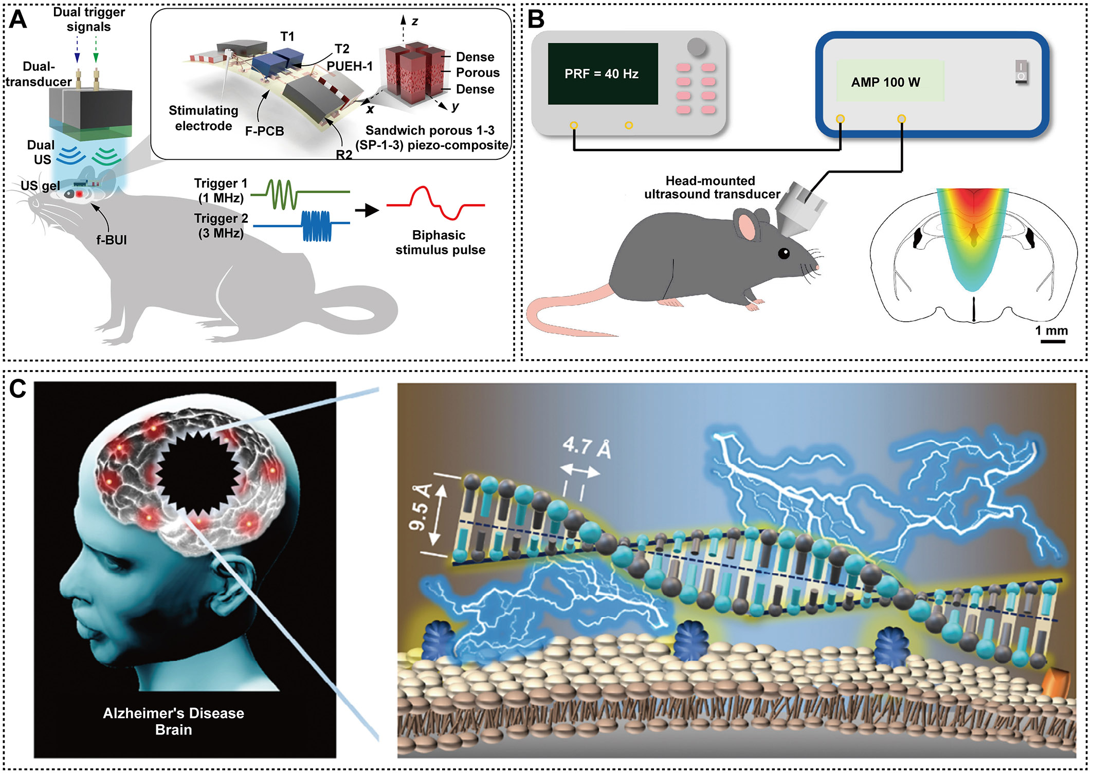
Figure 8. Representative piezoelectric mechanisms and ultrasound-enabled neuromodulation strategies relevant to Alzheimer’s disease. (A) Flexible lead-free dual-frequency piezoelectric ultrasound implant for wireless, biphasic deep-brain stimulation, adapted from Ref.[24] under CC BY 4.0 license. (B) Transcranial ultrasound stimulation pulsed at 40 Hz improves cognition and reduces neuroinflammation in female mice with Alzheimer's disease, adapted from Ref.[178], under CC BY 4.0 license. (C) Giant piezoelectricity of amyloid-β (Aβ) fibrils in Alzheimer's disease: monomeric Aβ peptides assemble into crystalline Aβ fibrils by forming dimers spaced 9.5 Å and stacking dimers spaced 4.7 Å, and the accumulation of these fibrils is associated with ion-channel hyperexcitability and neurodegeneration, adapted from Ref.[179], under CC BY 4.0 license.
Neural interfaces
Visual and auditory neural interfaces
Visual and auditory neural interfaces use piezoelectric transduction to convert images or acoustic vibrations into electrical signals that can be decoded by residual sensory pathways. Research in this area has progressed from single-site signal conversion toward spatial addressing, frequency discrimination, and validation of neural output. In the visual domain, Qian et al. fabricated a 3.1-MHz focused transducer from DL-47 piezoelectric ceramic to stimulate the retina of Royal College of Surgeons blind rats, achieving a spatial resolution of 250 μm and a frame rate of 5 Hz, as verified by neural responses recorded in the superior colliculus and primary visual cortex (V1)[184]. Jiang et al. subsequently constructed a 32-pixel flexible piezoelectric retinal array using a 1-3 PMN-PT/epoxy composite[113]. The independently addressable pixels provided tunable outputs ranging from 0 to 4.3 V and from 0 to 22.6 mW cm-2, encompassing the previously reported retinal stimulation charge threshold of 1-50 nC, and elicited spatially patterned neural responses in ex vivo retinas. Lu et al. developed a 16 × 16, 4.5-MHz integrated imaging-stimulation array that enabled ocular localization, stimulation with arbitrary two-dimensional patterns, and mapping of superior colliculus responses in blind rats[185]. The neural response maps exhibited a similarity of 0.91 ± 0.03 to the input patterns. When an independent 20-MHz focused transducer was used, the lateral full width at half maximum was further reduced to 81 μm. In the auditory domain, Yüksel et al. fabricated an eight-channel microelectromechanical systems transducer from thin-film PZT, covering a frequency range of 250-5,
Somatosensory interfaces
Somatosensory interfaces convert pressure, strain, and slip into neurally interpretable signals through PENG-based transduction and pulse encoding, with applications progressing from unit-level sensing toward tactile restoration and prosthetic control[190-192]. Kumar et al. constructed a piezoelectric synapse based on a NiO/ZnO heterostructure, in which mechanical strain modulated excitatory and inhibitory responses as well as short-term plasticity, thereby integrating tactile sensing with synaptic computation within a single functional unit[193]. Zhang et al. coupled a passive piezoelectric element to an NbOx Mott memristor to construct an artificial afferent nerve. The system exhibited a threshold voltage of approximately 2.05 V, consumed 38 pJ per pulse, and generated pulse trains spanning 0-1,100 Hz, thereby encoding pressure intensity as firing frequency[194]. Birkoben et al. fabricated a PiezoFET using a 500-nm Al0.73Sc0.27N film. Under a load of approximately 50 mN, the device produced adaptive pulses with a progressively decreasing firing frequency, mimicking the physiological adaptation to sustained tactile pressure[195]. Waseem et al. constructed rapidly and slowly adapting piezoelectric mechanoreceptors using intrinsic and Mg-doped GaN nanowires. At 1 Hz, the devices exhibited response and recovery times of 11 and 18 ms, respectively, and distinguished static grasping from dynamic texture exploration[196]. Wang et al. subsequently integrated PLLA, thin-film transistor (TFT), and PVDF-TrFE arrays to detect pressure, temperature, and slip, enabling the reconstruction of contact location, object shape, and surface texture[197]. Li et al. developed a tactile scanner by integrating a 10 × 10 PVDF array with TFT-based neuromorphic circuitry. At a scanning speed of 15 mm s-1, the system classified five texture categories with an accuracy of 85% and achieved a correlation coefficient of 0.68 between acquired and reproduced tactile signals. Moreover, more than 75% of participants successfully distinguished the reconstructed levels of surface roughness[198]. For prosthetic control, Jiang et al. fabricated a flexible PENG from porous P(VDF-TrFE), achieving a sensitivity of 38.4 mV kPa-1 and stable performance after 10,000 loading cycles. The device converted muscle deformation into control commands for robotic-hand grasping[199]. Wang et al. further developed a dual-modal electronic skin by integrating PVDF-TrFE/2DBP with a TFT array[200]. Across a pressure range of 0-310 kPa, the system achieved a sensitivity of 3.2 mV kPa-1, a spatial resolution of 50 μm, and a response time of 40 ms, while maintaining stable performance after 1,500 pressure-loading cycles and 4,000 bending cycles[201]. These interfaces have already enabled mechanical sensing, event encoding, and motor control. However, current studies remain largely confined to neuromorphic circuits, robotic systems, and body-surface interactions[202]. Future research should prioritize long-term, bidirectional closed-loop validation that integrates sensory detection, implanted stimulation of sensory nerves, and in vivo perceptual and behavioral outcomes.
CONCLUSIONS AND PERSPECTIVES
PENGs have advanced from energy-harvesting demonstrations to functional neural interfaces across the peripheral, central, and autonomic nervous systems, and cellular and animal studies have established their capacity to promote axonal regeneration, remyelination, and functional recovery. A growing number of systems now integrate sensing, closed-loop control, and biodegradable construction, yet the field has not produced a single clinical trial, and the reasons are instructive. Most studies still characterize devices by open-circuit voltage, short-circuit current, or power density, parameters that describe the generator rather than the stimulus. The biological response is governed by the charge delivered per phase, the charge density at the electrode-tissue interface, the field strength at the target, and the temporal structure of the pulse train, none of which can be recovered from a single voltage reading. Because PENGs, TENGs, magnetoelectric generators, and biofuel cells differ fundamentally in output impedance, waveform, and load dependence, their power figures are not interchangeable, and device-level output cannot be equated with the biologically effective dose[93-95]. The field therefore needs a reporting framework in which delivered charge, charge density, field strength, pulse parameters, cumulative dose, and electrode characteristics are stated together with functional outcomes and anchored to the safety limits established for conventional neurostimulation, in particular the Shannon criterion and the charge-injection capacities of electrode materials[114,116,117]. The checklist summarized in Table 3 is intended to provide such a framework, so that dose-response relationships become quantitatively comparable across studies and therapeutic windows can be defined before clinical protocols are designed.
Biodegradability illustrates a second, subtler tension. Resorbable PENGs eliminate the need for device removal, but the device must retain its piezoelectric output for the duration of the therapeutic window and then clear from the tissue without adverse effects. PLLA-based systems typically degrade over 12-24 months, whereas nerve regeneration in most experimental models requires electrical stimulation for only 3-6 months[203]. A device that loses its output before repair is complete fails therapeutically; one that persists beyond recovery leaves unnecessary foreign material in the body. Degradation kinetics can in principle be programmed to match the therapeutic window by tuning molecular weight, crystallinity, copolymer composition, crosslinking density, and encapsulation design, yet such programming remains largely empirical, and chronic studies must confront failure modes that are rarely reported, including encapsulation breakdown, electrode corrosion and delamination, and interfacial failure under cyclic mechanical loading, each of which can terminate function before nominal degradation is complete. Reporting degradation profiles, failure modes, and functional lifetime together would make the reliability of biodegradable PENGs directly comparable across studies and would allow device design to be matched to the repair process rather than to the material alone.
Extending PENGs from peripheral nerve repair to central neuromodulation raises the question of targeting. Deep brain regions, the spinal cord, and autonomic ganglia are heterogeneous in structure and function, and a diffusing electric field can readily activate off-target circuits. Focused ultrasound offers a spatially confined energy source, but acoustic access to deep structures is constrained by attenuation and phase aberration through the skull and intervening tissue, and stimulation parameters, including frequency, current density, and pulse width, remain difficult to regulate precisely without auxiliary circuitry. Progress will require the integration of acoustic-field modeling, image-guided targeting, and localized electric-field simulation with real-time electrophysiological feedback, so that the activated tissue volume can be predicted and verified. The chronic environment imposes further constraints: fluctuations in pH, ionic strength, enzymatic activity, and mechanical loading alter material structure and interfacial properties, so that in vitro performance frequently diverges from in vivo behavior. Standardized manufacturing procedures, quality-control specifications, and unified validation protocols are therefore prerequisites for consistent electromechanical performance, mechanical compliance, and long-term reliability.
The path forward is likely to be multimodal and closed-loop rather than a refinement of individual devices. Because no single transduction mechanism simultaneously provides high power and deep-tissue access, hybrid systems combining piezoelectric, triboelectric, magnetomechanical, and electromagnetic-induction elements with biofuel cells and microscale energy storage offer a rational response to the intermittency of physiological motion and the limited reach of external triggers[93-95]. Intelligent energy management can match harvested energy to stimulation demand, and closed-loop feedback can adjust stimulation parameters according to neural activity and tissue state, allowing device output to track biological response in real time. Mechanistically, the pathways through which piezoelectric stimulation acts, including voltage-gated channel recruitment, Ca2+ signaling, and downstream transcriptional programs, are understood in outline, but the interconnected neural, immune, and metabolic responses remain largely uncharted, and transcriptomic, proteomic, and metabolomic analyses will be needed to map them. Computational design that couples finite-element modeling of electromechanical and acoustic fields with machine-learning-based optimization could reduce the empirical iteration now required to match device output to a defined dose. The convergence of PENGs with iontronic logic and 2D-material biosensors points toward adaptive interfaces that combine energy harvesting, neural-state sensing, and closed-loop decision-making in a single biocompatible platform[204,205]. Integration with genetic, immune, and metabolic reprogramming, including sonogenetics, immune-modulatory vagus stimulation, and bioenergetic activation, offers therapeutic possibilities beyond electrical excitation alone. With coordinated progress in materials science, neurobiology, and clinical engineering, PENGs are well positioned to move from proof-of-concept devices toward clinically valuable self-powered neural interfaces.
DECLARATIONS
Acknowledgments
The graphical abstract was created using BioRender. Li, S. (2026) https://BioRender.com/7876h9j.
Authors’ contributions
Made substantial contributions to the conception and design of the study: Li, S.; Zhang, K.; Zhang, H.
Performed literature search, data organization and initial draft preparation: Li, S.; Li, W.; Yang, J.
Revised and polished the manuscript: Chen, S.; Li, W.
Provided funding acquisition and supervision: Zhang, K.; Zhang, H.
All authors read and approved the final manuscript.
Availability of data and materials
Not applicable.
AI and AI-assisted tools statement
Not applicable.
Financial support and sponsorship
This work was supported by Bingtuan Science and Technology Program Project (2025YD013, 2025AB083, 2024AB034); National Natural Science Foundation of China (32372973, 32060811); Tianchi Talent Project (CZ004311); Shihezi University International Science and Technology Cooperation Promotion Plan Project (Grant No. GJHZ202402); China Agriculture Research System earmarked fund (CARS-43-D13); Natural Science Foundation of Sichuan Province (Grant Number. 2026NSFSC1648); China Postdoctoral Science Foundation (Grant No.2025M782937); Sichuan Academy of Medical Sciences & Sichuan Provincial People’s Hospital Foundation (Grant Number. 25QNPY003).
Conflicts of interest
All authors declared that there are no conflicts of interest.
Ethical approval and consent to participate
Not applicable.
Consent for publication
Not applicable.
Copyright
© The Author(s) 2026.
REFERENCES
1. Xiao, L.; Yin, B.; Geng, Z.; Li, J.; Jia, R.; Zhang, K. Flexible wearable devices based on self-powered energy supply. Nano. Energy. 2025, 142, 111157.
2. Jin, S.; Yu, J.; Kan, C. W.; Zhang, C.; Tsoi, J. K. Self-powered and biocompatible electrospun PVDF/ZrO2 based piezoelectric energy harvesting device for oral health monitoring and dental care. Dent. Mater. 2026, 42, 1476-87.
3. Wang, Z. L.; Song, J. Piezoelectric nanogenerators based on zinc oxide nanowire arrays. Science 2006, 312, 242-6.
4. Pawar, O. Y.; Lu, B.; Lim, S. Enhanced flexible piezoelectric nanogenerators using ethanol-exfoliated g-C3N4/PVDF composites via 3D printing for self-powered applications. Nanomaterials 2024, 14, 1578.
5. Gołąbek, J.; Strankowski, M. A review of recent advances in human-motion energy harvesting nanogenerators, self-powering smart sensors and self-charging electronics. Sensors 2024, 24, 1069.
6. Li, Z.; Zhu, G.; Yang, R.; Wang, A. C.; Wang, Z. L. Muscle-driven in vivo nanogenerator. Adv. Mater. 2010, 22, 2534-7.
7. Marino, A.; Arai, S.; Hou, Y.; et al. Piezoelectric nanoparticle-assisted wireless neuronal stimulation. ACS. Nano. 2015, 9, 7678-89.
8. Qian, Y.; Cheng, Y.; Song, J.; et al. Mechano-informed biomimetic polymer scaffolds by incorporating self-powered zinc oxide nanogenerators enhance motor recovery and neural function. Small 2020, 16, e2000796.
9. Chen, P.; Wu, P.; Wan, X.; et al. Ultrasound-driven electrical stimulation of peripheral nerves based on implantable piezoelectric thin film nanogenerators. Nano. Energy. 2021, 86, 106123.
10. Wu, P.; Chen, P.; Xu, C.; et al. Ultrasound-driven in vivo electrical stimulation based on biodegradable piezoelectric nanogenerators for enhancing and monitoring the nerve tissue repair. Nano. Energy. 2022, 102, 107707.
11. Chen, P.; Cheng, C.; Yang, X.; et al. Wireless deep brain stimulation by ultrasound-responsive molecular piezoelectric nanogenerators. ACS. Nano. 2023, 17, 25625-37.
12. Li, T.; Wei, Z.; Jin, F.; et al. Soft ferroelectret ultrasound receiver for targeted peripheral neuromodulation. Nat. Commun. 2023, 14, 8386.
13. Zeng, Y.; Gong, C.; Lu, G.; et al. A programmable and self-adaptive ultrasonic wireless implant for personalized chronic pain management. Nat. Electron. 2025, 8, 437-49.
14. Dai, F.; Cheng, H.; Qi, H.; et al. Rochelle salt-based biodegradable piezoelectric devices for nerve regeneration and intestinal motility monitoring. Nat. Commun. 2026, 17, 2169.
15. Zhao, D.; Zhong, G.; Cao, Z.; et al. Piezoelectric neuromodulation of the subthalamic nucleus ameliorates motor and nonmotor symptoms of Parkinson’s disease. Sci. Adv. 2026, 12, eaec6955.
16. Yang, X.; Huo, N.; Zhou, H.; Li, S.; Fang, M.; Zhou, N. Application strategies of autologous and decellularized nerve grafts: structural and functional recovery. Neural. Regen. Res. 2026, 21, 2843-62.
17. Li, K.; Wu, X.; Xiong, P.; et al. Electroactive dual-drug hydrogel-PLA conduit for nerve regeneration. J. Biomed. Mater. Res. B. Appl. Biomater. 2026, 114, e70066.
18. Ghanem, W.; Ezzeddine, H.; Najjar, J.; et al. Vascularized nerve grafts: current concepts, indications, and future perspectives. Orthop. Rev. 2026, 18, 156463.
19. Gungordu Er, S.; Edirisinghe, M. Graphene for self-powered biosensors: a perspective. J. R. Soc. Interface. 2025, 22, 20250479.
20. Almas, T.; Haider, R.; Malik, J.; et al. Nanotechnology in interventional cardiology: a state-of-the-art review. Int. J. Cardiol. Heart. Vasc. 2022, 43, 101149.
21. Kim, T.; Kim, H. J.; Choi, W.; et al. Deep brain stimulation by blood-brain-barrier-crossing piezoelectric nanoparticles generating current and nitric oxide under focused ultrasound. Nat. Biomed. Eng. 2023, 7, 149-63.
22. Lans, J.; Eberlin, K. R.; Evans, P. J.; Mercer, D.; Greenberg, J. A.; Styron, J. F. A systematic review and meta-analysis of nerve gap repair: comparative effectiveness of allografts, autografts, and conduits. Plast. Reconstr. Surg. 2023, 151, 814e-27e.
23. Xu, D.; Fu, S.; Zhang, H.; et al. Ultrasound-responsive aligned piezoelectric nanofibers derived hydrogel conduits for peripheral nerve regeneration. Adv. Mater. 2024, 36, e2307896.
24. Wang, Q.; Zhang, Y.; Xue, H.; et al. Lead-free dual-frequency ultrasound implants for wireless, biphasic deep brain stimulation. Nat. Commun. 2024, 15, 4017.
25. Wang, L.; Li, X.; Jin, Q.; et al. Ultrasound-driven piezoelectret-based nanogenerator stimulates vagus nerve for myocardial infarction treatment. Nano. Energy. 2024, 128, 109864.
26. Chen, Y. X.; Feng, P. J.; Zhong, G.; et al. Piezoelectric nanogenerators enabled neuromodulation rescued dopaminergic neuron loss in Parkinson’s disease. Nano. Energy. 2024, 121, 109187.
27. Li, T.; Yuan, Y.; Gu, L.; et al. Ultrastable piezoelectric biomaterial nanofibers and fabrics as an implantable and conformal electromechanical sensor patch. Sci. Adv. 2024, 10, eadn8706.
28. Shan, Y.; Wang, E.; Cui, X.; et al. A biodegradable piezoelectric sensor for real-time evaluation of the motor function recovery after nerve injury. Adv. Funct. Mater. 2024, 34, 2400295.
29. Wu, R. T.; Lu, J. C. Y. Nerve autograft: preservation of a lost art. Hand. Clin. 2024, 40, 347-56.
30. Dishon Ben Ami, S.; Pinsk, N.; Hartstein, M.; et al. Piezoelectricity from dopant-induced structural distortions in molecular crystals revealed by Raman spectroscopy. J. Am. Chem. Soc. 2026, 148, 30029-36.
31. Pan, X.; Wu, Y.; Wang, Y.; Zhou, G.; Cai, H. Mechanical energy harvesting based on the piezoelectric materials: recent advances and future perspectives. Chem. Eng. J. 2024, 497, 154249.
32. Deng, W.; Zhou, Y.; Libanori, A.; Chen, G.; Yang, W.; Chen, J. Piezoelectric nanogenerators for personalized healthcare. Chem. Soc. Rev. 2022, 51, 3380-435.
33. Zhang, Z.; Wang, Z.; Li, X.; Zheng, Y.; Yang, Z. Design and manufacturing of piezoelectric biomaterials for bioelectronics and biomedical applications. Chem. Rev. 2025, 125, 9875-929.
34. Jeong, C. K.; Park, K. I.; Ryu, J.; Hwang, G. T.; Lee, K. J. Large-area and flexible lead-free nanocomposite generator using alkaline niobate particles and metal nanorod filler. Adv. Funct. Mater. 2014, 24, 2620-9.
35. Liu, Z.; Zhou, Y.; Qu, X.; et al. A self-powered optogenetic system for implantable blood glucose control. Research 2022, 2022, 9864734.
36. Mahapatra, S. D.; Mohapatra, P. C.; Aria, A. I.; et al. Piezoelectric materials for energy harvesting and sensing applications: roadmap for future smart materials. Adv. Sci. 2021, 8, e2100864.
37. Zhang, Y.; Kim, H.; Wang, Q.; et al. Progress in lead-free piezoelectric nanofiller materials and related composite nanogenerator devices. Nanoscale. Adv. 2020, 2, 3131-49.
38. Jeon, S.; Kim, D.; Jo, M. Y.; et al. Wireless acousto-piezoelectric conduit with aligned nanofibers for neural regeneration. Adv. Mater. 2025, 37, e03343.
39. Pathak, P. K.; Dasgupta Ghosh, B. Cellulose-based biodegradable and flexible piezoelectric materials toward energy-harvesting systems: a review. ACS. Biomater. Sci. Eng. 2026, 12, 2825-50.
40. KarbalaeiMahdi, A.; Moridi, K.; Esmaeili, Z.; Seifalian, N.; Chauhan, N. P. S.; Seifalian, A. M. Piezoelectric materials for tissue engineering and regenerative medicine: from fundamental principles to clinical applications. Acta. Biomater. 2026, 217, 44-70.
41. Nain, A.; Chakraborty, S.; Barman, S. R.; et al. Progress in the development of piezoelectric biomaterials for tissue remodeling. Biomaterials 2024, 307, 122528.
42. Krutko, M.; Poling, H. M.; Sheth, M.; et al. Thermal annealing enhances piezoelectricity and regenerative potential of PVDF-TrFE nanofiber scaffolds. Adv. Mater. Technol. 2025, 10, e01513.
43. Westphal, J. A.; Bryan, A. E.; Krutko, M.; Esfandiari, L.; Schutte, S. C.; Harris, G. M. Innervation of an ultrasound-mediated PVDF-TrFE scaffold for skin-tissue engineering. Biomimetics 2023, 9, 2.
44. Gryshkov, O.; Al Halabi, F.; Kuhn, A. I.; et al. PVDF and P(VDF-TrFE) electrospun scaffolds for nerve graft engineering: a comparative study on piezoelectric and structural properties, and in vitro biocompatibility. Int. J. Mol. Sci. 2021, 22, 11373.
45. Gaur, A.; Abebe, A.; Porwal, C.; et al. Interfacial Schottky junction induced enhanced piezocatalytic activity. Sci. Rep. 2026, 16, 22065.
46. Aphayvong, S.; Verma, A.; Nakamura, K.; et al. Polarity-dependent charge transport and resistance degradation enabled by oxygen-vacancy gradients in BiFeO3 films. ACS. Appl. Mater. Interfaces. 2026, 18, 36891-901.
47. Fan, L.; Dai, Z.; Bai, X.; et al. (K,Na)NbO3-based lead-free ceramic/polyvinylidene fluoride piezoelectric composite for biomimetic wound healing via endogenous electric field simulation. Biomater. Adv. 2026, 186, 214909.
48. Bian, T.; Jiang, Y.; Cao, J.; Wu, W.; Zhang, L.; Yang, Y. Fabrication of piezoelectric/conductive composite nerve conduits for peripheral nerve regeneration. Colloids. Surf. B. Biointerfaces. 2025, 250, 114544.
49. Kim, H.; Narayanan, K. B.; Kumar, V.; Han, S. S.; Bhaskar, R. Piezoelectric nanocomposite patch for self-powered bioelectrical stimulation for cardiac tissue engineering. Biomater. Adv. 2026, 182, 214717.
50. Mistewicz, K.; Jesionek, M.; Kim, H. J.; et al. Nanogenerator for determination of acoustic power in ultrasonic reactors. Ultrason. Sonochem. 2021, 78, 105718.
51. Wu, S.; Chen, M. S.; Maurel, P.; Lee, Y. S.; Bunge, M. B.; Arinzeh, T. L. Aligned fibrous PVDF-TrFE scaffolds with Schwann cells support neurite extension and myelination in vitro. J. Neural. Eng. 2018, 15, 056010.
52. Curry, E. J.; Le, T. T.; Das, R.; et al. Biodegradable nanofiber-based piezoelectric transducer. Proc. Natl. Acad. Sci. USA. 2020, 117, 214-20.
53. Demirbilek, M.; Sakar, M.; Karahaliloğlu, Z.; et al. Aligned bacterial PHBV nanofibrous conduit for peripheral nerve regeneration. Artif. Cells. Nanomed. Biotechnol. 2015, 43, 243-51.
54. Karbowniczek, J. E.; İlhan, P.; Szewczyk, P. K.; et al. A sustainable alternative to PVDF for neural tissue engineering via piezoelectric PHBV and cellulose acetate fibers. ACS. Biomater. Sci. Eng. 2026, 12, 2684-99.
55. Ceballos, D.; Navarro, X.; Dubey, N.; Wendelschafer-Crabb, G.; Kennedy, W. R.; Tranquillo, R. T. Magnetically aligned collagen gel filling a collagen nerve guide improves peripheral nerve regeneration. Exp. Neurol. 1999, 158, 290-300.
56. Bera, S.; Guerin, S.; Yuan, H.; et al. Molecular engineering of piezoelectricity in collagen-mimicking peptide assemblies. Nat. Commun. 2021, 12, 2634.
57. Jin, L.; Tai, Y.; Nam, J. Piezoelectric silk fibroin nanofibers: structural optimization to enhance piezoelectricity and biostability for neural tissue engineering. Nano. Energy. 2024, 132, 110367.
58. Zhen, W.; Chen, J.; Fan, S.; Zhang, Y. Piezoelectric fibers based on silk fibroin with excellent output performance. Chin. Phys. B. 2024, 33, 088701.
59. Zhu, G.; Wang, A. C.; Liu, Y.; Zhou, Y.; Wang, Z. L. Functional electrical stimulation by nanogenerator with 58 V output voltage. Nano. Lett. 2012, 12, 3086-90.
60. Ciofani, G.; Danti, S.; D’Alessandro, D.; et al. Enhancement of neurite outgrowth in neuronal-like cells following boron nitride nanotube-mediated stimulation. ACS. Nano. 2010, 4, 6267-77.
61. Eslami, R.; Malekkhouyan, A.; Santhirakumaran, P.; Mehrvar, M.; Zarrin, H. High-performance flexible nanogenerators based on piezoelectric hBN-induced polyvinylidene fluoride nanofibers. Mater. Today. Chem. 2023, 30, 101609.
62. Luo, C.; Omelchenko, I.; Manson, R.; et al. Direct intracochlear acoustic stimulation using a PZT microactuator. Trends. Hear. 2015, 19, 2331216515616942.
63. Wen, J.; Liu, M. Piezoelectric ceramic (PZT) modulates axonal guidance growth of rat cortical neurons via RhoA, Rac1, and Cdc42 pathways. J. Mol. Neurosci. 2014, 52, 323-30.
64. Jang, J.; Jang, J. H.; Choi, H. MEMS flexible artificial basilar membrane fabricated from piezoelectric aluminum nitride on an SU-8 substrate. J. Micromech. Microeng. 2017, 27, 075006.
65. Zhao, C.; Knisely, K. E.; Colesa, D. J.; Pfingst, B. E.; Raphael, Y.; Grosh, K. Voltage readout from a piezoelectric intracochlear acoustic transducer implanted in a living guinea pig. Sci. Rep. 2019, 9, 3711.
66. Cheng, Y.; Xu, Y.; Qian, Y.; Chen, X.; Ouyang, Y. M.; Yuan, W. E. 3D structured self-powered PVDF/PCL scaffolds for peripheral nerve regeneratione. Nano. Energy. 2020, 69, 104411.
67. You, Y. M.; Liao, W. Q.; Zhao, D.; et al. An organic-inorganic perovskite ferroelectric with large piezoelectric response. Science 2017, 357, 306-9.
68. Ramburrun, P.; Kumar, P.; Choonara, Y. E.; du Toit, L. C.; Pillay, V. Design and characterisation of PHBV-magnesium oleate directional nanofibers for neurosupport. Biomed. Mater. 2019, 14, 065015.
69. Ensarioğlu, K.; Şen, M.; Yüzer, E.; et al. Bifunctional peptide-conjugated vertically aligned tubular nanofibrous conduits for enhanced vascularization and nerve regeneration. Biomater. Adv. 2026, 185, 214879.
70. Azimi, S.; Golabchi, A.; Nekookar, A.; et al. Self-powered cardiac pacemaker by piezoelectric polymer nanogenerator implant. Nano. Energy. 2021, 83, 105781.
71. Zhang, Y.; Zhou, L.; Gao, X.; et al. Performance-enhanced flexible piezoelectric nanogenerator via layer-by-layer assembly for self-powered vagal neuromodulation. Nano. Energy. 2021, 89, 106319.
72. Ma, Y.; Wang, H.; Wang, Q.; Cao, X.; Gao, H. Piezoelectric conduit combined with multi-channel conductive scaffold for peripheral nerve regeneration. Chem. Eng. J. 2023, 452, 139424.
73. Tai, Y.; Tonmoy, T. I.; Win, S.; Brinkley, N. T.; Park, B. H.; Nam, J. Enhanced peripheral nerve regeneration by mechano-electrical stimulation. NPJ. Regen. Med. 2023, 8, 57.
74. Qi, T.; Wang, X.; Han, Y.; He, C. Suture-free piezoelectric band-aid membrane for complex peripheral nerve defects. ACS. Appl. Mater. Interfaces. 2026, 18, 38678-94.
75. Singh, A. N.; Nam, K. W. Gel-based self-powered nanogenerators: materials, mechanisms, and emerging opportunities. Gels 2025, 11, 451.
76. Kumar, V.; Alam, M. N.; Azam, S.; Park, S. S. Robust composites based on silicone rubber for self-powered piezoelectric nanogenerators. Polymers 2025, 17, 977.
77. Li, Y.; Li, Y.; Zhang, R.; et al. Progress in wearable acoustical sensors for diagnostic applications. Biosens. Bioelectron. 2023, 237, 115509.
78. Wang, D.; Liu, W.; Gu, L.; et al. Instantaneous piezoelectric nanogenerator for pacemaker applications. Nano. Energy. 2025, 138, 110828.
79. Lu, B.; Chen, Y.; Ou, D.; et al. Ultra-flexible piezoelectric devices integrated with heart to harvest the biomechanical energy. Sci. Rep. 2015, 5, 16065.
80. Cheng, X.; Xue, X.; Ma, Y.; et al. Implantable and self-powered blood pressure monitoring based on a piezoelectric thinfilm: simulated, in vitro and in vivo studies. Nano. Energy. 2016, 22, 453-60.
81. Yu, Y.; Sun, H.; Orbay, H.; et al. Biocompatibility and in vivo operation of implantable mesoporous PVDF-based nanogenerators. Nano. Energy. 2016, 27, 275-81.
82. Zhang, Z.; Yao, C.; Yu, Y.; Hong, Z.; Zhi, M.; Wang, X. Mesoporous piezoelectric polymer composite films with tunable mechanical modulus for harvesting energy from liquid pressure fluctuation. Adv. Funct. Mater. 2016, 26, 6760-5.
83. Yang, T.; Pan, H.; Tian, G.; et al. Hierarchically structured PVDF/ZnO core-shell nanofibers for self-powered physiological monitoring electronics. Nano. Energy. 2020, 72, 104706.
84. Mao, R.; Yu, B.; Cui, J.; et al. Piezoelectric stimulation from electrospun composite nanofibers for rapid peripheral nerve regeneration. Nano. Energy. 2022, 98, 107322.
85. Razbin, M.; Ruan, K.; Tahir, D.; et al. Sustainable P3HB:ZnO composite piezoelectric nanofibers for AI-driven gait monitoring. Small. Sci. 2026, 6, e70355.
87. Nelson, T. R.; Fowlkes, J. B.; Abramowicz, J. S.; Church, C. C. Ultrasound biosafety considerations for the practicing sonographer and sonologist. J. Ultrasound. Med. 2009, 28, 139-50.
88. Fry, F. J.; Barger, J. E. Acoustical properties of the human skull. J. Acoust. Soc. Am. 1978, 63, 1576-90.
89. Zhang, T.; Liang, H.; Wang, Z.; et al. Piezoelectric ultrasound energy-harvesting device for deep brain stimulation and analgesia applications. Sci. Adv. 2022, 8, eabk0159.
90. Lozano, A. M.; Lipsman, N.; Bergman, H.; et al. Deep brain stimulation: current challenges and future directions. Nat. Rev. Neurol. 2019, 15, 148-60.
91. Montgomery, K. L.; Yeh, A. J.; Ho, J. S.; et al. Wirelessly powered, fully internal optogenetics for brain, spinal and peripheral circuits in mice. Nat. Methods. 2015, 12, 969-74.
92. Cafarelli, A.; Marino, A.; Vannozzi, L.; et al. Piezoelectric nanomaterials activated by ultrasound: the pathway from discovery to future clinical adoption. ACS. Nano. 2021, 15, 11066-86.
93. Xu, S.; Manshaii, F.; Xiao, X.; Yin, J.; Chen, J. Triboelectric nanogenerators for self-powered neurostimulation. Nano. Res. 2024, 17, 8926-41.
94. Kozielski, K. L.; Jahanshahi, A.; Gilbert, H. B.; et al. Nonresonant powering of injectable nanoelectrodes enables wireless deep brain stimulation in freely moving mice. Sci. Adv. 2021, 7, eabc4189.
95. Cosnier, S.; Le Goff, A.; Holzinger, M. Towards glucose biofuel cells implanted in human body for powering artificial organs: review. Electrochem. Commun. 2014, 38, 19-23.
96. He, Y.; Yang, X.; Yuan, M.; et al. Wireless discharge of piezoelectric nanogenerator opens voltage-gated ion channels for calcium overload-mediated tumor treatment. Biomaterials 2025, 321, 123311.
97. Han, M.; Yildiz, E.; Bozuyuk, U.; et al. Janus microparticles-based targeted and spatially-controlled piezoelectric neural stimulation via low-intensity focused ultrasound. Nat. Commun. 2024, 15, 2013.
98. Genchi, G. G.; Ceseracciu, L.; Marino, A.; et al. P(VDF-TrFE)/BaTiO3 nanoparticle composite films mediate piezoelectric stimulation and promote differentiation of SH-SY5Y neuroblastoma cells. Adv. Healthc. Mater. 2016, 5, 1808-20.
99. Hoop, M.; Chen, X. Z.; Ferrari, A.; et al. Ultrasound-mediated piezoelectric differentiation of neuron-like PC12 cells on PVDF membranes. Sci. Rep. 2017, 7, 4028.
100. Liu, L.; Chen, B.; Liu, K.; et al. Wireless manipulation of magnetic/piezoelectric micromotors for precise neural stem-like cell stimulation. Adv. Funct. Mater. 2020, 30, 1910108.
101. Fan, C. H.; Tsai, H. C.; Tsai, Y. S.; et al. Selective activation of cells by piezoelectric molybdenum disulfide nanosheets with focused ultrasound. ACS. Nano. 2023, 17, 9140-54.
102. Zhang, Y.; Jiang, J.; Xie, X.; et al. Cell-anchored lead-free piezoelectric KNN NPs resisting washout for low-intensity ultrasound driven neuromodulation. Adv. Funct. Mater. 2024, 34, 2406919.
103. Kong, Y.; Liu, F.; Ma, B.; et al. Wireless localized electrical stimulation generated by an ultrasound-driven piezoelectric discharge regulates proinflammatory macrophage polarization. Adv. Sci. 2021, 8, 2100962.
104. Gao, H.; Wu, W.; Chen, X.; et al. A sono-piezoelectric scaffold prompts disc regeneration by activating Ca2+/CaMKII/Parkin-mediated mitophagy. Biomaterials 2026, 334, 124316.
105. Liu, J.; Wang, X.; Wu, Z.; et al. Dynamic stimulation of piezoelectric scaffolds enhances osteogenesis-related biological responses via electro-mechanical sensitive channels and cytoskeletal remodeling. Biomaterials 2026, 329, 123938.
106. Jiang, F.; Shan, Y.; Tian, J.; et al. Poly(l-lactic acid) nanofiber-based multilayer film for the electrical stimulation of nerve cells. Adv. Mater. Interfaces. 2023, 10, 2202474.
107. Wayman, G. A.; Impey, S.; Marks, D.; et al. Activity-dependent dendritic arborization mediated by CaM-kinase I activation and enhanced CREB-dependent transcription of Wnt-2. Neuron 2006, 50, 897-909.
108. Yan, X.; Liu, J.; Ye, Z.; et al. CaMKII-mediated CREB phosphorylation is involved in Ca2+-induced BDNF mRNA transcription and neurite outgrowth promoted by electrical stimulation. PLoS. One. 2016, 11, e0162784.
109. Wang, W.; Liu, W.; Zhu, H.; et al. Electrical stimulation promotes BDNF expression in spinal cord neurons through Ca2+- and ERK-dependent signaling pathways. Cell. Mol. Neurobiol. 2011, 31, 459-67.
110. Spencer, T. K.; Mellado, W.; Filbin, M. T. BDNF activates CaMKIV and PKA in parallel to block MAG-mediated inhibition of neurite outgrowth. Mol. Cell. Neurosci. 2008, 38, 110-6.
111. Cohen, S. M.; Suutari, B.; He, X.; et al. Calmodulin shuttling mediates cytonuclear signaling to trigger experience-dependent transcription and memory. Nat. Commun. 2018, 9, 2451.
112. Huang, J.; Ye, Z.; Hu, X.; Lu, L.; Luo, Z. Electrical stimulation induces calcium-dependent release of NGF from cultured Schwann cells. Glia 2010, 58, 622-31.
113. Jiang, L.; Lu, G.; Zeng, Y.; et al. Flexible ultrasound-induced retinal stimulating piezo-arrays for biomimetic visual prostheses. Nat. Commun. 2022, 13, 3853.
114. Cogan, S. F. Neural stimulation and recording electrodes. Annu. Rev. Biomed. Eng. 2008, 10, 275-309.
115. Li, X.; Yan, S.; Li, M.; et al. Piezoelectric nanoparticle-driven rhythmic ultrasound neuromodulation for treatment of early-stage Alzheimer’s disease. Biomaterials 2026, 328, 123905.
116. Shannon, R. V. A model of safe levels for electrical stimulation. IEEE. Trans. Biomed. Eng. 1992, 39, 424-6.
117. McCreery, D. B.; Agnew, W. F.; Yuen, T. G. H.; Bullara, L. A. Charge density and charge per phase as cofactors in neural injury induced by electrical stimulation. IEEE. Trans. Biomed. Eng. 1990, 37, 996-1001.
118. Zhang, J.; Li, F.; Gao, X.; et al. Bamboo-inspired composite conduit accelerates peripheral nerve regeneration through synergistic oriented structure and piezoelectricity. Adv. Mater. 2026, 38, e09425.
119. Pi, W.; Rao, F.; Cao, J.; et al. Sono-electro-mechanical therapy for peripheral nerve regeneration through piezoelectric nanotracts. Nano. Today. 2023, 50, 101860.
120. Huang, L.; Zhu, L.; Shi, X.; et al. A compound scaffold with uniform longitudinally oriented guidance cues and a porous sheath promotes peripheral nerve regeneration in vivo. Acta. Biomater. 2018, 68, 223-36.
121. Chen, G.; Hu, Q.; Tang, C.; et al. Biodegradable piezoelectric amino acid nerve guidance conduit repairs long-gap nerve defect under low frequency vibration from massage gun. Adv. Funct. Mater. 2026, 36, e10947.
122. Li, J.; Wu, Y.; Huang, X.; Zhang, S.; Chang, S.; Wang, P. Liquid metal-ZnO enhanced piezoelectric PVDF nanofibrous nerve guidance conduit: self-powered electrical stimulation for accelerated peripheral nerve regeneration. Biomater. Adv. 2026, 180, 214566.
123. Zhou, D.; Bian, M.; Wei, L.; et al. Stepwise regulation of cellular oxidative stress via conductive-piezoelectric integrated microstructured conduits for enhanced nerve regeneration. Adv. Sci. 2026, 13, e16124.
124. Qi, T.; Wang, X.; Wu, L.; et al. Multidimensional oriented piezoelectric conduits for peripheral nerve defect regeneration. Small 2026, 22, e14376.
125. Yin, X.; Liu, Y.; Zhan, G.; et al. A self-powered biodegradable electroactive conduit with sustained NGF release for enhanced peripheral nerve regeneration via mitochondrial bioenergetic activation. ACS. Appl. Mater. Interfaces. 2026, 18, 29676-94.
126. Jin, F.; Li, T.; Wei, Z.; et al. Biofeedback electrostimulation for bionic and long-lasting neural modulation. Nat. Commun. 2022, 13, 5302.
127. Hu, C.; Liu, B.; Huang, X.; et al. Sea cucumber-inspired microneedle nerve guidance conduit for synergistically inhibiting muscle atrophy and promoting nerve regeneration. ACS. Nano. 2024, 18, 14427-40.
128. Wang, Y.; Tian, J.; Xu, Z.; et al. A millimeter-scale implantable magneto-mechano-electric transducer based on BTO piezoelectric ceramics for remote wireless electrical stimulation of injured sciatic nerves. Adv. Sci. 2026, 13, e75833.
129. Lv, Y.; Zhan, L.; Yao, X.; et al. Piezoelectric polyvinylidene fluoride-trifluoroethylene/reduced graphene oxide/polycaprolactone fiber material: modulating neutrophil extracellular traps and reshaping the immune microenvironment in peripheral nerves. Adv. Fiber. Mater. 2025, 7, 645-63.
130. Tan, R.; Zhang, S.; Jia, W.; et al. Exercise-induced stimulation of self-powered nanofibers for aspirin/lysine delivery in the prevention of denervated muscle atrophy. ACS. Nano. 2025, 19, 31481-95.
131. Gu, H.; Zhao, S.; Gou, Y.; et al. Piezoelectric-hydrogen synergy in electrospun oriented fibrous scaffold enables multimodal repair of long-distance peripheral nerve defect. Regen. Biomater. 2026, 13, rbag156.
132. Nan, L.; Cao, W.; Fang, J.; et al. An implantable mechano-electro cascade platform synchronizes neuro-muscle repair. Nat. Commun. 2026, 17, 7033.
133. Lee, Y. S.; Wu, S.; Arinzeh, T. L.; Bunge, M. B. Enhanced noradrenergic axon regeneration into Schwann cell-filled PVDF-TrFE conduits after complete spinal cord transection. Biotechnol. Bioeng. 2017, 114, 444-56.
134. Li, Z.; Li, G.; Wang, X.; Zhao, Z. Piezoelectric composite hydrogel with wireless electrical stimulation enhances motor functional recovery of spinal cord injury. J. Mater. Sci. Technol. 2024, 172, 228-39.
135. Li, Z.; Wang, X.; Zhao, Z.; Liu, Y. A conductive piezoelectric hydrogel combined with perampanel and wireless electrical stimulation for spinal cord injury repair. Chem. Eng. J. 2024, 493, 152238.
136. Zhang, J.; Wang, Q.; Tang, X.; et al. A biodegradable piezoelectric scaffold promotes spinal cord injury nerve regeneration. Nano. Energy. 2024, 132, 110382.
137. Shi, G.; Su, T.; Li, J.; et al. Biomimetic piezoelectric hydrogel system for energy metabolism reprogramming in spinal cord injury repair. Theranostics 2025, 15, 4955-69.
138. Zhong, H.; Zhou, M.; Guo, J.; et al. Ultrasound-driven wireless piezoelectric hydrogel synergizes with cotransplantation of NSCs-hUCMSCs for structural and functional recovery in spinal cord injury. Mater. Today. Bio. 2025, 32, 101805.
139. Wang, H.; Zhang, W.; Ren, Y.; et al. Ultrasound-driven piezoelectric hydrogel enhances Schwann/neural stem cells co-transplantation for spinal cord injury repair. Mater. Des. 2025, 253, 113842.
140. Zhang, F.; He, X.; Dong, K.; et al. Combination therapy with ultrasound and 2D nanomaterials promotes recovery after spinal cord injury via Piezo1 downregulation. J. Nanobiotechnol. 2023, 21, 91.
141. Xiao, Z.; Li, T.; Zhang, L.; et al. Magnetically responsive piezoelectric nanocapacitors enhance neural recovery following spinal cord injury via targeted spinal magnetic stimulation. Adv. Sci. 2026, 13, e14954.
142. Cui, X.; Wu, L.; Zhang, C.; Li, Z. Implantable self-powered systems for electrical stimulation medical devices. Adv. Sci. 2025, 12, e2412044.
143. Zhao, T.; Han, Y.; Qin, L.; et al. Bidirectional modulation of neural plasticity by self-powered neural stimulation. Nano. Energy. 2021, 85, 106006.
144. Yang, G.; Tang, Y.; Lin, T.; et al. A self-powered closed-loop brain-machine-interface system for real-time detecting and rapidly adjusting blood glucose concentration. Nano. Energy. 2022, 93, 106817.
145. Liang, S.; Han, Y.; Zhang, W.; et al. A self-powered wearable body-detecting/brain-stimulating system for improving sports endurance performance. Nano. Energy. 2022, 93, 106851.
146. Xu, C.; Xie, Y.; Zhong, T.; et al. A self-powered wearable brain-machine-interface system for real-time monitoring and regulating body temperature. Nanoscale 2022, 14, 12483-90.
147. Xiang, W.; Xie, Y.; Han, Y.; et al. A self-powered wearable brain-machine-interface system for ceasing action. Nanoscale 2022, 14, 4671-8.
148. Lin, R.; Fan, Y.; Xie, Y.; et al. A self-powered wearable seizure-monitoring/brain-stimulating system for potential epilepsy treatment. Nano. Energy. 2023, 107, 108121.
149. Guan, H.; Tang, Y.; Long, Z.; et al. Cellphone remote intelligent neuroregulation with self-powered piezoelectric wireless brain probe. Nano. Energy. 2023, 106, 108105.
150. Tang, Q.; Li, D.; Chen, J.; et al. Ultrasound active piezoelectric nanostimulators for long-acting wireless deep brain electric stimulation to inhibit epileptic seizures. Adv. Mater. 2026, 38, e20447.
151. Hwang, G. T.; Park, H.; Lee, J. H.; et al. Self-powered cardiac pacemaker enabled by flexible single crystalline PMN-PT piezoelectric energy harvester. Adv. Mater. 2014, 26, 4880-7.
152. Li, J.; Hacker, T. A.; Wei, H.; et al. Long-term in vivo operation of implanted cardiac nanogenerators in swine. Nano. Energy. 2021, 90, 106507.
153. Sun, Y.; Chao, S.; Ouyang, H.; et al. Hybrid nanogenerator based closed-loop self-powered low-level vagus nerve stimulation system for atrial fibrillation treatment. Sci. Bull. 2022, 67, 1284-94.
154. Mathews, R. P.; Habibagahi, I.; Jafari Sharemi, H.; Challita, R.; Cha, S.; Babakhani, A. A closed loop fully automated wireless vagus nerve stimulation system. Sci. Rep. 2025, 15, 27856.
155. Jing, T.; Zhang, Y.; Li, Z.; et al. Magneto-responsive piezoelectric CoFe2O4-PVDF system for on-demand electrical modulation of infarcted myocardium. Mater. Today. Bio. 2026, 40, 103500.
156. Zhang, T.; Hu, H.; Jiang, H.; et al. KNN-based frequency-adjustable ferroelectric heterojunction and biomedical applications. Nat. Commun. 2025, 16, 7120.
157. Han, J.; Zhang, Y.; Wang, X.; et al. Ultrasound-mediated piezoelectric nanoparticle modulation of intrinsic cardiac autonomic nervous system for rate control in atrial fibrillation. Biomater. Sci. 2023, 11, 655-65.
158. Xu, X.; Qiao, J.; Wang, Y.; et al. PENG-based self-powered transcutaneous auricular vagus nerve stimulation attenuated myocardial infarction-induced heart-brain remodeling via ameliorating the neuroinflammatory response in central amygdala. Int. Immunopharmacol. 2025, 162, 115085.
159. Spencer, N. J.; Hu, H. Enteric nervous system: sensory transduction, neural circuits and gastrointestinal motility. Nat. Rev. Gastroenterol. Hepatol. 2020, 17, 338-51.
160. Eberhardson, M.; Levine, Y. A.; Tarnawski, L.; Olofsson, P. S. The brain-gut axis, inflammatory bowel disease and bioelectronic medicine. Int. Immunol. 2021, 33, 349-56.
161. Jiang, Y.; Soffer, E. Electroceuticals for neurogastroenterology and motility disorders. Curr. Gastroenterol. Rep. 2023, 25, 91-7.
162. Dagdeviren, C.; Javid, F.; Joe, P.; et al. Flexible piezoelectric devices for gastrointestinal motility sensing. Nat. Biomed. Eng. 2017, 1, 807-17.
163. Barth, B. B.; Travis, L.; Spencer, N. J.; Grill, W. M. Control of colonic motility using electrical stimulation to modulate enteric neural activity. Am. J. Physiol. Gastrointest. Liver. Physiol. 2021, 320, G675-87.
164. Poling, H. M.; Singh, A.; Kongsomros, S.; et al. Promoting human intestinal organoid formation and stimulation using piezoelectric nanofiber matrices. Tissue. Eng. Part. A. 2026, 32, 604-15.
165. Mac, C. H.; Tai, H. M.; Huang, S. M.; et al. Orally ingested self-powered stimulators for targeted gut-brain axis electrostimulation to treat obesity and metabolic disorders. Adv. Mater. 2024, 36, e2310351.
166. Mac, C. H.; Nguyen, G. L. T.; Nguyen, D. T. M.; et al. Noninvasive vagus nerve electrical stimulation for immune modulation in sepsis therapy. J. Am. Chem. Soc. 2025, 147, 8406-21.
167. Srinivasan, S.; Antonini, M. J.; Alshareef, A.; et al. Gastrointestinal neuroprosthesis for motility and metabolic neuromodulation. Nat. Commun. 2025, 16, 7374.
169. Zhao, D.; Feng, P. J.; Liu, J. H.; et al. Electromagnetized-nanoparticle-modulated neural plasticity and recovery of degenerative dopaminergic neurons in the mid-brain. Adv. Mater. 2020, 32, e2003800.
170. Hou, J. F.; Nayeem, M. O. G.; Caplan, K. A.; et al. An implantable piezoelectric ultrasound stimulator (ImPULS) for deep brain activation. Nat. Commun. 2024, 15, 4601.
171. Cui, C.; Gao, X.; Li, J.; et al. Aggregation-enhanced piezoelectric nanotransducers facilitate transgene-free wireless neurostimulation under low-intensity focused ultrasound. ACS. Nano. 2026, 20, 835-48.
172. Song, M.; Xu, Q.; Li, D.; et al. Ultrasound-driven piezoelectric nanomaterial platform enables wireless neuromodulation for Parkinson’s disease. Acta. Biomater. 2026, 220, 225-44.
174. Jack, C. R. Jr.; Andrews, J. S.; Beach, T. G.; et al. Revised criteria for diagnosis and staging of Alzheimer’s disease: Alzheimer’s association workgroup. Alzheimers. Dement. 2024, 20, 5143-69.
175. Jang, J.; Kim, K.; Yoon, J.; Park, C. B. Piezoelectric materials for ultrasound-driven dissociation of Alzheimer’s β-amyloid aggregate structure. Biomaterials 2020, 255, 120165.
176. Ning, S.; Sanchis-Gual, R.; Franco, C.; et al. Magnetic PiezoBOTs: a microrobotic approach for targeted amyloid protein dissociation. Nanoscale 2023, 15, 14800-8.
177. Sharma, M.; Choudhury, S.; Babu, A.; et al. Futuristic Alzheimer’s therapy: acoustic-stimulated piezoelectric nanospheres for amyloid reduction. Biomater. Sci. 2024, 12, 1801-21.
178. Yi, S.; Zou, J.; He, X.; et al. Transcranial ultrasound stimulation pulsed at 40 Hz improves cognition and neuroinflammation in female mice with Alzheimer's disease. Research 2026, 9, 1244.
179. Jang, J.; Joo, S.; Yeom, J.; et al. Lateral piezoelectricity of Alzheimer’s Aβ aggregates. Adv. Sci. 2024, 11, e2406678.
180. Das, S.; Dolai, J.; Mukherjee, B.; Maity, A.; Jana, N. R. Ultrasound-based mechanochemical generation of reactive oxygen species from nanoparticle-conjugated amyloid fibrils. RSC. Mechanochem. 2025, 2, 556-62.
181. Das, S.; Dolai, J.; Roy, D.; Maity, A.; Jana, N. R. BaTiO3 nanorod-mediated sono-piezocatalytic disintegration of amyloid fibrils. ACS. Appl. Mater. Interfaces. 2025, 17, 37539-47.
182. Luo, K.; Zhang, B.; Mei, Y.; et al. A piezoelectric enhanced and self-calibrating SERS platform based on Ag nanocubes-modified polyvinylidene fluoride for ultrasensitive and simultaneous detection of Aβ40 and Aβ42. Sens. Actuators. B. Chem. 2026, 446, 138685.
183. Luo, K.; Zhang, B.; Mei, Y.; et al. Actively-modulated SERS detection of multiple biomarkers for Alzheimer’s disease in various body fluids by piezoelectric-charge-plasmonic nanocomposite film. Sens. Actuators. B. Chem. 2026, 451, 139430.
184. Qian, X.; Lu, G.; Thomas, B. B.; et al. Noninvasive ultrasound retinal stimulation for vision restoration at high spatiotemporal resolution. BME. Front. 2022, 2022, 9829316.
185. Lu, G.; Gong, C.; Sun, Y.; et al. Noninvasive imaging-guided ultrasonic neurostimulation with arbitrary 2D patterns and its application for high-quality vision restoration. Nat. Commun. 2024, 15, 4481.
186. Yüksel, M. B.; Koyuncuoğlu, A.; Külah, H. Thin-film PZT-based multi-channel acoustic MEMS transducer for cochlear implant applications. IEEE. Sens. J. 2022, 22, 3052-60.
187. Uluşan, H.; Yüksel, M. B.; Topçu, Ö.; et al. A full-custom fully implantable cochlear implant system validated in vivo with an animal model. Commun. Eng. 2024, 3, 132.
188. Chang, J.; Maltby, T.; Moineddini, A.; et al. Piezoelectric nanofiber-based intelligent hearing system. Sci. Adv. 2025, 11, eadl2741.
189. Lu, G.; Qian, X.; Gong, C.; et al. Ultrasound retinal stimulation: a mini-review of recent developments. IEEE. Trans. Ultrason. Ferroelectr. Freq. Control. 2022, 69, 3224-31.
190. Yüksel, M. B.; Atik, A. C.; Külah, H. Piezoelectric multi-channel bilayer transducer for sensing and filtering ossicular vibration. Adv. Sci. 2024, 11, e2308277.
191. Ji, J.; Gong, C.; Lu, G.; et al. Potential of ultrasound stimulation and sonogenetics in vision restoration: a narrative review. Neural. Regen. Res. 2025, 20, 3501-16.
192. Kitsopoulos, P.; Grosh, K. Design of multi-bandwidth piezoelectric microelectromechanical systems accelerometers for totally implantable auditory prostheses: how many bandwidths are enough? J. Acoust. Soc. Am. 2025, 157, 4385-96.
193. Kumar, M.; Singh, R.; Kang, H.; Kim, S.; Seo, H. An artificial piezotronic synapse for tactile perception. Nano. Energy. 2020, 73, 104756.
194. Zhang, X.; Zhuo, Y.; Luo, Q.; et al. An artificial spiking afferent nerve based on Mott memristors for neurorobotics. Nat. Commun. 2020, 11, 51.
195. Birkoben, T.; Winterfeld, H.; Fichtner, S.; Petraru, A.; Kohlstedt, H. A spiking and adapting tactile sensor for neuromorphic applications. Sci. Rep. 2020, 10, 17260.
196. Waseem, A.; Abdullah, A.; Bagal, I. V.; Ha, J. S.; Lee, J. K.; Ryu, S. W. Self-powered and flexible piezo-sensors based on conductivity-controlled GaN nanowire-arrays for mimicking rapid- and slow-adapting mechanoreceptors. NPJ. Flex. Electron. 2022, 6, 58.
197. Wang, X.; Ling, X.; Hu, Y.; et al. Electronic skin based on PLLA/TFT/PVDF-TrFE array for multi-functional tactile sensing and visualized restoring. Chem. Eng. J. 2022, 434, 134735.
198. Li, H.; Lin, J.; Lin, S.; et al. A bioinspired tactile scanner for computer haptics. Nat. Commun. 2024, 15, 7632.
199. Jiang, Z.; Zhang, C.; Kwon, S. H.; Dong, L. Self-powered, soft and breathable human-machine interface based on piezoelectric sensors. Adv. Sens. Res. 2024, 3, 2400086.
200. Wang, F.; Yang, P.; Liu, W.; et al. Simultaneous visualization of dynamical and static tactile perception using piezoelectric-ultrasonic bimodal electronic skin based on in situ polarized PVDF-TrFE/2DBP composites and the TFT array. ACS. Appl. Mater. Interfaces. 2025, 17, 16057-71.
201. Valle, G.; Katic Secerovic, N.; Eggemann, D.; et al. Biomimetic computer-to-brain communication enhancing naturalistic touch sensations via peripheral nerve stimulation. Nat. Commun. 2024, 15, 1151.
202. Soghoyan, G.; Biktimirov, A.; Matvienko, Y.; Chekh, I.; Sintsov, M.; Lebedev, M. A. Peripheral nerve stimulation enables somatosensory feedback while suppressing phantom limb pain in transradial amputees. Brain. Stimul. 2023, 16, 756-8.
203. Ulery, B. D.; Nair, L. S.; Laurencin, C. T. Biomedical applications of biodegradable polymers. J. Polym. Sci. B. Polym. Phys. 2011, 49, 832-64.
204. Li, X.; Wang, Z. L.; Wei, D. Iontronic logic control driven by dynamic electrical double layer regulation. Iontronics 2025, 1, 2.
Cite This Article
 Open Access
How to Cite
Li, S.; Li, W.; Yang, J.; Chen, S.; Zhang, K.; Zhang, H. Self-powered neural intervention via piezoelectric nanogenerators. Energy Mater. 2026, 6, 600145. https://dx.doi.org/10.20517/energymater.2026.246
Download Citation
If you have the appropriate software installed, you can download article citation data to the citation manager of your choice. Simply select your manager software from the list below and click on download.
Export Citation File
Type of Import
Tips on Downloading Citation
Citation Manager File Format
Type of Import
Direct Import: When the Direct Import option is selected (the default state), a dialogue box will give you the option to Save or Open the downloaded citation data. Choosing Open will either launch your citation manager or give you a choice of applications with which to use the metadata. The Save option saves the file locally for later use.
Indirect Import: When the Indirect Import option is selected, the metadata is displayed and may be copied and pasted as needed.
About This Article
Copyright

Data & Comments
Data
0












Comments
Comments must be written in English. Spam, offensive content, impersonation, and private information will not be permitted. If any comment is reported and identified as inappropriate content by OAE staff, the comment will be removed without notice. If you have any queries or need any help, please contact us at [email protected].