

Pathophysiological mechanisms and evidence-based evaluation of extremely early in-hospital initiation of PCSK9 inhibitors in acute myocardial infarction
 0

Graphical Abstract
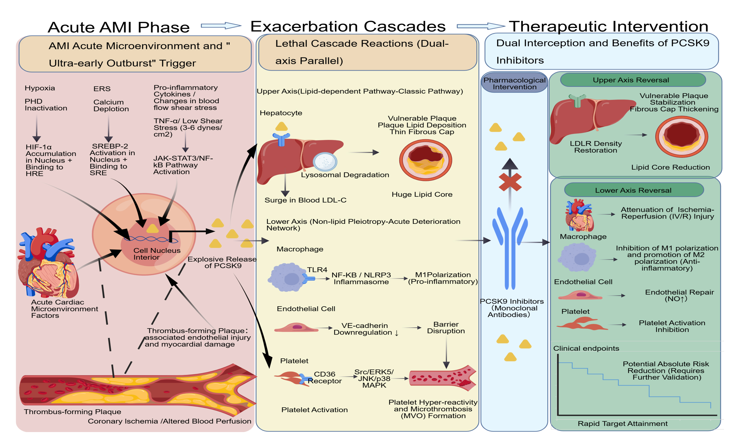
Abstract
Historically, lipid management in acute myocardial infarction (AMI) has predominantly followed a statin-based stepwise titration strategy, causing many patients to miss their target lipid levels during the critical early risk window. Deeper research revealed that under the stress of acute ischemia and inflammatory microenvironments in AMI, PCSK9 exhibits a specific "extremely early burst", which impairs low-density lipoprotein cholesterol
Keywords


 Download PDF
INTRODUCTION
For a long time, the clinical lipid management of atherosclerotic cardiovascular disease has universally followed a stepwise medication escalation strategy based on statins. This strategy necessitates evaluating lipid target attainment 4-6 weeks post-initiation of moderate-to-high-intensity statins, followed by the sequential adjunct of ezetimibe or PCSK9 (proprotein convertase subtilisin/kexin type 9) inhibitors upon target failure[1]. However, large real-world studies, including DA VINCI, reveal that among high-risk or very high-risk patients who do not meet lipid goals, over 60% still only receive moderate-intensity statin monotherapy, and the proportions combining ezetimibe or PCSK9 inhibitors are only 9% and 1%, respectively[2]. The inherent clinical inertia associated with this stepwise adjustment results in a substantial proportion of patients with acute myocardial infarction (AMI) enduring prolonged exposure to suboptimal lipid-lowering therapy during the highly vulnerable 90-day post-infarction period, a phase characterized by a peak incidence of recurrent cardiovascular events (CVEs)[3]. Propelled by the increasingly reinforced clinical consensus asserting that "lower is better and earlier is better" for low-density lipoprotein cholesterol (LDL-C), lipid-lowering strategies are currently undergoing a profound paradigm shift from conventional stepwise titration toward an "upfront combination" approach. This advocates the immediate deployment of potent combination regimens in cohorts prognosticated to fail target attainment on statin monotherapy[4]. Against this background, "in-hospital extremely early intervention" has become the core pathway for optimizing treatment. The "extremely early in-hospital" paradigm delineated herein refers to a substantial temporal advancement of the therapeutic window to within 0-72 h post-symptom onset or admission, extending to the immediate periprocedural phase of percutaneous coronary intervention (PCI)[5]. For descriptive purposes “early post-acute coronary syndrome (ACS)” as initiation > 72 h to 4 weeks after symptom onset or admission, and “later post-ACS” as initiation thereafter. This practice is to use the rapid lipid-lowering effect of the drug to shorten the “time to reach lipid goals”, and to stabilize plaque structure and protect the vascular endothelium in the earliest stage of plaque rupture and severe inflammatory reactions through intensive treatment in the acute phase[6], thereby potentially improving the short- and long-term clinical prognosis of patients with AMI. This article aimed to systematically explain the stress expression and mechanisms of action of PCSK9 in the acute microenvironment of patients with AMI, the dual blocking effect of PCSK9 inhibitors, compare the clinical evidence differences between extremely early and traditional dosing strategies, and consider practical barriers to continued treatment, to provide a clear theoretical basis and research gaps for establishing the optimal timing and precisely screening target populations in the future.
PATHOPHYSIOLOGICAL EVALUATION OF THE CELLULAR STRESS MICROENVIRONMENT DURING THE AMI ACUTE PHASE
The stress-induced eruption mechanism of PCSK9 in the acute microenvironment of AMI [Figure 1]
Ischemic-hypoxic microenvironment and the activation of the HIF-1α signaling cascade
The immediate physiological consequence of myocardial infarction is the abrupt cessation of regional tissue oxygen perfusion. The hypoxic environment is the core driving force that triggers the stress response of the body, and hypoxia-inducible factor 1 (HIF-1) is the molecular sensor that detects this change[7]. Under normal oxygen tension, the prolyl hydroxylase (PHD) activity of HIF-1 remains stable. This enzyme hydroxylates specific proline residues (for example, P402 and P564) on the HIF-1α protein, enabling their recognition by the von Hippel-Lindau protein and subsequent entry into the proteasome degradation pathway. However, under the acute hypoxic state of AMI, PHD loses its activity due to the lack of molecular oxygen as a substrate, which leads to rapid HIF-1α protein accumulation. The accumulated HIF-1α translocates into the nucleus and binds to constitutively expressed HIF-1β (also known as Aryl Hydrocarbon Receptor Nuclear Translocator) to form a heterodimeric transcriptionally active complex[8]. This complex can specifically recognize the hypoxia response element in the promoter region of target genes, whose core consensus sequence is usually 5'-RCGTG-3'. Evidence suggests that the PCSK9 gene is directly or indirectly regulated by HIF-1α. Within hypoxic and inflammatory microenvironments, PCSK9 protein expression is significantly upregulated, concomitant with an experimentally observed increase in HIF-1α expression[9]. This hypoxia-induced transcriptional activation and inflammatory signals cause PCSK9 to begin rising within hours after infarction. An animal study revealed that PCSK9 typically increases significantly within
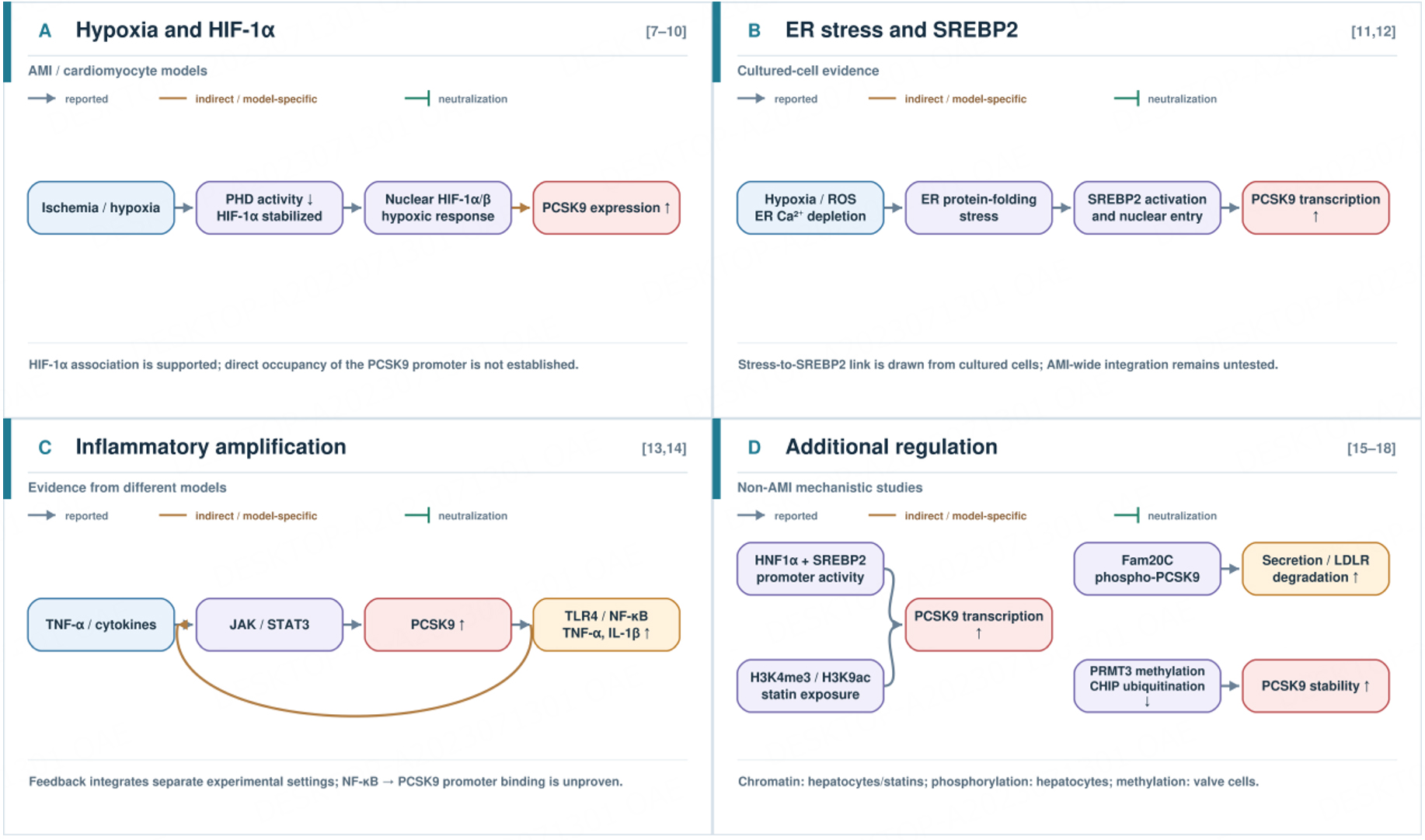
Figure 1. Stress-associated regulation of PCSK9 in acute myocardial infarction. (A) Hypoxia stabilizes HIF-1α and is associated with increased PCSK9 expression; direct binding of HIF-1α to the PCSK9 promoter has not been established[7-10]. (B) Endoplasmic reticulum stress and calcium depletion promote SREBP2-mediated PCSK9 transcription in cultured cells[11,12]. (C) Cytokine-associated JAK/STAT3 signaling and PCSK9-associated TLR4/NF-κB signaling may contribute to inflammatory amplification, although these pathways were studied in different models[13,14]. (D) Additional transcriptional and post-translational mechanisms include HNF1α/SREBP2 promoter regulation, histone modifications, Fam20C-mediated phosphorylation, and PRMT3-mediated methylation that reduces CHIP-dependent ubiquitination[15-18]. The mechanisms in panel D were investigated outside the acute myocardial infarction setting. Orange connections indicate indirect or model-specific links. AMI: Acute myocardial infarction; ER: endoplasmic reticulum; HIF-1α: hypoxia-inducible factor 1α; PCSK9: proprotein convertase subtilisin/kexin type 9; SREBP2: sterol regulatory element-binding protein 2; TLR4: Toll-like receptor 4; NF-κB: nuclear factor κB.
Endoplasmic reticulum stress (ERS)-mediated propagation of PCSK9 synthesis
AMI is accompanied by hypoxia and severe protein folding stress. The reactive oxygen species (ROS) generation, intracellular calcium homeostasis disruption, and nutrient deprivation collectively precipitate the excessive accumulation of unfolded or misfolded proteins in the endoplasmic reticulum (ER) lumen. This pathological accumulation compromises the structural integrity and functionality of the secretory proteome, a state defined as ERS[11]. In cultured-cell experiments, evidence indicates that in an AMI-triggered stress environment, calcium depletion inside the ER promotes the activation of the transcription factor Sterol Regulatory Element-Binding Protein 2 (SREBP-2). SREBP-2, which functions as a master transcriptional regulator of the PCSK9 gene, directly binds to the sterol regulatory element in the PCSK9 promoter[12], thereby orchestrating robust upregulation of PCSK9 expression in response to cellular stress.
Exacerbating cascade effects driven by inflammatory cytokines
Several pro-inflammatory cytokines are released in the early inflammatory phase of AMI, among which tumor necrosis factor (TNF)-α can upregulate PCSK9 expression through the Janus Kinase/signal transducer and activator of transcription 3 pathway[13]. Reciprocally, PCSK9 propagates the secretion of pro-inflammatory cytokines (for example, TNF-α and IL-1β) via the TLR4/NF-κB axis[14], establishing a positive feedback loop for inflammatory amplification. Direct binding of NF-κB to the PCSK9 promoter has not been confirmed by chromatin immunoprecipitation (ChIP) or equivalent promoter-occupancy experiments. The available evidence is mainly indirect intervention data, so an NF-κB-to-PCSK9 transcriptional pathway remains hypothetical.
Additional transcriptional and post-translational regulation of PCSK9
Beyond the stress-associated pathways described above, basic studies have identified additional levels of PCSK9 transcriptional control. In HepG2 cells, mutation of the hepatocyte nuclear factor 1α (HNF1α) binding site adjacent to the sterol regulatory element attenuated SREBP2-mediated activation of the PCSK9 promoter[15]. More recently, atorvastatin was shown to increase histone H3 lysine 4 trimethylation (H3K4me3) and histone H3 lysine 9 acetylation (H3K9ac) at this promoter in HepG2 cells; corresponding hepatic changes were also observed in high-fat-diet-fed mice[16]. These findings extend PCSK9 transcriptional regulation from transcription-factor binding to chromatin modification, although the latter experiments examined statin exposure rather than AMI itself.
Regulation also continues after PCSK9 protein synthesis. Fam20C-mediated phosphorylation of PCSK9 at Ser47, Ser666, Ser668, and Ser688 enhanced its secretion and LDL receptor degradation in hepatocyte experiments[17]. A recent study in valvular interstitial cells identified another mechanism: PRMT3-mediated methylation at PCSK9 Arg582 reduced CHIP-mediated ubiquitination at Lys575, thereby stabilizing PCSK9[18]. The methylation finding comes from a calcific aortic valve disease model and should not be interpreted as evidence that the same process drives PCSK9 elevation during AMI. Together, these studies show that PCSK9 abundance and activity depend on protein processing as well as transcription.
Acute hemodynamic injury and pathological crosstalk of local PCSK9 [Figure 2]
Local induction mechanisms under low shear stress states
In anatomical regions exhibiting coronary stenosis or decelerated perfusion, the laminar shear stress exerted on endothelial and vascular smooth muscle cells is markedly attenuated (3-6 dynes/cm2). This low-shear biomechanical environment induces nicotinamide adenine dinucleotide phosphate (NADPH) oxidase activation, resulting in augmented ROS generation. This sequence subsequently upregulates LOX-1 via the NF-κB pathway, thereby indirectly amplifying local PCSK9 expression in vascular endothelial and smooth muscle cells (with SMC expression typically predominating). Concurrently, ROS itself is also believed to be directly involved in regulating PCSK9 expression[19].
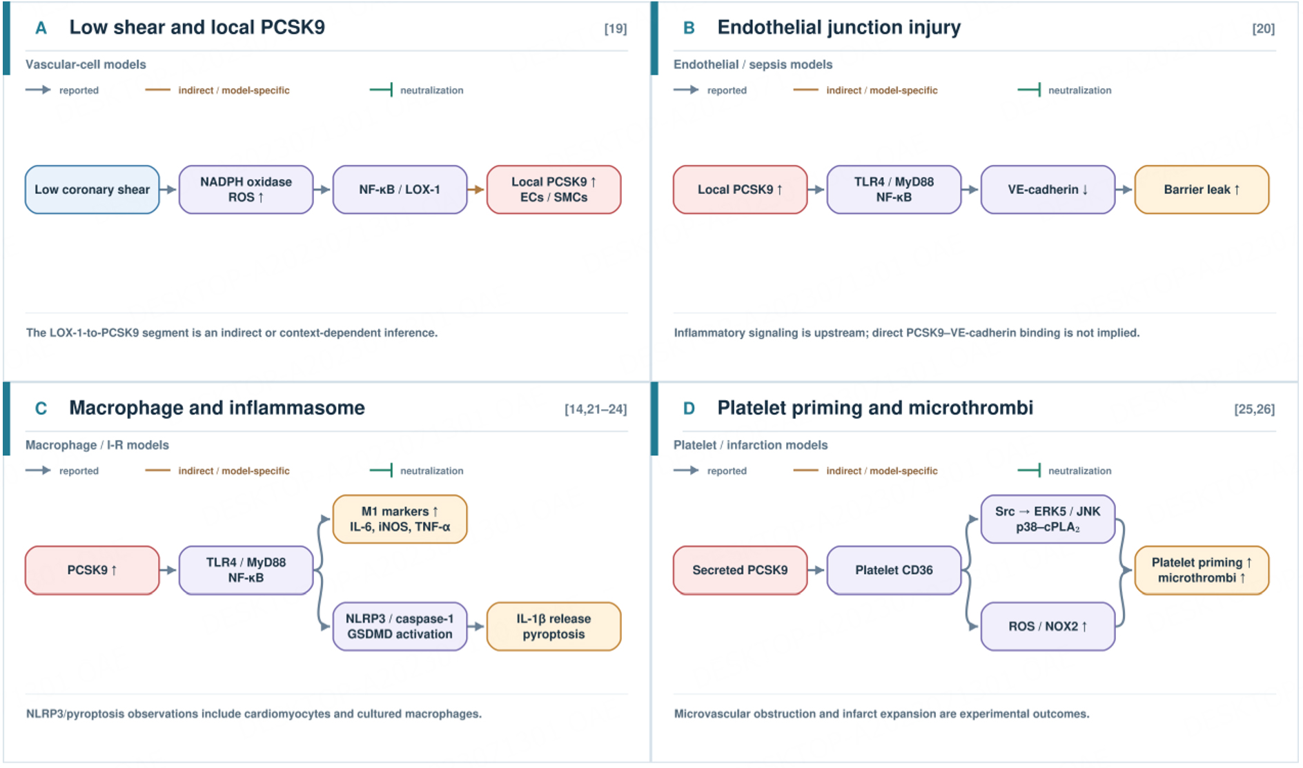
Figure 2. Local vascular and lipid-independent injury mechanisms associated with PCSK9. (A) Low shear stress promotes oxidant signaling and is associated with increased local PCSK9 expression in vascular cells; the LOX-1-to-PCSK9 link is indirect or context-dependent[19]. (B) In endothelial cell and sepsis models, PCSK9-associated TLR4/MyD88/NF-κB signaling reduces VE-cadherin expression and impairs endothelial junction integrity[20]. (C) Experimental studies link PCSK9 to pro-inflammatory macrophage polarization and NLRP3-associated inflammatory responses[14,21-24]. (D) PCSK9 binding to platelet CD36 activates kinase and oxidant signaling, increasing platelet reactivity and promoting microthrombus formation in experimental models[25,26]. These panels summarize findings from distinct experimental settings. Orange connections indicate indirect or model-specific links. LOX-1: Lectin-like oxidized low-density lipoprotein receptor 1; MyD88: myeloid differentiation primary response 88; NF-κB: nuclear factor κB; NLRP3: NOD-like receptor family pyrin domain-containing 3; PCSK9: proprotein convertase subtilisin/kexin type 9; TLR4: Toll-like receptor 4.
Endothelial integrity disruption by local PCSK9 overexpression via lipid-independent pathways
In endothelial cell and sepsis models, PCSK9 has been shown to significantly reduce VE-cadherin expression in endothelial cells. The more clearly understood mechanism currently is that PCSK9 induces an inflammatory response via the TLR4/MyD88/NF-κB pathway, thereby indirectly downregulating VE-cadherin protein levels and destroying the adhesion regulation function between cells[20].
"Lipid-independent" acute deterioration mechanisms [Figure 2]
Pro-inflammatory macrophage polarization and NLRP3 activation
PCSK9 is a potent endogenous regulator capable of inducing macrophage transition to a pro-inflammatory M1 phenotype in experimental models[21,22]. This mechanism is primarily mediated by the TLR4/MyD88/NF-κB axis[14]. Activated NF-κB further initiates the expression of M1 markers, including IL-6, iNOS, and TNF-α[21]. Moreover, under ischemia-reperfusion (I/R) stress, PCSK9 can promote NLRP3 inflammasome protein complex assembly, inducing pyroptosis and releasing mature IL-1β. This pathway has been systematically verified in cardiomyocytes and heart tissues in multiple late-stage preclinical models[23]. In macrophages, recent in vitro work reveals that the TLR4-NFκB-NLRP3 axis also exists and mediates the pro-inflammatory effects of PCSK9[24].
Induction of platelet hyperreactivity and microthrombogenic propensity
In mechanistic experiments, PCSK9 structurally engages with the CD36 scavenger receptor on the platelet surface, representing the core initiation step for platelet hyperreactivity[25,26]. Subsequent CD36 activation triggers phosphorylation cascades involving downstream Src kinases, ERK5, JNK, and p38 mitogen-activated protein kinase (MAPK), coupled with augmented ROS synthesis. These changes significantly lower the activation threshold of platelets to low concentrations of agonists (including ADP or collagen), putting platelets into a "primed" or "pre-activated" hyperreactive state[25]. In experimental infarction models, this pathologically heightened reactivity predisposes platelets to extensive adhesion and aggregation in the unstable ischemic milieu post-AMI, precipitating microthrombus formation, exacerbating microvascular obstruction, and directly expanding myocardial infarct size[25].
Synthesizing these multifaceted mechanisms, it is evident that from the initial hours to days post-AMI, PCSK9 functions as a homeostatic regulator of lipid metabolism and as a primary early instigator of the acute inflammatory cascade, endothelial degradation, and microthrombogenesis. This pathological characteristic of an "extremely early burst" suggests that clinical interventions must break away from the traditional "stable phase administration" mindset, significantly advancing the therapeutic window to intervene at the very forefront of the formation of the acute stress microenvironment. Based on the aforementioned multiplexed pathogenic network of PCSK9 in the acute microenvironment, the rationale for clinical intervention naturally shifts toward intercepting this malignant cascade. PCSK9 inhibitors, therefore, emerge as a focal point; their clinical utility is not confined to conventional lipid target attainment but extends to directly interfering with these acute pathological processes through a dual blockade of receptors and pathways.
DUAL INTERCEPTION MECHANISMS OF PCSK9 INHIBITORS: LIPID-DEPENDENT AND LIPID-INDEPENDENT PATHWAYS
Lipid-dependent effects of PCSK9 inhibitors: pathophysiological mechanisms underlying precipitous LDL-C attenuation and plaque stabilization [Figure 3]
PCSK9 is a natural negative regulator of LDLR. Following AMI, myocardial ischemia and inflammatory cascades precipitate a rapid elevation in serum PCSK9, although the temporal pattern varies across studies[10]. Subsequent PCSK9 binding to the extracellular domain of LDLR directs the receptor toward lysosomal degradation, thereby markedly depleting cell-surface LDLR abundance in hepatocytes and impairing LDL-C clearance. Conversely, PCSK9 inhibitors (monoclonal antibodies) directly sequester circulating PCSK9, competitively antagonizing the PCSK9-LDLR interaction. This intervention rescues LDLR from targeted degradation, facilitating the rapid restoration of hepatic surface LDLR density. Within 4 h of administration, this mechanism achieves profound suppression of circulating free PCSK9 (attaining an inhibition rate of 95%-97%)[27], thereby reinitiating the accelerated clearance of LDL-C. This precise mechanism supports the precipitous reduction in circulating LDL-C observed within days of therapeutic initiation. Because monoclonal antibodies target extracellular secreted PCSK9, their effects are not interchangeable with whole-gene or cell-specific knockout, which also removes intracellular PCSK9[28,29].
The acute attenuation of LDL-C exerts a direct structural impact on atherosclerotic plaque stability. The inherent vulnerability of coronary plaques in patients with AMI is predominantly dictated by a voluminous lipid core situated beneath a thin fibrous cap[30]. Cholesterol accumulation in the lipid core is the most important factor in plaque development and vulnerability[31]. PCSK9 inhibitors can significantly reduce the lipid core burden index and the abundance of lipid-core plaques in a very short period of 2-6 weeks[32]. The early morphological manifestations of lipid-lowering therapy on atheromata are primarily characterized by fibrous cap thickening, concomitant with a reduction in intraplaque lipid content and overall atheroma volume shrinkage[33]. Studies confirm that after lipid-lowering therapy, fibrous cap thickness (FCT) increases, lipid components decrease, and the plaque transforms from a vulnerable to a stable state[34].
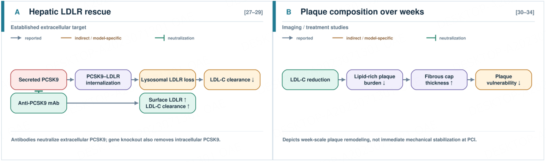
Figure 3. Lipid-dependent effects of extracellular PCSK9 neutralization. (A) Secreted PCSK9 promotes lysosomal degradation of the hepatic LDL receptor (LDLR), reducing LDL-C clearance. Anti-PCSK9 monoclonal antibodies neutralize extracellular PCSK9, preserve cell-surface LDLR, and enhance LDL-C clearance[27-29]. (B) Over subsequent weeks, LDL-C lowering is associated with reduced plaque lipid burden and increased fibrous cap thickness[30-34]. The plaque changes depicted in (B) should not be interpreted as immediate mechanical stabilization during percutaneous coronary intervention. The green T-bar indicates PCSK9 neutralization. LDL-C: Low-density lipoprotein cholesterol; LDLR: low-density lipoprotein receptor; PCSK9: proprotein convertase subtilisin/kexin type 9.
Lipid-independent pleiotropic efficacy: pathophysiological mechanisms of acute microcirculatory preservation [Figure 4]
Mitigation of I/R injury
The reperfusion phase of AMI inherently precipitates secondary I/R injury[35]. Multiple animal model studies demonstrate that PCSK9 inhibitors effectively mitigate this injury at cellular and histological levels by modulating multiple molecular pathways. First, at the microscopic level of inhibiting cardiomyocyte mortality, PCSK9 inhibitors manifest multidimensional protective efficacy. They significantly suppress NLRP3 inflammasome activation and attenuate cardiomyocyte apoptosis[36], while concurrently inhibiting cardiomyocyte cuproptosis by directly disrupting the interaction between PCSK9 and the LIAS protein[35]. Second, at the level of inhibiting macroscopic cardiac remodeling and structural damage, PCSK9 inhibitors mitigate I/R-induced myocardial fibrosis via TGF-β1/Smad3 signaling cascade suppression[37].
Meanwhile, by regulating the RIPK1/RIPK3/MLKL pathway, they effectively mitigate the significantly upregulated pathogenic damage after AMI, thereby restoring overall cardiac function[38]. Synthesizing these findings, myocardial PCSK9 expression is markedly upregulated under I/R or AMI conditions. Inhibiting PCSK9 can alleviate myocardial injury, suggesting that PCSK9 itself may have pathogenic effects and that PCSK9 inhibitors protect the myocardium by blocking this pathogenicity.
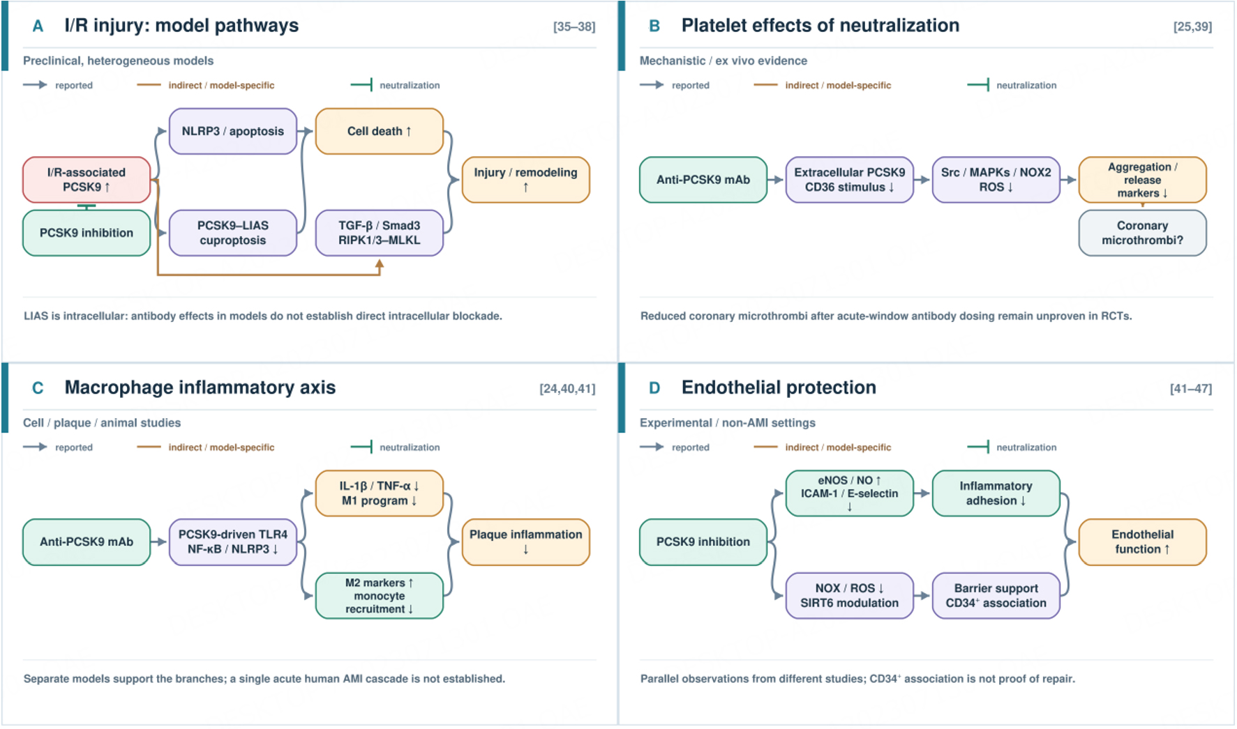
Figure 4. Experimental pathways associated with PCSK9 inhibition in ischemia-reperfusion injury. (A) Distinct preclinical studies implicate NLRP3-associated cell death, PCSK9-LIAS-associated cuproptosis, TGF-β/Smad3 signaling, and RIPK1/RIPK3/MLKL signaling in myocardial injury or remodeling[35-38]. The intracellular PCSK9-LIAS finding does not establish direct intracellular action of a circulating anti-PCSK9 antibody. (B) PCSK9 neutralization attenuates platelet CD36-associated signaling and platelet activation in mechanistic studies; a reduction in coronary microthrombi after acute antibody administration has not been established in a randomized trial[25,39]. (C) Macrophage and plaque models show attenuation of TLR4/NF-κB/NLRP3-associated inflammation[24,40,41]. (D) Experimental and non-AMI studies report changes in endothelial nitric oxide signaling, oxidative stress, adhesion molecules, and barrier-associated measures[41-47]. The pathways were investigated in different models and do not represent one verified cascade in human AMI. Orange connections indicate indirect or model-specific links; green T-bars indicate PCSK9 neutralization. AMI: Acute myocardial infarction; I/R: ischemia-reperfusion; LIAS: lipoic acid synthetase; NF-κB: nuclear factor κB; NLRP3: NOD-like receptor family pyrin domain-containing 3; PCSK9: proprotein convertase subtilisin/kexin type 9; TLR4: Toll-like receptor 4.
Suppression of platelet activation and microthrombogenesis
PCSK9 inhibitors suppress platelet activation via a dual mechanistic approach. First, it markedly reduces LDL-C, thereby mitigating lipid-driven platelet hyperreactivity[25]. Second, PCSK9 inhibitors may block the interaction between PCSK9 and the CD36 receptor on the platelet surface, effectively negating PCSK9-mediated direct platelet activation[39]. Specifically, PCSK9 inhibitors abrogate PCSK9-induced activation of downstream signaling cascades, including Src kinases, ERK5, JNK, and the p38MAPK/cPLA2 axis. This abrogation consequently attenuates platelet aggregation in response to physiological agonists (for example, ADP, thrombin, and collagen), diminishes P-selectin exocytosis from α-granules and ATP release from dense granules, and curtails ROS generation via NOX2 pathway inhibition[25]. These mechanistic insights substantiate that PCSK9 inhibitors effectively curtail PCSK9-mediated platelet activation and microthrombus formation, providing a rationale for clinical study; reduced coronary microthrombi or early recurrent events after antibody administration has not been established in an acute-window randomized trial.
Anti-inflammatory efficacy
PCSK9 inhibitors inhibit inflammatory responses by blocking the interaction between PCSK9 and the TLR4/NF-κB pathway. Within macrophages, PCSK9 upregulates TLR4 expression, propagating downstream NF-κB activation and increasing the secretion of pro-inflammatory cytokines, including IL-1β and TNF-α[14,21,23]. PCSK9 inhibitors can reduce MCP-1-mediated monocyte chemotaxis, indicating their role in regulating macrophage inflammation[40].
Concerning modulating macrophage polarization, evidence indicates that alirocumab effectively mitigates PCSK9-induced pro-inflammatory signal transduction within M1 macrophages. In vitro assays demonstrate that PCSK9 overexpression upregulates pro-inflammatory cytokines (for example, CXCL9, CXCL10, TNF-α, IL-1β, and IL-6) while concurrently downregulating anti-inflammatory mediators (for example, CCL17, TGM2, TGF-β1, and IL-10) in M1 macrophages. Furthermore, it potentiates NF-κB p65 nuclear translocation alongside the activation of NLRP3 and gasdermin-D. Crucially, these pathophenotypes are abrogated by TLR4 inhibition or targeted gene silencing. Independent of its potent LDL-C-lowering capacity, alirocumab exerts direct anti-inflammatory efficacy by modulating the TLR4-NFκB-NLRP3 axis within M1 macrophages and simultaneously fostering M2 phenotype polarization, thereby profoundly attenuating intraplaque inflammation[24].
In the context of oxidative stress regulation, evolocumab mitigates the expression of NADPH oxidase and NF-κB proteins within myocardial tissue, concomitantly decreasing lipid peroxidation markers, thereby demonstrating significant intrinsic antioxidant properties in that experimental setting[41].
Endothelial protective efficacy
The protective capacity of PCSK9 inhibitors on the endothelial barrier manifests as a multi-tiered, cascading interventional process. In the extreme pathological microenvironment induced by lipoprotein(a), PCSK9 inhibitors initially upregulate eNOS expression (corroborating prior findings wherein PCSK9 impairs NO biology via the NADPH oxidase pathway) and then restore phosphorylated eNOS protein levels and NO bioavailability, reversing endothelial dysfunction from the source[42-44]. Concurrently, this pharmacological agent effectively mitigates the intracellular oxidative stress burden by curtailing NADPH oxidase-derived ROS and modulating cytoprotective pathways, including SIRT6[41,45]. Consequent to the restoration of endothelial redox homeostasis, surface inflammatory activation is profoundly inhibited. Evolocumab and alirocumab have been associated with lower ICAM-1 and E-selectin expression in experimental or non-AMI settings, thereby cutting off the adhesion pathway for monocyte transendothelial infiltration[42]. Synergized by these mechanisms, targeted inhibition of endothelium-derived PCSK9 directly preserves the structural integrity of the endothelial barrier and significantly augments the peripheral concentration of CD34+ mature endothelial progenitor cells[46,47], thereby providing indispensable structural reparative support to the compromised vascular network.
In summary, the lipid-independent pleiotropic network of PCSK9 inhibitors (anti-inflammatory, antioxidant, endothelial, and microcirculation protection) addresses several pathways implicated in AMI reperfusion injury in preclinical models. However, it is imperative to objectively acknowledge that the prevailing mechanistic evidence supporting the lipid-independent protective efficacy of PCSK9 inhibitors predominantly derives from highly simplified in vivo models or in vitro cellular assays. The extent to which these microscopic pharmacological actions can truly equate to the extreme clinical microenvironment when human AMI occurs with severe hemodynamic disorders and severe internal environmental imbalances remains uncertain in translational medicine. Nonetheless, these insights elucidate, at a molecular level, the rationale for introducing this pharmacotherapy during the "extremely early" periprocedural window to test whether early target engagement yields clinically meaningful benefits. The preceding exposition of pathological and molecular mechanisms suggests a potential role for PCSK9 in the exacerbation network of acute AMI. Propelled by these micro-level breakthroughs, clinical investigators have initiated a critical reappraisal of the limitations of ambulatory stepwise therapy, redirecting the focus toward extremely early in-hospital protection. The following section systematically reviews the latest evidence-based map of the translation of this theory into clinical practice.
AN EVIDENCE-BASED CLINICAL LANDSCAPE TRANSCENDING CONVENTIONAL TEMPORAL LIMITATIONS [TABLE 1]
Dimensional comparison of intervention starting points, from "delayed medication after stabilization" to "immediate" perioperative protection
Therapeutic inertia within conventional paradigms (months-years)
In the landmark FOURIER trial, a cornerstone study of PCSK9 inhibitors, the cohort predominantly comprised stable patients with prior myocardial infarction[48]. Furthermore, historical clinical guidelines frequently recommended lipid reassessment 4-12 weeks post-discharge for patients with ACS to facilitate evaluation and subsequent adjustment of lipid-lowering algorithms. This "ambulatory therapeutic delay" inherently deprives patients of adequately potent lipid-lowering prophylaxis during the highly vulnerable 30-90 day post-discharge period.
Summary of major clinical trials and cardiovascular outcomes
| Study/sorce | Population/first dose | Intervention | Key quantitative result | Interpretation |
| Group A (refers to treatment initiation 0-72 h after symptom onset or hospital admission, including peri-PCI dosing within this interval): extremely early in-hospital initiation and related peri-PCI evidence | ||||
| EPIC-STEMI[5] | STEMI; alirocumab pre- or intra-PCI | Alirocumab vs. placebo | LDL-C -72.9% vs. -48.1% at ~6 weeks; difference -22.3 percentage points | Peri-PCI feasibility and early LDL-C lowering; no plaque or MACE endpoint |
| PACMAN-AMI[6] | AMI; first dose < 24 h after urgent culprit PCI | Alirocumab vs. placebo plus statin | Nonculprit PAV change at 52 weeks: -2.13% vs. -0.92% | Randomized plaque imaging; not a comparison of clinical event timing |
| Gim et al.[49] | ACS; observational peri-PCI cohort | Single-dose PCSK9i vs. control | 30-day MACE 4.0% vs. 10.2%; adjusted HR 0.38 (95% CI 0.18-0.80) | Association only; selection bias, confounding, and co-treatment remain |
| Ziogos et al.[59] | MI; in-hospital initiation | Evolocumab vs. placebo | Lower myocardial 18F-FDG PET inflammatory activity at 30 days | Imaging signal; no proven independent mechanism or MACE benefit |
| EVOPACS[50] | ACS; randomized during hospitalization | Evolocumab vs. placebo; high-intensity statin background | Week-8 LDL-C < 70 mg/dL: 95.7% vs. 37.6%; hs-CRP < 2 mg/L: 68.8% vs. 69.3% | Rapid LDL-C attainment; no significant systemic hs-CRP signal |
| Overlap groups A (refers to treatment initiation 0-72 h after symptom onset or hospital admission, including peri-PCI dosing within this interval) and B (refers to initiation > 72 h to 4 weeks after symptom onset or admission) | ||||
| HUYGENS[58] | NSTEMI; first dose within 7 days after baseline OCT/IVUS imaging during the index ACS evaluation | Evolocumab vs. placebo plus statin | Minimum FCT change at 50 weeks: +42.7 vs. +21.5 μm; P = 0.015 | OCT plaque phenotype endpoint; timing is measured from baseline imaging |
| Post-discharge initiation: a broader window that can overlap Groups B (refers to initiation > 72 h to 4 weeks after symptom onset or admission) and C (refers to initiation > 4 weeks after symptom onset or admission) | ||||
| VICTORION-INCEPTION[52] | Recent ACS; discharged ≤ 5 weeks before screening | Inclisiran + usual care vs. usual care | Day-330 LDL-C < 70 mg/dL: 66.7% vs. 28.1% | Post-discharge LDL-C management; outside the 0-72 h window |
| Group C(refers to initiation > 4 weeks after symptom onset or admission): Later post-ACS initiation | ||||
| REVERSAL[56] | Stable coronary disease; long-term treatment | Atorvastatin 80 mg vs. pravastatin 40 mg | Serial IVUS: less atheroma progression with intensive statin | Historical statin imaging comparator; not an acute timing trial |
| ASTEROID[55] | Coronary disease; long-term treatment | Rosuvastatin 40 mg; single arm | Serial IVUS: coronary atheroma regression | Historical nonrandomized statin imaging benchmark |
| EASY-FIT[57] | ACS; follow-up OCT | Atorvastatin 20 vs. 5 mg | Greater fibrous-cap thickening with higher statin dose | Statin dose and imaging comparator only |
| FOURIER[48] | Stable ASCVD; median follow-up 2.2 y | Evolocumab vs. placebo on statin | Primary composite CV endpoint: 9.8% vs. 11.3%; HR 0.85 | CVEs reduced; no general survival or acute-timing claim |
| ODYSSEY OUTCOMES[53] | ACS 1-12 months before randomization | Alirocumab vs. placebo on intensive statin | Primary composite endpoint: 9.5% vs. 11.1%; HR 0.85 | Event benefit in later post-ACS treatment, not 0-72 h initiation |
| DA VINCI[2] | European observational cross-sectional care | Prescribed lipid-lowering therapies | Incomplete 2019 ESC/EAS LDL-C goal attainment | Care gap; no randomized PCSK9 initiation effect |
Preemptive "first-dose" advantage of ultra-early administration (0-72 h)
First, advancing the therapeutic initiation to the emergent periprocedural phase: Diverging completely from conventional strategies, the EPIC-STEMI trial aggressively accelerated the therapeutic window to "Time Zero", administering the agent immediately pre- or intra-PCI in patients with STEMI, demonstrating the feasibility of periprocedural administration in this small study[5]. At approximately 6 weeks, LDL-C fell by 72.9% vs. 48.1% with control (between-group difference, 22.3 percentage points).
Second, achieving a profound reduction in short-term post-procedural cardiovascular risk: Gim et al.[49] reported that, in a 2025 Korean cohort analysis, administration of a single preemptive dose of a PCSK9 inhibitor at least one hour before PCI was associated with a lower incidence of one-month major adverse cardiovascular events (MACE) (4.0% vs. 10.2%). However, residual confounding, treatment selection bias, and concomitant therapies limit causal interpretation, and further randomized studies are required to validate the clinical benefit of extremely early PCSK9 inhibition.
Comparison of lipid target attainment speed and compliance: from "months of long-term titration" to "weeks of rapid lipid lowering"
Inherent therapeutic lag and adherence challenges in conventional stepwise lipid-lowering algorithms
Large real-world studies, including the DA VINCI study, have highlighted persistent limitations in conventional stepwise lipid-lowering strategies. Reliance on outpatient statin titration, delayed treatment intensification, or sequential addition of non-statin therapies often results in prolonged periods before achieving guideline-recommended LDL-C targets. In routine clinical practice, LDL-C goal attainment remains suboptimal despite widespread statin use, although the observational nature of DA VINCI precludes attributing this gap exclusively to the stepwise treatment paradigm. These findings emphasize that treatment inertia, delayed follow-up optimization, and implementation barriers may collectively contribute to residual lipid-related cardiovascular risk[2].
Expedited target attainment and adherence optimization via early in-hospital initiation
The major advantage of early lipid-lowering initiation is the ability to utilize the acute-care therapeutic window, allowing treatment optimization before or shortly after hospital discharge and facilitating structured follow-up. Evidence from randomized clinical trials has demonstrated that early initiation strategies can substantially accelerate LDL-C target attainment, although the timing of intervention differs among studies.
For true in-hospital initiation strategies, the EVOPACS and VCU-AlirocRT trials provided important evidence supporting the feasibility of initiating PCSK9 inhibition during the index hospitalization. In the EVOPACS trial, evolocumab administered within 24-72 h after ACS admission in patients receiving background statin therapy rapidly reduced LDL-C levels; at 8 weeks, 95.7% of patients in the evolocumab group achieved LDL-C < 70 mg/dL compared with 37.6% in the placebo group, demonstrating the ability of inpatient PCSK9 inhibition to accelerate lipid target attainment[50]. Similarly, the VCU-AlirocRT trial evaluated an in-hospital alirocumab initiation protocol and demonstrated substantial early lipid reduction during the vulnerable period following ACS, further supporting the feasibility of administering PCSK9 inhibitors before discharge in high-risk patients[51].
However, rapid LDL-C reduction does not necessarily translate into immediate modulation of systemic inflammatory biomarkers. In EVOPACS, despite significant LDL-C lowering, no significant difference was observed in the proportion of patients achieving high-sensitivity C-reactive protein (hs-CRP) < 2 mg/L at week 8 (68.8% vs. 69.3%; P = 0.89)[50]. Possible explanations include the timing of inflammatory biomarker assessment, concomitant high-intensity statin therapy, and differences between systemic inflammatory markers and local vascular inflammatory activity. These possibilities remain exploratory and require further mechanistic investigation.
In contrast to in-hospital initiation trials, the VICTORION-INCEPTION trial evaluated an early post-discharge treatment strategy rather than treatment initiation during the index hospitalization. This trial enrolled patients who had been discharged after ACS within the preceding 5 weeks and assessed the effectiveness of initiating inclisiran in combination with standard care. At day 330, 66.7% of patients receiving inclisiran achieved LDL-C < 70 mg/dL compared with 28.1% in the conventional-care group
Together, current evidence indicates that early implementation of intensive lipid-lowering strategies—whether during hospitalization or shortly after discharge—can accelerate LDL-C target achievement. The design limitations of this core evidence must be objectively examined. Whether EVOPACS or recent prospective RCTs, their research frameworks are generally limited by short follow-up periods (for example, 8 weeks) and relatively limited sample sizes. Nevertheless, most available studies remain focused on surrogate lipid endpoints rather than clinical outcomes, and adequately powered trials are still required to determine whether these timing strategies reduce MACE, recurrent myocardial infarction, and cardiovascular mortality.
Comparison of PCSK9-directed agents and lipid subfractions
Evolocumab and alirocumab neutralize secreted PCSK9, whereas inclisiran uses small interfering RNA to reduce hepatic PCSK9 synthesis[48,53,54]. In ORION-10 and ORION-11, inclisiran lowered LDL-C by 52.3% and 49.9%, respectively, relative to placebo at day 510; smaller changes in lipoprotein(a) [Lp(a)] and high-density lipoprotein cholesterol (HDL-C) were reported in ancillary analyses[54]. Monoclonal antibody outcome trials demonstrated reductions in composite CVEs in stable atherosclerotic disease or after ACS, but inclisiran had no completed event-driven acute-AMI timing trial in these datasets[48,53,54]. Differences in populations, follow-up, and background treatment preclude direct comparisons of LDL-C, HDL-C, Lp(a), or clinical outcomes between these drugs in the first 72 h.
Comparison of morphology and pathophysiological mechanisms, from "long-cycle onset" to "early rapid reversal"
Protracted vascular remodeling induced by conventional statins (18-24 months)
Although early landmark trials, including the IVUS-based REVERSAL and ASTEROID trials, proved that even using the highest intensity statin monotherapy required 1.5-2 years to achieve a significant reversal of percent atheroma volume (PAV)[55,56], critically, subsequent optical coherence tomography (OCT)-based statin monotherapy studies (including the EASY-FIT trial and control group data from PACMAN-AMI/HUYGENS) indicate that relying only on statins to achieve effective thickening of the fibrous cap and stabilization of the plaque microstructure often faces a long onset cycle. Consequently, during the most critical and vulnerable early phase post-infarction, atherosclerotic residual plaque vulnerability may remain despite background statin treatment[6,57,58].
Rapid reversal by early intensive intervention (days-52 weeks)
First, regarding short-term inhibition of acute myocardial inflammation, Ziogos et al.[59] reported in the EVACS trial that a single in-hospital dose of evolocumab reduced residual myocardial inflammatory activity after myocardial infarction, as assessed by 18F-FDG PET at 30 days. These findings suggest that early PCSK9 inhibition may exert anti-inflammatory effects beyond LDL-C reduction during the acute post-infarction phase.
Second, a pronounced compression of the plaque stabilization timeline to 52 weeks: The pivotal imaging-based RCTs, PACMAN-AMI and HUYGENS, showed favorable coronary imaging changes during approximately one year of treatment, with different starting points. Compared with the traditional statin monotherapy group, its speed and magnitude in stabilizing plaques have significant imaging advantages[6,58]. However, it must be observed that although studies, including PACMAN-AMI, observed significant reversal in imaging (including thin FCT), the degree to which these micro-anatomical benefits linearly correlate with long-term reductions in hard MACE endpoints (for example, cardiovascular mortality or non-fatal myocardial infarction) necessitates rigorous validation via large-scale, event-driven clinical trials. Methodologically, the exclusive reliance on imaging-derived surrogate endpoints to justify routine, extremely early administration may introduce logical vulnerabilities associated with the over-extrapolation of definitive clinical benefits. In PACMAN-AMI, the first alirocumab dose was given within 24 h after completion of urgent culprit-vessel PCI, and PAV changed by -2.13% vs. -0.92% at 52 weeks[6]. In HUYGENS, evolocumab was initiated within 7 days after baseline OCT, and minimum FCT rose by 42.7 vs. 21.5 μm at 50 weeks[58].
Temporal dependencies in absolute prognostic yield
Attenuation effect of absolute risk reduction (ARR) in late intervention
Clinical epidemiological data indicate that the initial months succeeding an ACS event represent the zenith of vulnerability for recurrent ischemic episodes. If intensive lipid-lowering therapy is initiated six months or even several years after the onset (as in the classic FOURIER trial), the patient has essentially passed the extremely high-risk period of CVEs and entered a relatively stable chronic phase. At this time, although the relative risk reduction (RRR) brought by PCSK9 inhibitors remains statistically significant, because the patient's "baseline absolute risk" has significantly dropped over time, the number of actual absolute events prevented by the same magnitude of RRR (that is, ARR) will exhibit a clear attenuation trend[48,60]. Consequently, while delayed administration retains pharmacological efficacy, it inherently forfeits the critical therapeutic window required to maximize absolute clinical yield.
Optimization of ARR via extremely early intervention
In stark juxtaposition to the attenuation observed in delayed intervention models, prespecified and subsequent post-hoc analyses of the ODYSSEY outcomes trial support alirocumab for secondary prevention in patients enrolled 1-12 months after ACS. The closer the intervention timing is to the early stage of ACS onset, the higher the hypercoagulable and highly inflammatory state (baseline absolute risk) of the patient. In the high-risk patient subgroup, a higher baseline risk may increase ARR, but ODYSSEY OUTCOMES did not directly compare treatment within 72 h with later initiation[53,61]. Moreover, a large ongoing prospective, pragmatic, randomized controlled trial, the EVOLVE-MI trial (whose outcome data were unavailable at the time of this revision), will further verify the benefits of this extremely early intervention. Although the launch of the EVOLVE-MI trial is highly anticipated, the hypothesis that "earlier intervention unequivocally confers augmented benefit" lacks robust, large-cohort RCT validation concerning its strict linearity, specifically in the extreme physiological stress window of 0-72 h. The benefits of early intervention may not be evenly distributed among all populations; however, they are highly concentrated in certain patients with high-risk phenotypes.
CLINICAL IMPLEMENTATION CONSIDERATIONS AND CHALLENGES FOR EXTREMELY EARLY IN-HOSPITAL APPLICATION OF PCSK9 INHIBITORS
Precise screening of target populations and current guideline status
Currently, the universal deployment of PCSK9 inhibitors as routine early interventional agents across the entire unselected AMI demographic lacks definitive evidence-based substantiation and ubiquitous guideline endorsement. Nonetheless, guideline directives pertaining to specifically delineated high-risk cohorts have undergone a paradigm shift. According to the 2025 ACC/AHA/ACEP/NAEMSP/SCAI Guidelines for the Management of Acute Coronary Syndromes, patients with ACS should start or intensify lipid-lowering therapy during hospitalization. High-intensity statin therapy is a Class I recommendation for all patients with ACS. For patients already taking maximally tolerated statin therapy who have LDL-C ≥ 70 mg/dL, adding a nonstatin lipid-lowering therapy is Class I; upfront addition of ezetimibe to statin therapy may be considered (Class 2b)[4]. At this stage, the priority benefit group with the most definite evidence base is patients who have received maximally tolerated statin therapy; however, they still have not met their LDL-C targets.
Furthermore, AMI subjects exhibiting concomitant familial hypercholesterolemia, profound multivessel coronary artery disease, or documented severe statin intolerance may warrant assessment for intensive lipid management, but comparative absolute benefit from initiation within 72 h has not been established. Translating into clinical reality, the indiscriminate generalization of these indications contravenes established evidence-based paradigms and remains fundamentally unfeasible. Once precise screening of target populations is achieved through strict medical indications, the next key challenge for extremely early intervention is how to effectively translate this intensive regimen established during hospitalization into long-term clinical prognostic benefits after discharge.
Optimizing long-term adherence via in-hospital therapeutic initiation
Physician-driven clinical inertia regarding therapeutic up-titration and progressive diminution in patient adherence during the post-discharge ambulatory phase constitute dual etiologies, supporting suboptimal lipid target attainment in ACS cohorts. Extensive real-world data delineate a pronounced, time-dependent attrition in lipid-lowering therapeutic adherence subsequent to hospital discharge[62]. The strategic initiation of lipid-lowering algorithm adjustments during the inpatient phase may support prescription of indicated lipid-lowering therapy at discharge; observational utilization data show that treatment gaps remain[63], although there is still some degree of compliance attenuation in the long run[64]. The theoretical foundations of the EVOPACS trial indicate that advancing the initiation of PCSK9 inhibitors to the in-hospital phase ensures that patients achieve LDL-C targets as early as possible during the post-onset hyper-vulnerable period and addresses the treatment delays present in previous outpatient initiation models. Nevertheless, while in-hospital initiation strategies possess unequivocal mechanistic and clinical superiority in enhancing therapeutic adherence and acute prognostic yield, the ubiquitous translation of this ultra-early, intensive pharmacological model into broad clinical practice remains inevitably obstructed by substantial health economic barriers.
Clinical feasibility and treatment access
The principal barrier to the ubiquitous deployment of PCSK9 inhibitors during the acute phase of AMI is their prohibitive acquisition costs[65]. Although widespread early use of monoclonal antibody drugs in the AMI acute phase can rapidly lower LDL-C, it remains unclear whether their high treatment costs can be offset by reducing short-term non-fatal MI recurrences and readmission rates, thereby achieving a "cost-benefit" balance in overall medical expenditure. Currently, there is a lack of large-scale health economics evaluation data specifically targeting "acute-phase inpatient medication"[65,66].
Several published modeling studies have examined the cost-effectiveness of PCSK9 inhibitors in post-ACS or post-MI populations, providing quantitative benchmarks for assessing economic feasibility under different conditions. Using a Markov model over a lifetime horizon from the Chinese healthcare perspective, Xi et al. found that PCSK9 inhibitors added to background statin therapy were associated with an incremental cost-effectiveness ratio of 87,050 yuan per QALY gained vs. ezetimibe plus statins in patients with recent ACS (LDL-C ≥ 100 mg/dL), well below the willingness-to-pay threshold of 217,341 yuan (three times per capita GDP, 2020)[67]. However, this favorable result depends critically on two conditions: (1) the magnitude of ARR—subgroup analyses from the FOURIER trial demonstrated that evolocumab provides a greater ARR in patients with multiple prior MIs (3.7% [95%CI: 0.8%-6.6%] vs. 1.3% [95%CI: 0.2%-2.7%]), and cost-effectiveness was only consistently achieved in high-risk subgroups such as those with recent MI, very high-risk ASCVD, or polyvascular disease[68]; (2) drug pricing—threshold analyses reveal that alirocumab would only become cost-effective in the general Chinese MI population if its annual cost were reduced by approximately 88% from the full official price (from 34,355 to ~6,071 yuan annually)[69]. Conversely, evolocumab was found to be cost-effective in patients with acute MI at ICERs ranging from $7,819 to $61,242 per QALY[70]. These findings highlight drug acquisition cost as an important determinant of modeled cost-effectiveness. A systematic review of 13 studies across developing countries confirmed that cost-effectiveness of PCSK9 inhibitors varies markedly across settings, driven primarily by drug prices, willingness-to-pay thresholds, and healthcare perspectives[70].
These modeling data collectively indicate that the economic feasibility of acute-phase PCSK9 inhibitor initiation is conditional upon both patient selection (high ARR populations) and drug pricing (substantial price reductions from list prices). In the absence of either condition—broad use in lower-risk patients or at current high acquisition cost-effectiveness remains uncertain.
Longitudinal safety surveillance in the context of profound LDL-C depletion
Regarding tolerability, short-duration clinical studies, notably EVOPACS and EPIC-STEMI, have reported no new short-term safety signal in their study populations. Concerning longitudinal safety, a comprehensive network meta-analysis of 16 RCTs elucidated that adverse event profiles are heterogeneously distributed among distinct PCSK9 inhibitors. Regarding the reduction in total adverse events, ongericimab performed best (surface under the cumulative ranking curve (SUCRA) 91.3%), whereas inclisiran ranked lowest (SUCRA 8.9%). Concerning injection-site reactogenicity, inclisiran demonstrated a comparatively elevated risk profile (SUCRA 36.9%). Concurrently, the relative risks for aggregate adverse events and specific injection-site reactions were notably augmented relative to placebo (RR > 1.2). Furthermore, regarding neurocognitive sequelae, alirocumab attained the highest safety ranking (SUCRA 89.5%), denoting the lowest propensity for neurocognitive perturbation. Synthesizing these metrics, evolocumab demonstrated maximal efficacy in LDL-C attenuation and CVE reduction, whereas ongericimab and alirocumab exhibited optimized safety profiles[71]. Another systematic review (n = 11,209) targeting very low LDL-C
LIMITATION
The evidence for PCSK9 inhibition initiated within the first 72 h of AMI is dominated by small trials with LDL-C or imaging surrogate endpoints and limited follow-up for clinical events. PACMAN-AMI and HUYGENS measured plaque morphology over about one year but used different reference points for the first dose and were not event-driven timing comparisons. Mechanistic studies vary by species, cell source, and genetic vs. extracellular interventions. The favorable early event association in the Gim et al. cohort may reflect confounding, selection bias, or concurrent treatment. No event-driven randomized trial has yet determined whether a first dose within 72 h prevents MACE more effectively than later initiation[49].
CONCLUSION AND FUTURE PERSPECTIVES
Here, extremely early in-hospital initiation refers to 0-72 h after symptom onset or admission, including peri-PCI dosing; “early post-ACS” as initiation > 72 h to 4 weeks after symptom onset or admission, and “later post-ACS” as initiation thereafter. The mechanism of this practice is that it can rapidly alleviate the lipid burden caused by elevated PCSK9 in the acute phase, inhibit inflammation, and improve endothelial damage. From an evidence-based standpoint, pivotal RCTs, including EVOPACS and PACMAN-AMI, showed faster LDL-C goal attainment in EVOPACS and favorable plaque imaging measures after early post-PCI administration in PACMAN-AMI; neither trial established that dosing in extremely early in-hospital and early post-ACS reduces short-term recurrent CVEs compared with later dosing.
Nevertheless, before the ubiquitous integration of this modality into standard clinical pathways, the paradigm of "early intervention coupled with intensive lipid attenuation" necessitates the resolution of critical evidentiary gaps. Rather than continuing to merely observe short-term efficacy, a more urgent priority is to construct an evidence-driven early risk stratification model tailored to the emergency PCI perioperative period. To make this model actionable, we propose that future investigations should move beyond single-biomarker strategies and integrate a multi-dimensional panel encompassing readily available clinical indices: baseline LDL-C, hs-CRP, lipoprotein(a), and the angiographic SYNTAX score. Their availability and measurement timing vary by center. This exploratory, hypothesis-generating panel might identify candidates for prospective evaluation, but its discrimination, calibration, thresholds, and treatment effect modification have not been validated. Furthermore, we advocate that this hypothesis-driven algorithmic framework requires prespecified validation in prospective datasets and, ultimately, an event-driven randomized comparison of initiation times, aiming to establish clear, guideline-ready cutoffs. Concurrently, the pronounced angiographic and morphological plaque improvements observed during extremely early intervention mandate definitive validation via large-scale, event-driven clinical trials with extended longitudinal follow-up, ensuring that these surrogate metrics successfully translate into statistically significant reductions in hard long-term prognostic endpoints, including non-fatal myocardial infarction and cardiovascular mortality. The completion of this targeted, variable-defined research agenda will furnish the key evidence base for future guideline updates and delineate a focused roadmap for intensive basic and clinical investigations. Admission LDL-C may be affected by the acute-phase response, fasting status, and assay or calculation method; repeat measurement and interpretation alongside pre-admission lipid results may improve treatment decisions.
DECLARATIONS
Acknowledgments
We acknowledge the use of the Generic Diagramming Platform (BioGDP.com) for the creation of the Graphical Abstract in this manuscript.
Authors' contributions
Wrote this article: Huang S
Completed the creation of Table 1: Yan Y
Completed critical review and revision: Pan G
Availability of data and materials
Not applicable.
AI and AI-assisted tools statement
The AI tool OpenAI Codex (version/model GPT-6 Sol, released 2026-09-22) was used to draft the modular molecular mechanism diagrams in Figures 1-4 based on the authors' revised mechanism text. The authors reviewed and edited the pathways, labels, and references and take full responsibility for the final figures.
Financial support and sponsorship
This research was funded by the National Clinical Key Specialty Major Scientific Research Project (Z2023048), the Hunan Provincial Natural Science Foundation (2023JJ50303), and the Hunan Provincial Natural Science Foundation (2025JJ70286).
Conflicts of interest
All authors declared that there are no conflicts of interest.
Ethical approval and consent to participate
Not applicable.
Consent for publication
Not applicable.
Copyright
© The Author(s) 2026.
REFERENCES
1. Mach F, Baigent C, Catapano AL, et al. 2019 ESC/EAS guidelines for the management of dyslipidaemias: lipid modification to reduce cardiovascular risk. Eur Heart J 2020;41:111-88.
2. Ray KK, Molemans B, Schoonen WM, et al. EU-wide cross-sectional observation al study of lipid-modifying therapy use in sec ondary and pri mary care: the DA VINCI study. Eur J Prev Cardiol 2021;28:1279-89.
3. Chi G, Lee JJ, Kazmi SHA, et al. Early and late recurrent cardiovascular events among high-risk patients with an acute coronary syndrome: Meta-analysis of phase III studies and implications on trial design. Clin Cardiol 2022;45:299-307.
4. Rao SV, O’donoghue ML, Ruel M, et al. 2025 ACC/AHA/ACEP/NAEMSP/SCAI guideline for the management of patients with acute coronary syndromes: a report of the American College of Cardiology/American Heart Association Joint Committee on clinical practice guidelines. Circulation 2025;151:e771-862.
5. Mehta SR, Pare G, Lonn EM, et al. Effects of routine early treatment with PCSK9 inhibitors in patients undergoing primary percutaneous coronary intervention for ST-segment elevation myocardial infarction: a randomised, double-blind, sham-controlled trial. EuroIntervention 2022;18:e888-96.
6. Räber L, Ueki Y, Otsuka T, et al. Effect of alirocumab added to high-intensity statin therapy on coronary atherosclerosis in patients with acute myocardial infarction: the PACMAN-AMI randomized clinical trial. JAMA 2022;327:1771.
7. Semenza GL. Hypoxia-inducible factor 1 and cardiovascular disease. Annu Rev Physiol 2014;76:39-56.
8. Ball AT, Mohammed S, Doigneaux C, et al. Identification and development of cyclic peptide inhibitors of hypoxia inducible factors 1 and 2 that disrupt hypoxia-response signaling in cancer cells. J Am Chem Soc 2024;146:8877-86.
9. Ning L, Zou Y, Li S, et al. Anti-PCSK9 treatment attenuates liver fibrosis via inhibiting hypoxia-induced autophagy in hepatocytes. Inflammation 2023;46:2102-19.
10. Zhang Y, Liu J, Li S, et al. Proprotein convertase subtilisin/kexin type 9 expression is transiently up-regulated in the acute period of myocardial infarction in rat. BMC Cardiovasc Disord 2014;14:192.
11. Ma C, Liu Y, Fu Z. Implications of endoplasmic reticulum stress and autophagy in aging and cardiovascular diseases. Front Pharmacol 2024;15:1413853.
12. Lebeau P, Al-Hashimi A, Sood S, et al. Endoplasmic reticulum stress and Ca2+ depletion differentially modulate the sterol regulatory protein PCSK9 to control lipid metabolism. J Biol Chem 2017;292:1510-23.
13. Ruscica M, Ricci C, Macchi C, et al. Suppressor of cytokine signaling-3 (SOCS-3) Induces proprotein convertase subtilisin kexin type 9 (PCSK9) expression in hepatic HepG2 cell line. J Biol Chem 2016;291:3508-19.
14. Tang Z, Peng J, Ren Z, et al. New role of PCSK9 in atherosclerotic inflammation promotion involving the TLR4/NF-κB pathway. Atherosclerosis 2017;262:113-22.
15. Li H, Dong B, Park SW, Lee H, Chen W, Liu J. Hepatocyte nuclear factor 1α plays a critical role in PCSK9 gene transcription and regulation by the natural hypocholesterolemic compound berberine. J Biol Chem 2009;284:28885-95.
16. Duddu S, Katakia YT, Chakrabarti R, Sharma P, Shukla PC. New epigenome players in the regulation of PCSK9-H3K4me3 and H3K9ac alterations by statin in hypercholesterolemia. J Lipid Res 2025;66:100699.
17. Ben Djoudi Ouadda A, Gauthier M, Susan-Resiga D, et al. Ser-phosphorylation of PCSK9 (proprotein convertase subtilisin-kexin 9) by Fam20C (family with sequence similarity 20, member C) kinase enhances its ability to degrade the LDLR (low-density lipoprotein receptor). Arterioscler Thromb Vasc Biol 2019;39:1996-2013.
18. Zhang X, Hao Y, Han D, et al. PRMT3-mediated arginine methylation stabilizes PCSK9 to promote aortic valve calcification. Circulation 2026;153:1637-60.
19. Ding Z, Liu S, Wang X, et al. Hemodynamic shear stress via ROS modulates PCSK9 expression in human vascular endothelial and smooth muscle cells and along the mouse aorta. Antioxid Redox Signal 2015;22:760-71.
20. Huang L, Li Y, Cheng Z, Lv Z, Luo S, Xia Y. PCSK9 promotes endothelial dysfunction during sepsis via the TLR4/MyD88/NF-κB and NLRP3 pathways. Inflammation 2022;46:115-28.
21. Wang F, Li M, Zhang A, Li H, Jiang C, Guo J. PCSK9 modulates macrophage polarization-mediated ventricular remodeling after myocardial infarction. J Immunol Res 2022;2022:1-18.
22. Wang C, Zhang W, Liu Y, Guo Y. Targeting PCSK9 suppresses NF-κB-mediated M1 polarization in alveolar macrophages and lung injury induced by low-dose systemic PM2.5 exposure. Toxicol Lett 2026;419:111887.
23. Wang X, Li X, Liu S, et al. PCSK9 regulates pyroptosis via mtDNA damage in chronic myocardial ischemia. Basic Res Cardiol 2020;115:66.
24. Espadas C, Soto-Catalán M, Romero-Cote M, et al. Alirocumab attenuated plaque inflammation and PCSK9-induced proinflammatory signalling in M1 macrophages independently of lipid lowering. Biomolecules 2026;16:397.
25. Qi Z, Hu L, Zhang J, et al. PCSK9 (proprotein convertase subtilisin/kexin 9) enhances platelet activation, thrombosis, and myocardial infarct expansion by binding to platelet CD36. Circulation 2021;143:45-61.
26. Cammisotto V, Pastori D, Nocella C, et al. PCSK9 regulates Nox2-mediated platelet activation via CD36 receptor in patients with atrial fibrillation. Antioxidants 2020;9:296.
27. Ferri N, Ruscica M, Lupo MG, Vicenzi M, Sirtori CR, Corsini A. Pharmacological rationale for the very early treatment of acute coronary syndrome with monoclonal antibodies anti-PCSK9. Pharmacol Res 2022;184:106439.
28. Laudette M, Lindbom M, Arif M, et al. Cardiomyocyte-specific PCSK9 deficiency compromises mitochondrial bioenergetics and heart function. Cardiovasc Res 2023;119:1537-52.
29. Laudette M, Lindbom M, Cinato M, et al. PCSK9 regulates cardiac mitochondrial cholesterol by promoting TSPO degradation. Circ Res 2025;136:924-42.
30. Sakakura K, Nakano M, Otsuka F, Ladich E, Kolodgie FD, Virmani R. Pathophysiology of atherosclerosis plaque progression. Heart Lung Circ 2013;22:399-411.
31. Biccirè FG, Gatto L, La Porta Y, Pignatelli P, Prati F, Pastori D. Effects of lipid lowering therapies on vulnerable plaque features: an updated narrative review of the literature. J Cardiovasc Dev Dis 2023;10:260.
32. Kataoka T, Morishita T, Uzui H, et al. Very short-term effects of a single dose of a proprotein convertase subtilisin/kexin 9 inhibitor before percutaneous coronary intervention: A single-arm study. Atherosclerosis 2024;399:118581.
33. Fujimoto D, Fuster V, Jang I. The sequence of changes in intravascular imaging findings during lipid-lowering therapy and its implication. Nat Cardiovasc Res 2025;4:661-76.
34. Barbieri L, Tumminello G, Fichtner I, et al. PCSK9 and coronary artery plaque—new opportunity or red herring? Curr Atheroscler Rep 2024;26:589-602.
35. Li Z, Guo L, An Y, et al. Evolocumab attenuates myocardial ischemia/reperfusion injury by blocking PCSK9/LIAS-mediated cuproptosis of cardiomyocytes. Basic Res Cardiol 2025;120:301-20.
36. Zhang M, Liu F, Gao Y, et al. PCSK9 inhibition protects against myocardial ischemia-reperfusion injury in type 2 diabetes rats via suppressing inflammation and apoptosis. Anatol J Cardiol 2025;30:146-56.
37. Huang Q, Zhou Z, Xu L, Zhan P, Huang G. PCSK9 inhibitor attenuates cardiac fibrosis in reperfusion injury rat by suppressing inflammatory response and TGF-β1/Smad3 pathway. Biochem Pharmacol 2024;230:116563.
38. An H, Zhu J, Li Q. PCSK9 inhibitor improved cardiac function after acute myocardial infarction in rats. Microvasc Res 2025;162:104847.
39. Puteri MU, Azmi NU, Kato M, Saputri FC. PCSK9 promotes cardiovascular diseases: recent evidence about its association with platelet activation-induced myocardial infarction. Life 2022;12:190.
40. Jin P, Ma J, Wu P, et al. PCSK9 inhibition mitigates vulnerable plaque formation induced by hyperhomocysteinemia through regulating lipid metabolism and inflammation. Biochem Pharmacol 2025;239:117031.
41. Cebova M, Bulkova R, Pechanova O. Evolocumab reduces oxidative stress and lipid peroxidation in obese zucker rats. Pathophysiology 2025;32:5.
42. Zulkapli R, Muid SA, Wang SM, Putera Mohd Yusof MY, Nawawi H. Modulation of atherogenesis biomarkers by PCSK9 inhibitors in Lp(a)-stimulated human coronary artery endothelial cells. BMC Cardiovasc Disord 2025;25:537.
43. Desouky MA, El-Magd MA, Mousa MA, Mallick AH, Khan D, Desouky DA. PCSK9 molecular bridge between hyperlipidemia and vascular inflammation: novel insights into PCSK9-centered pathways in atherosclerosis: a comprehensive review. Beni-Suef Univ J Basic Appl Sci 2025;14:105.
44. Leucker T, Amat-Codina N, Chelko S, Gerstenblith G. Proprotein convertase subtilisin/kexin type 9 links inflammation to vascular endothelial cell dysfunction. Eur Heart J 2021;42:ehab724.3436.
45. D’onofrio N, Donisi I, Del Vecchio V, et al. PCSK9 inhibition ameliorates microplastic-induced endothelial redox imbalance via SIRT6 modulation. Cell Mol Biol Lett 2025;31:9.
46. Wang P, Wang H, Yan D, Zhang Z. Endothelial cell-derived PCSK9 in atherosclerosis: pathophysiological roles and therapeutic perspectives. Rev Cardiovasc Med 2025;26:44885.
47. Itzhaki Ben Zadok O, Mager A, Leshem-Lev D, Lev V, Kornowski R, Eisen A. The effect of proprotein convertase subtilisin kexin type 9 inhibitors on circulating endothelial progenitor cells in patients with cardiovascular disease. Cardiovasc Drugs Ther 2021;36:85-92.
48. Sabatine MS, Giugliano RP, Keech AC, et al. Evolocumab and clinical outcomes in patients with cardiovascular disease. N Engl J Med 2017;376:1713-22.
49. Gim DH, Choi IY, Ki Y, et al. Short-term effects of PCSK-9 inhibitors on percutaneous coronary intervention in patients with acute coronary syndrome. Korean J Intern Med 2025;40:438-48.
50. Koskinas KC, Windecker S, Pedrazzini G, et al. Evolocumab for early reduction of LDL cholesterol levels in patients with acute coronary syndromes (EVOPACS). J Am Coll Cardiol 2019;74:2452-62.
51. Trankle CR, Wohlford G, Buckley LF, et al. Alirocumab in acute myocardial infarction: results from the virginia commonwealth university alirocumab response trial (VCU-AlirocRT). J Cardiovasc Pharmacol 2019;74:266-9.
52. Knowlton KU, Navar AM, Anderson JL, et al. Low-density lipoprotein cholesterol lowering with inclisiran plus usual care in recent acute coronary syndrome: VICTORION-INCEPTION, a randomized, controlled, open-label trial. J Am Heart Assoc 2026;15:e043655.
53. Schwartz GG, Steg PG, Szarek M, et al. Alirocumab and cardiovascular outcomes after acute coronary syndrome. N Engl J Med 2018;379:2097-107.
54. Ray KK, Wright RS, Kallend D, et al. Two phase 3 trials of inclisiran in patients with elevated LDL cholesterol. N Engl J Med 2020;382:1507-19.
55. Nissen SE, Nicholls SJ, Sipahi I, et al. Effect of very high-intensity statin therapy on regression of coronary atherosclerosis: the ASTEROID trial. JAMA 2006;295:1556.
56. Nissen SE, Tuzcu EM, Schoenhagen P, et al. Effect of intensive compared with moderate lipid-lowering therapy on progression of coronary atherosclerosis: a randomized controlled trial. JAMA 2004;291:1071.
57. Komukai K, Kubo T, Kitabata H, et al. Effect of atorvastatin therapy on fibrous cap thickness in coronary atherosclerotic plaque as assessed by optical coherence tomography: the EASY-FIT study. J Am Coll Cardiol 2014;64:2207-17.
58. Nicholls SJ, Kataoka Y, Nissen SE, et al. Effect of evolocumab on coronary plaque phenotype and burden in statin-treated patients following myocardial infarction. JACC Cardiovasc Imaging 2022;15:1308-21.
59. Ziogos E, Harb T, Valenta I, et al. Impact of in-hospital PCSK9 inhibition on myocardial inflammation after myocardial infarction: a randomized clinical trial. JACC Basic Transl Sci 2025;10:709-20.
60. Gencer B, Mach F, Murphy SA, et al. Efficacy of evolocumab on cardiovascular outcomes in patients with recent myocardial infarction: a prespecified secondary analysis from the FOURIER trial. JAMA Cardiol 2020;5:952.
61. Ray KK, Szarek M, Bhatt DL, et al. Lipoprotein(a) levels and time to cardiovascular benefit with alirocumab in patients with recent acute coronary syndrome. J Am Coll Cardiol 2025;86:2071-6.
62. Khatib R, Yeh EJ, Glowacki N, et al. Lipid-lowering therapy utilization and dosage among patients with acute coronary syndrome events: a retrospective cohort from 12 community hospitals. Clin Epidemiol 2023;15:547-57.
63. Gargiulo P, Basile C, Galasso G, et al. Strike early-strike strong lipid-lowering strategy with proprotein convertase subtilisin/kexin type 9 inhibitors in acute coronary syndrome patients: real-world evidence from the AT-TARGET-IT registry. Eur J Prev Cardiol 2024;31:1806-16.
64. Blais JE, Zhong W, Li CYV, Kongkaew C, Chan AHY. Adherence to PCSK9 inhibitors in clinical practice : systematic review and meta-analysis of observational studies. JACC Adv 2026;5:102733.
65. Hosseini K, Soleimani H, Maleki S, et al. Early administration of proprotein convertase subtilisin/kexin type 9 (PCSK9) inhibitors in patients with acute coronary syndrome: a systematic review and meta-analysis. BMC Cardiovasc Disord 2024;24:395.
66. Kazi DS, Penko J, Coxson PG, Guzman D, Wei PC, Bibbins-Domingo K. Cost-effectiveness of alirocumab: a just-in-time analysis based on the ODYSSEY outcomes trial. Ann Intern Med 2019;170:221-9.
67. Xi X, Wang X, Xie W, et al. Comparison of evolocumab and ezetimibe, both combined with statin therapy, for patients with recent acute coronary syndrome: a cost-effectiveness analysis from the Chinese healthcare perspective. Cardiovasc Drugs Ther 2022;37:905-16.
68. Sabatine MS, De Ferrari GM, Giugliano RP, et al. Clinical benefit of evolocumab by severity and extent of coronary artery disease: analysis from FOURIER. Circulation 2018;138:756-66.
69. Liang Z, Chen Q, Wei R, et al. Cost-effectiveness of alirocumab for the secondary prevention of cardiovascular events after myocardial infarction in the Chinese setting. Front Pharmacol 2021;12:648244.
70. Azari S, Pourasghari H, Rezaei MA, et al. Fair pricing, fair access; a systematic review of cost-effectiveness of new hyperlipidemia injectable medication in developing countries. Cost Eff Resour Alloc 2025;23:68.
71. Luo B, Sun Z, Luo H. Efficacy and safety of different proprotein convertase subtilisin/kexin type 9 inhibitors in the general population and solid organ transplant recipients: a network meta-analysis. Front Pharmacol 2025;16:1584612.
Cite This Article
 Open Access
How to Cite
Huang S, Yan Y, Pan G. Pathophysiological mechanisms and evidence-based evaluation of extremely early in-hospital initiation of PCSK9 inhibitors in acute myocardial infarction. Vessel Plus. 2026;10:60. https://dx.doi.org/10.20517/2574-1209.2026.63
Download Citation
If you have the appropriate software installed, you can download article citation data to the citation manager of your choice. Simply select your manager software from the list below and click on download.
Export Citation File
Type of Import
Tips on Downloading Citation
Citation Manager File Format
Type of Import
Direct Import: When the Direct Import option is selected (the default state), a dialogue box will give you the option to Save or Open the downloaded citation data. Choosing Open will either launch your citation manager or give you a choice of applications with which to use the metadata. The Save option saves the file locally for later use.
Indirect Import: When the Indirect Import option is selected, the metadata is displayed and may be copied and pasted as needed.
About This Article
Copyright

Data & Comments
Data
0









Comments
Comments must be written in English. Spam, offensive content, impersonation, and private information will not be permitted. If any comment is reported and identified as inappropriate content by OAE staff, the comment will be removed without notice. If you have any queries or need any help, please contact us at [email protected].