

fig3
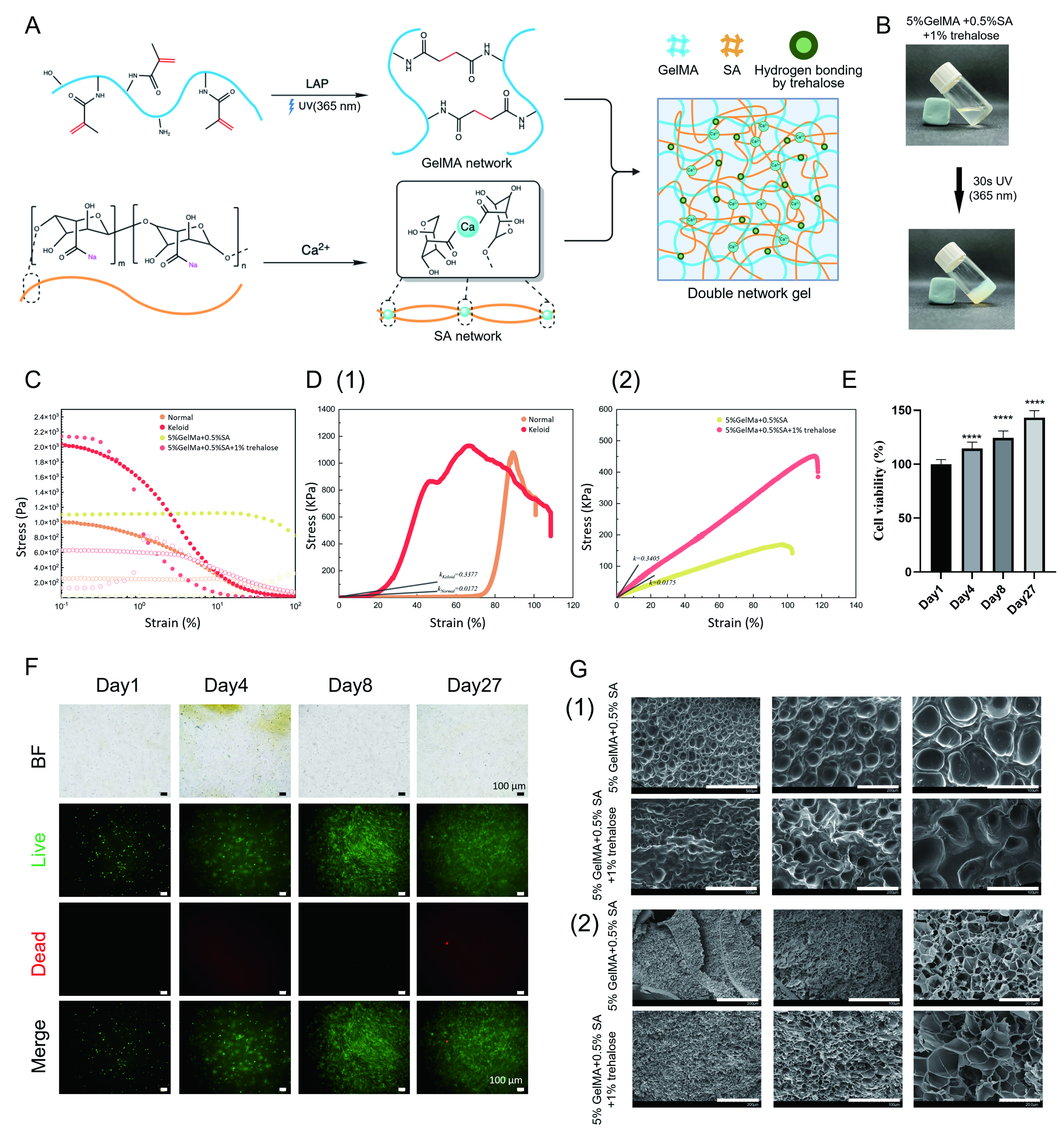
Figure 3. Characterization of the soft and stiff hydrogel. (A) Schematic illustration of the fabrication of the mechanically tunable GelMA/SA dual-network hydrogel. Created with BioRender.com; (B) Gelation process of keloid stiff hydrogel (author’s original photograph); (C) Strain sweeps of keloid tissue and GelMA/SA hydrogels confirm their similar rheological characteristics; (D) Tensile test of normal skin and keloid tissue demonstrates higher stiffness in skin fibrosis. Different GelMA/SA hydrogel systems were used to mimic the distinctions of stiffness (in Young’s modulus); (E) Cell viability and (F) distribution of keloid cells within the hydrogel after 4 weeks (live: green, dead: red); (G) Surface and inner structure of hydrogel. Statistical significance was determined by one-way ANOVA with Tukey’s multiple comparisons test. Data are shown as mean ± SD. Analyses are based on technical replicates (n = 3 per group). ****P < 0.0001; ns, not significant. Scale bar: (F) 100 μm; (G) from left to right: (1) 500, 200 and 100 μm, (2) 200, 100 and 20 μm. GelMA: Gelatin methacryloyl; SA: sodium alginate; ANOVA: analysis of variance; SD: standard deviation; LAP: lithium phenyl-2,4,6-trimethylbenzoylphosphinate; UV: ultraviolet; BF: bright field.