

Hydrogel-driven construction of integrated cancer theranostic platforms: from diagnosis to therapy
 0
0 Graphical Abstract
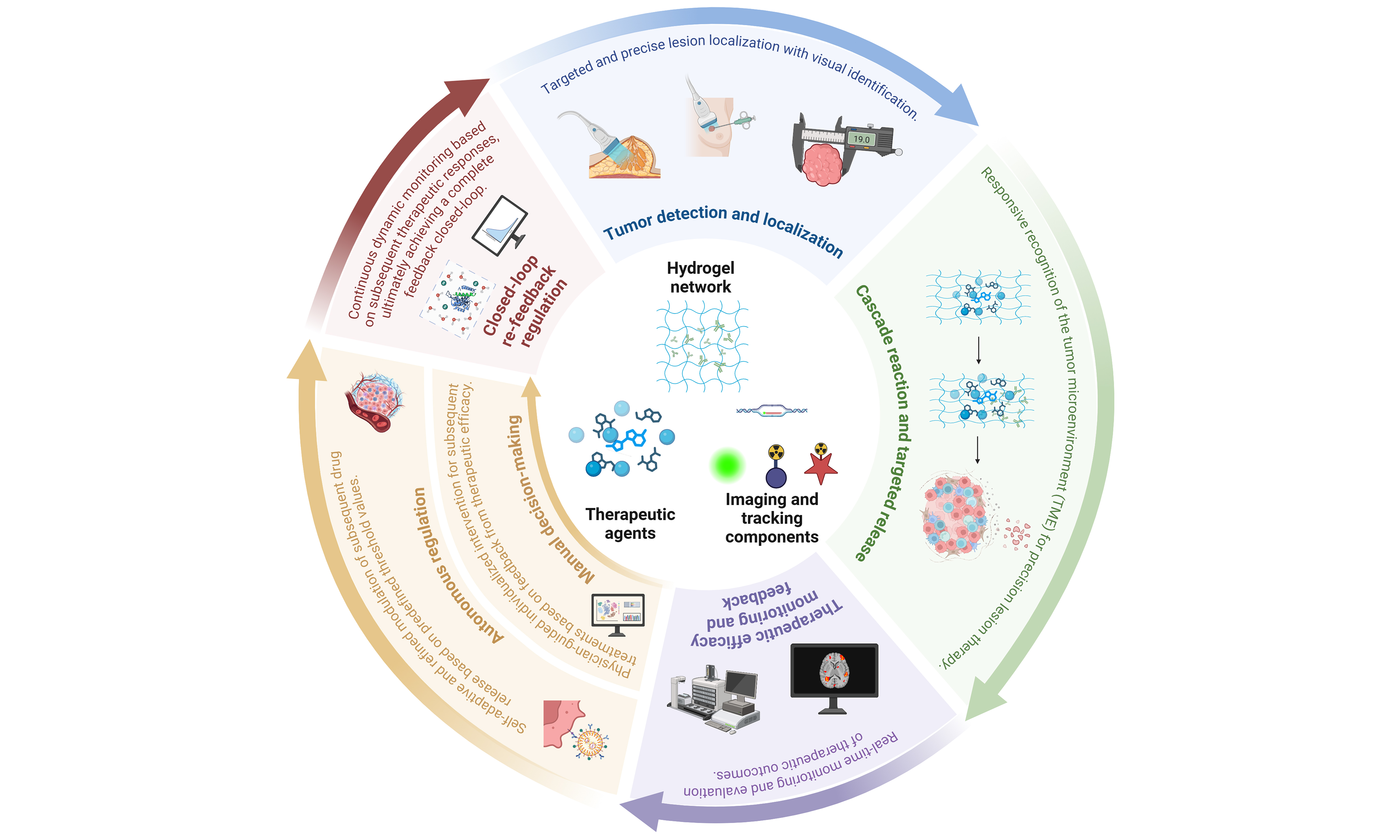
Abstract
Current cancer treatment still relies largely on stage-specific diagnosis and relatively fixed therapeutic regimens, while diagnostic information, therapeutic intervention, and efficacy feedback are often separated across different time windows. This fragmentation limits the ability to meet the demands of precision and individualized therapy. Theranostic platforms integrate disease diagnosis with therapeutic decision-making and can provide concurrent feedback on, or prediction of, treatment efficacy, enabling continuous assessment throughout the therapeutic course. Hydrogels are soft materials with tissue-like three-dimensional networks whose biofunctional properties permit stable loading of imaging probes and therapeutic agents, as well as controlled release and treatment monitoring at lesion sites. These features make hydrogels promising material platforms for integrated cancer theranostics. This review systematically summarizes hydrogel construction strategies for different diagnostic and therapeutic scenarios, emphasizing how matrix composition, crosslinking mode, and emerging materials influence platform performance. We further examined advances in hydrogel research for precision oncology over the past three years, with particular attention to emerging applications in cancer diagnosis and treatment. Building on this overview, we summarized the key oncological applications of hydrogel-based theranostic platforms according to distinct feedback modes. We also assessed the major limitations that currently hinder their development and discussed potential strategies to address them. Finally, we examined the challenges that continue to hinder real-world implementation and considered how integration with emerging technologies may shape the future of the field. Overall, this review provides a systematic framework for designing next-generation hydrogel-based cancer theranostic platforms that combine personalization, continuous monitoring, and realistic potential for clinical translation.
Keywords


 Download PDF
INTRODUCTION
Cancer has become one of the leading causes of death and disease burden worldwide. According to projections from the Global Burden of Disease Study 2023, the global cancer burden is expected to reach approximately 30.5 million new cases and 18.6 million cancer-related deaths annually by 2050, representing increases of 60.7% and 74.5%, respectively, compared with the 2024 levels[1]. As the global cancer burden intensifies, conventional clinical pathways that distribute diagnosis, treatment, and efficacy feedback across separate time windows are increasingly unable to meet the demands for precision and timeliness in oncologic care. Precision oncology emphasizes delivering the right treatment to the right patient at the right time[2]. However, in routine practice, diagnosis and treatment often occur as two temporally distinct stages: the former provides subtype stratification and target identification, while the latter involves systemic administration or local intervention[3,4]. Cancer is not a static entity but a dynamic system characterized by pronounced spatial heterogeneity and temporal evolution[5-7]. Under therapeutic selection pressure, the composition of tumor subclones, molecular profiles, and microenvironmental states continues to change[6,7]. Consequently, one-time, static diagnostic information is insufficient to support long-term therapeutic decisions[3,8], thereby limiting efficacy optimization and the realization of true individualized therapy.
In this context, theranostic platforms capable of simultaneously supporting molecular recognition, local intervention, and dynamic monitoring are emerging as a critical direction in precision cancer medicine. Theranostics, an increasingly important branch of medicine, integrates disease diagnosis with therapeutic decision-making to advance individualized healthcare[9,10]. Built on biomarker detection and early disease identification, this strategy tailors interventions to the patient’s specific condition and provides real-time feedback or prediction of therapeutic efficacy[3,10], thereby enabling more targeted medical strategies. In this process, clinicians utilize modern imaging, genomics, and other biotechnological approaches to comprehensively evaluate disease status[11,12] and select appropriate targeted drugs or treatment modalities.
Hydrogels, three-dimensional (3D) networks formed by crosslinked hydrophilic polymer chains, possess high water content, tunable pore architectures, and mechanical properties resembling those of soft tissues. These features enable them to closely mimic the physicochemical environment of the native extracellular matrix (ECM)[13]. This not only provides a stable local environment for the retention and controlled release of bioactive components, but also allows fine control over drug diffusion, degradation behavior, and tissue compatibility by regulating material composition, crosslinking mode, and network structure[14]. Compared with conventional systemic formulations, injectable and in situ gelling hydrogels can establish local drug depots through minimally invasive administration, prolong intratumoral retention, reduce off-target toxicity, and improve therapeutic precision[15]. Furthermore, hydrogel matrices can carry a wide range of functional molecules, including chemotherapeutic agents, proteins, nucleic acids, immunomodulators, exosomes, and imaging probes[14], thereby integrating diagnostic and therapeutic functions within a single material system. Their responsiveness to signals in the tumor microenvironment (TME), such as pH, reactive oxygen species (ROS), enzymes, temperature, and light[16], further permits spatiotemporally controlled signal activation and drug release at tumor sites. Collectively, these features make hydrogels ideal materials for constructing multifunctional platforms that connect molecular recognition, targeted intervention, and real-time monitoring [Figure 1].
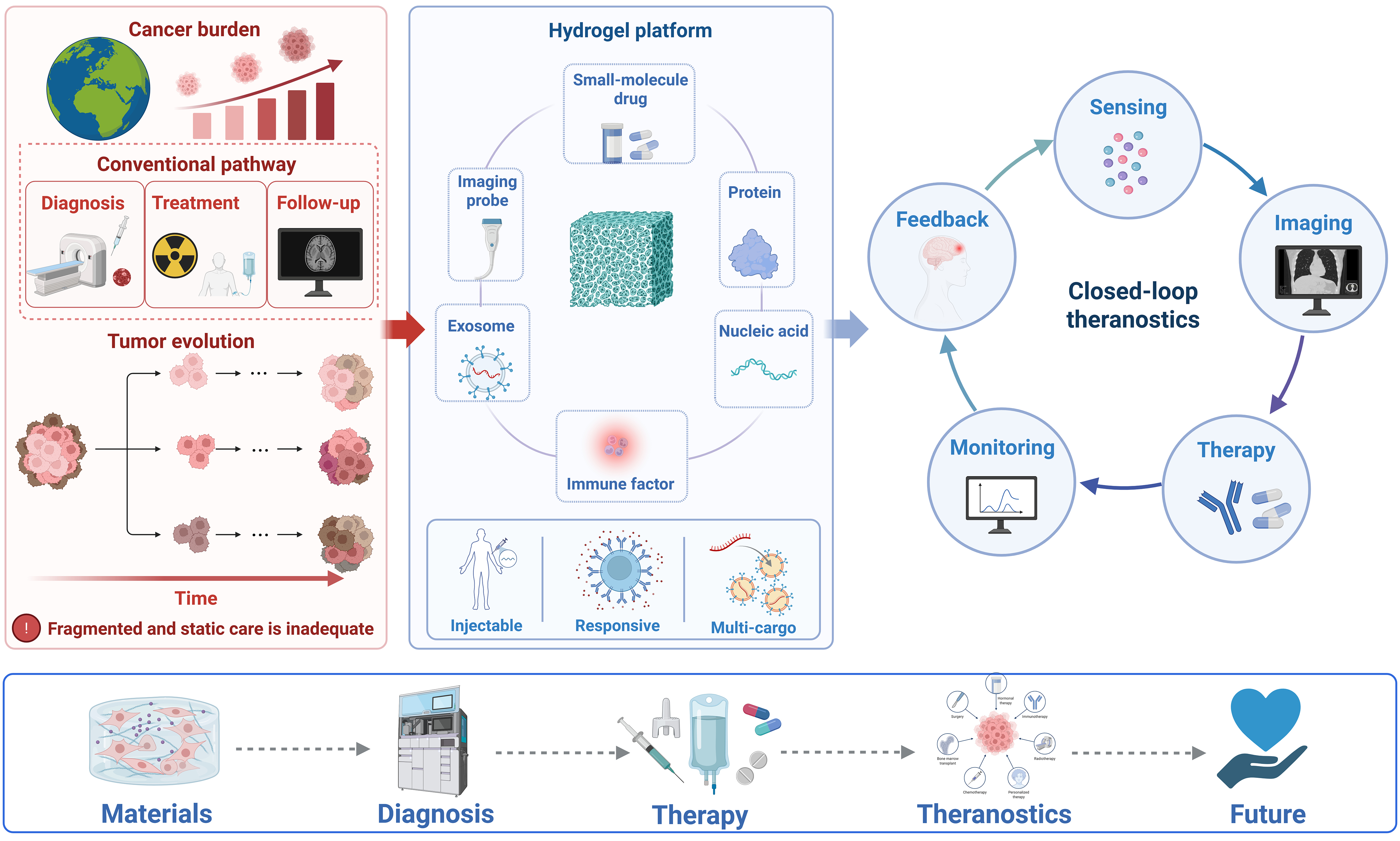
Figure 1. Schematic diagram illustrating how the hydrogel-based theranostic platform overcomes the current fragmentation between diagnosis and therapy in cancer care, thereby enabling integrated tumor diagnosis and therapy [Created in BioRender. Mao, X. (2026) https://BioRender.com/tb8z975].
In recent years, as the application of hydrogels in cancer diagnosis and therapy has continued to deepen, they have gradually evolved toward integrated platforms capable of simultaneously performing both diagnostic and therapeutic functions. Existing reviews of hydrogel-based theranostics, however, have largely focused on a particular cancer type, signal, or technological modality [Table 1][17-19]. A systematic review that breaks through these limitations and highlights the transition of hydrogels from cancer diagnosis and treatment toward practical applications as integrated theranostic platforms is still lacking. To address this gap, we begin with the structural design of hydrogels and examine how matrix materials and crosslinking strategies influence diagnostic signal generation, therapeutic payload delivery, and stimulus-responsive behavior. We then discuss advances reported since 2024 in hydrogel-enabled molecular cancer diagnosis, molecular therapeutics, and theranostic platforms operating through different feedback modes. To our knowledge, this is the first review to classify a broad range of hydrogel-based theranostic platforms according to their feedback modes after systematically comparing their underlying mechanisms. This framework highlights the distinct design principles and potential applications associated with each mode. Finally, we examine the challenges and limitations that may hinder the practical implementation of integrated hydrogel-based cancer theranostics and consider how emerging technologies, including artificial intelligence and bioelectronic interfaces, may contribute to the next generation of personalized hydrogel theranostic systems.
Comparison between recent representative reviews and this review
| Review | Main scope and organizational logic | Key comparison with the present review | |
| Masaeli et al.[17] | This review summarizes methods for fluorescent labeling of hydrogels and discusses recent applications of fluorescent hydrogels in cancer therapy, biosensing, and visual imaging | Starting from the needs of cancer diagnosis, functional imaging, drug delivery, and biosensing, this review places greater emphasis on the fabrication procedures of fluorescent hydrogels | The present review places greater emphasis on the practical applications of hydrogel-based theranostic platforms in cancer diagnosis, treatment, and the construction of integrated theranostic systems |
| Sampath et al.[18] | A breast cancer-focused review of magnetic hydrogels, covering their composition, functional properties, and therapeutic applications in breast cancer | This review emphasizes the functional properties of magnetic hydrogels and uses breast cancer treatment examples to examine how the integration of magnetic nanoparticles can improve therapeutic precision and multifunctionality | The present review is not restricted to a single cancer type or a specific technological modality. Instead, it synthesizes practical advances reported over the past three years and places greater emphasis on their current applications |
| Lee et al.[19] | A pan-cancer review of multifunctional hydrogel systems, covering the applications of multifunctional hydrogels integrated with diagnostic modalities in cancer theranostics | This review primarily focuses on design strategies that integrate multifunctional hydrogels with advanced diagnostic imaging technologies, followed by a brief discussion of their applications across different cancer treatment modalities. However, it provides relatively few detailed examples of practical applications | Starting from practical applications, the present review systematically synthesizes frontier studies from the past three years and places greater emphasis on analyzing the feedback-regulation mechanisms of different theranostic platforms through representative examples |
HYDROGEL MATRICES AND DELIVERY STRATEGIES
Hydrogel matrix materials
The key to using hydrogels in integrated cancer theranostic platforms lies in their highly hydrated 3D networks, which can simultaneously support biomarker enrichment, signal transduction, drug release, and efficacy monitoring[20-26]. Matrix composition determines biocompatibility and payload compatibility; crosslinking strategy governs gelation behavior and release kinetics; loaded functional components dictate recognition, imaging, and therapeutic outputs; and hydrogel network design determines whether these modules can respond on demand in vivo in a controllable temporal and spatial sequence[20,24-26].
Hydrogel material types and structural classification
The advantages of natural polymer hydrogels stem primarily from their ECM-mimetic properties, mild gelation conditions, and degradable chemical structures[21,27-30] [Figure 2]. Alginate forms “egg-box” physical networks via divalent cations, making it suitable for encapsulating protein drugs, living cells, or nanoscale components under mild conditions. However, because it lacks mammalian enzymatically degradable sites and cell-adhesion sequences, alginate typically requires modification through Arg-Gly-Asp (RGD) grafting, oxidative functionalization, or combination with natural ECM materials to enhance cell interactions[27-29]. Chitosan possesses a cationic backbone, mucoadhesive properties, and amino-reactive sites, enabling electrostatic complexation with negatively charged nucleic acids, exosomal membrane components, or anionic polymers. Nevertheless, its solubility, mechanical stability, and pH-dependent degradation require further regulation through polyelectrolyte complexation or dynamic covalent crosslinking[30,31]. Hyaluronic acid (HA), a representative glycosaminoglycan, combines high hydration, hyaluronidase-mediated degradability, and cluster of differentiation 44 (CD44)-mediated recognition, making it particularly valuable for tumor-targeted delivery and cell capture[32,33]. Collagen, gelatin/gelatin methacryloyl (GelMA), and silk fibroin more closely resemble proteinaceous ECM; they provide integrin recognition, matrix metalloproteinase-sensitive degradation, and tunable mechanical support, making them well-suited for constructing 3D tumor models, postoperative local fillers, and delivery systems compatible with tissue repair[34-37].

Figure 2. Schematic diagram illustrating the classification of hydrogel material components and the characteristics of hydrogels with varying matrix compositions [Created in BioRender. Mao, X. (2026) https://BioRender.com/6hifl6t]. ECM: Extracellular matrix; PLGA: poly(lactic-co-glycolic acid); PAAm: polyacrylamide; PVA: polyvinyl alcohol; PEG: poly(ethylene glycol).
Synthetic polymer hydrogels emphasize programmable structures and batch-to-batch consistency, with poly(ethylene glycol) (PEG), polyvinyl alcohol (PVA), polyacrylamide (PAAm), and degradable block copolymers being the most common representatives[38,39]. PEG networks can regulate pore size, mechanical strength, and the release rate of encapsulated components through terminal functionalization, arm number, and crosslink density[40,41]. However, their bioinert nature necessitates the introduction of adhesive peptides, enzyme-sensitive linkers, or natural polymer segments to enable recognition by cells and the TME[42,43]. PVA forms stable networks through freeze–thaw cycling or chemical crosslinking and is suitable for patches, microneedles, and local covering-type delivery systems[44-47]. PAAm is commonly used in mechanically tunable in vitro models[48], while thermosensitive copolymers such as poly(lactic-co-glycolic acid) (PLGA)-PEG-PLGA undergo in situ gelation at body temperature, enabling long-acting local release[49,50].
Short or long peptides and oligonucleotides can translate their sequence information into hydrogel functionality by forming reversible networks through hydrogen bonding, π–π stacking, hydrophobic interactions, host–guest interactions, or base complementarity[51-55]. Compared with conventional polymers, these building blocks can incorporate targeting sequences, enzyme-cleavable segments, immunomodulatory motifs, or nucleic acid-recognition structures at the molecular level. Consequently, they are better suited for molecular recognition, local responsiveness, and reversible assembly[51-54]. Their primary limitation is that noncovalent networks are relatively sensitive to concentration and ionic strength; thus, they usually require combination with natural or synthetic polymers, or the introduction of secondary crosslinking, to improve structural stability in vivo[52-55].
Hydrogel crosslinking strategies
The crosslinking strategy dictates the transformation of a hydrogel from an injectable solution into a local network and governs the in vivo release profile of payloads[56,57]. Physical crosslinking relies on reversible interactions, such as ionic bonds, hydrogen bonds, and metal coordination[58]. This approach generally offers shear-thinning behavior, self-healing capability, and minimally invasive injectability, making it suitable for applications requiring rapid spreading, local adhesion, or short- to medium-term release[58,59]. However, the gel network is highly sensitive to ionic conditions and interstitial fluid exchange. Consequently, when delivering high loads of chemotherapeutics, photosensitizers, or immune adjuvants, burst release often must be mitigated through the incorporation of nanocarriers or secondary crosslinking[60,61].
Chemical crosslinking forms relatively stable covalent networks via free-radical polymerization, photocrosslinking, click reactions, and related methods, thereby enabling precise control over gel strength, degradation rate, and payload diffusion[62]. These robust networks minimize the risks of premature material disintegration and excessively rapid drug release. Therefore, they are widely employed in postoperative cavity administration, implantable sustained-release systems, and local treatments requiring high shape fidelity[63,64]. Nevertheless, conventional small-molecule crosslinkers or initiation systems may leave toxic residues in vivo. Furthermore, crosslinking processes involving free radicals or other highly reactive agents may compromise the bioactivity of loaded therapeutics[65,66].
Dynamic covalent crosslinking combines chemical stability with reversible responsiveness, allowing bond exchange or cleavage under stimuli such as pH, redox conditions, or glucose. This makes it suitable for constructing adaptive release systems, tissue-adhesive materials, and degradable networks[67]. However, because dynamic covalent bonds are intrinsically reversible, hydrogel networks in body fluids may be influenced by pH, redox state, and reaction conditions, leading to fluctuations in gelation efficiency, network strength, and drug-release behavior[68]. In practice, optimizing crosslink density, bond-exchange kinetics, and local delivery conditions is often necessary to balance responsiveness and durability[68].
Biological crosslinking primarily involves constructing hydrogel networks under mild, physiologically compatible conditions by harnessing catalytic or recognition mechanisms derived from biological systems. Compared with traditional chemical crosslinking, these strategies can usually be conducted at physiological pH and temperature without ultraviolet irradiation or highly reactive crosslinkers, and are therefore more conducive to preserving the functional integrity of active components, such as cells, proteins, and nucleic acids, during gelation[69,70]. Beyond classical enzyme-mediated crosslinking, biomolecular recognition systems based on DNA base pairing or aptamer–target recognition are often regarded as functional extensions of biological crosslinking[71,72]. A defining feature of such systems is that the crosslinking units not only participate in network formation but also mediate molecular recognition, signal response, and payload regulation, thereby facilitating integration with tumor diagnostic applications[73]. However, the gelation efficiency and final network properties of these hydrogels often depend on enzyme activity, substrate structure, and the local microenvironment. Consequently, gelation speed, crosslink density, and mechanical performance may fluctuate across material batches or in complex body-fluid environments[69,70].
Multifunctional hydrogels with composite architectures
In hydrogel-mediated cancer theranostic design, the matrix composition and crosslinking strategy together determine the hydrogel’s recognition of tumor-associated signals, responsiveness, and therapeutic outputs. Natural polysaccharide or protein matrices mimic the ECM, offering tissue adhesion and enzymatic degradability, which facilitates the immobilization of cells, vesicles, nucleic acid probes, or protein factors. Conversely, synthetic networks such as PEG and PVA ensure stable local retention and act as diffusion barriers through the regulation of terminal reactions, crosslinking density, and pore size[27,28,38-40]. Integrating distinct matrices into composite networks via diverse crosslinking mechanisms allows hydrogels to maintain structural stability while generating controllable responses to the TME or exogenous stimuli, thereby rendering both gelation and cargo release tunable[56-58,67,68].
Nanogels are hydrogel structures with crosslinked networks reduced to the nanoscale. They retain the high water content and swelling properties of bulk hydrogels while exhibiting the favorable diffusibility, ease of modification, and responsive controllability characteristics of nanocarriers[74,75]. Compared with bulk hydrogels, nanogels diffuse more readily through blood or the tumor interstitium and can be engineered to recognize biomarkers via antibodies, aptamers, or peptide ligands[76]. Their network structures respond to the TME or external stimuli, translating material-level structural changes into signal enhancement or payload release[75,77-79]. In diagnostics, nanogels serve as microreactors or signal-enrichment units to increase the local concentration of low-abundance biomarkers. In therapy, they function as drug reservoirs embedded within bulk hydrogels; the bulk gel ensures lesion retention, while the nanogels mediate tissue penetration, cellular uptake, and microenvironment-triggered release[76-79].
Liposome–hydrogel composite systems combine the drug-loading capacity of liposomes with the local retention and sustained-release properties of hydrogels[80]. Lipid bilayers encapsulate drugs or functional molecules, improving payload stability and delivery compatibility, whereas hydrogel networks restrict diffusion at injection or implantation sites, reduce premature leakage, and prolong local exposure[81-83]. By tuning pore size, crosslink density, and degradable linkages, liposome release, drug extravasation, and gel degradation can be coordinated across different timescales[82]; with the further introduction of stimulus-responsive modules, synergistic triggering by exogenous stimuli and the TME can also be achieved[84]. Metal–drug coordination networks, two-dimensional (2D) nanomaterials, and carbon-based nanostructures can also be embedded within hydrogels to provide metal-ion imaging, catalytic therapy, photothermal conversion, or electrical/acoustic responsiveness. However, risks related to metal accumulation, long-term degradation, nanomaterial migration, and local inflammation must be evaluated concurrently[85-87].
Delivery strategies for hydrogels with different form factors
Beyond matrix composition and crosslinking chemistry, the physical dimensions and structural format of a hydrogel also shape its role in cancer diagnosis and treatment. Based on the organization of their final networks, hydrogels can be broadly divided into bulk and particulate systems[88]. Bulk hydrogels contain a continuous polymer network that extends throughout the material, allowing them to serve as stable local drug depots[89]. Particulate systems, by contrast, consist of discrete gel particles at the micro- or nanoscale. These particles can also be assembled through various strategies to form granular hydrogels[90]. Each format has its own advantages and limitations, depending on the location of the tumor and the specific therapeutic requirements.
Delivery of bulk hydrogels to tumor surfaces and postsurgical residual disease
Bulk hydrogels are formed from continuous 3D crosslinked networks. They may take the form of preformed gel blocks, films, patches, implantable scaffolds, or monolithic gels generated in situ after injection[91]. For tumor surfaces that can be directly exposed during surgery, as well as postsurgical resection cavities, in situ-crosslinked bulk hydrogels can remain in close contact with the margins of the lesion[91-95]. Their continuous networks allow multiple therapeutic and diagnostic components to be encapsulated within a single depot[92]. Therapeutic payloads can then be delivered locally through mechanisms such as stimulus-responsive release or diffusion along concentration gradients[94,95].
This approach is particularly suited to preventing local recurrence after the resection of gliomas, breast tumors, liver tumors, and intraperitoneal malignancies[91,92]. For cutaneous and other superficial tumors, hydrogel patches or films can be placed directly over the treatment area and loaded with photothermal agents, photosensitizers, chemotherapeutic drugs, or related components[96]. External light can pass through the thin hydrogel layer and activate these agents, producing photothermal or photodynamic effects at the tumor surface and within shallow tissues[95,96]. Thus, when bulk hydrogels are applied to tumor surfaces, they do not actively penetrate the intact tumor mass. Instead, they form a relatively stable therapeutic depot at the lesion interface, from which the incorporated payloads subsequently diffuse into the tumor tissue through different release pathways.
In situ intratumoral delivery of injectable bulk hydrogels
For solid tumors that cannot be directly exposed or are located deep within tissues, bulk hydrogels can be introduced into the tumor as precursor solutions under endoscopic or image guidance. They subsequently form continuous networks through temperature-triggered gelation, dynamic covalent reactions, or light-induced crosslinking[97-99]. Before injection, these systems remain sufficiently fluid to pass through a fine-gauge needle. Once gelation occurs, however, they become localized depots that resist rapid leakage from the injection site, thereby prolonging the intratumoral retention of therapeutic components[99]. Compared with the direct intratumoral injection of free drugs, in situ gelation can reduce the risk of rapid drug clearance. Stimuli-responsive structures can also be incorporated to regulate the timing and amount of drug release[100,101].
Delivery of discrete hydrogel particles into tumors
Particulate hydrogel systems can be divided by particle size into nanogels and microgels[102]. Each particle consists of a crosslinked polymer network with finite dimensions and can independently carry therapeutic agents or diagnostic probes. Compared with continuous bulk hydrogels, particulate systems offer a larger specific surface area and a shorter intraparticle diffusion distance. Their circulation profiles and cellular uptake can also be tuned through various design strategies. Nanogels can be administered intravenously, intra-arterially, or by local injection. Passive accumulation or ligand-mediated recognition may further enhance their selective localization within tumors[102-104]. Once they enter the tumor interstitium, their small size can facilitate uptake by tumor cells. The payload can then be released in response to relevant cues within the TME.
Microgels and hydrogel microspheres are better suited to intratumoral, peritumoral, or postoperative local injection[103-105]. Rather than forming a single continuous depot, a large number of dispersed microgels can establish multiple independent drug microdepots throughout the lesion. This arrangement increases the interfacial area between the payload and tissue fluid while shortening the distance that drugs must travel from the hydrogel interior to the surrounding tumor tissue[105,106]. Different microgel populations can also be loaded with distinct drugs and mixed at predefined ratios, allowing combination therapy and differential release profiles to be achieved.
APPLICATIONS OF HYDROGELS IN TUMOR DIAGNOSIS
Early, rapid, and noninvasive cancer detection is essential for effective treatment and has become a major focus of precision medicine. As hydrogel research has advanced, the distinctive roles of these materials in biosensing and diagnostic imaging have opened new avenues for detecting cancer with high sensitivity, strong selectivity, and rapid readout. Meanwhile, diagnostic hydrogels capable of sensing disease biomarkers and changes in the local microenvironment are continuing to emerge. These advances also lay an important foundation for the future development of hydrogel-based theranostic systems.
Biosensing and molecular biomarker recognition
In recent years, the role of hydrogels in tumor diagnosis has evolved from passive carriers to diagnostic platforms that integrate sample enrichment, activity preservation, controlled release, and signal amplification. This evolution is particularly evident in liquid biopsy, where biomarkers such as circulating tumor cells (CTCs), circulating tumor DNA (ctDNA), microRNAs (miRNAs), and exosomes/extracellular vesicles (EVs) are typically dispersed in complex matrices such as blood, urine, saliva, or tissue fluid, and are generally characterized by low abundance, strong heterogeneity, susceptibility to interference from background components, and, for some molecules, poor stability[107-109]. Owing to their highly hydrated 3D networks and tissue-like softness, hydrogels can provide a gentler microenvironment for the capture of cells, vesicles, and nucleic acids[109-112]. At the same time, recognition modules such as antibodies, aptamers, and nucleic acid probes can be introduced into the network to enable selective enrichment of target biomarkers[111,112]. More importantly, the pore architecture, swelling/shrinking behavior, and degradability or stimulus responsiveness of hydrogels can also support biomarker release and signal amplification when needed[113-116] [Figure 3]. Lu et al. proposed stimulus-responsive signal-amplifying hydrogel microparticles termed Novabeads[117]. In this system, poly(ethylene glycol) diacrylate (PEGDA) served as the network matrix, and pH-responsive acrylic acid motifs were incorporated, resulting in approximately 5-fold volumetric contraction under pH regulation. Coupled with high-density bioreceptor loading, this contraction effect markedly enhanced fluorescence signals and enabled enzyme-free detection of the cancer-associated miR-16. Compared with conventional hydrogel microparticles, this platform achieved more than a 30-fold signal enhancement and a substantially lower limit of detection, demonstrating that the hydrogel itself can function directly as a signal-amplifying element in the diagnostic process.
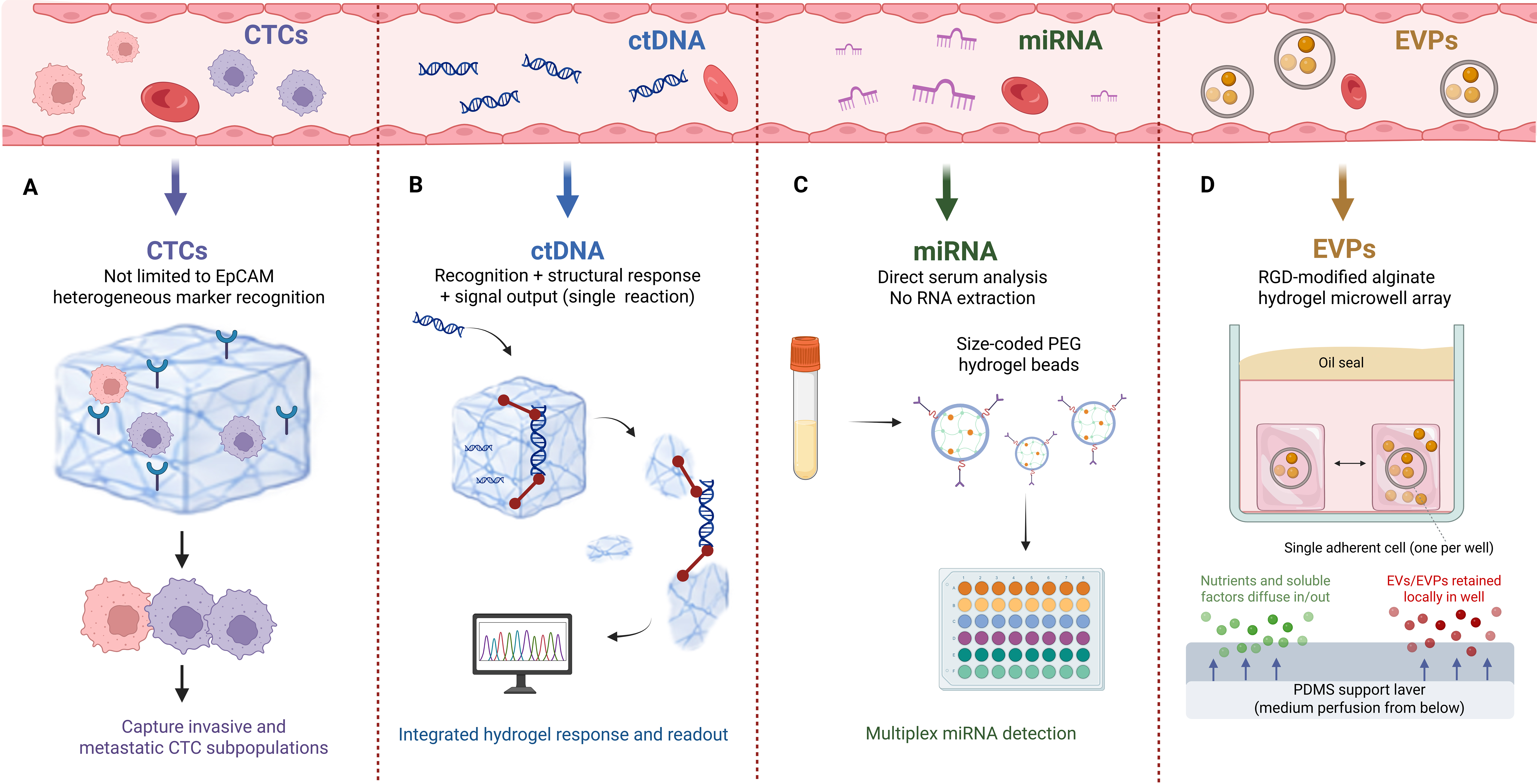
Figure 3. Schematic diagram illustrating capture methods for different liquid biopsy biomarkers. (A) Current CTC capture methods are no longer limited by the heterogeneity caused by EpCAM downregulation; (B) Recognition of target ctDNA, structural changes of the material, and signal output have been achieved within a hydrogel platform; (C) Using a size-encoded PEG hydrogel microbead system, extraction-free multiplex miRNA liquid biopsy analysis can be performed in authentic serum samples; (D) By integrating oil-sealed isolation with hydrogel microwells, single cells and their secreted EVPs can be independently isolated and confined during extended culture periods [Created in BioRender. Mao, X. (2026) https://BioRender.com/nok9rig]. CTC: Circulating tumor cell; EpCAM: epithelial cell adhesion molecule; ctDNA: circulating tumor DNA; PEG: poly(ethylene glycol); miRNA: microRNA; EVPs: extracellular vesicles and particles; EVs: extracellular vesicles.
CTCs are important liquid-biopsy biomarkers for monitoring metastasis and recurrence, yet they are extremely rare in peripheral blood, and distinct CTC subpopulations exhibit pronounced heterogeneity in epithelial–mesenchymal plasticity (EMP) and stemness-related phenotypes/marker expression[118,119]. Accordingly, the frontier of hydrogel-based CTC capture platforms has shifted from simply increasing capture numbers to emphasizing heterogeneous biomarker recognition, cell viability preservation, and subsequent release-based analysis. In the context of heterogeneous biomarker recognition, one study developed a CXCL12-loaded HA hydrogel (CLG) to capture metastasis-prone CTCs[120]. This system exploits the chemokine (C-X-C motif) ligand 12/C-X-C chemokine receptor type 4 (CXCL12/CXCR4) axis to attract invasive CXCR4-positive cells into the hydrogel network. Compared with conventional hydrogel platforms that rely on epithelial cell adhesion molecule (EpCAM), this strategy is less constrained by EpCAM downregulation during epithelial–mesenchymal transition (EMT)[120]. It preferentially enriches CTC subpopulations with hybrid epithelial–mesenchymal phenotypes, stem cell-related features, and invasive metastatic potential, thereby providing a basis for metastasis risk assessment and downstream phenotypic analysis. With respect to preserving cell viability for downstream analysis, another representative advance is a bis-histidine-mediated cell-imprinted hydrogel for the precise capture and dynamic release of circulating hepatocellular carcinoma (HCC) cells[121]. This system improves recognition of target tumor cells through the combined use of sialylated glycans (SGs) and cell-imprinted morphology. Specifically, L-histidine-L-histidine (HH) was integrated into a low-swelling poly(ethylene glycol) dimethacrylate (PEGDMA) cell-imprinted hydrogel to generate imprinted sites on the material surface that closely match the size and contour of the target cells, thereby coupling molecular-scale affinity recognition with cell-scale spatial matching[121]. Because the interaction between HH and SGs is tunable, intact release of captured CTCs can be achieved under the mildly alkaline conditions of trypsin–ethylenediaminetetraacetic acid (trypsin-EDTA), while preserving membrane integrity, proliferative capacity, and subsequent passaging potential to the greatest extent possible.
Compared with CTCs, ctDNA is better suited to reflecting mutational spectra, minimal residual disease, and the evolution of therapeutic resistance, but its short fragment length and low abundance, together with dilution by large amounts of normal cell-free DNA (cfDNA), pose major analytical challenges[122,123]. Current hydrogel platforms are therefore evolving not only toward higher-efficiency ctDNA enrichment but also toward programmable structures and integrated signal transduction. A notable example has already demonstrated, in breast cancer, the coupling of target ctDNA recognition, material structural transformation, and signal output within a single homogeneous reaction, breaking away from the conventional workflow of hydrogel capture, washing/sorting, and subsequent polymerase chain reaction (PCR) or instrument-based amplification readout[124]. In that study, a rolling circle amplification (RCA)-derived DNA hydrogel was integrated with DNA@Cu2+ nanospheres to simultaneously analyze two hotspot mutations in breast cancer, PIK3CA and ESR1, within a single system[124]. Upon ctDNA recognition, the DNA hydrogel actively disassembles to expose a G-quadruplex structure that generates a thioflavin T (ThT) turn-on signal; simultaneously, the nanomaterial releases Cu2+, causing a quantum-dot turn-off. Target recognition is thus converted into dual orthogonal signal outputs through hydrogel disassembly and nanosphere decomposition, reducing sample loss and improving the detection efficiency of low-abundance ctDNA. DNA methylation analysis represents another frontier in ctDNA-based liquid biopsy. Hou et al. constructed a thermosensitive hydrogel-enhanced one-pot recombinase polymerase amplification (RPA)-CRISPR/Cas12a system[125], combined with methylation-sensitive restriction endonucleases, for the ultrasensitive detection of specific methylation sites in breast cancer ctDNA. By employing a thermosensitive hydrogel, this method achieved spatial separation while maintaining reaction connectivity within a single tube for the first time, thereby alleviating target depletion and reduced sensitivity caused by premature Cas12a cleavage. Thus, hydrogels no longer function merely as reaction vessels but as integrated reaction systems capable of regulating amplification, nuclease digestion, and signal readout through spatial organization.
Current quantitative techniques for multiple miRNAs typically require RNA extraction from body fluids as a preprocessing step[126]. This process is labor-intensive and may compromise analytical accuracy due to target miRNA loss and degradation[127]. There is therefore a pressing need for reaction systems that can directly process body fluids without preprocessing. Recently reported size-coded PEG hydrogel bead systems have advanced hydrogel platforms toward extraction-free, multiplex miRNA liquid biopsy analysis in real serum samples[128-131]. In one study, capture probes for miR-21, miR-205, and miR-375 were immobilized in beads of different diameters, allowing target miRNAs to diffuse directly from serum into the beads without RNA extraction and to be selectively retained[129]. RCA amplification was then completed within the beads, and parallel quantification of the three miRNAs was achieved by size-based grouping via flow cytometry; when combined with machine-learning analysis, this strategy enabled lung cancer diagnosis and subtype classification[129]. This also reflects the broader shift of liquid biopsy toward automation, multiparametric measurement, and algorithm-assisted diagnosis.
Resolving the heterogeneity of extracellular vesicles and particles (EVPs) derived from single cells is of major importance for elucidating mechanisms of tumor metastasis and advancing cancer medicine[132]. Existing single-cell EVP microfluidic systems generally struggle to simultaneously achieve source purity and long-term culture[133,134], and technologies capable of independently isolating and sealing single cells along with their secreted EVPs over extended culture periods remain lacking. The emergence of a microwell array composed of oil-sealed, RGD-modified alginate hydrogel has helped address this gap[135]. By integrating oil-sealed isolation with RGD-modified alginate hydrogel microwells, this study established a gel system capable of simultaneously achieving independent single-cell confinement and in situ retention of secreted EVPs during long-term culture. The oil layer isolates each microwell from the others, preventing inter-well mixing of EVPs; the size-selective permeability of the alginate hydrogel mesh allows nutrients and soluble factors to pass while blocking EVP escape; and the RGD motif provides adhesion sites for adherent cells, with culture medium continuously replenished through a polydimethylsiloxane (PDMS) spacer layer at the base. This platform provides a foundation for future liquid biopsy studies in which single-cell-derived EVs can be traced to their source.
Visual imaging and image guidance
Because of their hydrated 3D network structure, hydrogels can locally confine imaging probes, reduce premature probe leakage, and protect signal-generating components from rapid inactivation or nonspecific diffusion, making them highly important imaging matrices in tumor diagnosis[136-138]. Compared with freely diffusing small-molecule probes or nanoparticles, hydrogel-based systems generally exhibit longer retention at tumor sites or in postoperative cavities and more readily couple signal generation to endogenous cues, such as adenosine triphosphate (ATP), pH, and enzymes, or to externally applied optical stimulation[139,140]. Consequently, hydrogels have evolved from passive contrast agent carriers into programmable materials for lesion localization, intelligent response, and therapeutic monitoring and prediction [Figure 4].
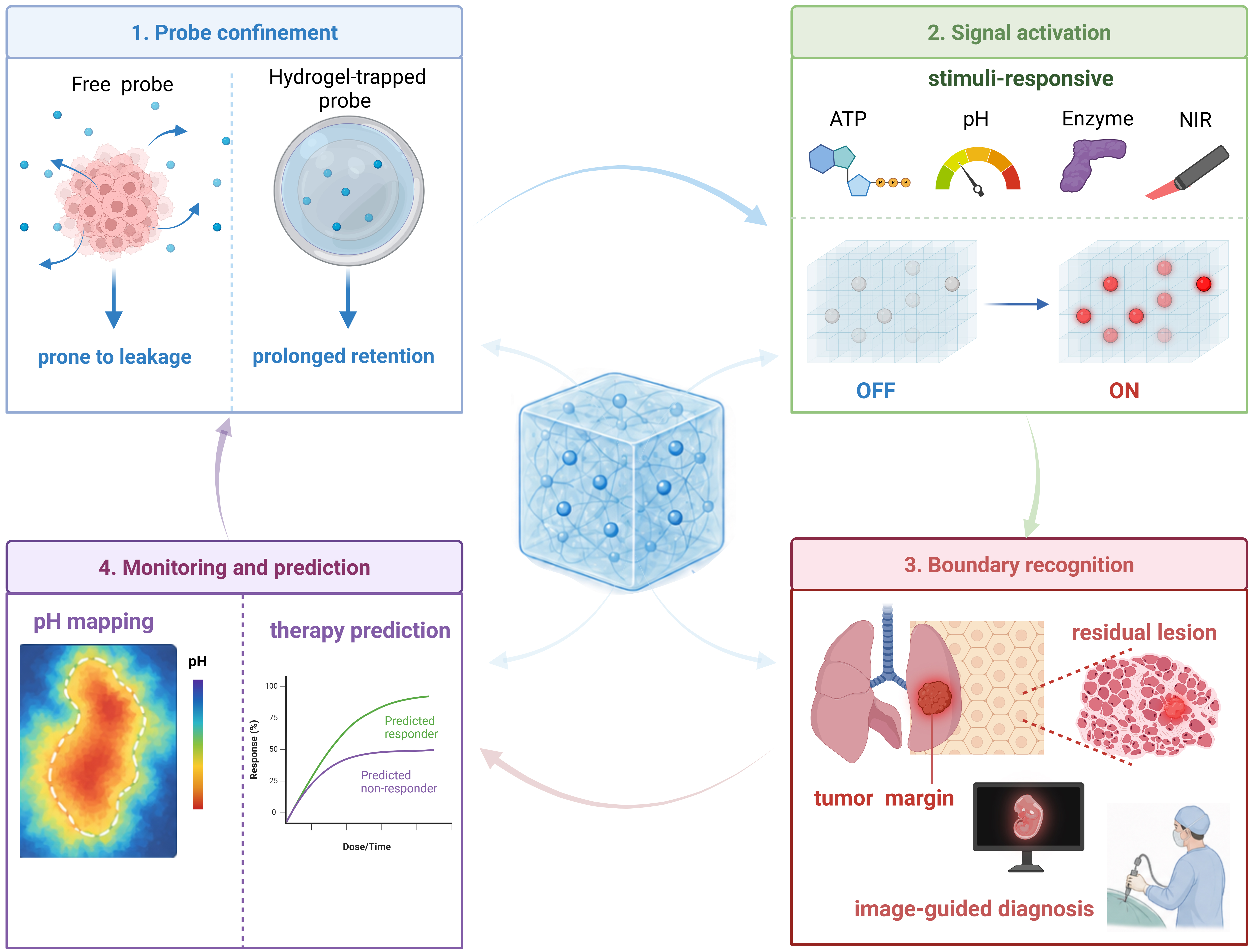
Figure 4. Schematic diagram of hydrogel-based tumor visualization imaging and image-guided modalities. The hydrogel network confines the retention of imaging components and can integrate with the TME to confer smart responsive properties, enabling tumor visualization and the reflection or prediction of therapeutic outcomes [Created in BioRender. Mao, X. (2026) https://BioRender.com/4kts57u]. TME: Tumor microenvironment; ATP: adenosine triphosphate; NIR: near-infrared.
Fluorescence imaging remains one of the most accessible diagnostic modalities due to its high sensitivity, operational simplicity, and compatibility with intraoperative visualization[141]. However, the diagnostic utility of conventional fluorescent probes is often limited by rapid diffusion, photobleaching, aggregation-caused quenching, and insufficient retention at lesion sites[142,143]. Hydrogel encapsulation partially addresses these limitations by providing a hydrated microenvironment that stabilizes organic fluorescent dyes, aggregation-induced emission nanoparticles, and other luminophores[144,145]. In 2013, Park et al. developed a near-infrared-sensitive molecular imaging probe based on a hydrogel composite[146]. In this system, HA served as the matrix, conferring CD44-targeting capability, while a near-infrared-sensitive organic dye was encapsulated. Upon near-infrared excitation, the hydrogel emitted fluorescence, enabling the imaging of gastric cancer cells expressing CD44. From the standpoint of current frontier research, the development of fluorescence-optical imaging hydrogels has shifted from early systems that relied on single-cell surface receptor recognition to highly sensitive, stimulus-responsive tumor molecular imaging platforms. Yang et al. further constructed a TME-responsive DNA hybrid hydrogel, termed AHB Gel[147]. By coupling a PAAm hydrogel network with an ATP-responsive aptamer, this system can directly sense abnormally elevated extracellular ATP levels in the TME while maintaining close tissue contact without tissue invasion. It rapidly activates fluorescence under high-ATP conditions but produces almost no obvious fluorescence against the low-ATP background of normal tissues. As a result, this hydrogel not only enables highly selective recognition of tumor tissue but also creates a clear boundary between tumor and normal tissue, thereby improving the accuracy of lesion localization and margin delineation.
On this basis, further integrating responsive fluorescent probes into hydrogel systems that can be locally coated or formed in situ can establish a transiently stable imaging interface within the surgical cavity for intraoperative navigation and residual-disease detection. Walker et al. developed a locally spreadable thermosensitive imaging gel in which AKRO-QC-ICG was loaded into a Pluronic F-127 matrix for the rapid identification of residual breast cancer tissue within surgical cavities[148]. After local tissue contact, this gel generated sufficient fluorescence contrast within 10 min to distinguish the tumor from the surrounding normal tissue, and high signal-to-noise ratios were retained even after washing. In both murine orthotopic models and patient-derived xenograft (PDX) models, it demonstrated high sensitivity and specificity for lesion detection. For cancers with highly heterogeneous molecular markers, the recent emergence of fluorescent gels targeting tumor-microenvironment components offers a new solution[149]. Their design no longer relies primarily on highly heterogeneous tumor-cell surface molecules but instead shifts toward using relatively stable collagen components in the TME as anchoring sites. This study showed that collagen-adhesive probes can improve the sensitivity and stability of intraoperative fluorescence imaging for bladder cancer, thereby providing a new imaging strategy for lesions such as non-muscle-invasive bladder cancer (NMIBC), in which accurate boundary delineation is particularly critical.
Beyond intraoperative residual disease detection, hydrogel fluorescence imaging platforms are increasingly applied to the localization of deep lesions during endoscope-assisted or minimally invasive procedures. Near-infrared fluorescence imaging offers deeper tissue penetration, lower autofluorescence, and greater suitability for solid tumors located in complex or relatively deep anatomical regions. Accurate localization of deep lesions remains a major challenge in endoscope-assisted minimally invasive surgery. Lee et al. developed a thermosensitive hydrogel loaded with near-infrared dyes that can safely, durably, and visibly localize deep lesions in real time during endoscopic surgery[150]. In this system, indocyanine green (ICG) or ICG-human serum albumin (HSA) was incorporated into a thermosensitive hydrogel. Because the material remains fluid at low temperatures but undergoes a sol–gel transition at physiological temperatures after in vivo administration, it can gel in situ after local injection into a lesion, thereby confining the dye locally and reducing its diffusion into surrounding tissues. This provides a more stable preoperative-to-intraoperative imaging reference for sustained localization of deep lesions and fluorescence-guided minimally invasive surgery.
Unlike near-infrared fluorescence imaging, which is mainly used for spatial localization, second near-infrared window (NIR-II) photoacoustic imaging emphasizes converting differences in optical absorption within tumor tissues into acoustic signals that can be detected at depth[151], and is therefore better suited for coupling with tumor-microenvironment parameters such as pH, temperature, or metabolic state[152,153]. Conventional always-on photoacoustic probes, however, continuously emit signals in normal tissues, making them susceptible to interference from endogenous chromophores such as hemoglobin and thereby limiting lesion specificity. To address this, Sun et al. constructed a low-pH-triggered, thermal-responsive volume phase-transition nanogel, gold nanorod@poly(N-isopropylacrylamide)-vinyl acetic acid (AuNR@PNIPAM-VAA)[152]. Gold nanorods (AuNRs) provide photothermal conversion and generate photoacoustic signals, whereas the PNIPAM-VAA shell acquires low-pH-dependent thermoresponsive volume-phase transition behavior through the introduction of vinyl acetic acid[152]. Following laser irradiation, AuNRs convert light into heat and raise the local temperature; because tumor regions generally exhibit lower pH than normal tissues, the shell undergoes a swelling-to-collapse phase transition specifically within the tumor[152]. During this process, the local refractive index around the AuNRs increases, the longitudinal plasmon resonance red-shifts, and the NIR-II photoacoustic signal is substantially amplified. Furthermore, Folz et al. constructed tumor-targeted PAAm hydrogel nanoparticles in combination with photoacoustic chemical imaging to predict the chemotherapeutic response of breast cancer by monitoring pH changes in the TME[154]. Their group embedded the pH-sensitive dye seminaphthorhodafluor-5/6F (SNARF-5/6F) into the hydrogel and generated quantitative pH maps using multiwavelength ratiometric photoacoustic chemical imaging, thereby monitoring tumor-microenvironment pH in patient-derived breast cancer xenografts during alkalinization therapy and chemotherapy to achieve efficacy monitoring and prediction.
APPLICATIONS OF HYDROGELS IN TUMOR THERAPY
In therapeutic settings, hydrogels place greater emphasis on actively regulating local tumor pharmacokinetics, the spatiotemporal distribution of different treatment modalities, and the post-treatment microenvironment[155-159]. Leveraging their highly hydrated 3D networks, injectable in situ gelation, tunable crosslink densities, and stimulus responsiveness, hydrogels can not only serve as sustained-release depots that prolong the retention of therapeutic agents within tumors or postoperative cavities[155], but can also integrate chemotherapy, immunomodulation, and photo-, sono-, and magnetically mediated therapies to reduce systemic exposure and enhance local treatment intensity[156,158,159]. Hydrogels are therefore now widely used throughout the course of cancer therapy [Figure 5].
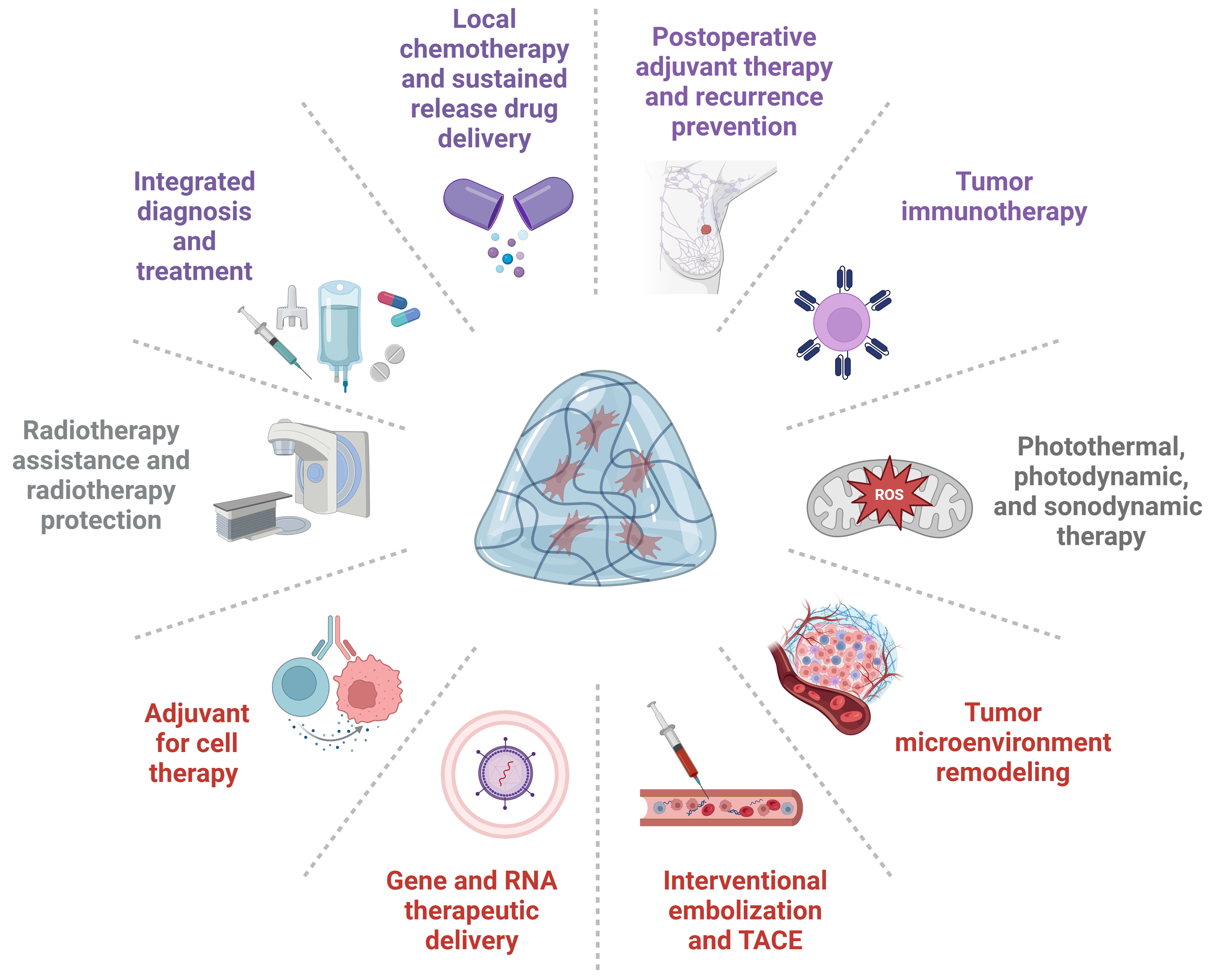
Figure 5. Schematic summary of current application strategies of hydrogels in tumor therapy [Created in BioRender. Mao, X. (2026) https://BioRender.com/qze9yuz]. TACE: Transarterial chemoembolization; ROS: reactive oxygen species.
Chemotherapeutic drug delivery
Chemotherapy remains an essential component of treatment for many solid tumors, but small-molecule chemotherapeutics often face problems such as nonspecific distribution, fluctuations in plasma concentration, insufficient intratumoral retention, and dose-limiting toxicity[160]. The core value of hydrogel-based delivery systems lies in changing the mode of drug exposure: through local injection, postoperative cavity filling, mucoadhesion, or transdermal delivery, hydrogels can transform transient systemic peak concentrations into sustained and relatively stable drug release at the tumor site, and can further achieve TME-responsive release by exploiting differences in pH, enzyme activity, redox status, or temperature[155,156,161-163].
Doxorubicin (DOX) exhibits well-established broad-spectrum antitumor activity, but its clinical utility is limited by cardiotoxicity, myelosuppression, and the risk of resistance[160]. One study developed an injectable pH-responsive polypeptide hydrogel, FOK, for the local sustained release of DOX[161]. This system maintains a stable gel structure at physiological pH but degrades gradually in the acidic tumor environment, thereby promoting DOX release and prolonging its therapeutic effect at the tumor site. Compared with free DOX, the DOX/FOK hydrogel better maintains effective local cytotoxic concentrations by reducing premature drug diffusion and systemic exposure[161]. Shi et al. further reported an HA-based injectable DOX-loaded hydrogel, DOX-MCHAgel, for locoregional breast cancer therapy and metastasis inhibition[162]. In a 4T1 breast cancer model, this hydrogel achieved sustained DOX release for more than 10 d and significantly suppressed primary tumor growth as well as liver and lung metastases, without evident systemic toxicity. These findings indicate that the therapeutic value of hydrogels lies not only in the local sustained release of chemotherapeutic drugs, but also in confining drug exposure to tumors and adjacent high-risk areas, thereby reducing distal organ toxicity and diminishing opportunities for metastatic dissemination. For solid tumors with a high risk of postoperative recurrence, hydrogels can also function as local therapeutic depots within postoperative cavities to eliminate residual tumor cells and improve the tissue-repair microenvironment. Zhang et al. developed a composite system, ZC-DOX@GEL, in which a GelMA hydrogel was loaded with DOX@zeolitic imidazolate framework-8 (ZIF-8)/cerium dioxide (CeO2) nanoparticles for postoperative tumor treatment and wound repair[163]. In this platform, ZIF-8 serves as a DOX nanocarrier to improve drug loading and acid-responsive release, whereas CeO2 nanoparticles alleviate postoperative local oxidative stress and promote wound healing; the GelMA network further immobilizes the nanoparticles within the postoperative cavity, endowing the system with residual tumor eradication, controlled drug release, and tissue-repair functions.
In superficial lesions, such as skin tumors, hydrogel microneedles (HMNs) offer a new structural platform for the local delivery of chemotherapeutic agents or combination therapeutics. HMNs can penetrate the stratum corneum and form continuous hydrogel microchannels, thereby overcoming the skin-barrier limitation of conventional transdermal delivery while being better suited than standard injections for localized, repeatable, and minimally invasive administration[145]. As part of transdermal drug delivery systems (TDDS), microneedles perforate the epidermis and create microchannels; because of their hydrophilicity, they swell to form continuous hydrogel conduits[164-166]. These channels facilitate drug diffusion and ensure stable and sustained therapeutic effects. HMNs have now evolved into integrated drug-delivery systems capable of loading multiple drugs or immune adjuvants for combination cancer therapy. Zhang et al. constructed a bifunctional HA-based HMN system in which the needle tips were loaded with CCa@TF/chlorin e6 (Ce6) nanocomposites and rapidly dissolved after insertion into tumors to release therapeutic components[167]. The hydrogel base, HB-PVA, formed via boronate ester bonds between 3-aminophenylboronic acid-grafted HA and PVA, remained locally to support subsequent skin repair[167]. During treatment, CCa@TF/Ce6 mediated photothermal therapy (PTT) and photodynamic therapy (PDT), respectively, under dual-laser irradiation. More importantly, ROS and thermal stimulation also induced immunogenic cell death (ICD), promoted dendritic cell (DC) maturation, and drove macrophage polarization from the M2 to the M1 phenotype, thereby converting local physical/oxidative injury into antitumor immune activation.
In urologic malignancies, hydrogel delivery systems can also address the problem of intracavitary drug clearance caused by continuous urine dilution, periodic bladder emptying, and insufficient mucosal retention. After surgery for NMIBC, intravesical instillation is often limited by urine dilution, voiding, and poor mucosal retention, resulting in insufficient effective drug-contact time[168]. Mucoadhesive or reverse thermosensitive hydrogels can be instilled as liquids at low temperature and form local drug depots after entering the bladder at body temperature, thereby prolonging the contact of chemotherapeutic agents with urothelial tumor tissue. Zheng et al. proposed an adhesion-to-penetration-transformable nanomotors-in-hydrogel system for intravesical therapy[169], combining the intracavitary retention of conventional hydrogels with the active penetration capability of nanomotors. This platform used poloxamer 407 (PLX), HA, and sodium bicarbonate to construct a self-disintegrating composite hydrogel. PLGA nanoparticles loaded with gemcitabine (GEM) were coated with bladder cancer cell membranes and conjugated with urease to form urease-powered membrane nanomotors (UMN), which were then encapsulated within the hydrogel network. After intravesical instillation, the PLX hydrogel underwent in situ gelation at body temperature and adhered to the urothelial surface, thereby prolonging GEM retention in the bladder lumen and enriching UMN at the mucosal surface. Subsequently, urea in urine diffused into the hydrogel, creating a concentration gradient. The UMN acquired motility by catalyzing urea, allowing them to migrate directionally along the gradient and actively traverse the urinary mucus layer and the dense urothelial barrier, thereby delivering GEM into deeper bladder tissues [Figure 6].
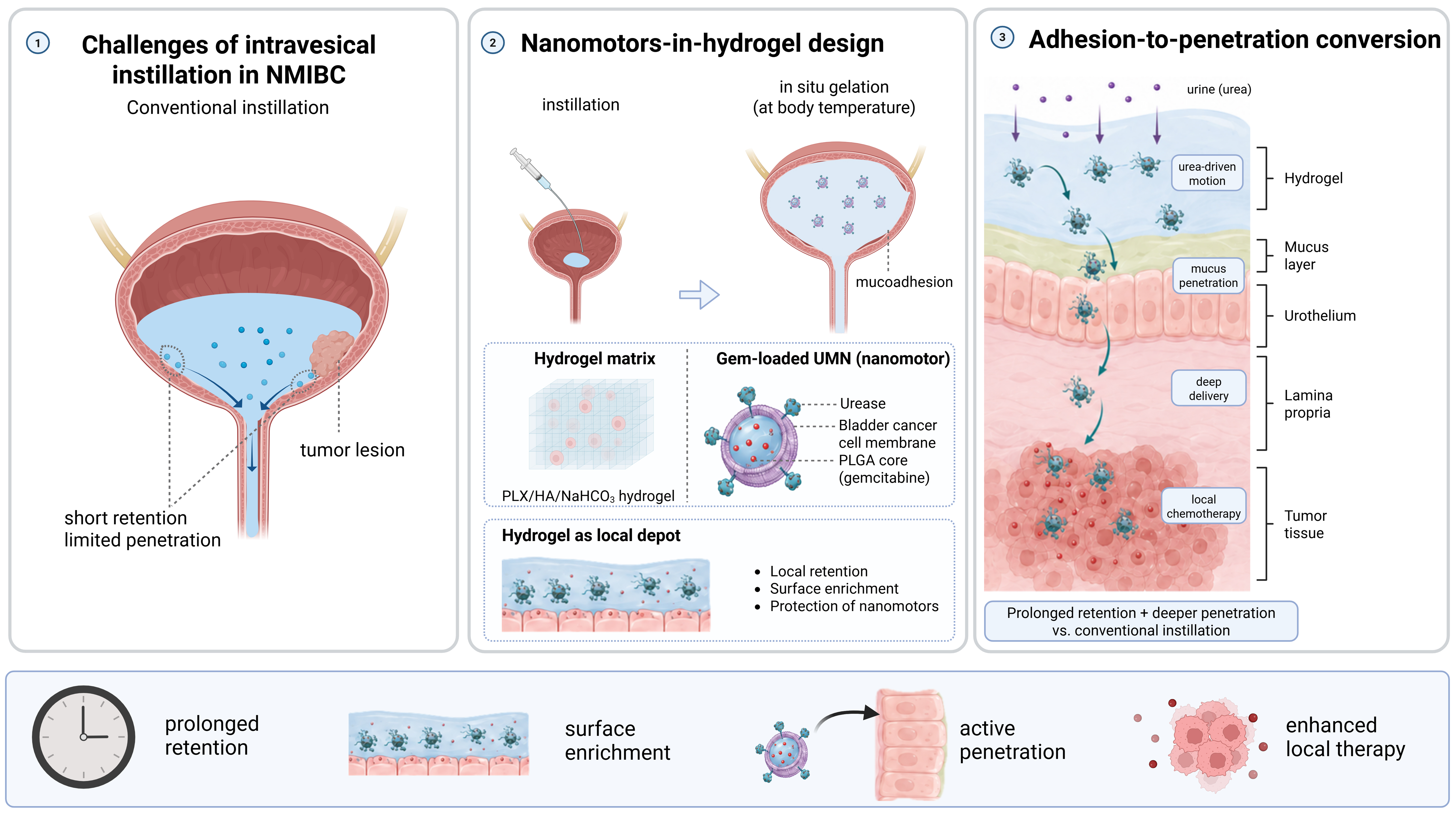
Figure 6. Schematic diagram illustrating the combination of hydrogels and nanomotors for mitigating intravesical drug clearance during intravesical administration. ① Conventional intravesical drug delivery is susceptible to urine flushing, leading to poor therapeutic efficacy. ② Drug-loaded hydrogels enable favorable intravesical retention of therapeutic components. ③ Nanomotors embedded within the hydrogel can actively migrate, achieving better penetration capability [Created in BioRender. Mao, X. (2026) https://BioRender.com/bdbj9ux]. NMIBC: Non-muscle-invasive bladder cancer; UMN: urease-powered membrane nanomotors; PLGA: poly(lactic-co-glycolic acid); PLX: poloxamer 407; HA: hyaluronic acid.
Cancer vaccine carriers
With the continued evolution of vaccine technologies, cancer vaccines can now be designed for patients with established malignancies to induce tumor-specific immune activation and thereby promote targeted recognition and clearance of tumor cells[170,171]. Nevertheless, the development of effective cancer vaccines still faces many challenges. For example, insufficient release of tumor antigens by tumor cells can limit antigen uptake by immune cells. Inadequate antigen processing by antigen-presenting cells restricts the generation of antigen-specific T-cell responses[172]. In addition, the immunosuppressive TME can lead to effector-cell exhaustion and death[173]. Rationally designed delivery technologies, including lipid nanoparticles, hydrogels, scaffolds, and polymeric nanoparticles, can target therapeutic agents to tumor cells, immune cells, or the ECM and thereby effectively overcome these challenges[174-177]. In cancer therapy, hydrogels hold particular promise for maximizing the efficacy of cancer vaccines. On the one hand, the highly porous network structure of hydrogels can protect vaccines from in vivo degradation; on the other hand, hydrogels enable not only minimally invasive injection near the tumor site, but also spatiotemporally controlled release of vaccine formulations[174,175].
In situ tumor vaccines represent one of the most characteristic directions in hydrogel vaccine systems. Their basic concept is that individualized antigens released after local tumor-cell death are recognized by the immune system in situ. Because postoperative tumor-injury regions are often accompanied by bleeding and tissue-fluid exudation, conventional in situ vaccines are readily diluted, displaced, or lost, leading to insufficient local drug exposure. Qiu et al. reported a rapidly hemostatic in situ hydrogel tumor vaccine for chemotherapy-immunotherapy in colorectal cancer (CRC)[178]. This system can rapidly achieve hemostasis and local retention at tumor-resection or injury sites while continuously releasing oxaliplatin to induce ICD in tumor cells and stimulate the release of individualized antigens, thereby locally amplifying the antigen source and activating antitumor immunity. In vivo experiments showed that it not only suppressed the growth of orthotopic CRC but also inhibited distant untreated tumors. The modular hydrogel vaccine proposed by Ji et al. further illustrates the logic of spatiotemporally ordered release design[179]. In this system, chemokine (C-C motif) ligand 21a (CCL21a) and tumor-cell-derived exosomes loaded with granulocyte-macrophage colony-stimulating factor (GM-CSF) messenger RNA and Ce6 were co-assembled into a nanoclay/GelMA hydrogel. CCL21a was released earlier to guide metastatic cells from tumor-draining lymph nodes into the hydrogel, whereas subsequently produced GM-CSF continuously recruited and activated DCs; after sonodynamic therapy killed trapped tumor cells, the dead cells within the gel themselves became an antigen source, ultimately forming a closed-loop immune process.
With the development of antigen screening and immune checkpoint blockade (ICB) therapy, hydrogel vaccines are also moving toward defined antigens and combination ICB strategies. HCC itself elicits relatively weak immune responses, and ICB alone is often insufficient to fully activate antitumor T cells. Zhao et al. used transcriptomic analysis to identify kinesin family member 20A (KIF20A) as an overexpressed antigen in HCC and then constructed a KIF20A protein/resiquimod (R848) liposome-loaded thermosensitive PLGA-PEG-PLGA hydrogel vaccine, K/RLip@Gel[180]. This hydrogel forms a sustained local depot for antigen and adjuvant release, enhances DC maturation and T-cell infiltration, and improves the therapeutic efficacy of anti-programmed death-ligand 1 (anti-PD-L1) treatment in subcutaneous, orthotopic, and immune-humanized PDX models of liver cancer. Wang et al., by contrast, developed an injectable thermosensitive oncolytic hydrogel, LC-Gel, for the co-delivery of the oncolytic peptide LTX-315 and the chemokine CCL21 to tumor sites[181]. LTX-315 induces tumor-cell ICD and exposes antigens, whereas the sustained release of CCL21 recruits DCs to capture these antigens and thereby promotes T-cell activation. In an orthotopic breast cancer model, this system inhibited tumor growth while inducing long-term immune memory and distant antitumor effects[181].
Hydrogel-based 3D tumor models and drug screening
The role of hydrogels in cancer therapy is not limited to in vivo drug-delivery systems but also extends to the in vitro construction of 3D tumor models for the discovery, screening, and optimization of therapeutic strategies. Conventional 2D culture cannot recapitulate tumor 3D architecture, cell–ECM interactions, or oxygen/nutrient gradients, and its drug-response profiles therefore often deviate from in vivo efficacy[182,183]. Although PDX models are closer to clinical reality, they are time-consuming and costly and cannot readily satisfy high-throughput screening demands[184]. Compared with traditional animal-derived matrices such as Matrigel, recent research has increasingly emphasized hydrogel systems with chemically defined composition, tunable mechanical properties, and intrinsic bioactivity[185-188]. Hydrogels modulate ECM signals through diverse material matrices and simulate tissue- or stage-specific stiffness by adjusting crosslinking density[189,190]. Stiffness modulation is particularly critical for tumors such as pancreatic cancer, where extensive fibrosis and matrix stiffness significantly influence therapeutic response[191]. One study developed a synthetic hydrogel based on sodium alginate (NaA) and HA for pancreatic cancer organoid culture and drug testing[192]. By tuning hydrogel stiffness, this model mimicked the pancreatic cancer stromal environment, demonstrating that biomimetic stiffness enhanced resistance to conventional chemotherapeutics while increasing the sensitivity of epidermal growth factor receptor (EGFR)-mutant organoids to erlotinib. Furthermore, hydrogel-based 3D tumor models have been employed to reconstruct invasive phenotypes in breast cancer and evaluate drug responses in CRC, thereby supporting subsequent individualized screening[193,194].
Hydrogel-based 3D models encapsulate patient-derived tumor cells within a controllable TME, enabling the construction of patient-derived organoid (PDO) systems. By tuning the mechanical strength, chemical composition, and bioactive ligands of hydrogels, researchers can more closely recapitulate ECM states across different tissues or tumor types[195,196]. Regarding co-culture with stromal components, Luo et al. previously developed an HA-gelatin hydrogel-based co-culture model of CRC PDOs and cancer-associated fibroblasts (CAFs)[197]. In this system, CRC PDOs were embedded in an HA-gelatin hydrogel and co-cultured with patient-derived CAFs, thereby better preserving the molecular features of primary tumors and tumor–stroma interactions for the evaluation of standard chemotherapeutic drugs. More recently, Zhao et al. developed HADR, a chemically defined, mechanically tunable, and bioactive HA/alginate double-network hydrogel for HCC organoid construction[198]. This system comprises methacrylated HA, NaA, methacrylamide dopamine, and c(RGDfC); its stiffness can be regulated to approximate physiological liver tissue while supporting the formation of HCC organoids in a 3D environment. Compared with 2D culture, HepG2 organoids grown in HADR exhibited a higher half-maximal inhibitory concentration for DOX, indicating that a 3D ECM-like environment more faithfully reflects in vivo drug tolerance.
Hydrogel-based 3D models also facilitate high-throughput drug screening, promoting individualized drug selection and accelerating precision medicine across diverse tumor types. Jung et al. combined engineered ECM-mimetic hydrogels with high-throughput 3D bioprinting to construct 3D models of patient-derived neuroblastoma and sarcoma[199]. This platform rapidly generates multiwell-format 3D tumor models from limited patient samples while preserving the genetic and phenotypic features of primary tumors, thereby enabling individualized drug-sensitivity analysis. Miebach et al. proposed an in vitro 3D hydrogel-based tumor-resection model in which pancreatic cancer cells were used to create standardized resection cavities within hydrogels, thereby simulating narrow surgical margins and evaluating the responses of residual cells after resection to different treatments[200]. This further indicates that hydrogel-based drug models are no longer confined to drug screening alone, but are also being extended to clinical evaluation of postoperative recurrence risk and margin-directed therapeutic strategies.
Imaging-assisted hydrogel therapeutic platforms
X-ray fluoroscopy provides direct procedural guidance in interventional oncology, particularly by enabling real-time visualization during transarterial catheter delivery and embolization[201]. Incorporating hydrophilic X-ray contrast agents into injectable hydrogels can therefore render the gels radiopaque during intravascular delivery. This makes it possible to combine image guidance, local embolization, and multimodal therapy within a single interventional platform [Figure 7].
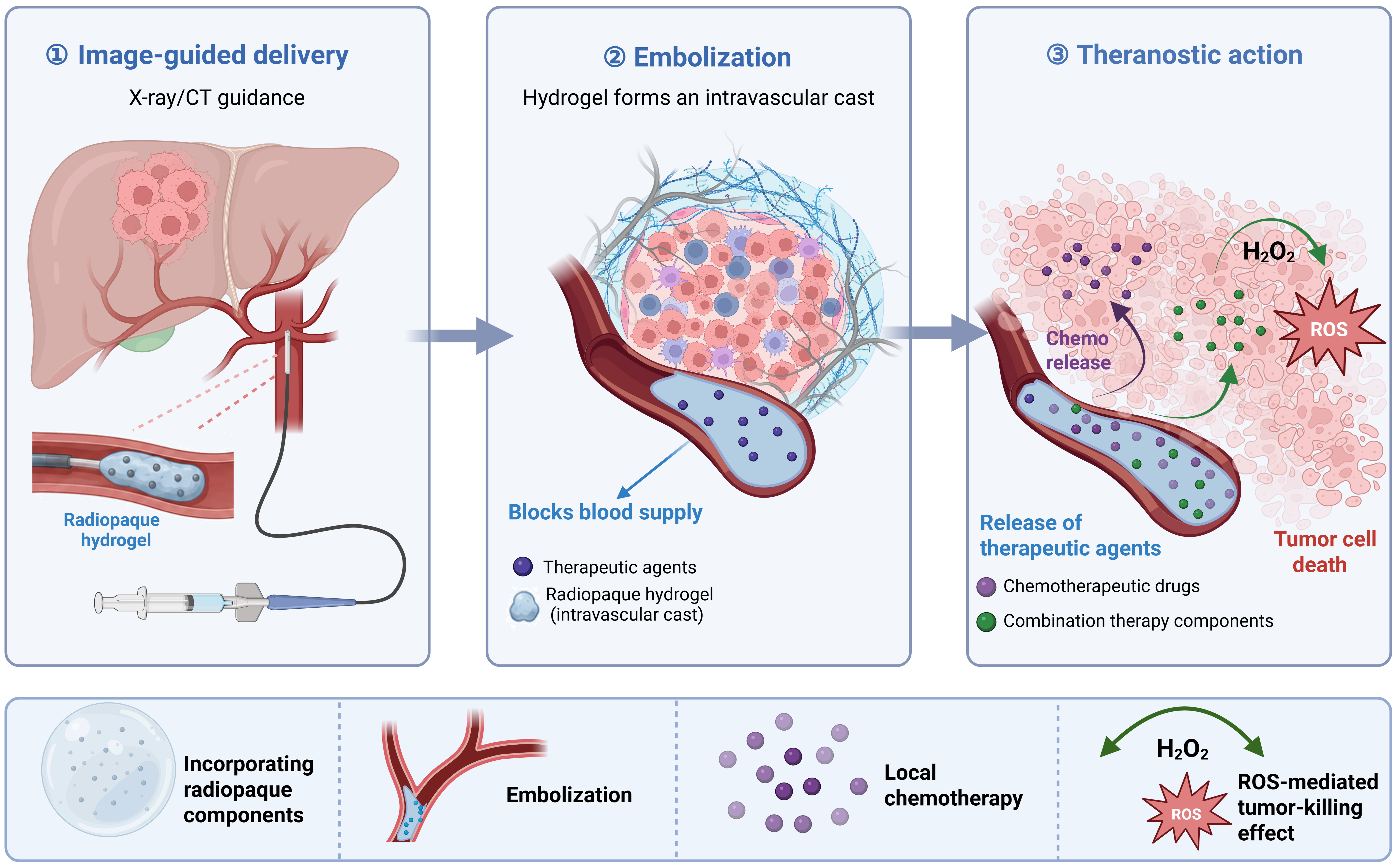
Figure 7. Schematic diagram of radiopaque hydrogel applied to tumor embolization. ① Injectable radiopaque hydrogel enables localization under imaging modalities such as X-ray. ② After injection, the hydrogel occludes the blood vessel. ③ Concurrently, therapeutic components are released for tumor eradication [Created in BioRender. Mao, X. (2026) https://BioRender.com/jwpprzc]. CT: Computed tomography; ROS: reactive oxygen species.
Lee et al. developed an iopamidol (IPD)-doped radiopaque hydrogel for X-ray fluoroscopy-guided transarterial chemo/starvation/chemodynamic embolization (TACSCE) of HCC[202]. The gel consisted of a hyaluronic acid–dopamine (HD) network crosslinked by Mn ions and incorporated glucose oxidase (GOx), DOX, and IPD. IPD provided X-ray visibility, whereas catechol–Mn4+ coordination between HD and MnO2 drove the sol-to-gel transition required for local vascular embolization. On the therapeutic side, DOX provided sustained local chemotherapy. GOx depleted glucose in tumor cells while generating hydrogen peroxide (H2O2). MnO2 further contributed to a cascade tumor-killing effect through glutathione (GSH) depletion, O2 generation, and the production of hydroxyl radicals (·OH) via Mn2+-mediated Fenton-like reactions. This study showed that radiopaque hydrogels can integrate image guidance, vascular embolization, local chemotherapy, and TME-responsive cascade catalysis into a transarterial theranostic platform for HCC.
By 2019, imaging agents and therapeutic drugs had already been co-encapsulated within liposomes for cancer diagnosis and treatment[203]. Conventional liposomal systems, however, remain susceptible to cargo leakage, particle instability, and burst release. The incorporation of hydrogels offers a possible way around these limitations. By constructing a hydrogel core within liposomes and co-loading an iodinated contrast agent and a chemotherapeutic drug, such platforms can simultaneously improve contrast-agent retention, controlled drug release, and passive tumor accumulation, thereby creating a CT-visible theranostic nanoplatform.
Lee et al. developed a radiopaque hydrogel-in-liposome (RHL) nanoplatform in which IPD and DOX were co-encapsulated within a liposome–PEG hydrogel composite structure as the micro-computed tomography (μCT) contrast agent and chemotherapeutic drug, respectively[204]. Its particle size of approximately 160 nm favored passive tumor accumulation through the enhanced permeability and retention (EPR) effect. Meanwhile, the PEG hydrogel core inside the liposomes reduced premature DOX burst release and IPD leakage, improving particle stability and in vivo delivery efficiency. In vivo experiments showed that RHLs enhanced μCT contrast at tumor sites in SCCVII tumor-bearing mice and produced greater antitumor effects than conventional radiopaque liposomes and free DOX.
CONSTRUCTION OF HYDROGEL-BASED INTEGRATED THERANOSTIC PLATFORMS
As precision medicine continues to advance, the conventional model in which diagnosis and treatment are handled as separate processes is becoming increasingly inadequate for patients with rapidly progressing tumors[205,206]. In recent years, as research on hydrogels in the field of tumor diagnosis and therapy has continued to deepen, they have evolved into integrated platforms capable of combining complete diagnostic and therapeutic functions. By incorporating therapeutic agents together with imaging or tracing components into the hydrogel network, such systems can couple targeted and sustained treatment with diagnostic imaging[207,208]. This strategy, which brings diagnostic or monitoring functions together with therapeutic intervention, is generally referred to as theranostics[209,210]. Importantly, a hydrogel-based theranostic system should do more than simply contain both diagnostic and therapeutic components. The information obtained through detection or imaging must contribute to the localization and implementation of tumor treatment. This distinguishes hydrogel-based theranostic platforms from the imaging-assisted hydrogel therapeutic platforms described above. The latter mainly use imaging to visualize or guide hydrogel delivery, whereas true theranostic platforms place greater emphasis on how diagnostic information informs subsequent treatment. In practical terms, tumor detection, diagnostic imaging, and anticancer therapy are integrated within the same hydrogel system. The resulting information can then support lesion identification, treatment localization, and the tracking of drug delivery, while also informing subsequent assessment of therapeutic efficacy or adjustment of the treatment regimen. Based on whether these later therapeutic adjustments require human intervention, we classify hydrogel-based theranostic platforms into two categories: feedback-informed platforms and closed-loop adaptive platforms.
Feedback-informed hydrogel-based theranostic platforms
Feedback-informed hydrogel platforms emphasize the feedback of therapeutic monitoring information to inform further treatment decisions. However, subsequent adjustments still require a physician or an externally operated controller to adjust the timing of treatment or later dosing regimens based on this information. At present, feedback-informed theranostic platforms are mainly used in two settings. First, imaging techniques can be used to monitor the treatment process in real time. Changes in relevant imaging parameters may indicate the therapeutic response or help predict treatment outcomes, providing a basis for the subsequent personalization of treatment strategies. Second, the sensing functions of hydrogels can be used to monitor treated tumor sites for signs of recurrence. Once the detected signal reaches a predefined threshold, the hydrogel sensor generates feedback or an alert, prompting further evaluation and subsequent treatment.
Imaging-guided hydrogel-based theranostic platforms
The defining feature of imaging-guided hydrogel-based theranostic platforms is that they do more than visualize drug delivery in real time. The imaging readouts can also be used to assess or predict therapeutic response, providing individualized information for refining subsequent treatment strategies[211-213]. Among the imaging modalities most commonly integrated into these platforms, three broad categories predominate: fluorescence imaging[214-221] [Table 2], magnetic resonance imaging (MRI), and X-ray/CT imaging[222-227] [Table 3].
Summary of image-guided hydrogel theranostic platforms reported in the past five years
| Representative platform | Specific theranostic approach: diagnostic/monitoring component → influence on treatment | Advantages | Limitations | Innovation | Research stage |
| Mantooth et al.[214] | Fluorescently labeled therapeutic cytokines were used to verify intratumoral retention and compare release rates among hydrogel formulations, thereby guiding selection of a sustained-release formulation and suitable dosing intervals | Changing the formulation tuned the release of multiple cytokines from the same hydrogel scaffold, providing a basis for cargo-specific release strategies that address tumor heterogeneity | Fluorescence served only as a drug-tracing label; dosing was not adjusted using individualized feedback signals | Hydrogel matrix composition was adjusted to achieve tunable release of therapeutic components | Small-animal preclinical model |
| Vu et al.[215] | Gel-encapsulated coumarin enabled fluorescence tracking. Preprogrammed 808 nm irradiation then cleaved the crosslinks and released DOX, while fluorescence monitored the release process | External irradiation initiated gel degradation and treatment, enabling temporal control and reducing burst release when irradiation was absent | Red fluorescence has limited penetration and quantitative accuracy in deep tissues, and its attenuation does not directly represent the effective DOX concentration | Coumarin simultaneously functioned as a crosslinker, fluorophore, and NIR-cleavable moiety | Small-animal preclinical model |
| Liang et al.[216] | Cy3-IgG and Cy5-IgG were used as fluorescent model antibodies to assess in vitro antibody release and in vivo local retention, respectively, indirectly indicating the delivery behavior of anti-PD-L1 | The MMP-cleavable design may better align release with the TME and reduce systemic antibody exposure | Cy3-IgG and Cy5-IgG were surrogate antibodies rather than directly labeled anti-PD-L1, so they could only approximate the therapeutic antibody’s behavior | Copper-free click gelation and an MMP-cleavable peptide enabled one polypeptide depot to co-deliver a small-molecule drug and an antibody | Small-animal preclinical model |
| Gierlich et al.[217] | The photosensitizer was incorporated directly into the PEG/chitosan network, so fluorescence indicated the location of therapeutically active photosensitizer and guided the timing of subsequent treatment | The same component provided diagnostic and therapeutic functions, while sustained intratumoral release enabled multiple PDT sessions after one injection | Deep-tumor accessibility is limited, and the system lacks a defined signal threshold, real-time quantification, and an algorithm for individualized irradiation adjustment | Temoporfin was modified into a functional node combining network formation, fluorescence tracking, and PDT | Small-animal preclinical model |
| Wang et al.[218] | The reaction of genipin with tumor proteins simultaneously generated fluorescence and photothermal capability; in vivo fluorescence intensity was then used to select the timing of photothermal treatment | Fluorescence confirmed whether therapeutic photothermal capability had formed, avoiding premature ineffective treatment. In situ photothermal-agent generation also reduced long-term retention and manufacturing complexity | Fluorescence and laser irradiation are both depth-limited, making the system more suitable for superficial or interventionally accessible lesions | Tumor proteins generated a fluorescent and photothermal therapeutic complex in situ, allowing one chemical reaction to report system status and create therapeutic capability | Small-animal preclinical model |
| Xu et al.[219] | PEG-MeoTTPy provided near-infrared FLI, while infrared thermal imaging (PTI) mapped the temperature generated by MXene photothermal conversion, enabling treatment tracking and thermal monitoring | FLI and PTI monitored hydrogel location, degradation, and treatment temperature, thereby improving safety | Imaging results were not converted into fully autonomous closed-loop treatment control; human intervention remained necessary | AIE fluorescence, MXene PTT, PDT, and controlled release were integrated, with pyroptosis underlying mild dual-phototherapy synergy | Small-animal preclinical model |
| Heng et al.[220] | Temperature-sensitive fluorescence informed the PTT thermal dose. Under 808 nm irradiation, Nd3+ produced near-infrared fluorescence while the material generated therapeutic heat; monitoring the fluorescence peak helped maintain approximately 50 °C | Real-time temperature monitoring across treatment groups clarified the relationship between thermal dose and therapeutic efficacy | In vivo quantitative fluorescence thermometry, depth dependence, real-time control, and long-term rare-earth-element safety remain insufficiently established | Ion co-doping made whitlockite a temperature sensor, photothermal agent, and bone substitute; the 50 °C window demonstrated theranostic parameter optimization | Small-animal preclinical model |
| Lee et al.[221] | The PEG hydrogel co-encapsulated ICG/PTX/BSA-Ce6 nanoparticles and a PFC-containing albumin nanoemulsion for hypoxic 4T1 tumors. ICG/Ce6 enabled fluorescence and phototherapy, thermography monitored temperature, and photoacoustic imaging assessed HbO2 changes | Three monitoring modalities jointly tracked the treatment process and therapeutic efficacy | The many materials, nanoformulations, ultrasound procedures, dual phototherapies, and imaging devices create complex manufacturing and clinical workflows | Albumin nanomedicines, a PFC oxygen carrier, and an injectable PEG depot were integrated to combine local hypoxia relief, multimodal therapy, and multimodal verification | Small-animal preclinical model |
Summary of image-guided hydrogel theranostic platforms
| Study/platform | Specific theranostic mechanism: diagnostic information → therapy | Advantages | Limitations | Innovation | Research stage |
| Peng et al.[222] | Meglumine diatrizoate renders the hydrogel radiopaque, allowing confirmation of the intratumoral injection site and material retention; PVP-I locally releases iodine in a sustained manner and induces tumor-cell apoptosis | Gel formation and sustained release reduce the risk of rapid diffusion of free iodine, while X-ray visibility decreases the uncertainty associated with blind injection | The hydrogel has insufficient mechanical strength to simultaneously provide structural support for bone defects | The system integrates PVP-I as a local antitumor component, silk fibroin as the sustained-release matrix, and meglumine diatrizoate as a procedural visualization component, while linking material release to biological effects through the Fas/FasL death-receptor pathway | Small-animal preclinical model |
| Liu et al.[223] | Iohexol enables CT tracking of the liquid embolic agent; DSA assesses whether embolization is complete, whereas CECT, CTA, and ultrasonography monitor perfusion and recanalization during follow-up. DOX is continuously released while embolization is maintained | Its liquid state, shear-thinning behavior, and low injection force make it compatible with microcatheters. High drug loading and prolonged release address the rapid drug release associated with lipiodol emulsions | Immune changes after TACE were not systematically investigated, and the optimal balance between material degradation rate and embolization durability has not been established | This study was the first to use a drug-loaded liquid coacervate for tumor chemoembolization, combining distal embolization, visualization, and prolonged drug release | Small-animal preclinical model |
| Peng et al.[224] | Ta nanoparticles render the embolic material visible under X-ray/CT and simultaneously provide radiosensitization; imaging is used to confirm TACE deposition and retention | The carrier-free design increases the proportion of therapeutically active constituents and reduces the amount of inert excipients | The rabbit experiments did not complete an integrated in vivo evaluation of TACE plus radiotherapy in an orthotopic liver cancer model, limiting translation to the interventional scale | Two small-molecule drugs are directly assembled to construct the fibrous hydrogel scaffold | Small-animal preclinical study (mice/rats/rabbits) |
| Dong et al.[225] | AuNPs provide CT contrast; FLASH-generated ROS cleave the selenocystamine crosslinks and release IMQ/AuNPs. CT is used for localization before radiotherapy and for monitoring hydrogel volume and degradation after treatment | AuNPs support CT imaging and potential radiation interactions, while enabling longitudinal observation of material volume. The design is well aligned with the radiotherapy workflow | Only one subcutaneous melanoma model in female mice was used in vivo, and FLASH was not directly compared with a conventional dose rate in vivo | The principal innovation is the extension of FLASH from a stand-alone radiotherapy modality to a trigger for hydrogel-mediated drug release, with subsequent material changes observable by CT | Small-animal preclinical study (mice) |
| Delgado et al.[226] | Iodixanol makes the gel visible under fluoroscopy/CBCT. The initial iodine distribution corresponds to the DOX fluorescence distribution, enabling quantitative assessment of the 3D coverage produced by different needle types, injection volumes, and flow rates | The use of clinical fluoroscopy and CBCT, rather than specialized experimental imaging, facilitates integration into interventional workflows | Ex vivo normal bovine liver lacks blood flow, perfusion, tissue pressure, and tumor heterogeneity; therefore, the findings cannot be extrapolated to gel leakage, pharmacokinetics, safety, or antitumor efficacy | The study combines an imageable thermosensitive drug gel with a quantitative 3D “delivery engineering” assessment of needle selection and injection parameters | In vitro/ex vivo study (including ex vivo bovine liver) |
| Som et al.[227] | IPD makes the IMQ-loaded thermosensitive hydrogel visible on CT, allowing confirmation of intratumoral drug-depot delivery; cryoablation releases tumor antigens and is combined with immunotherapy | A single injection replaces repeated dosing and may improve the practical feasibility of the treatment regimen | Large-animal percutaneous cryoablation workflows, long-term polymer and iodine burden, and systemic immune safety were not evaluated | A CT-visible, single-dose immunotherapeutic depot spatially and temporally integrates cryoablation-induced antigen release, TLR7 stimulation, and systemic ICB | Small-animal preclinical study (mice) |
Fluorescence-enabled drug-delivery hydrogels offer a powerful means of combining cancer treatment with real-time imaging. These systems can often respond to exogenous or endogenous cues, including light, temperature, pH, and specific molecular targets, making them well suited to targeted and stimuli-responsive cancer therapy. Another important feature is that fluorescence signals can visualize the local delivery and distribution of therapeutic agents. This allows the treatment process to be tracked in real time and therapeutic responses to be monitored noninvasively. Such dual functionality not only improves treatment precision but also provides timely feedback while therapy is under way. Ranjbar-Navazi et al. developed a theranostic platform based on an InP/ZnS quantum dot–nanohydrogel composite[228]. Aptamer functionalization enabled selective targeting of cancer cells, whereas the quantum dots provided fluorescence imaging capability for monitoring cellular drug uptake. The system was co-loaded with paclitaxel and sodium oxamate, combining conventional chemotherapy with metabolic intervention. Together, these treatments induced mitochondria-mediated apoptosis in MCF-7 cells. Notably, many fluorescent materials themselves also possess photothermal conversion or photodynamic capabilities, enabling them to mediate therapy while simultaneously visualizing the tumor. Wang et al. further developed a near-infrared-activated and ATP-responsive injectable upconversion nano-jelly theranostic system[229]. Upon NIR excitation, its upconversion core emitted green light at 540 nm for tumor imaging. This fluorescence signal enabled real-time tracking of therapeutic payload distribution, helped delineate the region for subsequent PDT, and indicated an appropriate treatment window. Under fluorescence guidance, the concomitant 660 nm red emission generated by NIR excitation was absorbed by methylene blue, leading to the production of singlet oxygen. The resulting photodynamic effect acted synergistically with ATP-responsive chemotherapy. Serial imaging over 48 h also provided information on the clearance of the nanogel system and could help determine whether additional dosing was required.
MRI is a widely used, noninvasive clinical imaging modality with high spatial resolution and excellent soft-tissue contrast. Integrating MRI capability into multifunctional hydrogel systems can couple real-time imaging with localized therapeutic delivery, thereby facilitating the development of theranostic platforms[230]. Paramagnetic ions can enhance contrast on T1-weighted MRI. When incorporated into hydrogels, they enable real-time MRI monitoring of the material after administration. Meanwhile, polar water molecules retained within the gel undergo dipolar rotation under a microwave field and generate heat through dielectric loss, thereby enhancing microwave ablation (MWA)[231]. Building on this concept, researchers developed a minimalist manganese–alginate (ALG–Mn) hydrogel. After intratumoral injection, the ALG network locally retained both water and Mn2+, producing marked signal enhancement on T1-weighted MRI. This allowed the spatial distribution and retention of the hydrogel within the tumor to be visualized over time. The Mn2+ ions and water molecules within the gel also enhanced microwave heating, enabling rapid ablation of the bulk tumor. Subsequently, the sustained release of Mn2+ catalyzed endogenous H2O2 to generate ROS, thereby inducing chemodynamic therapy (CDT). The MRI readouts could indicate whether the hydrogel adequately covered the tumor, helping to define the region for MWA and evaluate the therapeutic window for post-ablation CDT.
Beyond conventional T1-weighted imaging, chemical exchange saturation transfer (CEST) MRI generates contrast through the exchange of selectively saturated protons between molecules and bulk water. Hydrogels containing exchangeable functional groups, such as hydroxyl or amine groups, can therefore produce endogenous CEST contrast without the need for exogenous contrast agents[232,233]. Under physiological conditions, these CEST-active hydrogels enable sensitive monitoring of hydrogel degradation, spatial localization, and drug-release behavior[234]. Park et al. developed a CEST MRI-detectable liposomal alginate hydrogel loaded with methotrexate and gemcitabine[235]. After the hydrogel was injected adjacent to the tumor, three distinct CEST contrasts enabled longitudinal monitoring of drug release and tumor response at a clinical field strength of 3 T. These signals also provided spatial information on regional differences in treatment efficacy. Analysis of the CEST signals within the treated tumor showed that changes in amide proton transfer (APT) could reveal a local therapeutic response earlier than changes in tumor volume, allowing treatment outcomes to be assessed and predicted at an earlier stage. Combining drug-associated CEST signals with APT readouts enables early identification and localization of inadequately treated regions, thereby providing a basis for subsequent guidance on additional hydrogel administration, adjustment of the implantation site, or combination with other therapeutic modalities.
X-rays are high-frequency electromagnetic waves, and CT can generate clear, detailed cross-sectional images. X-ray-based techniques therefore play a central role in clinical diagnosis and treatment[236]. Radiopaque hydrogels further extend material visualization to X-ray-guided interventions and CT-assisted imaging, allowing their distribution to be monitored in vivo[237].
Intrahepatic tumors often show limited contrast against the surrounding normal liver on conventional unenhanced CT. Local delivery of a radiopaque hydrogel can improve lesion conspicuity, which is particularly important for accurate radiotherapy of liver tumors. In one study, intrinsically radiopaque LC Bead LUMI hydrogel microspheres were used to construct a theranostic platform[238]. The microspheres could be loaded with DOX and delivered into tumor-feeding arteries, where they produced vascular embolization while sustaining local drug release.
Following transarterial chemoembolization (TACE), the resulting high-density microsphere deposits improved lesion visibility and served as internal fiducial markers for subsequent stereotactic body radiotherapy (SBRT). CBCT acquired after embolization visualized the regions reached by the embolic material and therapeutic payload. Because the microspheres remained visible within the tumor, they also provided internal localization landmarks before each subsequent SBRT fraction. This information assisted target delineation and patient positioning, effectively carrying the spatial information obtained during the initial interventional procedure forward into subsequent precision radiotherapy.
The radiopacity of hydrogel microspheres does more than enhance contrast under X-ray imaging. It has also been explored as a means of estimating local drug delivery and treatment coverage from the distribution of bead-associated components[239]. After transarterial administration, the microspheres blocked the tumor blood supply and continuously released DOX. At the same time, iodine permanently incorporated into the microsphere structure allowed their intratumoral deposition to be visualized on CBCT.
In this study, prototype image-segmentation software was used to quantify the amount of bead-associated iodine. A correlation model was then established between the iodine signal and the concentration of DOX measured in tumor tissue. This transformed the CBCT readout from a simple map of material localization into an estimate of local drug dose and spatial coverage. The system could therefore identify tumor regions receiving insufficient drug delivery and provide evidence for intraprocedural catheter repositioning or the administration of additional drug-loaded microspheres.
Theranostic platforms for monitoring tumor recurrence
Postoperative tumor recurrence remains a major cause of cancer treatment failure[240]. In current clinical practice, recurrence is mainly monitored through imaging examinations and tumor biomarker testing[241]. Imaging, however, may not detect recurrence at its earliest stage and can produce false-positive findings because of postoperative inflammation and other nonmalignant changes[242,243]. Blood-based biomarker tests also have limited sensitivity when only a small number of recurrent tumor cells are present and tumor-specific biomarkers remain at very low concentrations[244]. Consequently, metastasis may already have occurred in many patients before recurrence can be confirmed[245,246]. Postoperative consolidation therapy is just as important as recurrence surveillance. Radiotherapy and systemic chemotherapy remain widely used after tumor resection. Yet patients often have limited tolerance immediately after surgery, and these treatments are usually delayed. This delay may allow the optimal window for eliminating residual tumor cells to pass[247,248]. There is therefore a pressing need for theranostic systems that can enrich tumor cells in situ, generate an early local warning signal, and capture and eliminate residual or recurrent cells at the same site.
As discussed above, hydrogels can support both biosensing and the local delivery of therapeutic payloads. Their tissue compatibility, injectability, and ability to form gels in situ make them particularly attractive for postoperative implantation. On this basis, Wang et al. developed an implantable DNA hydrogel for early warning of tumor recurrence and combined photodynamic–immune therapy [Figure 8A-C][249]. The hydrogel contained PD-L1 aptamers that captured and enriched residual or recurrent tumor cells at the surgical site [Figure 8D]. As these cells accumulated, the local ATP concentration increased. ATP binding then induced dehybridization of a Cy5/BHQ2-labeled bicyclic ATP sensor anchored within the hydrogel network, restoring Cy5 fluorescence and generating an early warning signal [Figure 8E]. Once this signal was detected, an external 660 nm laser was applied to activate Ce6 within the hydrogel, initiating PDT and producing ROS [Figure 8F]. The generated ROS not only damaged the captured tumor cells but also cleaved the DNA hydrogel backbone. This photoinduced self-disassembly released CpG and PD-L1 aptamers, thereby combining local PDT with immune stimulation and immune-checkpoint blockade. Similarly, another study utilized the capture of carcinoembryonic antigen (CEA) within the TME to monitor recurrence[250]. In this study, a molecular-beacon hydrogel sensor was constructed using mesoporous polydopamine (MPDA)-based nanoprobes. When sufficient CEA accumulated around the sensor, molecular recognition restored Cy5 fluorescence, converting the local biomarker signal into an optical indication of residual tumor tissue. After this diagnostic information was obtained, the MPDA component within the same platform generated heat under 808 nm laser irradiation. The resulting photothermal effect ablated tumor cells remaining in the resection cavity, including those locally enriched near the hydrogel, thereby linking postoperative tumor detection with image-guided treatment.
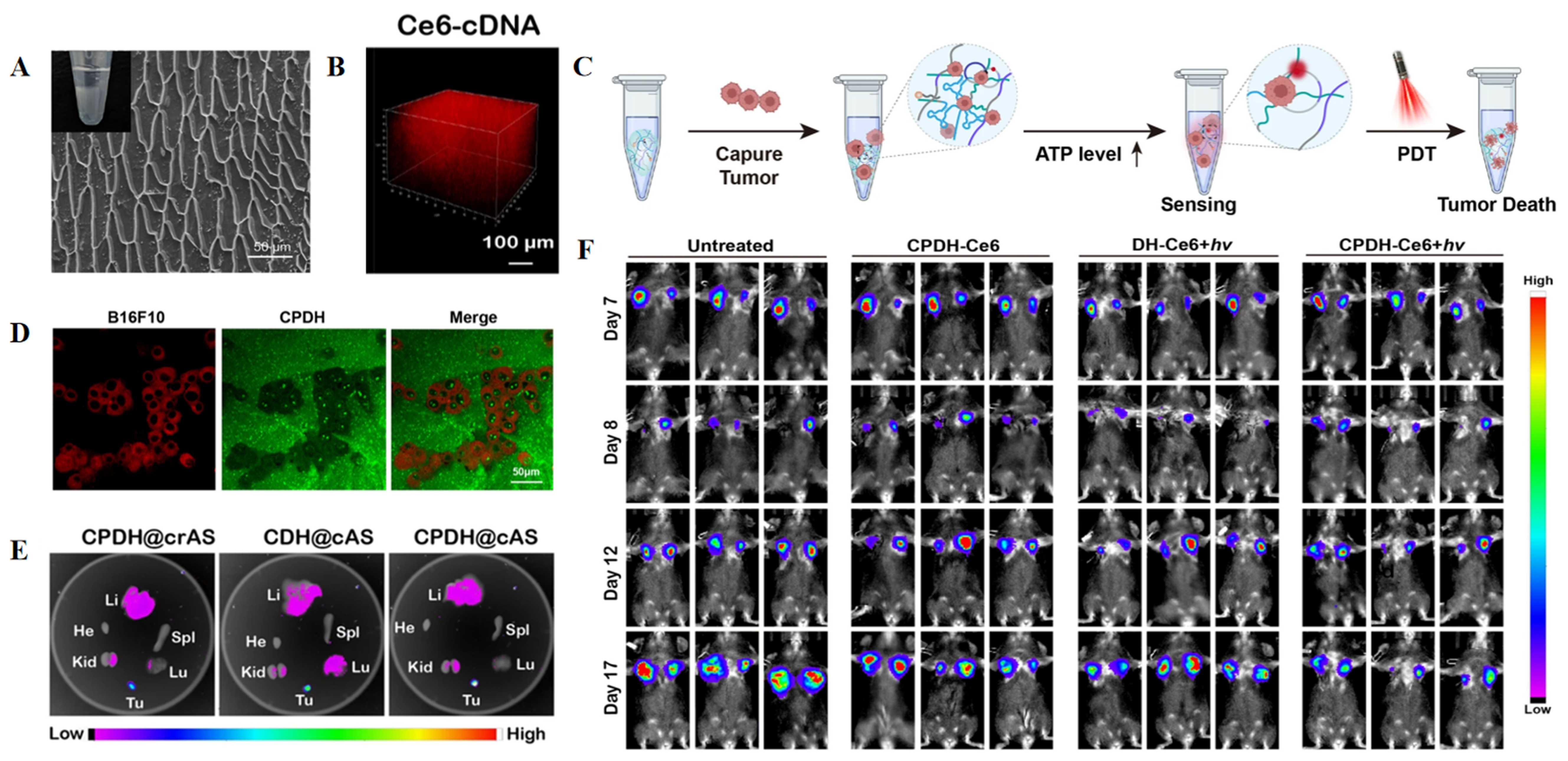
Figure 8. Hydrogel-based theranostic platform for monitoring postoperative tumor recurrence. (A) Cryo-scanning electron microscopy image showing the microstructure of CPDH; (B) 3D stacked fluorescence image showing Ce6-cDNA; (C) Schematic illustration of tumor-cell capture by CPDH-Ce6@cAS, followed by PDT-mediated tumor-cell killing; (D) Confocal fluorescence image of B16F10 cells captured within CPDH-Ce6; (E) Ex vivo fluorescence images of tumors and major organs harvested 48 h after implantation of the indicated hydrogel formulations; (F) Representative bioluminescence images showing the response of B16F10 tumors to local CPDH-Ce6 treatment. (A-F) Adapted with permission from Ref.[249]. In accordance with the Creative Commons Attribution 4.0 International License (CC BY 4.0). © The Author(s) 2023. The original panels were selected, relabeled, and rearranged. CPDH: CpG- and PD-L1 aptamer-containing DNA hydrogel (CpG: unmethylated cytosine-phosphate-guanine motifs; PD-L1: programmed death-ligand 1; DNA: deoxyribonucleic acid); 3D: three-dimensional; Ce6: chlorin e6; cDNA: complementary DNA; cAS: circular ATP sensor; PDT: photodynamic therapy; ATP: adenosine triphosphate.
Closed-loop adaptive hydrogel-based theranostic platforms
Unlike feedback-informed theranostic platforms, closed-loop adaptive systems place greater emphasis on the autonomous use of therapeutic-response feedback, without requiring direct human intervention. By continuously sensing key signals associated with treatment response, these platforms are expected to adjust therapy dynamically at the relevant site. Such adjustments may include changing the rate or spatial location of drug release or regulating the temperature used for local ablation[251]. Monitoring continues after each therapeutic intervention, allowing new feedback to be generated and treatment output to be modified until the desired therapeutic endpoint is reached. The central requirement is therefore a self-correcting loop in which dynamic diagnostic and treatment-response information continuously drives autonomous local therapy. In its ideal form, this process follows a recurring “sense–treat–feedback–adjust” cycle.
Fully closed-loop adaptive hydrogel-based theranostic platforms remain at an early stage of development. More substantial progress has so far been made in areas such as glucose regulation, coagulation control, and wound healing[252-257]. Yang et al., for example, incorporated glucose-responsive phenylboronic acid groups, negatively charged insulin, and a positively charged glucagon analog into the same hydrogel network[257]. As blood glucose fluctuated, reversible binding between glucose and the phenylboronic acid groups dynamically altered the release of insulin and the glucagon analog, thereby helping maintain glucose within a controlled range. The resulting changes in blood glucose were then sensed again by the glucose-responsive units in the material. In this way, glucose sensing, signal transduction, therapeutic output, and therapeutic effect feedback occurred continuously within the same material network, enabling multiple rounds of reversible regulation as glucose levels changed.
Tumors, however, are not single-variable, rapidly reversible therapeutic targets in the way that blood glucose is. Most sensing signals currently used in hydrogel-based cancer theranostic systems reflect features of the TME, yet the mechanisms underlying changes in this environment are highly complex[251]. Acidosis, oxidative stress, and hypoxia within a tumor may indicate disease progression, but they may also arise transiently after PDT, PTT, or chemotherapy[258]. A controller may therefore be unable to determine the next therapeutic action reliably from a single microenvironmental measurement acquired at one time point. In most existing systems, treatment-related information is instead returned to the physician in one form or another. The physician then integrates this information with other clinical findings before making the next decision. These systems are more appropriately classified as feedback-informed theranostic platforms, which are currently much more mature than autonomous closed-loop systems[258-265].
Solid tumors also display pronounced spatial heterogeneity and continue to evolve over time. Blood flow, oxygenation, metabolic activity, and drug sensitivity may differ substantially across regions of the same lesion[262-264]. Moreover, a measurable treatment response may take hours, days, or even weeks to emerge. This delay makes tumors far more difficult to regulate in real time than rapidly changing, comparatively low-dimensional variables such as blood glucose or abnormal neuronal electrical activity[263,266].
A further limitation lies in the way therapeutic payloads are released from most current hydrogel systems. Drug delivery commonly depends on network degradation or passive diffusion[14,267,268]. Once a drug has left the hydrogel, it cannot be withdrawn. Similarly, release driven by irreversible gel degradation is difficult to pause, restart, or repeatedly modulate[268]. Consequently, even when diagnostic information can be collected continuously, the platform may still lack an actuator capable of translating that feedback into precise, repeated adjustments of subsequent treatment[269].
The integration of hydrogels with flexible bioelectronics offers several possible routes around these limitations[270,271]. In a complex TME, multiple independent sensing units could be positioned within or on the surface of a hydrogel, allowing several types of information to be acquired simultaneously[266]. These signals could include measures of tumor burden, such as tumor volume and metabolic activity; indicators of treatment response, such as local drug concentration and cell death; and safety-related parameters, including the temperature of surrounding normal tissue during PTT and cumulative therapeutic output[253-256].
However, its value does not simply lie in increasing the number of sensors; rather, it lies in identifying the temporal sequence of signal changes through continuous time‑series measurements, thereby enabling more accurate assessment of treatment efficacy[271]. For example, if ROS increase shortly after PDT begins and are followed by sustained reductions in tumor volume and metabolic activity, the increase in ROS is more likely to represent an effective treatment response. By contrast, if ROS levels rise while the tumor continues to enlarge, the same signal may indicate inadequate treatment or ongoing disease progression rather than successful therapy[266,267]. The flexible autonomous sensor measuring tumor volume regression, or FAST, has already demonstrated that changes in tumor burden can be converted into continuous electronic feedback. In subcutaneous tumor models, the flexible sensor monitored tumor regression on a minute-scale basis and distinguished treated tumors from controls approximately 5 h after therapy was initiated[266]. In addition, other flexible hydrogel sensors utilize changes in mechanical properties, fluorescence, and impedance induced by ROS or H2O2 in the TME for wireless monitoring, providing a material basis for local sensing at the molecular level[272].
A major limitation of current hydrogel‐based theranostic platforms is the difficulty of achieving multiple rounds of adjustable therapeutic output. The integration of flexible electronics offers a potential solution by enabling the release of therapeutic payloads to be controlled via electrical signals, thereby translating dynamically acquired diagnostic information into electronically guided autonomous regulation of drug delivery[269-271,273]. Recently, an ionic diode‐based drug delivery system achieved potential‐controlled release of drugs at the nanogram‐to‐microgram scale in tumor‐bearing mice, with the drug being continuously delivered to the lesion through a hydrogel reservoir. Using DOX as a model drug, this system demonstrated superior antitumor efficacy and reduced off‐target immunotoxicity compared with intratumoral injection of the free drug[274].
One of the most representative examples of a hydrogel-based autonomous therapeutic loop is the hydroElex antiseizure system developed by Qu et al.[273]. HydroElex was fabricated from an interpenetrating-network conductive hydrogel and patterned into a flexible microneedle array. Each microneedle was connected through an independent circuit, allowing the array to conform to curved brain tissue, penetrate the cortex, and record local field potentials continuously at multiple sites. This enabled real-time recognition and spatial localization of seizure-like pathological activity. The recorded neural signals were transmitted to a signal-acquisition and processing unit. Once a predefined abnormality threshold was exceeded, the program automatically generated a therapeutic command. Only the drug-delivery channel closest to the origin of the abnormal electrical activity was activated, while the remaining electrodes continued to record neural signals. The amount of drug released could also be adjusted according to the intensity of the pathological signal. Importantly, hydroElex did not stop operating after the initial drug release. It continued to record local field potentials. If abnormal activity persisted or reappeared, the system initiated another round of drug delivery. Once neural activity returned to a predefined safe range, treatment was stopped automatically. This system provides a useful architectural blueprint for future hydrogel-mediated cancer theranostics. A comparable cancer system could use a microneedle or flexible sensor array to monitor several tumor-associated parameters at different spatial locations. The collected signals could then be converted into electrical outputs and evaluated against predefined response thresholds. These electronic commands could selectively activate drug release in regions showing inadequate response, while post-treatment sensing would determine whether treatment should be intensified, repeated, or stopped.
CHALLENGES AND PERSPECTIVES
Current challenges
Safety challenges
Hydrogel-based cancer theranostic platforms must first address the balance between local retention and long-term safety. Prolonged local residence is essential for sustained drug release and longitudinal imaging follow-up. However, safety risks may arise when the residence time of the material does not match the intended therapeutic window. Excessively rapid release can produce high local drug concentrations while shortening the duration of therapeutic activity. By contrast, overly slow degradation may provoke foreign-body reactions, chronic inflammation, and related complications[275-277].
Importantly, hydrogel degradation does not necessarily mean that the material has been safely cleared from the body. Hydrogel networks can break down through several pathways into soluble polymer chains and low-molecular-weight products. These products may subsequently enter the blood or lymphatic circulation and be eliminated through renal or hepatobiliary routes. Larger, poorly degradable, or protein-adsorbing fragments, however, may be taken up by macrophages or accumulate over extended periods within the mononuclear phagocyte system of the liver and spleen[278,279]. This distinction is particularly important for theranostic hydrogels containing metallic components, because disappearance of the hydrogel matrix does not demonstrate that the incorporated metals have also been degraded or excreted[278-280]. Likewise, once fluorescent dyes or imaging contrast components dissociate from the gel, their biodistribution and clearance may no longer parallel those of the hydrogel itself. This may increase exposure in off-target organs and, at the same time, make the imaging signal an unreliable indicator of the amount of hydrogel that remains in the body[280].
The stimulus-dependent activation of drugs, nanoparticles, or imaging probes may introduce another layer of risk. Theranostic hydrogels commonly rely on endogenous cues within the TME. Yet the intensity and distribution of these cues vary among patients, across different regions of the same tumor, and over the course of treatment. Consequently, unintended activation may occur in normal tissues, insufficient drug release may happen in poorly responsive tumor regions, and abrupt disruption of the local network may trigger burst release[275,276]. For payloads with narrow therapeutic windows, including certain chemotherapeutic agents and immune agonists, rapid release of a large dose may cause local tissue necrosis or excessive immune activation. Insufficient triggering presents a different problem: residual drugs may remain trapped in the gel and undergo delayed release as the material subsequently degrades[275,276]. Biocompatibility is also influenced by chemical modification of the hydrogel matrix, crosslinking density, and the nature of the resulting degradation products[281-283]. Implanted materials can promote protein adsorption, inflammatory-cell recruitment, and macrophage fusion. Excessive crosslinking may further intensify proinflammatory and profibrotic responses[281,282]. Studies of GelMA hydrogels have shown that highly crosslinked formulations can induce stronger inflammation and greater profibrotic cellular activity than less densely crosslinked materials. Softer, lightly crosslinked gels, in contrast, are generally more permissive to cellular infiltration and phagocytic clearance[282]. This also illustrates that improving hydrogel stability may conflict with its long-term biocompatibility.
The long-term safety of a hydrogel-based cancer theranostic platform can therefore be considered adequately supported only when several conditions are met. The therapeutic payload must be activated at the intended dose; the hydrogel and all incorporated components must be traceable throughout their residence and eventual clearance; and the entire platform must avoid irreversible local or systemic toxicity over its full period of use.
FDA regulatory status and approval prospects for hydrogels in cancer diagnosis and treatment
Hydrogel-based products currently cleared by the FDA for cancer-related diagnostic applications mainly serve as tissue markers, biopsy-site localization aids, and image-guidance tools[284-286]. For example, the TraceIT Tissue Marker received 510(k) clearance. It consists primarily of an injectable PEG hydrogel that remains visible on MRI, CT, and ultrasound after implantation and is intended for radiographic marking of soft tissue during surgery or before a subsequent procedure[284]. The HydroMARK Breast Biopsy Site Marker was likewise cleared through the 510(k) pathway. It comprises a resorbable hydrogel and a permanent metallic marker and can be used to mark biopsy sites in breast tissue and axillary lymph nodes[285]. By contrast, diagnostic platforms that use hydrogels to enrich tumor-associated molecular biomarkers remain largely confined to laboratory research. Recent systematic reviews indicate that hydrogel-based platforms for early cancer detection and molecular classification have focused mainly on the capture and detection of CTCs and protein biomarkers. Commonly used materials include polyacrylamide, PEG, DNA, and alginate[286,287]. Most studies, however, remain at the proof-of-concept stage or have been evaluated only in small sets of biological samples[287].
Hydrogel-based therapeutics with explicit anticancer indications have also received FDA approval[288-292]. JELMYTO was approved through a new drug application in 2020 for adults with low-grade upper tract urothelial cancer[288]. In 2025, the FDA further approved ZUSDURI for adults with recurrent low-grade intermediate-risk NMIBC[289]. Both products use reverse-thermal hydrogel formulations to deliver a chemotherapeutic agent locally. Beyond drug-delivery systems, the FDA has authorized several hydrogel medical devices that support interventional oncology and radiotherapy[290-292]. The Embrace Hydrogel Embolic System received premarket approval in 2025 for the embolization of hypervascular tumors in peripheral arteries[290]. In 2015, the SpaceOAR System was granted De Novo classification as a Class II absorbable perirectal hydrogel spacer. By temporarily separating the prostate from the rectum, it reduces the radiation dose delivered to the anterior rectal wall during prostate cancer radiotherapy[291]. Barrigel subsequently received 510(k) clearance and performs a similar tissue-protection function during radiotherapy[292]. In addition to these FDA-authorized products, several hydrogel delivery systems are undergoing clinical evaluation and have shown potential therapeutic activity in NMIBC, basal cell carcinoma, and hypervascular tumors[293,294].
Although hydrogel-mediated platforms have already produced successful examples of FDA authorization in cancer diagnosis and treatment, their progression into clinical practice and eventual regulatory approval remains difficult. Nevertheless, their favorable biocompatibility and tunable physicochemical properties give them distinctive advantages in precision cancer diagnosis and therapy[295,296]. To date, searches of publicly available FDA records and recent clinical studies have not identified an approved or cleared product that uses a single hydrogel system to complete tumor-signal detection, diagnostic-information output, therapeutic release, and treatment-response feedback. Most integrated hydrogel theranostic platforms remain at the preclinical stage. Although these systems have considerable translational and commercial potential, several barriers still need to be addressed[297]. Quality control during scale-up is one of the major obstacles. Critical quality attributes, including dose uniformity and product stability, must be tightly controlled throughout the large-scale manufacture of hydrogel delivery systems[287,297]. Most platforms, however, are prepared only in small batches during preclinical studies. Quality-control methods established at this stage may not transfer directly to industrial-scale production, potentially creating new safety risks. Theranostic hydrogels are also commonly stimulus-responsive and sensitive to specific environmental or external triggers. Unintended exposure to these stimuli may disrupt the hydrogel matrix prematurely. Manufacturing and storage protocols specifically suited to smart hydrogels must therefore be established before clinical translation. The regulatory complexity of theranostic platforms also exceeds that of products designed solely for diagnosis or therapy[298]. A single platform may incorporate a hydrogel device, an imaging or detection probe, a therapeutic drug, a biological product, a sensor, and control software. The FDA classifies products composed of drug, device, and/or biological-product components as combination products. The lead review center and the principal marketing application pathway are determined according to the primary mode of action - the constituent function expected to make the greatest contribution to the overall therapeutic effect[298]. The resulting cross-center review and additional evidentiary requirements may extend development timelines, reduce commercial feasibility, and increase the cost of clinical translation for hydrogel delivery systems.
From a regulatory perspective, hydrogel-based theranostic platforms are more likely to follow a phased translational pathway[295,296,298]. The first stage could build on an already cleared localization hydrogel by incorporating a therapeutic payload. Imaging would be used to confirm the position, spatial coverage, and degradation of the gel, while treatment decisions would remain under physician control. In the second stage, imaging or sensing signals could be used to reflect drug release and therapeutic response, creating a feedback-informed platform in which diagnostic information supports clinical decision-making. Only after these functions have been adequately validated would it be reasonable to advance toward a closed-loop system capable of autonomously adjusting therapeutic output in response to local signals.
Compared with hydrogel platforms, recent biotherapeutics, including antibody- and cell-based therapies, have established more mature clinical-development and regulatory pathways[299-302]. Numerous monoclonal antibodies have been approved for solid tumors and hematological malignancies. Immune-checkpoint antibodies targeting PD-1, PD-L1, and CTLA-4 now have indications spanning breast, lung, bladder, colorectal, liver, and kidney cancers, as well as melanoma and other malignancies[299,300]. Antibody products can generally be manufactured at scale as standardized formulations and incorporated into existing oncology workflows through intravenous or subcutaneous administration.
Cell therapy has followed another clearly defined, although highly specialized, translational pathway. The FDA has authorized multiple cellular therapies, including chimeric antigen receptor T-cell products, tumor-infiltrating lymphocyte therapies, and engineered T-cell receptor products. Among these, CAR-T therapy has become an important treatment option for selected patients with relapsed or refractory hematological malignancies[301-303].
Challenges to practical implementation
There is an inherent contradiction between hydrogel network stability and mass transport[304]. Network stability is regulated by factors such as the polymer backbone, the nature of the crosslinking bonds, and the crosslinking density. These same parameters also determine the permeability of the gel network[305-307]. Consequently, increasing the polymer concentration or crosslinking density to improve the overall stability of a hydrogel may simultaneously hinder the capture of target molecules and the release of therapeutic payloads[305]. On the one hand, high polymer concentrations and dense crosslinking can restrict the premature diffusion of drugs and functional nanocomponents, which is a key advantage of hydrogel-based delivery over the administration of free drugs. On the other hand, the same network may impede the entry of low-abundance biomarkers, the diffusion of therapeutic agents toward tumor margins, immune-cell infiltration, and metabolite exchange[308-310]. In diagnostic settings, inappropriate design of mesh size and surface recognition sites may reduce the capture efficiency of CTCs, EVs, or nucleic acid biomarkers. In therapeutic settings, excessively strong local immobilization may lead to uneven drug distribution, causing overexposure in tissues adjacent to the gel while leaving distal tumor regions underdosed. For integrated theranostic systems, mass-transfer limitations may also create discrepancies between imaging signals and actual drug exposure, thereby compromising the accuracy of efficacy evaluation.
The mismatch between stimulus-responsive mechanisms and the actual TME remains a major limitation in the design of smart hydrogels. Many hydrogel systems rely on predefined thresholds of tumor-microenvironment cues to trigger payload release. Tumors, however, do not behave as single-variable systems. Their microenvironment varies markedly across cancer types, among patients, and even between different regions of the same tumor. These conditions also continue to evolve following chemotherapy, PTT, or immunotherapy[311,312]. If the response threshold is set too low, the hydrogel may be activated prematurely outside the lesion. If it is set too high, the material may respond insufficiently within the tumor. More importantly, in an integrated theranostic platform, the same stimulus-responsive process may simultaneously govern drug release, signal activation, and the assessment of therapeutic efficacy. A single response mode is therefore unlikely to accommodate the continuously changing microenvironment throughout treatment.
Hydrogel-guided integrated theranostic platforms may also exhibit performance interference among diagnostic, therapeutic, and feedback modules. The value of hydrogel-based theranostics lies not in integrating all functional components into one material, but in functionally coupling localization, release, treatment, and monitoring. Enhancing imaging signals may require the introduction of inorganic or metallic components, yet such additions may increase residual risk or alter hydrogel mechanics. Increasing drug loading benefits local therapy but may compromise the stability of fluorescence, magnetic resonance, or photoacoustic signals. Similarly, incorporating photothermal, photodynamic, or immunomodulatory modules may enhance therapeutic efficacy, but the resulting increase in system complexity raises additional concerns regarding the stability and potency of incorporated biomolecules and the viability and phenotypic stability of loaded cells[283,296,313,314].
Future perspectives
Material stability and functional lifetime control
Material stability and functional lifetime are key determinants of whether hydrogel platforms can perform their intended diagnostic, therapeutic, or theranostic functions[275,315,316]. To establish a robust safety profile and facilitate regulatory approval, future materials should be designed with an in vivo residence time that matches the specific clinical task. At the same time, the contradiction between network stability and mass transport must be minimized as much as possible. One rational strategy is to securely retain therapeutic payloads within the hydrogel through chemical conjugation or surface tethering[306,317,318]. The underlying network structure can then be adjusted according to the size and physicochemical properties of the target biomarkers. This would preserve sufficient biomarker permeability without allowing the diagnostic components to leak from the gel. Theranostic platforms must also address the coupling between diagnostic signals and therapeutic functions[319]. Simply co-encapsulating a contrast agent and a drug within the same hydrogel does not ensure that they will share the same in vivo retention and release profiles[320]. In multicomponent systems, different payloads could instead be assigned to separate layers, distinct nanocarriers, or structural domains with different bond-cleavage rates. Such compartmentalization would enable delayed, sequential, or stage-specific release. Future platforms should also be designed so that the diagnostic signal directly reports the amount of therapeutic cargo remaining in the hydrogel or reflects the actual treatment response, rather than merely indicating the location of the material. This could be achieved by coupling the diagnostic and therapeutic components through a shared cleavable linkage or by allowing drug release to activate a fluorescence or MRI signal. Closed-loop systems intended for repeated sensing and treatment cycles will require additional properties, including reversible responsiveness, self-healing capacity, and resistance to fatigue. Basal leakage before the first stimulus and progressive loss of responsiveness over repeated cycles must also be carefully controlled.
For biodegradable hydrogels, lifetime control should follow a simple principle: the material must remain stable until its intended function has been completed and should then break down into nontoxic products that can be metabolized or excreted[321]. However, in actual clinical practice, the TME exhibits significant inter-individual and inter-stage variability, making it difficult to directly predict the true in vivo lifetime from degradation profiles obtained in vitro[316,322]. Particularly for enzyme-responsive or oxidation-responsive hydrogels, their degradation rates may fluctuate markedly with the patient’s metabolic status[323,324]. Future hydrogel networks should therefore not rely entirely on a single microenvironmental signal for degradation control. A more reliable approach would be to establish a multifactor-responsive control system. Double-network and interpenetrating-network designs are particularly well suited to this purpose. A slowly degrading network could maintain mechanical stability, while a faster, stimulus-responsive network would cleave first in response to signals at the lesion and initiate payload release. This division of functions could reduce the risk of complete network collapse and abrupt drug release when local endogenous stimuli become excessively strong.
Spatially and temporally graded response design for heterogeneous lesions
As discussed above, the pronounced heterogeneity of tumors remains a major barrier to translating hydrogel-based cancer theranostic platforms from in vitro studies to in vivo applications. In the future, efforts should be focused on establishing a hierarchically organized response system for these platforms in both space and time. Specifically, at the spatial level, the crosslinking density, pore size, or distribution of therapeutic payloads can be adjusted across different regions of the hydrogel, thereby enabling differential degradation and release behaviors in distinct regions. Studies have shown that the composition, structure, mechanical properties, and functional molecules of hydrogel materials can be varied either continuously along a predefined direction or in discrete zones, providing a material basis for mimicking the complex spatial heterogeneity within tissues[275,306,317]. Temporally, both the TME and its therapeutic requirements often change in stages as treatment proceeds. Different drugs can be loaded into separate hydrogel layers or into nanocarriers embedded within the gel. Linkages with different cleavage rates can then be used to control the release time of each component. In this way, multiple drugs delivered in a single administration could act sequentially according to a predefined schedule.
Huang et al. reported a Janus-inspired core–shell hydrogel scaffold[325]. Through differential drug loading and graded degradation of the core and shell, the material released melatonin at a higher concentration during the early postoperative stage to eliminate residual tumor cells. This was followed by sustained release at a lower concentration to support subsequent bone repair. Multiresponsive systems should also account for the logical relationships among different tumor-associated signals, rather than treating every stimulus as an independent switch. AND, OR, and other logic-gate operations have already been introduced into multistimuli-responsive hydrogel designs to improve the conditional selectivity of drug release[326-328]. A relatively permissive OR response could be assigned to the outer hydrogel layer to recognize and cover infiltrative tumor margins, whereas the inner layer or a reservoir containing highly toxic drugs could adopt a more stringent AND response, ensuring that the therapeutic components are released only when multiple tumor features are present simultaneously.
Dynamic covalent bonds, hydrogen bonds, host–guest interactions, and other reversible interactions enable the hydrogel network to undergo continuous dissociation, reorganization, and mechanical adaptation in response to environmental changes, providing a basis for accommodating the constantly evolving TME during treatment[329,330]. The further integration of imaging probes, biosensors, or flexible electronic devices could enable the system to convert the acquired information into regulatory commands that initiate or adjust therapy, thereby advancing multiresponsive hydrogels from passive material responses toward feedback-informed treatment and, ultimately, autonomous closed-loop regulation. Finally, the rational design of gradient and hierarchical structures depends on a quantitative understanding of the response signals within each patient’s lesion. Before material fabrication, future studies should map the spatial distribution of relevant stimuli within the tumor using multiregion biopsies, spatial omics, or other analytical methods. Release profiles across different tumor regions and treatment stages should then be evaluated using PDOs and models that more closely reproduce the human immune microenvironment.
Emerging technologies and their future applications
In response to these challenges, future hydrogel theranostic platforms will advance not merely by increasing material complexity, but by prioritizing individualization, clinical adaptability, and dynamic feedback. Unlike earlier validation strategies that relied primarily on standardized cell lines and animal models, next-generation hydrogel platforms must more accurately reflect patient-specific tumor heterogeneity, treatment-stage evolution, and actual delivery environments. A key foundation for this shift is the use of PDOs. Organoids partially preserve the tissue architecture, molecular characteristics, and drug-response patterns of primary tumors in vitro[331,332]. Consequently, they hold promise for pre-evaluating the compatibility between hydrogel theranostic platforms with diverse matrices and crosslinking strategies and specific patients, thereby reducing reliance on experience-driven material screening.
At the same time, advances in spatial omics provide higher-dimensional tissue information for the precise deployment of hydrogel platforms. By resolving intratumoral matrix density, immune-cell distribution, hypoxic regions, and boundaries at high risk of recurrence, spatial omics can help define the delivery range, response threshold, and release sequence of hydrogels, thereby shifting material design from a focus on single lesion characteristics to fine adaptation to tumor spatial heterogeneity. On this basis, artificial intelligence will become an important tool linking patient information with material parameters. By integrating material parameters, organoid drug-sensitivity data, spatial microenvironment information, and imaging/sensing data, artificial intelligence may predict how different design schemes will affect release behavior, imaging readouts, and therapeutic responses, thereby improving the predictability and personalization of hydrogel-platform development.
Finally, bioelectronic interfaces may endow hydrogel platforms with the capacity for continuous monitoring and feedback regulation. By combining conductive or injectable hydrogels with flexible electronic components, it may become possible to continuously record temperature, pH, electrochemical, and inflammation-related signals after implantation or local administration. This assists in assessing material degradation, drug release, and therapeutic changes. Furthermore, biosafety must remain a core design constraint for next-generation hydrogel theranostic platforms. Developing hydrogel systems with clearly defined degradation pathways, low-toxicity degradation products, and efficient in vivo clearance is essential to avoid long-term residual risks caused by local retention and to promote the translation of hydrogels into clinically viable platforms that combine therapeutic efficacy with safety.
In summary, the future of hydrogel-driven integrated cancer theranostic platforms depends not only on the continued optimization of material composition, crosslinking architecture, and functional components, but also on achieving a deeper understanding of the complex interplay among hydrogel networks, the TME, and clinical delivery pathways. As technologies such as PDOs, spatial omics, artificial intelligence, and bioelectronic interfaces are progressively integrated into material design and efficacy evaluation, hydrogel platforms are expected to evolve into patient-information-driven, monitorable, and dynamically adjustable individualized theranostic systems, ultimately becoming core soft-material platforms that connect tumor recognition, local intervention, and therapeutic feedback.
DECLARATIONS
Acknowledgment
The graphical abstract was created in BioRender. Mao, X. (2026) https://BioRender.com/tvog32m.
Authors’ contributions
Conceptualization and design of the review: Chen, Z. S.; Wang, S.; Sun, G.
Literature search, data collection, and organization of the manuscript structure: Mao, X.; Cui, M.; Cao, L. Q.; Zhao, Y.; Cai, L.; Liu, Y.
Drafting of the manuscript: Mao, X.; Cui, M.; Cao, L. Q.
Preparation and refinement of figures/tables: Mao, X.; Cui, M.; Xie, Y.; Chen, X.
Provided critical revisions and refined the manuscript for important intellectual content: Wang, S.; Chen, Z. S.; Sun, G.
Availability of data and materials
Not applicable.
AI and AI-assisted tools statement
During the preparation of this manuscript, the AI tool ChatGPT (version 5.5, released 2026-04-24) was used solely for language editing. The tool did not influence the study design, data collection, analysis, interpretation, or the scientific content of the work. All authors take full responsibility for the accuracy, integrity, and final content of the manuscript.
Financial support and sponsorship
This work was supported by the Natural Science Foundation of Hebei Province (Grant No. H2023209083) and the National Natural Science Foundation of China (Grant No. 82472636).
Conflicts of interest
Chen, Z. S. is the Guest Editor of the Special Issue entitled “Smart Hydrogel-Based Micro/Nanoscale Delivery Systems for Advanced Therapeutics and Bioelectronic Integration” in Soft Science. He had no involvement in the review or editorial process of this manuscript, including, but not limited to, reviewer selection, evaluation, and the final decision. The other authors declare that they have no conflicts of interest.
Ethical approval and consent to participate
Not applicable.
Consent for publication
Not applicable.
Copyright
© The Author(s) 2026.
REFERENCES
1. GBD 2023 Cancer Collaborators. The global, regional, and national burden of cancer, 1990-2023, with forecasts to 2050: a systematic analysis for the Global Burden of Disease Study 2023. Lancet 2025, 406, 1565-86.
2. Schwartzberg, L.; Kim, E. S.; Liu, D.; Schrag, D. Precision oncology: who, how, what, when, and when not? Am. Soc. Clin. Oncol. Educ. Book. 2017, 37, 160-9.
3. El-Deiry, W. S.; Goldberg, R. M.; Lenz, H. J.; et al. The current state of molecular testing in the treatment of patients with solid tumors, 2019. CA. Cancer. J. Clin. 2019, 69, 305-43.
4. Kelkar, S. S.; Reineke, T. M. Theranostics: combining imaging and therapy. Bioconjug. Chem. 2011, 22, 1879-903.
6. Gerlinger, M.; Rowan, A. J.; Horswell, S.; et al. Intratumor heterogeneity and branched evolution revealed by multiregion sequencing. N. Engl. J. Med. 2012, 366, 883-92.
7. Marusyk, A.; Janiszewska, M.; Polyak, K. Intratumor heterogeneity: the rosetta stone of therapy resistance. Cancer. Cell. 2020, 37, 471-84.
8. Bardelli, A.; Pantel, K. Liquid biopsies, what we do not know (yet). Cancer. Cell. 2017, 31, 172-9.
9. Collins, F. S.; Varmus, H. A new initiative on precision medicine. N. Engl. J. Med. 2015, 372, 793-5.
10. Choi, K. Y.; Liu, G.; Lee, S.; Chen, X. Theranostic nanoplatforms for simultaneous cancer imaging and therapy: current approaches and future perspectives. Nanoscale 2012, 4, 330-42.
11. Rowe, S. P.; Pomper, M. G. Molecular imaging in oncology: current impact and future directions. CA. Cancer. J. Clin. 2022, 72, 333-52.
12. O’Connor, J. P. B.; Aboagye, E. O.; Adams, J. E.; et al. Imaging biomarker roadmap for cancer studies. Nat. Rev. Clin. Oncol. 2017, 14, 169-86.
13. Mehta, P.; Sharma, M.; Devi, M. Hydrogels: an overview of its classifications, properties, and applications. J. Mech. Behav. Biomed. Mater. 2023, 147, 106145.
14. Li, J.; Mooney, D. J. Designing hydrogels for controlled drug delivery. Nat. Rev. Mater. 2016, 1, 16071.
15. Sun, Z.; Song, C.; Wang, C.; Hu, Y.; Wu, J. Hydrogel-based controlled drug delivery for cancer treatment: a review. Mol. Pharm. 2020, 17, 373-91.
16. Solanki, R.; Bhatia, D. Stimulus-responsive hydrogels for targeted cancer therapy. Gels 2024, 10, 440.
17. Masaeli, E.; Srinivasan, S.; Rajabzadeh, A. R. Transforming cancer diagnosis and therapy through fluorescent hydrogels: a review. Adv. Healthc. Mater. 2026, 15, e02577.
18. Sampath, B.; Charles, J. Theranostic magnetic hydrogels for treatment, recurrence prevention, tumour modelling and emerging applications in breast cancer management: a review. Int. J. Biol. Macromol. 2026, 359, 151874.
19. Lee, K. K.; Go, K.; Lee, E.; et al. Multifunctional hydrogels for advanced cancer treatment: diagnostic imaging and therapeutic modalities. Gels 2025, 11, 426.
20. Hoare, T. R.; Kohane, D. S. Hydrogels in drug delivery: progress and challenges. Polymer 2008, 49, 1993-2007.
21. Peppas, N. A.; Bures, P.; Leobandung, W.; Ichikawa, H. Hydrogels in pharmaceutical formulations. Eur. J. Pharm. Biopharm. 2000, 50, 27-46.
22. Tibbitt, M. W.; Anseth, K. S. Hydrogels as extracellular matrix mimics for 3D cell culture. Biotechnol. Bioeng. 2009, 103, 655-63.
23. Drury, J. L.; Mooney, D. J. Hydrogels for tissue engineering: scaffold design variables and applications. Biomaterials 2003, 24, 4337-51.
24. Zhu, H.; Zheng, J.; Oh, X. Y.; et al. Nanoarchitecture-integrated hydrogel systems toward therapeutic applications. ACS. Nano. 2023, 17, 7953-78.
25. Lu, P.; Ruan, D.; Huang, M.; et al. Harnessing the potential of hydrogels for advanced therapeutic applications: current achievements and future directions. Signal. Transduct. Target. Ther. 2024, 9, 166.
26. Guo, L.; Fu, Z.; Li, H.; et al. Smart hydrogel: a new platform for cancer therapy. Adv. Colloid. Interface. Sci. 2025, 340, 103470.
27. Lee, K. Y.; Mooney, D. J. Alginate: properties and biomedical applications. Prog. Polym. Sci. 2012, 37, 106-26.
28. Augst, A. D.; Kong, H. J.; Mooney, D. J. Alginate hydrogels as biomaterials. Macromol. Biosci. 2006, 6, 623-33.
29. Rowley, J. A.; Madlambayan, G.; Mooney, D. J. Alginate hydrogels as synthetic extracellular matrix materials. Biomaterials 1999, 20, 45-53.
30. Dash, M.; Chiellini, F.; Ottenbrite, R.; Chiellini, E. Chitosan - a versatile semi-synthetic polymer in biomedical applications. Prog. Polym. Sci. 2011, 36, 981-1014.
31. Croisier, F.; Jérôme, C. Chitosan-based biomaterials for tissue engineering. Eur. Polym. J. 2013, 49, 780-92.
32. Sockett, K. A.; Jiang, N. C.; Ungolan, P.; Niu, J.; Grinstaff, M. W. Advancements in hyaluronic acid synthetic methodologies and their translational relevance. Chem. Rev. 2026, 126, 3224-86.
33. Burdick, J. A.; Prestwich, G. D. Hyaluronic acid hydrogels for biomedical applications. Adv. Mater. 2011, 23, H41-56.
34. Highley, C. B.; Prestwich, G. D.; Burdick, J. A. Recent advances in hyaluronic acid hydrogels for biomedical applications. Curr. Opin. Biotechnol. 2016, 40, 35-40.
35. Yue, K.; Trujillo-de Santiago, G.; Alvarez, M. M.; Tamayol, A.; Annabi, N.; Khademhosseini, A. Synthesis, properties, and biomedical applications of gelatin methacryloyl (GelMA) hydrogels. Biomaterials 2015, 73, 254-71.
36. Caliari, S. R.; Burdick, J. A. A practical guide to hydrogels for cell culture. Nat. Methods. 2016, 13, 405-14.
37. Gonzalez-Obeso, C.; Jane Hartzell, E.; Albert Scheel, R.; Kaplan, D. L. Delivering on the promise of recombinant silk-inspired proteins for drug delivery. Adv. Drug. Deliv. Rev. 2023, 192, 114622.
38. Canal, T.; Peppas, N. A. Correlation between mesh size and equilibrium degree of swelling of polymeric networks. J. Biomed. Mater. Res. 1989, 23, 1183-93.
39. Zustiak, S. P.; Leach, J. B. Hydrolytically degradable poly(ethylene glycol) hydrogel scaffolds with tunable degradation and mechanical properties. Biomacromolecules 2010, 11, 1348-57.
40. Raeber, G. P.; Lutolf, M. P.; Hubbell, J. A. Molecularly engineered PEG hydrogels: a novel model system for proteolytically mediated cell migration. Biophys. J. 2005, 89, 1374-88.
41. Rehmann, M. S.; Skeens, K. M.; Kharkar, P. M.; et al. Tuning and predicting mesh size and protein release from step growth hydrogels. Biomacromolecules 2017, 18, 3131-42.
42. Lutolf, M.; Raeber, G.; Zisch, A.; Tirelli, N.; Hubbell, J. Cell‐responsive synthetic hydrogels. Adv. Mater. 2003, 15, 888-92.
43. Gill, B. J.; Gibbons, D. L.; Roudsari, L. C.; et al. A synthetic matrix with independently tunable biochemistry and mechanical properties to study epithelial morphogenesis and EMT in a lung adenocarcinoma model. Cancer. Res. 2012, 72, 6013-23.
44. Li, J. K.; Wang, N.; Wu, X. S. Poly(vinyl alcohol) nanoparticles prepared by freezing-thawing process for protein/peptide drug delivery. J. Control. Release. 1998, 56, 117-26.
45. Oh, N. G.; Hwang, S. Y.; Na, Y. H. Fabrication of a PVA-based hydrogel microneedle patch. ACS. Omega. 2022, 7, 25179-85.
46. Nguyen, H. X.; Bozorg, B. D.; Kim, Y.; et al. Poly (vinyl alcohol) microneedles: fabrication, characterization, and application for transdermal drug delivery of doxorubicin. Eur. J. Pharm. Biopharm. 2018, 129, 88-103.
47. Jain, M.; Pansuriya, R.; Thakur, R.; et al. PVA/guanidinium oleate transdermal patch as a pH-responsive drug delivery system for the localized and targeted delivery of anticancer drugs. Mater. Adv. 2024, 5, 1998-2011.
48. Tilghman, R. W.; Cowan, C. R.; Mih, J. D.; et al. Matrix rigidity regulates cancer cell growth and cellular phenotype. PLoS. One. 2010, 5, e12905.
49. Gao, Y.; Ren, F.; Ding, B.; et al. A thermo-sensitive PLGA-PEG-PLGA hydrogel for sustained release of docetaxel. J. Drug. Target. 2011, 19, 516-27.
50. Ma, H.; He, C.; Cheng, Y.; et al. PLK1shRNA and doxorubicin co-loaded thermosensitive PLGA-PEG-PLGA hydrogels for osteosarcoma treatment. Biomaterials 2014, 35, 8723-34.
51. Zhang, S.; Holmes, T.; Lockshin, C.; Rich, A.; New Collective Author. Spontaneous assembly of a self-complementary oligopeptide to form a stable macroscopic membrane. Proc. Natl. Acad. Sci. U. S. A. 1993, 90, 3334-8.
52. Hartgerink, J. D.; Beniash, E.; Stupp, S. I. Self-assembly and mineralization of peptide-amphiphile nanofibers. Science 2001, 294, 1684-8.
53. Um, S. H.; Lee, J. B.; Park, N.; Kwon, S. Y.; Umbach, C. C.; Luo, D. Enzyme-catalysed assembly of DNA hydrogel. Nat. Mater. 2006, 5, 797-801.
54. Xing, Y.; Cheng, E.; Yang, Y.; et al. Self-assembled DNA hydrogels with designable thermal and enzymatic responsiveness. Adv. Mater. 2011, 23, 1117-21.
55. Rodell, C. B.; Kaminski, A. L.; Burdick, J. A. Rational design of network properties in guest-host assembled and shear-thinning hyaluronic acid hydrogels. Biomacromolecules 2013, 14, 4125-34.
56. Lin, C. C.; Anseth, K. S. PEG hydrogels for the controlled release of biomolecules in regenerative medicine. Pharm. Res. 2009, 26, 631-43.
57. Censi, R.; Di Martino, P.; Vermonden, T.; Hennink, W. E. Hydrogels for protein delivery in tissue engineering. J. Control. Release. 2012, 161, 680-92.
58. Ding, X.; Wang, Y. Weak bond-based injectable and stimuli responsive hydrogels for biomedical applications. J. Mater. Chem. B. 2017, 5, 887-906.
59. Jalalvandi, E.; Shavandi, A. Shear thinning/self-healing hydrogel based on natural polymers with secondary photocrosslinking for biomedical applications. J. Mech. Behav. Biomed. Mater. 2019, 90, 191-201.
60. Shi, Y.; Li, D.; He, C.; Chen, X. Design of an injectable polypeptide hydrogel depot containing the immune checkpoint blocker anti-PD-L1 and doxorubicin to enhance antitumor combination therapy. Macromol. Biosci. 2021, 21, e2100049.
61. Siboro, S. A. P.; Anugrah, D. S. B.; Ramesh, K.; Park, S. H.; Kim, H. R.; Lim, K. T. Tunable porosity of covalently crosslinked alginate-based hydrogels and its significance in drug release behavior. Carbohydr. Polym. 2021, 260, 117779.
62. Fairbanks, B. D.; Schwartz, M. P.; Bowman, C. N.; Anseth, K. S. Photoinitiated polymerization of PEG-diacrylate with lithium phenyl-2,4,6-trimethylbenzoylphosphinate: polymerization rate and cytocompatibility. Biomaterials 2009, 30, 6702-7.
63. Shin, G. R.; Kim, H. E.; Ju, H. J.; et al. Injectable click-crosslinked hydrogel containing resveratrol to improve the therapeutic effect in triple negative breast cancer. Mater. Today. Bio. 2022, 16, 100386.
64. Cao, D.; Zhang, X.; Akabar, M. D.; et al. Liposomal doxorubicin loaded PLGA-PEG-PLGA based thermogel for sustained local drug delivery for the treatment of breast cancer. Artif. Cells. Nanomed. Biotechnol. 2019, 47, 181-91.
65. Nguyen, A. K.; Goering, P. L.; Reipa, V.; Narayan, R. J. Toxicity and photosensitizing assessment of gelatin methacryloyl-based hydrogels photoinitiated with lithium phenyl-2,4,6-trimethylbenzoylphosphinate in human primary renal proximal tubule epithelial cells. Biointerphases 2019, 14, 021007.
66. Nguyen, A. K.; Goering, P. L.; Elespuru, R. K.; Sarkar Das, S.; Narayan, R. J. The photoinitiator lithium phenyl (2,4,6-trimethylbenzoyl) phosphinate with exposure to 405 nm light is cytotoxic to mammalian cells but not mutagenic in bacterial reverse mutation assays. Polymers 2020, 12, 1489.
67. Yesilyurt, V.; Webber, M. J.; Appel, E. A.; Godwin, C.; Langer, R.; Anderson, D. G. Injectable self-healing glucose-responsive hydrogels with pH-regulated mechanical properties. Adv. Mater. 2016, 28, 86-91.
68. Huang, Z.; Delparastan, P.; Burch, P.; Cheng, J.; Cao, Y.; Messersmith, P. B. Injectable dynamic covalent hydrogels of boronic acid polymers cross-linked by bioactive plant-derived polyphenols. Biomater. Sci. 2018, 6, 2487-95.
69. Deng, C. C.; Brooks, W. L. A.; Abboud, K. A.; Sumerlin, B. S. Boronic acid-based hydrogels undergo self-healing at neutral and acidic pH. ACS. Macro. Lett. 2015, 4, 220-4.
70. Lee, F.; Chung, J. E.; Kurisawa, M. An injectable enzymatically crosslinked hyaluronic acid- hydrogel system with independent tuning of mechanical strength and gelation rate. Soft. Matter. 2008, 4, 880-7.
71. Kurisawa, M.; Lee, F.; Wang, L.; Chung, J. E. Injectable enzymatically crosslinked hydrogel system with independent tuning of mechanical strength and gelation rate for drug delivery and tissue engineering. J. Mater. Chem. 2010, 20, 5371.
72. Cheng, E.; Xing, Y.; Chen, P.; et al. A pH-triggered, fast-responding DNA hydrogel. Angew. Chem. Int. Ed. Engl. 2009, 48, 7660-3.
73. Yang, H.; Liu, H.; Kang, H.; Tan, W. Engineering target-responsive hydrogels based on aptamer-target interactions. J. Am. Chem. Soc. 2008, 130, 6320-1.
74. Katopodi, T.; Petanidis, S.; Floros, G.; Porpodis, K.; Kosmidis, C. Hybrid nanogel drug delivery systems: transforming the tumor microenvironment through tumor tissue editing. Cells 2024, 13, 908.
75. Liu, Y.; Chen, L.; Shi, Q.; Zhao, Q.; Ma, H. Tumor microenvironment-responsive polypeptide nanogels for controlled antitumor drug delivery. Front. Pharmacol. 2021, 12, 748102.
76. Zhang, H.; Ba, S.; Lee, J. Y.; Xie, J.; Loh, T. P.; Li, T. Cancer biomarker-triggered disintegrable DNA nanogels for intelligent drug delivery. Nano. Lett. 2020, 20, 8399-407.
77. Xu, X.; Jiang, Y.; Lu, C. Self-assembled ATP-responsive DNA nanohydrogel for specifically activated fluorescence imaging and chemotherapy in cancer cells. Anal. Chem. 2022, 94, 10221-6.
78. Shang, J.; Yu, S.; Li, R.; He, Y.; Wang, Y.; Wang, F. Bioorthogonal disassembly of hierarchical DNAzyme nanogel for high-performance intracellular microRNA imaging. Nano. Lett. 2023, 23, 1386-94.
79. Su, M.; Zhu, Y.; Chen, J.; et al. Microfluidic synthesis of manganese-alginate nanogels with self-supplying H2O2 capability for synergistic chemo/chemodynamic therapy and boosting anticancer immunity. Chem. Eng. J. 2022, 435, 134926.
80. Sanati, M.; Amin, Yavari. S. Liposome-integrated hydrogel hybrids: promising platforms for cancer therapy and tissue regeneration. J. Control. Release. 2024, 368, 703-27.
81. Binaymotlagh, R.; Hajareh Haghighi, F.; Chronopoulou, L.; Palocci, C. Liposome-hydrogel composites for controlled drug delivery applications. Gels 2024, 10, 284.
82. GuhaSarkar, S.; Pathak, K.; Sudhalkar, N.; et al. Synergistic locoregional chemoradiotherapy using a composite liposome-in-gel system as an injectable drug depot. Int. J. Nanomedicine. 2016, 11, 6435-48.
83. Mao, Y.; Li, X.; Chen, G.; Wang, S. Thermosensitive hydrogel system with paclitaxel liposomes used in localized drug delivery system for in situ treatment of tumor: better antitumor efficacy and lower toxicity. J. Pharm. Sci. 2016, 105, 194-204.
84. Bouché, M.; Dong, Y. C.; Sheikh, S.; et al. Novel treatment for glioblastoma delivered by a radiation responsive and radiopaque hydrogel. ACS. Biomater. Sci. Eng. 2021, 7, 3209-20.
85. Huang, S.; Hong, X.; Zhao, M.; et al. Nanocomposite hydrogels for biomedical applications. Bioeng. Transl. Med. 2022, 7, e10315.
86. Wang, C.; Zhao, N.; Huang, Y.; He, R.; Xu, S.; Yuan, W. Coordination of injectable self-healing hydrogel with Mn-Zn ferrite@mesoporous silica nanospheres for tumor MR imaging and efficient synergistic magnetothermal-chemo-chemodynamic therapy. Chem. Eng. J. 2020, 401, 126100.
87. Yue, J.; Miao, P.; Li, L.; Yan, R.; Dong, W. F.; Mei, Q. Injectable carbon dots-based hydrogel for combined photothermal therapy and photodynamic therapy of cancer. ACS. Appl. Mater. Interfaces. 2022, 14, 49582-91.
88. Vaziri, A.; Maia, R.; Zhang, P.; et al. Granular hydrogels as modular biomaterials: from structural design to biological responses. Adv. Healthc. Mater. 2026, 15, e02462.
89. Daly, A. C.; Riley, L.; Segura, T.; Burdick, J. A. Hydrogel microparticles for biomedical applications. Nat. Rev. Mater. 2020, 5, 20-43.
90. Sinclair, A.; O’Kelly, M. B.; Bai, T.; Hung, H. C.; Jain, P.; Jiang, S. Self-healing zwitterionic microgels as a versatile platform for malleable cell constructs and injectable therapies. Adv. Mater. 2018, 30, e1803087.
91. Zhong, Z.; Gan, L.; Feng, Z.; et al. Hydrogel local drug delivery systems for postsurgical management of tumors: Status Quo and perspectives. Mater. Today. Bio. 2024, 29, 101308.
92. Zhang, J.; Chen, C.; Li, A.; et al. Immunostimulant hydrogel for the inhibition of malignant glioma relapse post-resection. Nat. Nanotechnol. 2021, 16, 538-48.
93. Fu, Z.; Li, H.; Xue, P.; et al. Implantable bioresponsive hydrogel prevents local recurrence of breast cancer by enhancing radiosensitivity. Front. Bioeng. Biotechnol. 2022, 10, 881544.
94. Yan, X.; Sun, T.; Song, Y.; et al. In situ thermal-responsive magnetic hydrogel for multidisciplinary therapy of hepatocellular carcinoma. Nano. Lett. 2022, 22, 2251-60.
95. Zhou, J.; Wang, H.; Chen, H.; et al. pH-responsive nanocomposite hydrogel for simultaneous prevention of postoperative adhesion and tumor recurrence. Acta. Biomater. 2023, 158, 228-38.
96. Amatya, R.; Hwang, S.; Park, T.; et al. BSA/silver nanoparticle-loaded hydrogel film for local photothermal treatment of skin cancer. Pharm. Res. 2021, 38, 873-83.
97. Seo, J. Y.; Lee, B.; Kang, T. W.; et al. Electrostatically interactive injectable hydrogels for drug delivery. Tissue. Eng. Regen. Med. 2018, 15, 513-20.
98. Akther, F.; Little, P.; Li, Z.; Nguyen, N. T.; Ta, H. T. Hydrogels as artificial matrices for cell seeding in microfluidic devices. RSC. Adv. 2020, 10, 43682-703.
99. Shin, G. R.; Kim, H. E.; Kim, J. H.; Choi, S.; Kim, M. S. Advances in injectable in situ-forming hydrogels for intratumoral treatment. Pharmaceutics 2021, 13, 1953.
100. Han, X.; Meng, X.; Wu, Z.; Wu, Z.; Qi, X. Dynamic imine bond cross-linked self-healing thermosensitive hydrogels for sustained anticancer therapy via intratumoral injection. Mater. Sci. Eng. C. Mater. Biol. Appl. 2018, 93, 1064-72.
101. Wang, Y.; Wang, S.; Hu, W.; et al. In situ hydrogels prepared by photo-initiated crosslinking of acrylated polymers for local delivery of antitumor drugs. J. Pharm. Sci. 2023, 112, 1863-71.
102. Attama, A. A.; Nnamani, P. O.; Onokala, O. B.; Ugwu, A. A.; Onugwu, A. L. Nanogels as target drug delivery systems in cancer therapy: a review of the last decade. Front. Pharmacol. 2022, 13, 874510.
103. Li, X.; Li, H.; Zhang, C.; Pich, A.; Xing, L.; Shi, X. Intelligent nanogels with self-adaptive responsiveness for improved tumor drug delivery and augmented chemotherapy. Bioact. Mater. 2021, 6, 3473-84.
104. Qian, K.; Ma, Y.; Wan, J.; et al. The studies about doxorubicin-loaded p(N-isopropyl-acrylamide-co-butyl methylacrylate) temperature-sensitive nanogel dispersions on the application in TACE therapies for rabbit VX2 liver tumor. J. Control. Release. 2015, 212, 41-9.
105. Ma, X.; Zhou, W.; Zhang, R.; et al. Minimally invasive injection of biomimetic Nano@Microgel for in situ ovarian cancer treatment through enhanced photodynamic reactions and photothermal combined therapy. Mater. Today. Bio. 2023, 20, 100663.
106. Agrawal, G.; Agrawal, R. Functional microgels: recent advances in their biomedical applications. Small 2018, 14, e1801724.
108. Raza, A.; Khan, A. Q.; Inchakalody, V. P.; et al. Dynamic liquid biopsy components as predictive and prognostic biomarkers in colorectal cancer. J. Exp. Clin. Cancer. Res. 2022, 41, 99.
109. Kim, Y. J.; Min, J. Hydrogel-based technologies in liquid biopsy for the detection of circulating clinical markers: challenges and prospects. Anal. Bioanal. Chem. 2024, 416, 2065-78.
110. Luchini, A.; Geho, D. H.; Bishop, B.; et al. Smart hydrogel particles: biomarker harvesting: one-step affinity purification, size exclusion, and protection against degradation. Nano. Lett. 2008, 8, 350-61.
111. Tang, J.; Jia, X.; Li, Q.; et al. A DNA-based hydrogel for exosome separation and biomedical applications. Proc. Natl. Acad. Sci. U. S. A. 2023, 120, e2303822120.
112. Chen, B.; Wang, G.; Huang, C.; et al. A light-induced hydrogel responsive platform to capture and selectively isolate single circulating tumor cells. Nanoscale 2022, 14, 3504-12.
113. Kim, Y. J.; Cho, Y. H.; Min, J.; Han, S. W. Circulating tumor marker isolation with the chemically stable and instantly degradable (CSID) hydrogel immunospheres. Anal. Chem. 2021, 93, 1100-9.
114. Kaniewska, K.; Kowalczyk, A.; Karbarz, M.; Nowicka, A. M. Changes in the volume phase transition temperature of hydrogels for detection of the DNA hybridization process. Analyst 2016, 141, 5815-21.
115. Xu, Y.; Zhang, X.; Luan, C.; Wang, H.; Chen, B.; Zhao, Y. Hybrid hydrogel photonic barcodes for multiplex detection of tumor markers. Biosens. Bioelectron. 2017, 87, 264-70.
116. Sun, S.; Chen, J. Recent advances in hydrogel-based biosensors for cancer detection. ACS. Appl. Mater. Interfaces. 2024, 16, 46988-7002.
117. Lu, H.; Samman, F.; Hasan, E.; Alsulaiman, D. Novabeads: stimuli-responsive signal-amplifying hydrogel microparticles for enzymeless fluorescence-based detection of microRNA biomarkers. Small 2025, 21, e2503990.
118. Ring, A.; Nguyen-Sträuli, B. D.; Wicki, A.; Aceto, N. Biology, vulnerabilities and clinical applications of circulating tumour cells. Nat. Rev. Cancer. 2023, 23, 95-111.
119. Capuozzo, M.; Ferrara, F.; Santorsola, M.; Zovi, A.; Ottaiano, A. Circulating tumor cells as predictive and prognostic biomarkers in solid tumors. Cells 2023, 12, 2590.
120. Portella, L.; Bertolini, G.; Guardascione, G.; et al. CXCL12-loaded-hydrogel (CLG): a new device for metastatic circulating tumor cells (CTCs) capturing and characterization. Heliyon 2024, 10, e35524.
121. Sun, W.; You, X.; Zhao, X.; et al. Precise capture and dynamic release of circulating liver cancer cells with dual-histidine-based cell imprinted hydrogels. Adv. Mater. 2024, 36, e2402379.
122. Dawson, S. J.; Tsui, D. W.; Murtaza, M.; et al. Analysis of circulating tumor DNA to monitor metastatic breast cancer. N. Engl. J. Med. 2013, 368, 1199-209.
123. Garcia-Murillas, I.; Schiavon, G.; Weigelt, B.; et al. Mutation tracking in circulating tumor DNA predicts relapse in early breast cancer. Sci. Transl. Med. 2015, 7, 302ra133.
124. Zhao, X.; Meng, X.; Zeng, X.; He, T.; Chen, J.; Chen, P. Homogeneous extraction-free dual-ctDNA detection via DNA nanomaterial fusion for rapid breast cancer diagnosis. ACS. Nano. 2025, 19, 42066-78.
125. Hou, J.; Wang, Y.; Yuan, W.; et al. Thermosensitive hydrogel-enhanced RPA-CRISPR/Cas12a biosensor for ultrasensitive detection of methylated loci in breast cancer ctDNA. Anal. Chim. Acta. 2026, 1388, 345101.
126. McAlexander, M. A.; Phillips, M. J.; Witwer, K. W. Comparison of methods for miRNA extraction from plasma and quantitative recovery of RNA from cerebrospinal fluid. Front. Genet. 2013, 4, 83.
127. Sourvinou, I. S.; Markou, A.; Lianidou, E. S. Quantification of circulating miRNAs in plasma: effect of preanalytical and analytical parameters on their isolation and stability. J. Mol. Diagn. 2013, 15, 827-34.
128. Jang, W.; Kim, Y. J.; Roh, H. K.; Song, E. L.; Bong, K. W. Hydrogel-based in situ DNA extension assay for multiplexed and rapid detection of microRNA. Anal. Chem. 2025, 97, 7317-24.
129. Chen, D.; Wang, Y.; Wei, Y.; et al. Size-coded hydrogel microbeads for extraction-free serum multi-miRNAs quantifications with machine-learning-aided lung cancer subtypes classification. Nano. Lett. 2025, 25, 453-60.
130. Lin, Y.; Xing, G.; Wu, Z.; et al. Microfluidic-enabled self-directed hydrogel microspheres for multiplexed microRNA assays. Anal. Chem. 2025, 97, 1972-6.
131. Yao, C.; Wang, Q.; Lu, X.; Chen, X.; Li, Z. Hydrogel-based microdroplet ensembles encapsulating multiplexed EXPAR assays for trichromic digital profiling of microRNAs and in-depth classification of primary urethral cancers. Nano. Lett. 2024, 24, 15861-9.
132. Yu, W.; Hurley, J.; Roberts, D.; et al. Exosome-based liquid biopsies in cancer: opportunities and challenges. Ann. Oncol. 2021, 32, 466-77.
133. Xu, X.; Zhang, M.; Zhang, X.; et al. Decoding expression dynamics of protein and transcriptome at the single-cell level in paired picoliter chambers. Anal. Chem. 2022, 94, 8164-73.
134. Miwa, H.; Dimatteo, R.; de Rutte, J.; Ghosh, R.; Di Carlo, D. Single-cell sorting based on secreted products for functionally defined cell therapies. Microsyst. Nanoeng. 2022, 8, 84.
135. Yamagata, C.; Hamazaki, Y.; Nakazato, T.; et al. Oil-sealed RGD-modified alginate hydrogel microwell array for analysis of single-cell-derived extracellular vesicles and particles. Microsyst. Nanoeng. 2025, 11, 238.
136. Lee, S. S.; Kim, H.; Sohn, D. K.; et al. Indocyanine green-loaded injectable alginate hydrogel as a marker for precision cancer surgery. Quant. Imaging. Med. Surg. 2020, 10, 779-88.
137. Linders, D. G. J.; Bijlstra, O. D.; Walker, E.; et al. Ex vivo fluorescence-guided resection margin assessment in breast cancer surgery using a topically applied, cathepsin-activatable imaging agent. Pharmacol. Res. 2024, 209, 107464.
138. Yang, H. Y.; Jang, M. S.; Sun, X. S.; et al. CD44-mediated tumor homing of hyaluronic acid nanogels for hypoxia-activated photodynamic therapy against tumor. Colloids. Surf. B. Biointerfaces. 2023, 228, 113395.
139. Song, P.; Ye, D.; Zuo, X.; et al. DNA hydrogel with aptamer-toehold-based recognition, cloaking, and decloaking of circulating tumor cells for live cell analysis. Nano. Lett. 2017, 17, 5193-8.
140. Chen, T.; Su, L.; Lin, L.; et al. Mesoporous radiosensitized nanoprobe for enhanced NIR-II photoacoustic imaging-guided accurate radio-chemotherapy. Nano. Res. 2022, 15, 4154-63.
141. Vahrmeijer, A. L.; Hutteman, M.; van der Vorst, J. R.; van de Velde, C. J.; Frangioni, J. V. Image-guided cancer surgery using near-infrared fluorescence. Nat. Rev. Clin. Oncol. 2013, 10, 507-18.
142. Saxena, V.; Sadoqi, M.; Shao, J. Degradation kinetics of indocyanine green in aqueous solution. J. Pharm. Sci. 2003, 92, 2090-7.
143. Andreiuk, B.; Reisch, A.; Bernhardt, E.; Klymchenko, A. S. Fighting aggregation-caused quenching and leakage of dyes in fluorescent polymer nanoparticles: universal role of counterion. Chem. Asian. J. 2019, 14, 836-46.
144. Liu, C.; Ruan, C.; Shi, R.; Jiang, B. P.; Ji, S.; Shen, X. C. A near infrared-modulated thermosensitive hydrogel for stabilization of indocyanine green and combinatorial anticancer phototherapy. Biomater. Sci. 2019, 7, 1705-15.
145. Donnelly, R. F.; Singh, T. R.; Garland, M. J.; et al. Hydrogel-forming microneedle arrays for enhanced transdermal drug delivery. Adv. Funct. Mater. 2012, 22, 4879-90.
146. Park, J.; Ku, M.; Kim, E.; et al. CD44-specific supramolecular hydrogels for fluorescence molecular imaging of stem-like gastric cancer cells. Integr. Biol. 2013, 5, 669-72.
147. Yang, J.; Qu, J.; Teng, X.; et al. Tumor microenvironment-responsive hydrogel for direct extracellular ATP imaging-guided surgical resection with clear boundaries. Adv. Healthc. Mater. 2023, 12, e2301084.
148. Walker, E.; Linders, D. G. J.; Abenojar, E.; et al. Formulation of a thermosensitive imaging hydrogel for topical application and rapid visualization of tumor margins in the surgical cavity. Cancers 2022, 14, 3459.
149. Guo, P.; Qi, A.; Shang, W.; et al. Targeting tumour surface collage with hydrogel probe: a new strategy to enhance intraoperative imaging sensitivity and stability of bladder cancer. Eur. J. Nucl. Med. Mol. Imaging. 2024, 51, 4165-76.
150. Lee, S. S.; Choi, Y. Near-infrared dye-loaded thermosensitive hydrogels as novel fluorescence tissue markers. Gels 2025, 11, 649.
151. Upputuri, P. K.; Pramanik, M. Photoacoustic imaging in the second near-infrared window: a review. J. Biomed. Opt. 2019, 24, 1-20.
152. Sun, X.; Li, Y.; Liu, X.; Cui, D.; Shi, Y.; Huang, G. Tumor-specific enhanced NIR-II photoacoustic imaging via photothermal and low-pH coactivated AuNR@PNIPAM-VAA nanogel. J. Nanobiotechnology. 2024, 22, 326.
153. Jo, J.; Folz, J.; Gonzalez, M. E.; et al. Personalized oncology by in vivo chemical imaging: photoacoustic mapping of tumor oxygen predicts radiotherapy efficacy. ACS. Nano. 2023, 17, 4396-403.
154. Folz, J.; Jo, J.; Eido, A.; et al. Photoacoustic chemical imaging of tumor microenvironment pH for prediction of chemotherapeutic efficacy in breast cancer. Nano. Lett. 2025, 25, 14808-16.
155. Tyler, B.; Fowers, K. D.; Li, K. W.; et al. A thermal gel depot for local delivery of paclitaxel to treat experimental brain tumors in rats. J. Neurosurg. 2010, 113, 210-7.
156. Wang, C.; Wang, J.; Zhang, X.; et al. In situ formed reactive oxygen species-responsive scaffold with gemcitabine and checkpoint inhibitor for combination therapy. Sci. Transl. Med. 2018, 10, eaan3682.
157. Chen, Q.; Wang, C.; Zhang, X.; et al. In situ sprayed bioresponsive immunotherapeutic gel for post-surgical cancer treatment. Nat. Nanotechnol. 2019, 14, 89-97.
158. Liu, Y.; Han, Y. Y.; Lu, S.; et al. Injectable hydrogel platform with biodegradable Dawson-type polyoxometalate and R848 for combinational photothermal-immunotherapy of cancer. Biomater. Sci. 2022, 10, 1257-66.
159. Zhao, J.; Li, J.; Zhu, C.; et al. Design of phase-changeable and injectable alginate hydrogel for imaging-guided tumor hyperthermia and chemotherapy. ACS. Appl. Mater. Interfaces. 2018, 10, 3392-404.
160. Shi, J.; Kantoff, P. W.; Wooster, R.; Farokhzad, O. C. Cancer nanomedicine: progress, challenges and opportunities. Nat. Rev. Cancer. 2017, 17, 20-37.
161. Guo, Y.; Chen, Y.; Wu, Y.; et al. Injectable pH-responsive polypeptide hydrogels for local delivery of doxorubicin. Nanoscale. Adv. 2024, 6, 6420-32.
162. Shi, Y.; Zhu, H.; Xu, S.; et al. Injectable doxorubicin-loaded hyaluronic acid-based hydrogel for locoregional therapy and inhibiting metastasis of breast cancer. Colloids. Surf. B. Biointerfaces. 2025, 247, 114433.
163. Zhang, Q.; Zhang, Y.; Chen, H.; et al. Injectable hydrogel with doxorubicin-loaded ZIF-8 nanoparticles for tumor postoperative treatments and wound repair. Sci. Rep. 2024, 14, 9983.
164. Mohite, P.; Puri, A.; Munde, S.; et al. Hydrogel-forming microneedles in the management of dermal disorders through a non-invasive process: a review. Gels 2024, 10, 719.
165. Zhu, L.; Qiao, G.; Gao, H.; Jiang, A.; Zhang, L.; Wang, X. Enhancing melanoma therapy with hydrogel microneedles. Front. Oncol. 2025, 15, 1590534.
166. Wang, C.; Ye, Y.; Hochu, G. M.; Sadeghifar, H.; Gu, Z. Enhanced cancer immunotherapy by microneedle patch-assisted delivery of anti-PD1 antibody. Nano. Lett. 2016, 16, 2334-40.
167. Zhang, Z.; Zhang, Z.; Zeng, W.; Li, Y.; Zhu, C. A hyaluronic acid-based dual-functional hydrogel microneedle system for sequential melanoma ablation and skin regeneration. Int. J. Biol. Macromol. 2024, 283, 138039.
168. Messing, E. M.; Tangen, C. M.; Lerner, S. P.; et al. Effect of intravesical instillation of gemcitabine vs saline immediately following resection of suspected low-grade non-muscle-invasive bladder cancer on tumor recurrence: SWOG S0337 randomized clinical trial. JAMA 2018, 319, 1880-8.
169. Zheng, B.; Zhang, H.; Wang, J.; et al. A mucoadhesive-to-penetrating nanomotors-in-hydrogel system for urothelium-oriented intravesical drug delivery. J. Nanobiotechnology. 2024, 22, 560.
170. Sahin, U.; Derhovanessian, E.; Miller, M.; et al. Personalized RNA mutanome vaccines mobilize poly-specific therapeutic immunity against cancer. Nature 2017, 547, 222-6.
171. Ott, P. A.; Hu, Z.; Keskin, D. B.; et al. An immunogenic personal neoantigen vaccine for patients with melanoma. Nature 2017, 547, 217-21.
172. Gong, N.; Alameh, M. G.; El-Mayta, R.; Xue, L.; Weissman, D.; Mitchell, M. J. Enhancing in situ cancer vaccines using delivery technologies. Nat. Rev. Drug. Discov. 2024, 23, 607-25.
173. Sakuishi, K.; Apetoh, L.; Sullivan, J. M.; Blazar, B. R.; Kuchroo, V. K.; Anderson, A. C. Targeting Tim-3 and PD-1 pathways to reverse T cell exhaustion and restore anti-tumor immunity. J. Exp. Med. 2010, 207, 2187-94.
174. Fan, S.; Han, H.; Yan, Z.; Lu, Y.; He, B.; Zhang, Q. Lipid-based nanoparticles for cancer immunotherapy. Med. Rev. 2023, 3, 230-69.
175. Mikhail, A. S.; Morhard, R.; Mauda-Havakuk, M.; Kassin, M.; Arrichiello, A.; Wood, B. J. Hydrogel drug delivery systems for minimally invasive local immunotherapy of cancer. Adv. Drug. Deliv. Rev. 2023, 202, 115083.
176. Pechnikova, N. A.; Aggeli, A.; Latypova, A. A.; et al. Implantable biomaterials for cancer immunotherapies. Adv. Funct. Mater. 2025, 35, 2416813.
177. Jia, W.; Wu, Y.; Xie, Y.; Yu, M.; Chen, Y. Advanced polymeric nanoparticles for cancer immunotherapy: materials engineering, immunotherapeutic mechanism and clinical translation. Adv. Mater. 2025, 37, e2413603.
178. Qiu, W.; Zheng, Y.; Shen, F.; et al. Rapid hemostasis tumor in situ hydrogel vaccines for colorectal cancer chemo-immunotherapy. ACS. Appl. Mater. Interfaces. 2024, 16, 61679-91.
179. Ji, P.; Sun, W.; Zhang, S.; et al. Modular hydrogel vaccine for programmable and coordinate elicitation of cancer immunotherapy. Adv. Sci. 2023, 10, e2301789.
180. Zhao, X.; Xuan, F.; Li, Z.; et al. A KIF20A-based thermosensitive hydrogel vaccine effectively potentiates immune checkpoint blockade therapy for hepatocellular carcinoma. NPJ. Vaccines. 2025, 10, 1.
181. Wang, Z. L.; Qiu, S. Y.; Sun, Y. Q.; et al. An injectable oncolytic hydrogel platform for in situ dendritic cell vaccination to boost antitumor immunity. Biomater. Sci. 2025, 13, 3016-29.
182. Kenny, H. A.; Krausz, T.; Yamada, S. D.; Lengyel, E. Use of a novel 3D culture model to elucidate the role of mesothelial cells, fibroblasts and extra-cellular matrices on adhesion and invasion of ovarian cancer cells to the omentum. Int. J. Cancer. 2007, 121, 1463-72.
183. Fischbach, C.; Chen, R.; Matsumoto, T.; et al. Engineering tumors with 3D scaffolds. Nat. Methods. 2007, 4, 855-60.
184. Gao, H.; Korn, J. M.; Ferretti, S.; et al. High-throughput screening using patient-derived tumor xenografts to predict clinical trial drug response. Nat. Med. 2015, 21, 1318-25.
185. Tian, Y. F.; Ahn, H.; Schneider, R. S.; et al. Integrin-specific hydrogels as adaptable tumor organoids for malignant B and T cells. Biomaterials 2015, 73, 110-9.
186. Duan, Y.; Jin, Y.; Jia, Y.; et al. Fully synthetic nanofibrillar hydrogel for the growth and enzyme-free release of patient-derived ovarian tumor organoids. Biomaterials 2026, 326, 123723.
187. Gjorevski, N.; Sachs, N.; Manfrin, A.; et al. Designer matrices for intestinal stem cell and organoid culture. Nature 2016, 539, 560-4.
188. Ng, S.; Tan, W. J.; Pek, M. M. X.; Tan, M. H.; Kurisawa, M. Mechanically and chemically defined hydrogel matrices for patient-derived colorectal tumor organoid culture. Biomaterials 2019, 219, 119400.
189. Cruz-Acuña, R.; Kariuki, S. W.; Sugiura, K.; et al. Engineered hydrogel reveals contribution of matrix mechanics to esophageal adenocarcinoma and identifies matrix-activated therapeutic targets. J. Clin. Invest. 2023, 133, e168146.
190. Mosquera, M. J.; Kim, S.; Bareja, R.; et al. Extracellular matrix in synthetic hydrogel-based prostate cancer organoids regulate therapeutic response to EZH2 and DRD2 Inhibitors. Adv. Mater. 2022, 34, e2100096.
191. Rice, A. J.; Cortes, E.; Lachowski, D.; et al. Matrix stiffness induces epithelial-mesenchymal transition and promotes chemoresistance in pancreatic cancer cells. Oncogenesis 2017, 6, e352.
192. Tao, T.; Liu, H.; Gan, Z.; et al. A synthetic hydrogel with tunable stiffness for engineering pancreatic cancer organoids and drug testing. ACS. Biomater. Sci. Eng. 2025, 11, 5000-11.
193. Cavo, M.; Caria, M.; Pulsoni, I.; Beltrame, F.; Fato, M.; Scaglione, S. A new cell-laden 3D Alginate-Matrigel hydrogel resembles human breast cancer cell malignant morphology, spread and invasion capability observed “in vivo”. Sci. Rep. 2018, 8, 5333.
194. Jung, Y. H.; Choi, D. H.; Park, K.; et al. Drug screening by uniform patient derived colorectal cancer hydro-organoids. Biomaterials 2021, 276, 121004.
195. Prince, E.; Cruickshank, J.; Ba-Alawi, W.; et al. Biomimetic hydrogel supports initiation and growth of patient-derived breast tumor organoids. Nat. Commun. 2022, 13, 1466.
196. LeSavage, B. L.; Zhang, D.; Huerta-López, C.; et al. Engineered matrices reveal stiffness-mediated chemoresistance in patient-derived pancreatic cancer organoids. Nat. Mater. 2024, 23, 1138-49.
197. Luo, X.; Fong, E. L. S.; Zhu, C.; et al. Hydrogel-based colorectal cancer organoid co-culture models. Acta. Biomater. 2021, 132, 461-72.
198. Zhao, Y.; Gong, J.; Liu, H.; Huang, H.; Tan, W. S.; Cai, H. A chemically defined, mechanically tunable, and bioactive hyaluronic acid/alginate double-network hydrogel for liver cancer organoid construction. Int. J. Biol. Macromol. 2024, 282, 136707.
199. Jung, M.; Poltavets, V.; Skhinas, J. N.; et al. High-throughput 3D engineered paediatric tumour models for precision medicine. Mol. Syst. Biol. 2025, 21, 1748-77.
200. Miebach, L.; Hagge, M.; Hübner, L.; Bekeschus, S. Protocol for generating a 3D hydrogel-based tumor resection model in vitro using pancreatic cancer cells. STAR. Protoc. 2026, 7, 104416.
201. Zhang, S.; Lv, R.; Zhang, Z.; Wang, Z.; Jin, Z. Advancements in hydrogel-based embolic agents: categorized by therapeutic mechanisms. Cancer. Med. 2024, 13, e70183.
202. Lee, S. Y.; Choi, J. W.; Hwang, C.; et al. Intravascular casting radiopaque hydrogel systems for transarterial chemo/cascade catalytic/embolization therapy of hepatocellular carcinoma. Small 2024, 20, e2400287.
203. Xu, H.; Ohulchanskyy, T. Y.; Yakovliev, A.; et al. Nanoliposomes co-encapsulating CT imaging contrast agent and photosensitizer for enhanced, imaging guided photodynamic therapy of cancer. Theranostics 2019, 9, 1323-35.
204. Lee, S. M.; Yoo, S. Y.; Kim, T.; et al. Radiopaque hydrogel-in-liposomes towards theranostic applications for malignant tumors. Biomed. Pharmacother. 2025, 183, 117822.
205. Wang, B.; Hu, S.; Teng, Y.; et al. Current advance of nanotechnology in diagnosis and treatment for malignant tumors. Signal. Transduct. Target. Ther. 2024, 9, 200.
206. Hamad, H. T. Benzothiazole derivatives in cancer treatment: synthesis and therapeutic potential: review. MedMat 2025, 2, 17-32.
207. Lee, K. K.; Lee, J. H.; Lee, S. C.; Lee, C. S. MnCO3-mineralized polydopamine nanoparticles as an activatable theranostic agent for dual-modality imaging-guided photothermal therapy of cancers. Theranostics 2022, 12, 6762-78.
208. Ryu, J. H.; Koo, H.; Sun, I. C.; et al. Tumor-targeting multi-functional nanoparticles for theragnosis: new paradigm for cancer therapy. Adv. Drug. Deliv. Rev. 2012, 64, 1447-58.
209. Dessale, M.; Mengistu, G.; Mengist, H. M. Nanotechnology: a promising approach for cancer diagnosis, therapeutics and theragnosis. Int. J. Nanomedicine. 2022, 17, 3735-49.
210. Annigeri, N. C.; Mohan, R.; Vijayakumar, D. B.; Lingaraj, A. J. Theragnosis using fluorescence: a review. J. Adv. Dental. Pract. Res. 2024, 2, 59-61.
211. Jirvankar, P.; Agrawal, S.; Chambhare, N.; Agrawal, R. Harnessing biopolymer gels for theranostic applications: imaging agent integration and real-time monitoring of drug delivery. Gels 2024, 10, 535.
212. Gao, Z.; Zhang, Y.; Liu, Q.; Ding, D. Mechanism and design of organic afterglow luminescent probes for cancer theranostics. MedMat 2024, 1, 27-39.
213. Wang, S.; Ma, R.; Mei, Z.; Hou, Y. Metalloptosis: metal ions-induced programmed cell death based on nanomaterials for cancer therapy. MedMat 2024, 1, 6-26.
214. Mantooth, S. M.; Green, J. M.; Green, W. D.; et al. An injectable hydrogel enhances intratumoral retention and antitumor efficacy of cytokine immunotherapy in murine triple negative breast tumor models. J. Control. Release. 2025, 383, 113761.
215. Vu, T. T.; Jo, S. H.; Kim, S. H.; Kim, B. K.; Park, S. H.; Lim, K. T. Injectable and multifunctional hydrogels based on poly(N-acryloyl glycinamide) and alginate derivatives for antitumor drug delivery. ACS. Appl. Mater. Interfaces. 2024, 16, 15322-35.
216. Liang, S.; Wang, T.; Ding, J.; Yang, J.; He, C.; Rong, Y. Injectable matrix metalloproteinase-responsive polypeptide hydrogels as drug depots for antitumor chemo-immunotherapy. Pharmaceutics 2025, 17, 1453.
217. Gierlich, P.; Donohoe, C.; Behan, K.; Kelly, D. J.; Senge, M. O.; Gomes-da-Silva, L. C. Antitumor immunity mediated by photodynamic therapy using injectable chitosan hydrogels for intratumoral and sustained drug delivery. Biomacromolecules 2024, 25, 24-42.
218. Wang, Y.; Pan, H.; Meng, Z.; Zhang, C. In situ biosynthesis of photothermal parasite for fluorescence imaging-guided photothermal therapy of tumors. Gels 2022, 8, 754.
219. Xu, Y.; Wang, F.; Liu, W.; et al. Aggregation-induced-emission luminogens functionalized MXene nanosheets for stimuli-responsive hydrogel in pyroptosis-mediated choroidal melanoma therapy. Nanomicro. Lett. 2026, 18, 282.
220. Heng, C.; Zheng, X.; Hui, J.; Ma, X.; Fan, D. Neodymium and manganese ions co-doped whitlockite for temperature monitoring, photothermal therapy, and bone tissue repair in osteosarcoma. J. Colloid. Interface. Sci. 2024, 653, 1488-503.
221. Lee, W. T.; Yoon, J.; Kim, S. S.; et al. Combined antitumor therapy using in situ injectable hydrogels formulated with albumin nanoparticles containing indocyanine green, chlorin e6, and perfluorocarbon in hypoxic tumors. Pharmaceutics 2022, 14, 148.
222. Peng, Z.; Li, M.; Wang, Y.; et al. Self-assembling imageable silk hydrogels for the focal treatment of osteosarcoma. Front. Cell. Dev. Biol. 2022, 10, 698282.
223. Liu, M.; Sun, Y.; Zhou, Y.; et al. A novel coacervate embolic agent for tumor chemoembolization. Adv. Healthc. Mater. 2024, 13, e2304488.
224. Peng, Y.; Liu, H.; Liang, X.; et al. Self-assembling chemodrug fiber-hydrogel for transarterial chemoembolization and radiotherapy-enhanced antitumor immunity. J. Control. Release. 2025, 380, 1-16.
225. Dong, Y. C.; Nieves, L. M.; Hsu, J. C.; et al. Novel combination treatment for melanoma: FLASH radiotherapy and immunotherapy delivered by a radiopaque and radiation responsive hydrogel. Chem. Mater. 2023, 35, 9542-51.
226. Delgado, J. F.; Pritchard, W. F.; Varble, N.; et al. X-ray imageable, drug-loaded hydrogel that forms at body temperature for image-guided, needle-based locoregional drug delivery. Sci. Rep. 2024, 14, 13352.
227. Som, A.; Rosenboom, J. G.; Wehrenberg-Klee, E.; et al. Percutaneous intratumoral immunoadjuvant Gel increases the abscopal effect of cryoablation for checkpoint inhibitor resistant cancer. Adv. Healthc. Mater. 2024, 13, e2301848.
228. Ranjbar-Navazi, Z.; Fathi, M.; Abdolahinia, E. D.; Omidi, Y.; Davaran, S. MUC-1 aptamer conjugated InP/ZnS quantum dots/nanohydrogel fluorescent composite for mitochondria-mediated apoptosis in MCF-7 cells. Mater. Sci. Eng. C. Mater. Biol. Appl. 2021, 118, 111469.
229. Wang, Y.; Wu, J.; Chen, M.; et al. Application of near-infrared-activated and ATP-responsive trifunctional upconversion nano-jelly for in vivo tumor imaging and synergistic therapy. Biosens. Bioelectron. 2024, 250, 116094.
230. Shuhendler, A. J.; Staruch, R.; Oakden, W.; et al. Thermally-triggered ‘off-on-off’ response of gadolinium-hydrogel-lipid hybrid nanoparticles defines a customizable temperature window for non-invasive magnetic resonance imaging thermometry. J. Control. Release. 2012, 157, 478-84.
231. Zhou, Y.; Shu, G.; Luo, Y.; et al. Achieving complete tumor clearance: a minimalist manganese hydrogel for magnetic resonance imaging-guided synergetic microwave ablation and chemodynamic therapy. Adv. Healthc. Mater. 2024, 13, e2303268.
232. Shazeeb, M. S.; Corazzini, R.; Konowicz, P. A.; et al. Assessment of in vivo degradation profiles of hyaluronic acid hydrogels using temporal evolution of chemical exchange saturation transfer (CEST) MRI. Biomaterials 2018, 178, 326-38.
233. Dorsey, S. M.; Haris, M.; Singh, A.; et al. Visualization of injectable hydrogels using chemical exchange saturation transfer MRI. ACS. Biomater. Sci. Eng. 2015, 1, 227-37.
234. Zhu, W.; Chu, C.; Kuddannaya, S.; et al. In vivo imaging of composite hydrogel scaffold degradation using CEST MRI and two-color NIR imaging. Adv. Funct. Mater. 2019, 29, 1903753.
235. Park, S. W.; Lai, J. H. C.; Han, X.; et al. Preclinical application of CEST MRI to detect early and regional tumor response to local brain tumor treatment. Pharmaceutics 2024, 16, 101.
236. Lusic, H.; Grinstaff, M. W. X-ray-computed tomography contrast agents. Chem. Rev. 2013, 113, 1641-66.
237. Li, X.; Ullah, M. W.; Li, B.; Chen, H. Recent progress in advanced hydrogel-based embolic agents: from rational design strategies to improved endovascular embolization. Adv. Healthc. Mater. 2023, 12, e2202787.
238. Isaac, E. I.; Hall, J.; Dault, J. B.; Elbich, J.; McGhee, A.; Fields, E. C. Transarterial chemoembolization with LC bead LUMI followed by stereotactic body radiation therapy in treatment of hepatocellular carcinoma. Adv. Radiat. Oncol. 2022, 7, 100830.
239. Mikhail, A. S.; Pritchard, W. F.; Negussie, A. H.; et al. Cone-beam computed tomography-based spatial prediction of drug dose after transarterial chemoembolization using radiopaque drug-eluting beads in woodchuck hepatocellular carcinoma. Invest. Radiol. 2022, 57, 495-501.
240. Bakos, O.; Lawson, C.; Rouleau, S.; Tai, L. H. Combining surgery and immunotherapy: turning an immunosuppressive effect into a therapeutic opportunity. J. Immunother. Cancer. 2018, 6, 86.
241. Pantel, K.; Alix-Panabières, C. Minimal residual disease as a target for liquid biopsy in patients with solid tumours. Nat. Rev. Clin. Oncol. 2025, 22, 65-77.
242. Løberg, M.; Lousdal, M. L.; Bretthauer, M.; Kalager, M. Benefits and harms of mammography screening. Breast. Cancer. Res. 2015, 17, 63.
243. Schiffman, J. D.; Fisher, P. G.; Gibbs, P. Early detection of cancer: past, present, and future. Am. Soc. Clin. Oncol. Educ. Book. 2015, 57-65.
244. Feng, Z.; Wu, J.; Lu, Y.; et al. Circulating tumor cells in the early detection of human cancers. Int. J. Biol. Sci. 2022, 18, 3251-65.
245. Crosby, D.; Bhatia, S.; Brindle, K. M.; et al. Early detection of cancer. Science 2022, 375, eaay9040.
246. Zhang, Y.; Wang, T.; Tian, Y.; et al. Gold nanorods-mediated efficient synergistic immunotherapy for detection and inhibition of postoperative tumor recurrence. Acta. Pharm. Sin. B. 2021, 11, 1978-92.
247. Sun, L.; Shen, F.; Tian, L.; et al. ATP-responsive smart hydrogel releasing immune adjuvant synchronized with repeated chemotherapy or radiotherapy to boost antitumor immunity. Adv. Mater. 2021, 33, e2007910.
248. Zhao, H.; Song, Q.; Zheng, C.; et al. Implantable bioresponsive nanoarray enhances postsurgical immunotherapy by activating pyroptosis and remodeling tumor microenvironment. Adv. Funct. Mater. 2020, 30, 2005747.
249. Wang, D.; Liu, J.; Duan, J.; et al. Enrichment and sensing tumor cells by embedded immunomodulatory DNA hydrogel to inhibit postoperative tumor recurrence. Nat. Commun. 2023, 14, 4511.
250. Geng, S.; Guo, P.; Wang, J.; et al. Ultrasensitive optical detection and elimination of residual microtumors with a postoperative implantable hydrogel sensor for preventing cancer recurrence. Adv. Mater. 2024, 36, e2307923.
251. Li, X.; Li, R.; Wang, F.; et al. Theranostic hydrogels: construction strategies and applications. Chem. Eng. J. 2025, 505, 159545.
252. Maitz, M. F.; Freudenberg, U.; Tsurkan, M. V.; Fischer, M.; Beyrich, T.; Werner, C. Bio-responsive polymer hydrogels homeostatically regulate blood coagulation. Nat. Commun. 2013, 4, 2168.
253. Jiang, Y.; Trotsyuk, A. A.; Niu, S.; et al. Wireless, closed-loop, smart bandage with integrated sensors and stimulators for advanced wound care and accelerated healing. Nat. Biotechnol. 2023, 41, 652-62.
254. Mostafalu, P.; Tamayol, A.; Rahimi, R.; et al. Smart bandage for monitoring and treatment of chronic wounds. Small 2018, 14, e1703509.
255. Xu, G.; Lu, Y.; Cheng, C.; et al. Battery‐free and wireless smart wound dressing for wound infection monitoring and electrically controlled on‐demand drug delivery. Adv. Funct. Mater. 2021, 31, 2100852.
256. Ge, Z.; Guo, W.; Tao, Y.; et al. Wireless and closed-loop smart dressing for exudate management and on-demand treatment of chronic wounds. Adv. Mater. 2023, 35, e2304005.
257. Yang, C.; Sheng, T.; Hou, W.; et al. Glucose-responsive microneedle patch for closed-loop dual-hormone delivery in mice and pigs. Sci. Adv. 2022, 8, eadd3197.
258. Zheng, X.; Sun, W.; Ju, M.; Wu, J.; Huang, H.; Shen, B. A chemical biology toolbox to overcome the hypoxic tumor microenvironment for photodynamic therapy: a review. Biomater. Sci. 2022, 10, 4681-93.
259. Corbet, C.; Feron, O. Tumour acidosis: from the passenger to the driver’s seat. Nat. Rev. Cancer. 2017, 17, 577-93.
260. Gorrini, C.; Harris, I. S.; Mak, T. W. Modulation of oxidative stress as an anticancer strategy. Nat. Rev. Drug. Discov. 2013, 12, 931-47.
261. Singleton, D. C.; Macann, A.; Wilson, W. R. Therapeutic targeting of the hypoxic tumour microenvironment. Nat. Rev. Clin. Oncol. 2021, 18, 751-72.
262. Zhao, F.; Zhao, Z.; Gao, H.; et al. Cuproptosis: an emerging domain for copper-based nanomaterials mediated cancer therapy. MedMat 2024, 1, 74-94.
263. Chen, J.; Larsson, L.; Swarbrick, A.; Lundeberg, J. Spatial landscapes of cancers: insights and opportunities. Nat. Rev. Clin. Oncol. 2024, 21, 660-74.
264. Beeghly, G. F.; Shimpi, A. A.; Riter, R. N.; Fischbach, C. Measuring and modelling tumour heterogeneity across scales. Nat. Rev. Bioeng. 2023, 1, 712-30.
265. Brindle, K. New approaches for imaging tumour responses to treatment. Nat. Rev. Cancer. 2008, 8, 94-107.
266. Abramson, A.; Chan, C. T.; Khan, Y.; et al. A flexible electronic strain sensor for the real-time monitoring of tumor regression. Sci. Adv. 2022, 8, eabn6550.
267. Shah, B. A.; Sardar, A.; Li, Y.; Yang, B. Shining light on immunotherapy in metastatic urothelial carcinoma: trends and prospects. MedMat 2025, 2, 240-52.
268. Lin, C. C.; Metters, A. T. Hydrogels in controlled release formulations: network design and mathematical modeling. Adv. Drug. Deliv. Rev. 2006, 58, 1379-408.
269. Murdan, S. Electro-responsive drug delivery from hydrogels. J. Control. Release. 2003, 92, 1-17.
271. Kim, H. J.; Koo, J. H.; Lee, S.; Hyeon, T.; Kim, D. Materials design and integration strategies for soft bioelectronics in digital healthcare. Nat. Rev. Mater. 2025, 10, 654-73.
272. Subba, S. H.; Park, S. Y. In situ cancer-cell-triggered visible changes in mechanical properties, electroconductivity, and adhesiveness of a MnO2@PD-based mineralized hydrogel. ACS. Appl. Mater. Interfaces. 2023, 15, 38357-66.
273. Qu, J.; Xie, K.; Chen, S.; et al. Multifunctional hydrogel electronics for closed-loop antiepileptic treatment. Sci. Adv. 2024, 10, eadq9207.
274. Yoo, H.; Kang, S. B.; Kim, J.; et al. Ionic diode-based drug delivery system. Adv. Mater. 2025, 37, e2412377.
275. Cui, H.; An, B.; Li, Z.; Li, J. Dynamic bonds enabled hydrogel adhesives: advancing ophthalmic repair and regeneration. MedMat 2025, 2, 188-94.
276. Wang, J.; Hou, J.; Lu, H.; et al. Engineering programmable nanobiomaterials (PNBMs) for cancer nanotherapy. MedMat 2025, 2, 1-16.
277. Mndlovu, H.; Kumar, P.; du Toit, L. C.; Choonara, Y. E. A review of biomaterial degradation assessment approaches employed in the biomedical field. npj. Mater. Degrad. 2024, 8, 66.
278. Longmire, M.; Choyke, P. L.; Kobayashi, H. Clearance properties of nano-sized particles and molecules as imaging agents: considerations and caveats. Nanomedicine 2008, 3, 703-17.
279. Tsoi, K. M.; MacParland, S. A.; Ma, X. Z.; et al. Mechanism of hard-nanomaterial clearance by the liver. Nat. Mater. 2016, 15, 1212-21.
280. Dong, Y. C.; Bouché, M.; Uman, S.; Burdick, J. A.; Cormode, D. P. Detecting and monitoring hydrogels with medical imaging. ACS. Biomater. Sci. Eng. 2021, 7, 4027-47.
281. Mariani, E.; Lisignoli, G.; Borzì, R. M.; Pulsatelli, L. Biomaterials: foreign bodies or tuners for the immune response? Int. J. Mol. Sci. 2019, 20, 636.
282. Butenko, S.; Nagalla, R. R.; Guerrero-Juarez, C. F.; et al. Hydrogel crosslinking modulates macrophages, fibroblasts, and their communication, during wound healing. Nat. Commun. 2024, 15, 6820.
283. Gavish Moscovitz, A.; Simaan Yameen, H.; Bar-Am, O.; Seliktar, D. Evaluating cross-linking efficiency and cytocompatibility of three commonly used photoinitiators across different cell-compatible hydrogel platforms. Biomacromolecules 2025, 26, 6817-33.
284. U.S. Food and Drug Administration. 510(k) premarket notification: MARKIT Tissue Marker (K121964). https://www.accessdata.fda.gov/scripts/cdrh/cfdocs/cfpmn/pmn.cfm?ID=K121964. (accessed 2026-09-16).
285. U.S. Food and Drug Administration. 510(k) premarket notification: HydroMARK Breast Biopsy Site Marker (K212158). https://www.accessdata.fda.gov/scripts/cdrh/cfdocs/cfpmn/pmn.cfm?id=K212158. (accessed 2026-09-16).
286. Mandal, A.; Clegg, J. R.; Anselmo, A. C.; Mitragotri, S. Hydrogels in the clinic. Bioeng. Transl. Med. 2020, 5, e10158.
287. Saputri, R. I.; Mensah, A.; Rodgers, A. M.; et al. Hydrogel-based devices for cancer diagnostics: a systematic review of materials and biomarkers. Future. Sci. OA. 2025, 11, 2587305.
288. U.S. Food and Drug Administration. Orphan drug designations and approvals: JELMYTO (Mitomycin). https://www.accessdata.fda.gov/scripts/opdlisting/oopd/detailedIndex.cfm?cfgridkey=445114. (accessed 2026-09-16).
289. U.S. Food and Drug Administration. FDA approves mitomycin intravesical solution for recurrent low-grade intermediate-risk non-muscle-invasive bladder cancer. 2025. https://www.fda.gov/drugs/resources-information-approved-drugs/fda-approves-mitomycin-intravesical-solution-recurrent-low-grade-intermediate-risk-non-muscle. (accessed 2026-09-16).
290. U.S. Food and Drug Administration. Premarket approval: Embrace Hydrogel Embolic System (P250003). https://www.accessdata.fda.gov/scripts/cdrh/cfdocs/cfpma/pma.cfm?ID=P250003. (accessed 2026-09-16).
291. U.S. Food and Drug Administration. Device classification under section 513(f)(2) (de novo): SpaceOAR System (DEN140030). https://www.accessdata.fda.gov/scripts/cdrh/cfdocs/cfpmn/denovo.cfm?id=DEN140030. (accessed 2026-09-16).
292. U.S. Food and Drug Administration. 510(k) premarket notification: Barrigel Injectable Gel (K220641). https://www.accessdata.fda.gov/scripts/cdrh/cfdocs/cfpmn/pmn.cfm?id=K220641. (accessed 2026-09-16).
293. Clegg, J. R.; Adebowale, K.; Zhao, Z.; Mitragotri, S. Hydrogels in the clinic: an update. Bioeng. Transl. Med. 2024, 9, e10680.
294. Abi-Jaoudeh, N.; Chen, J.; Dey, C.; et al. Multicenter randomized trial evaluating the safety and effectiveness of the instylla hydrogel embolic system for vascular occlusion of hypervascular tumors. J. Vasc. Interv. Radiol. 2026, 37, 108946.
295. Wang, F.; Chen, J.; Liu, J.; Zeng, H. Cancer theranostic platforms based on injectable polymer hydrogels. Biomater. Sci. 2021, 9, 3543-75.
296. Wu, H.; Piao, G.; Chen, Q.; et al. Hydrogels: current biomedical applications and future directions. Mol. Biomed. 2026, 7, 123.
297. Carballo-Pedrares, N.; Giménez, V. M. M.; Alonso, M. J. Clinical translation of injectable hydrogels: from bioactive polymers to long-acting drug delivery systems. Drug. Deliv. Transl. Res. 2026, 16, 1685-708.
298. U.S. Food and Drug Administration. Frequently asked questions about combination products. https://www.fda.gov/combination-products/about-combination-products/frequently-asked-questions-about-combination-products. (accessed 2026-09-16).
299. National Cancer Institute. Immune checkpoint inhibitors. https://www.cancer.gov/about-cancer/treatment/types/immunotherapy/checkpoint-inhibitors. (accessed 2026-09-16).
300. Viola, M.; Sequeira, J.; Seiça, R.; et al. Subcutaneous delivery of monoclonal antibodies: how do we get there? J. Control. Release. 2018, 286, 301-14.
301. U.S. Food and Drug Administration. Approved cellular and gene therapy products. https://www.fda.gov/vaccines-blood-biologics/cellular-gene-therapy-products/approved-cellular-and-gene-therapy-products. (accessed 2026-09-16).
302. National Cancer Institute. CAR T cells: engineering patients’ immune cells to treat their cancers. https://www.cancer.gov/about-cancer/treatment/research/car-t-cells. (accessed 2026-09-16).
303. Kelley, B. Industrialization of mAb production technology: the bioprocessing industry at a crossroads. MAbs 2009, 1, 443-52.
304. Gao, Y.; Cho, H. J. Quantifying the trade-off between stiffness and permeability in hydrogels. Soft. Matter. 2022, 18, 7735-40.
305. Chavda, H.; Patel, C. Effect of crosslinker concentration on characteristics of superporous hydrogel. Int. J. Pharm. Investig. 2011, 1, 17-21.
306. Lee, S.; Tong, X.; Yang, F. The effects of varying poly(ethylene glycol) hydrogel crosslinking density and the crosslinking mechanism on protein accumulation in three-dimensional hydrogels. Acta. Biomater. 2014, 10, 4167-74.
307. Lee, S.; Tong, X.; Yang, F. Effects of the poly(ethylene glycol) hydrogel crosslinking mechanism on protein release. Biomater. Sci. 2016, 4, 405-11.
308. Alix-Panabières, C.; Pantel, K. Liquid biopsy: from discovery to clinical application. Cancer. Discov. 2021, 11, 858-73.
309. Liu, L.; Liu, J.; Zhou, W.; et al. An artificial antibody for exosome capture by dull template imprinting technology. J. Mater. Chem. B. 2022, 10, 6655-63.
310. Liu, L.; Dong, C.; Li, X.; et al. Antibody-free hydrogel with the synergistic effect of cell imprinting and boronate affinity: toward the selective capture and release of undamaged circulating tumor cells. Small 2020, 16, e1904199.
311. Zuo, H.; Jiao, Y.; Chen, J.; Tong, S.; Li, Y.; Zhao, W. Recent advances in smart stimulus-responsive hydrogels for precision drug delivery in tumours. Gels 2026, 12, 98.
312. Ding, H.; Tan, P.; Fu, S.; et al. Preparation and application of pH-responsive drug delivery systems. J. Control. Release. 2022, 348, 206-38.
313. Xue, W.; Wang, L.; Yi, K.; Sun, L.; Ren, H.; Bian, F. Hepatocellular carcinoma biomarkers screening based on hydrogel photonic barcodes with tyramine deposition amplified ELISA. Biosens. Bioelectron. 2024, 255, 116270.
314. Zorrón, M.; Cabrera, A. L.; Sharma, R.; et al. Emerging 2D nanomaterials-integrated hydrogels: advancements in designing theragenerative materials for bone regeneration and disease therapy. Adv. Sci. 2024, 11, e2403204.
315. Shen, C.; Wang, Y.; Xiao, X.; et al. Hydrogels for in vivo biomedical applications: recent advances and future perspectives. J. Mater. Chem. B. 2026, 14, 2386-406.
316. Rodriguez-Rivera, G. J.; Green, M.; Shah, V.; Leyendecker, K.; Cosgriff-Hernandez, E. A user’s guide to degradation testing of polyethylene glycol-based hydrogels: from in vitro to in vivo studies. J. Biomed. Mater. Res. A. 2024, 112, 1200-12.
317. Schneider, E. L.; Henise, J.; Reid, R.; Ashley, G. W.; Santi, D. V. Hydrogel drug delivery system using self-cleaving covalent linkers for once-a-week administration of exenatide. Bioconjug. Chem. 2016, 27, 1210-5.
318. Herrmann, A.; Haag, R.; Schedler, U. Hydrogels and their role in biosensing applications. Adv. Healthc. Mater. 2021, 10, e2100062.
319. Han, X.; Huang, J.; To, A. K. W.; et al. CEST MRI detectable liposomal hydrogels for multiparametric monitoring in the brain at 3T. Theranostics 2020, 10, 2215-28.
320. Guan, Y.; Zhao, J.; Hou, S.; et al. Fluorescent self-reporting theranostic platforms for tumour therapy: from cytotoxin release monitoring to therapeutic feedback evaluation. J. Mater. Chem. B. 2026, 14, 5836-58.
321. Garcia-Garcia, A.; Muñana-González, S.; Lanceros-Mendez, S.; Ruiz-Rubio, L.; Alvarez, L. P.; Vilas-Vilela, J. L. Biodegradable natural hydrogels for tissue engineering, controlled release, and soil remediation. Polymers 2024, 16, 2599.
322. Dagogo-Jack, I.; Shaw, A. T. Tumour heterogeneity and resistance to cancer therapies. Nat. Rev. Clin. Oncol. 2018, 15, 81-94.
323. Sobczak, M. Enzyme-responsive hydrogels as potential drug delivery systems-state of knowledge and future prospects. Int. J. Mol. Sci. 2022, 23, 4421.
324. Zhang, Y.; Xu, Y.; Wei, C.; et al. Diselenide-containing poly(ε-caprolactone)-based thermo-responsive hydrogels with oxidation and reduction-triggered degradation. Mater. Today. Chem. 2017, 4, 172-9.
325. Huang, W.; Wu, X.; Zhao, Y.; et al. Janus-inspired core-shell structure hydrogel programmatically releases melatonin for reconstruction of postoperative bone tumor. ACS. Appl. Mater. Interfaces. 2023, 15, 2639-55.
326. Komatsu, H.; Matsumoto, S.; Tamaru, S.; Kaneko, K.; Ikeda, M.; Hamachi, I. Supramolecular hydrogel exhibiting four basic logic gate functions to fine-tune substance release. J. Am. Chem. Soc. 2009, 131, 5580-5.
327. Badeau, B. A.; Comerford, M. P.; Arakawa, C. K.; Shadish, J. A.; DeForest, C. A. Engineered modular biomaterial logic gates for environmentally triggered therapeutic delivery. Nat. Chem. 2018, 10, 251-8.
328. Liu, G. F.; Ji, W.; Feng, C. L. Installing logic gates to multiresponsive supramolecular hydrogel co-assembled from phenylalanine amphiphile and bis(pyridinyl) derivative. Langmuir 2015, 31, 7122-8.
329. Webber, M. J.; Tibbitt, M. W. Dynamic and reconfigurable materials from reversible network interactions. Nat. Rev. Mater. 2022, 7, 541-56.
330. Picchioni, F.; Muljana, H. Hydrogels based on dynamic covalent and non covalent bonds: a chemistry perspective. Gels 2018, 4, 21.
331. Cao, L. Q.; Xie, Y.; Liu, Y.; Wurpel, J.; Zeng, L.; Chen, Z. S. Application of cancer organoids: the forefront of personalized oncology and preclinical testing. Organoid. Res. 2025, 1, 025050008.
Cite This Article
 Open Access
How to Cite
Mao, X.; Cui, M.; Cao, L.Q.; Zhao, Y.; Xie, Y.; Cai, L.; Chen, X.; Liu, Y.; Wang, S.; Chen, Z.S.; Sun, G. Hydrogel-driven construction of integrated cancer theranostic platforms: from diagnosis to therapy. Soft Sci. 2026, 6, 88. https://dx.doi.org/10.20517/ss.2026.146
Download Citation
If you have the appropriate software installed, you can download article citation data to the citation manager of your choice. Simply select your manager software from the list below and click on download.
Export Citation File
Type of Import
Tips on Downloading Citation
Citation Manager File Format
Type of Import
Direct Import: When the Direct Import option is selected (the default state), a dialogue box will give you the option to Save or Open the downloaded citation data. Choosing Open will either launch your citation manager or give you a choice of applications with which to use the metadata. The Save option saves the file locally for later use.
Indirect Import: When the Indirect Import option is selected, the metadata is displayed and may be copied and pasted as needed.
About This Article
Special Topic
Copyright

Data & Comments
Data
0












Comments
Comments must be written in English. Spam, offensive content, impersonation, and private information will not be permitted. If any comment is reported and identified as inappropriate content by OAE staff, the comment will be removed without notice. If you have any queries or need any help, please contact us at [email protected].