

fig4
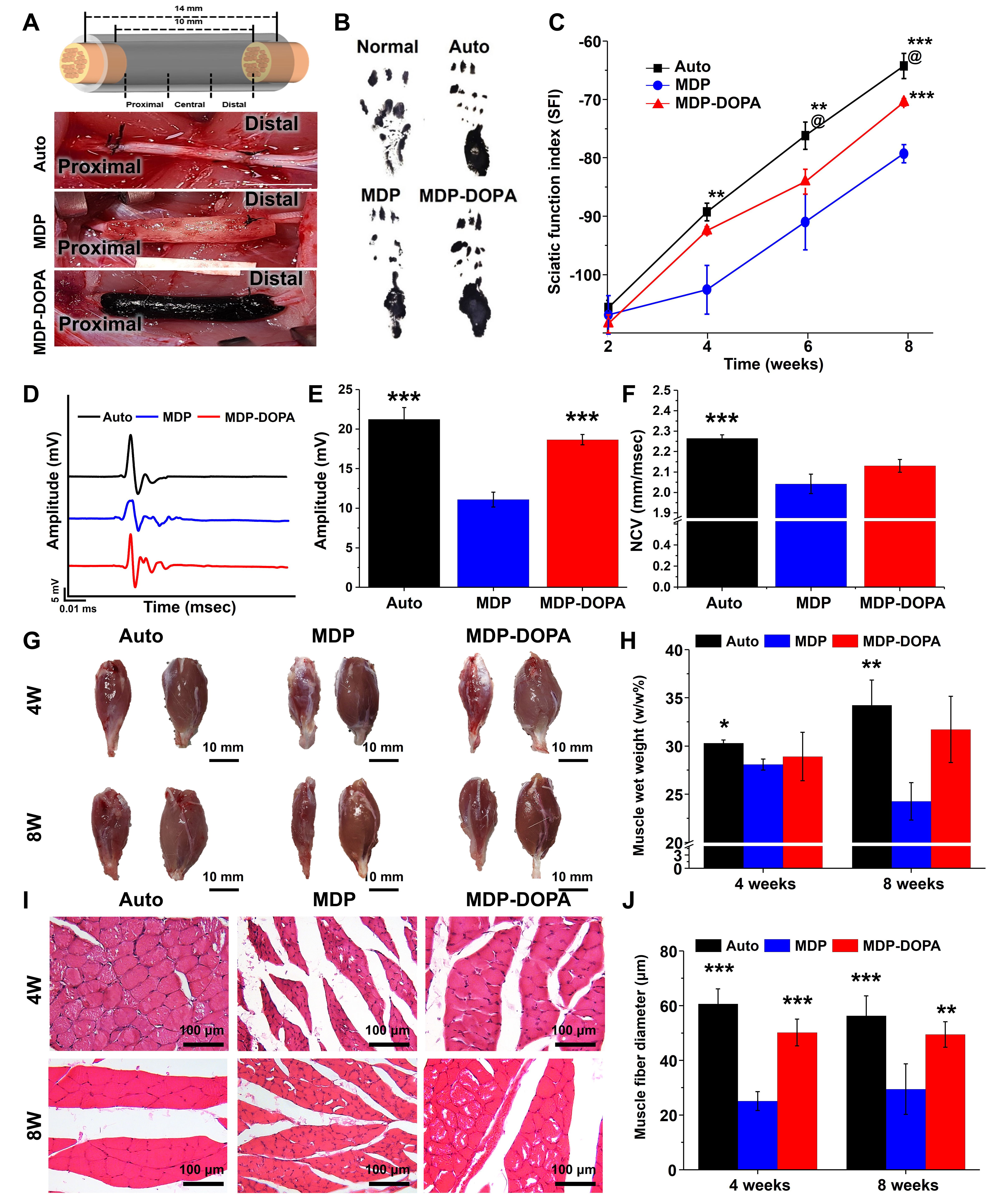
Figure 4. (A) Schematic illustration of NGC implantation and macroscopic images of implanted NGCs; (B) Paw printing at 8 weeks post-implantation and (C) quantification of sciatic functional index (**P < 0.05 and ***P < 0.001 vs. MDP, @P < 0.05 vs. MDP-DOPA); (D) Electrophysiological evaluation of compound muscle action potentials, (E) peak amplitude measurements, and (F) nerve conduction velocity of each group (***P < 0.001 vs. MDP); (G) Gross view of gastrocnemius muscle (scale bar = 10 mm) and (H) quantification of muscle wet weight ratio; (I) H&E histological sections of isolated gastrocnemius muscle (Scale bar = 100 μm) and (J) quantification of muscle fiber diameter. (*P < 0.05, **P < 0.01, and ***P < 0.001 vs. MDP). Data are presented as mean ± SD (biological n = 4). NGC: Nerve guidance conduit; MDP: milk-derived protein; DOPA: dihydroxyphenylalanine; H&E: hematoxylin and eosin; SD: standard deviation.





