


fig4
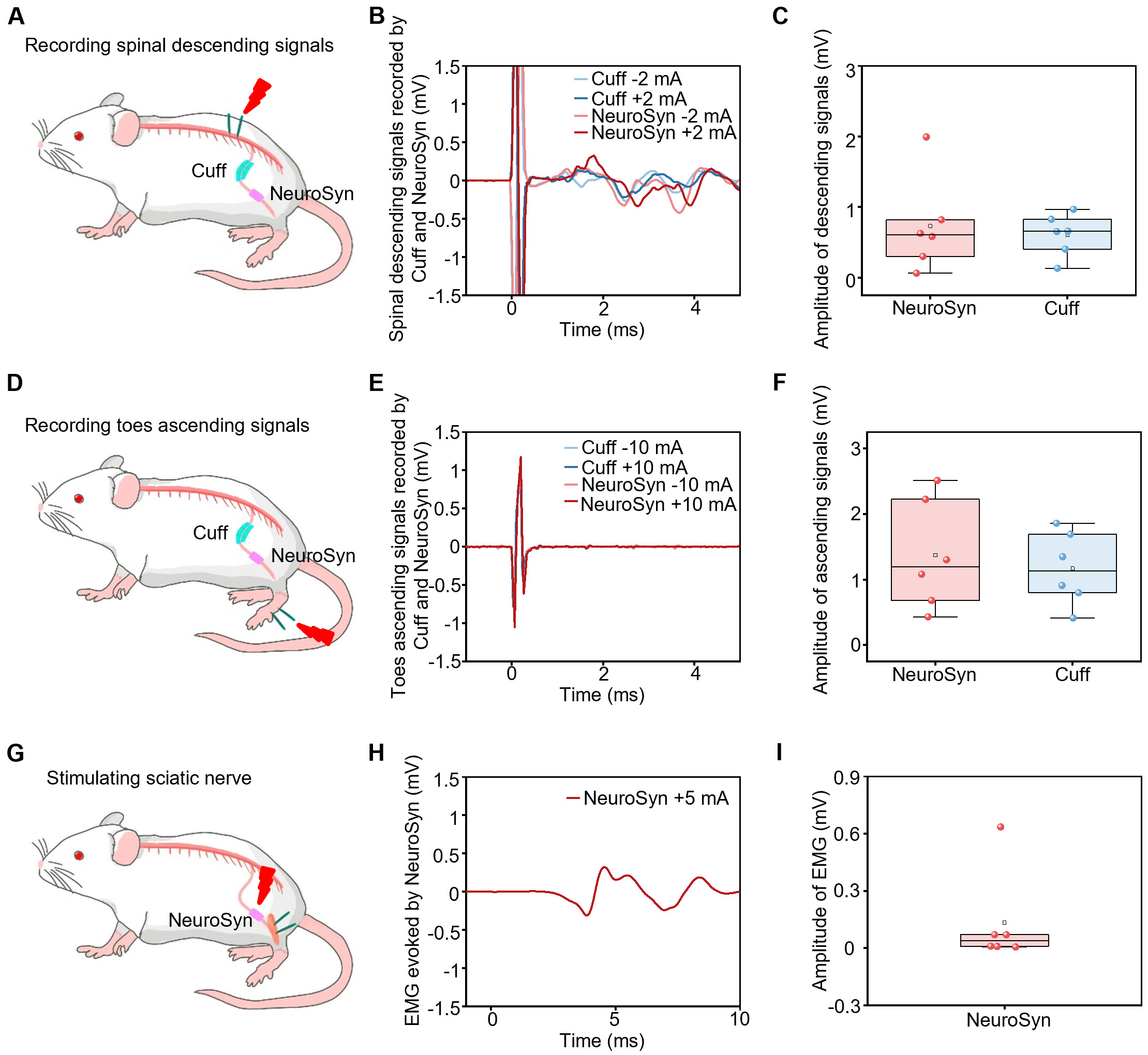
Figure 4. NeuroSyn as a bidirectional neural interface. (A) Schematic diagram of recording descending signals by commercial cuff electrodes and NeuroSyn upon stimulation at the spinal cord at 8 weeks post-implantation. Stimulation parameters: ±2 mA, 1 Hz, pulse width 0.1 ms; (B) Representative descending signals recorded by cuff electrodes and NeuroSyn; (C) The statistical results of the descending signals; (D) Schematic diagram of recording ascending signals by commercial cuff electrodes and NeuroSyn upon stimulation at the feet at 8 weeks post-implantation. Electrical stimulation parameters: ±10 mA, 1 Hz, pulse width 0.1 ms; (E) Representative ascending signals recorded by cuff electrodes and NeuroSyn; (F) The statistical results of the ascending signals; (G) Schematic diagram of stimulating the sciatic nerve by NeuroSyn at 8 weeks post-implantation. Electrical stimulation parameters: ±5 mA, 1 Hz, pulse width


