


fig2
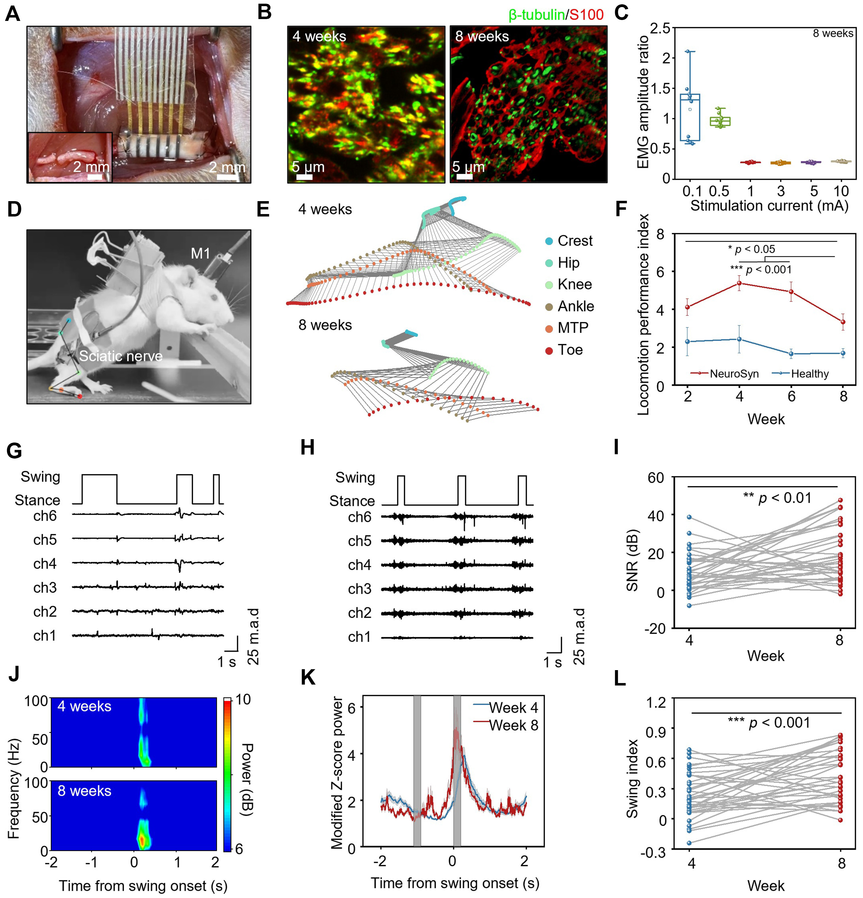
Figure 2. The electrophysiological signals recorded by NeuroSyn during treadmill walking. (A) NeuroSyn is implanted at the injured site, bridging the proximal and distal ends. The inset shows the sciatic nerve defect (6 mm) created by transection; (B) Immunofluorescent images of transverse section at 2 mm from the proximal end of the nerve segment at 4 and 8 weeks post-implantation. Immunohistochemical staining: axons (β-tubulin, green), Schwann cells (S100, red); (C) The normalized EMG amplitude relative to contralateral control generated by different stimulation currents at the proximal side on the operated side at 8 weeks post-implantation. n = 5 animals in each group. The box plot presents the median (center line), lower quartile (lower border), upper quartile (upper border), maximum (upper whisker) and minimum (lower whisker), which are ≤ 1.5 times the interquartile range; (D) Photograph of neural signals and gait recording during treadmill walking of the rat; (E) Stick diagram illustrating leg movements of a rat with an injured leg during one swing event at 4 and 8 weeks post-implantation; (F) Locomotion performance index of healthy rats and rats with NeuroSyn at 2, 4, 6, 8 weeks post-implantation. Data are presented as mean ± SD; (G) Recorded peripheral nerve signal traces during locomotion by NeuroSyn at 4 weeks post-implantation; (H) Recorded peripheral nerve signal traces during locomotion by NeuroSyn at 8 weeks post-implantation; (I) Signal to noise ratio (SNR) of the signals recorded by NeuroSyn at 4 and 8 weeks post-implantation; (J) Frequency-time spectrogram of the recorded signals at 4 and 8 weeks post-implantation; (K) Modified peripheral nerve signal power at 4 and 8 weeks post-implantation; (L) Swing index at 4 and 8 weeks post-implantation. In (D) to (L), n = 6 animals in each group. Statistics is analyzed through SPSS (version 27.0), followed by one way ANOVA (*P < 0.05, **P < 0.01, ***P < 0.001). In (F), Tukey’s post-hoc test is used for pairwise comparisons. In (G) to (H), signal intensities were normalized into units of median absolute deviation (m.a.d.) to the median to mitigate different noise levels in each time point. NeuroSyn: A biosynchronized, transient and flexible regenerative peripheral neural interface; EMG: electromyography; SD: standard deviation; SPSS: statistical product and service solutions; ANOVA: analysis of variance; M1: primary motor cortex; MTP: metatarsophalangeal.


