


fig5
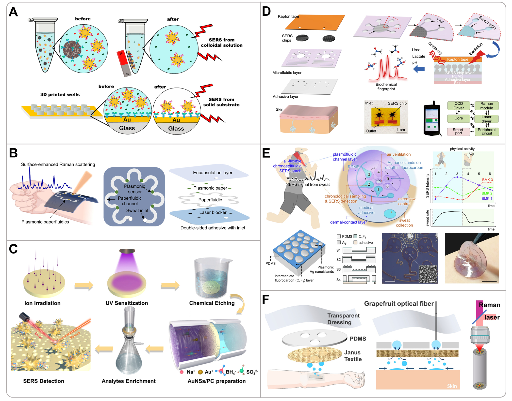
Figure 5. (A) Illustration of two SERS immunoassay approaches: magnetic-bead suspensions bearing cortisol antibodies for magnetically assisted detection, and gold-coated glass substrates functionalized with cortisol antibodies for rigid-support detection. Reproduced with permission[78]. Copyright 2020, Elsevier; (B) Conceptual, overhead, and stacked representations of the wearable plasmonic paper microfluidic system for sweat collection, storage, and in situ SERS measurement. Reproduced with permission[67]. Copyright 2022, Science; (C) Schematic illustration depicting the fabrication process of the Au NSs/ion-track-etched polycarbonate membrane-based SERS substrate and its subsequent detection procedure. Reproduced with permission[94]. Copyright 2023, American Chemical Society; (D) Schematic diagram of the stacked structure of functional layers, an integrated microfluidic plasmonic device enabling biofluid flow, storage and SERS analysis, alongside a system-level block diagram of the internal functional modules within the portable Raman analyzer. Reproduced with permission[99]. Copyright 2022, Nature Publishing Group; (E) Schematic illustration of the fabrication and initiation of the flexible nanoplasmonic SERS patch comprising plasmonic fluid-channel layer and dermal-contact layer. Reproduced with permission[71]. Copyright 2025, Nature Publishing Group; (F) Schematic diagram of the sweat collection principle of Janus-based wearable textiles and the generation of inverse Raman signals by microstructured optical fiber cores. Reproduced with permission[66]. Copyright 2023, American Chemical Society. 3D: 3-dimensional; SERS: surface-enhanced Raman scattering; PDMS: polydimethylsiloxane.


