fig9

Electrochemical biosensing across microsampled blood and interstitial fluid for metabolic, hormonal, and therapeutic monitoring

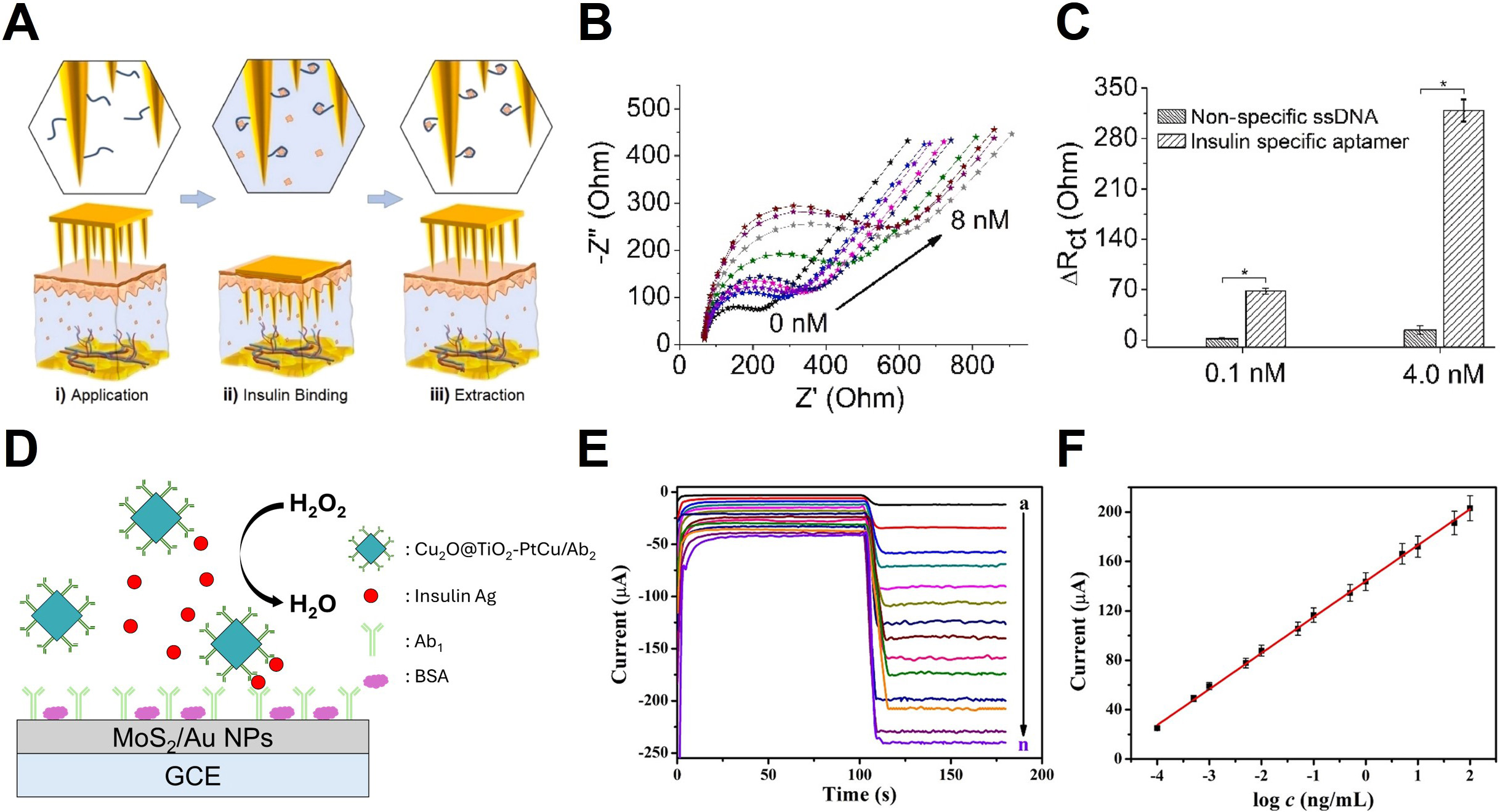
Figure 9. Microneedle base electrochemical insulin sensing. (A) Steps involved in aptasensor operation: (i) Extraction of insulin from ISF, (ii) binding of the extracted insulin to the immobilized aptamer, and (iii) EIS; (B) Electrochemical response of the aptasensor during cortisol detection; (C) Comparison of ∆Rct (change in charge-transfer resistance) for insulin-specific aptamers and non-specific ssDNA. *P < 0.05, One-way analysis of variance; (D) Schematic illustration of the sandwich-type immunosensor. Insulin is sequentially captured through antibody-antigen binding on a MoS2/Au NP-modified GCE, while the Cu2O@TiO2-PtCu/Ab2 nanocomposite functions as a catalytic label to enhance the electrochemical signal via H2O2 reduction; (E) Amperometric responses of the immunosensor toward increasing insulin concentrations (0-100 ng/mL); (F) Nyquist plots illustrate stepwise impedance changes during immunosensor assembly. The error bars in (C) and (F) show the standard deviation. (A-C) Reproduced with permission Copyright 2025, Biosensors and Bioelectronics[25]. (E and F) Reproduced with permission Copyright 2019, ACS Applied Materials & Interfaces[128]. ISF: Interstitial fluid; EIS: electrochemical impedance spectroscopy; ssDNA: single-stranded DNA; NPs: nanoparticles; GCE: glassy carbon electrode; BSA: bovine serum albumin.

Soft Science
ISSN 2769-5441 (Online)

Portico

All published articles are preserved here permanently:

https://www.portico.org/publishers/oae/

Portico

All published articles are preserved here permanently:

https://www.portico.org/publishers/oae/