


fig8
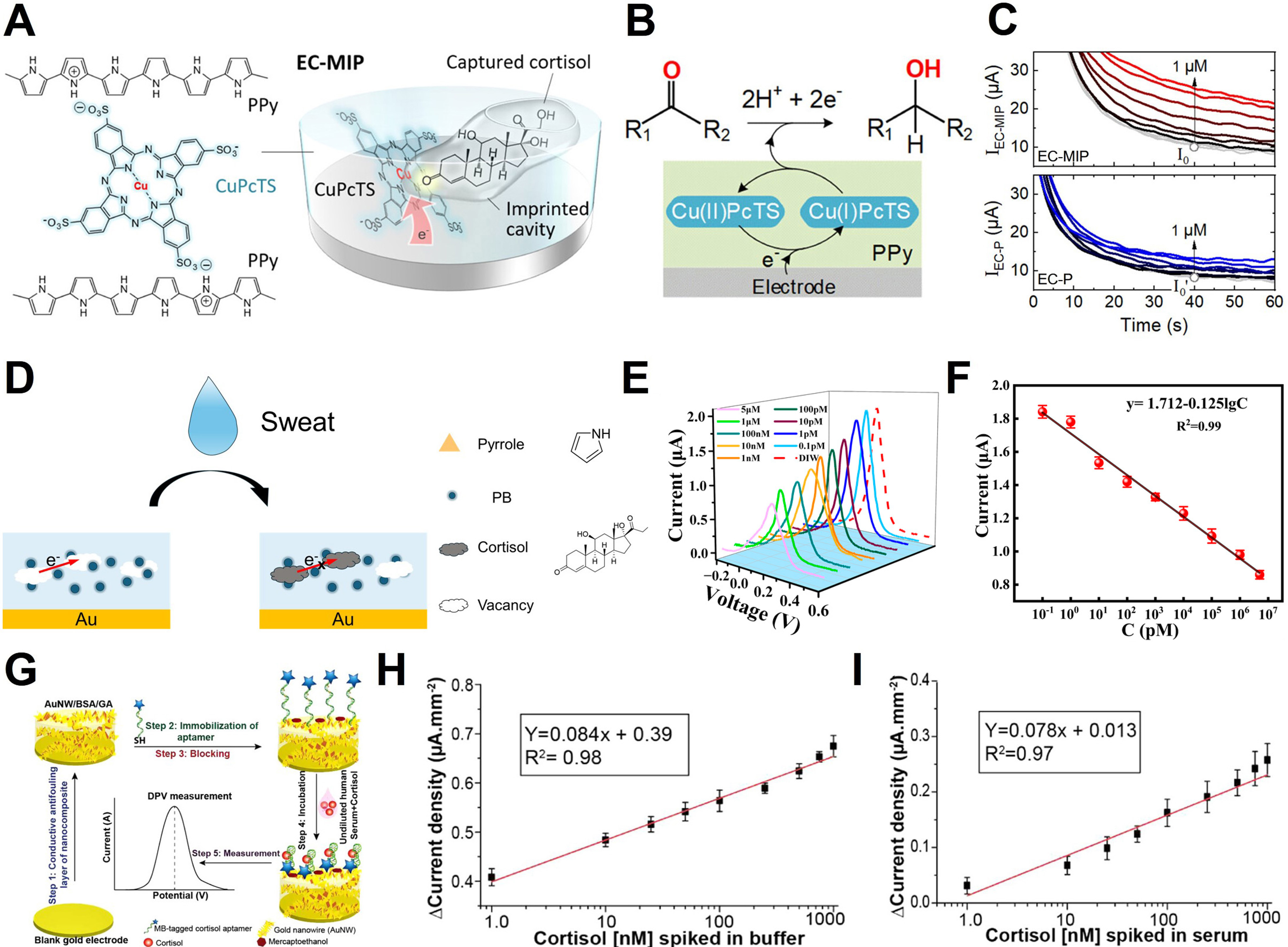
Figure 8. Cortisol sensing mechanism and applications. (A) Structure of the EC-MIP sensor for cortisol detection, using CuPcTS-doped polypyrrole (PPy); (B) Electrocatalytic hydrogenation mechanism at the EC-P electrode; (C) Amperometric responses of the sensor at cortisol concentrations (0 to 1 μM). (Top: EC-MIP sensor, showing a clear increase in current; Bottom: EC-P electrode, showing a less responsive current); (D) Schematic representation of the cortisol detection process using the MIP@PI sensor; (E) DPV current responses of the MIP@PI sensor at various cortisol concentrations; (F) Calibration curve of the MIP@PI sensor; (G) Schematic overview of the aptasensor fabrication process: (Step 1) Conductive antifouling layer synthesis, (Step 2) Aptamer immobilization, (Step 3) Blocking, (Step 4) Incubation with sample, (Step 5) DPV measurement for cortisol detection; (H) DPV current density response to varying cortisol concentrations spiked in buffer; (I) DPV current density response to varying cortisol concentrations spiked in undiluted human serum. The error bars in (F), (H) and (I) represent the standard deviation. (A-C) Reproduced with permission Copyright 2023, ACS Applied Materials & Interfaces[133]. (D-F) Reproduced with permission Copyright 2025, Biosensors[35]. (G-I) Reproduced with permission Copyright 2021, ACS Omega[36]. EC: Electrocatalytic; MIP: molecularly imprinted polymer; PI: polyimide; DPV: differential pulse voltammetry; PB: Prussian blue; MB: methylene blue.


