


fig11
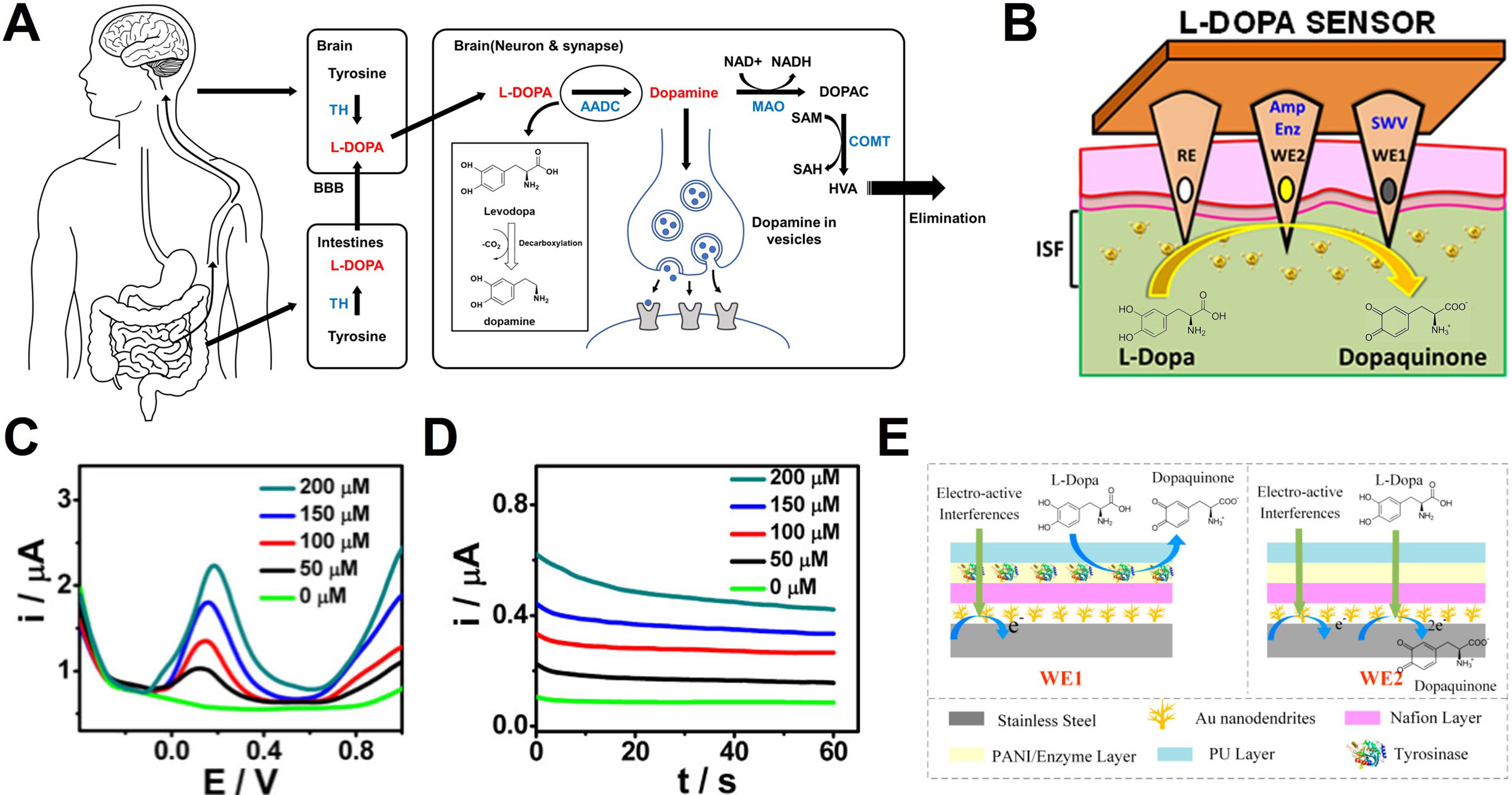
Figure 11. Dopamine modulation by L-Dopa and real-time biosensing. (A) Schematic diagram illustrating the in vivo administration route and metabolic process of L-Dopa, leading to the formation and excretion of dopamine; (B) Schematic illustration of the sensor configuration and sensing principle for L-Dopa detection. The sensor detects the electron transfer generated at the WE during the oxidation of L-Dopa to dopaquinone, utilizing a dual-electrode system for enzymatic and non-enzymatic measurements; (C) SWV and (D) chronoamperometric results corresponding to increasing L-Dopa concentrations (0-200 µM); (E) Schematic of the differential sensing mechanism and the applied sensor. WE1 selectively detects interferences by blocking the L-Dopa reaction, while WE2 detects all electro-active substances. The specific signal of L-Dopa is extracted by calculating the difference between the two signals. (B-D) Reproduced with permission Copyright 2019, ACS Sensors[222]. (E) Reproduced with permission Copyright 2022, Biosensors[223]. L-Dopa: Levodopa; WE: working electrode; SWV: square wave voltammetry; TH: tyrosine hydroxylase; L-DOPA: levodopa; BBB: blood-brain barrier; AADC: aromatic L-amino acid decarboxylase; DOPAC: 3,4-dihydroxyphenylacetic acid; MAO: monoamine oxidase; NAD+: nicotinamide adenine dinucleotide (oxidized form); NADH: nicotinamide adenine dinucleotide (reduced form); SAM: S-adenosyl-L-methionine; SAH: S-adenosyl-L-homocysteine; COMT: catechol-O-methyltransferase; HVA: homovanillic acid; PANI: polyaniline; PU: polyurethane.


