


fig3
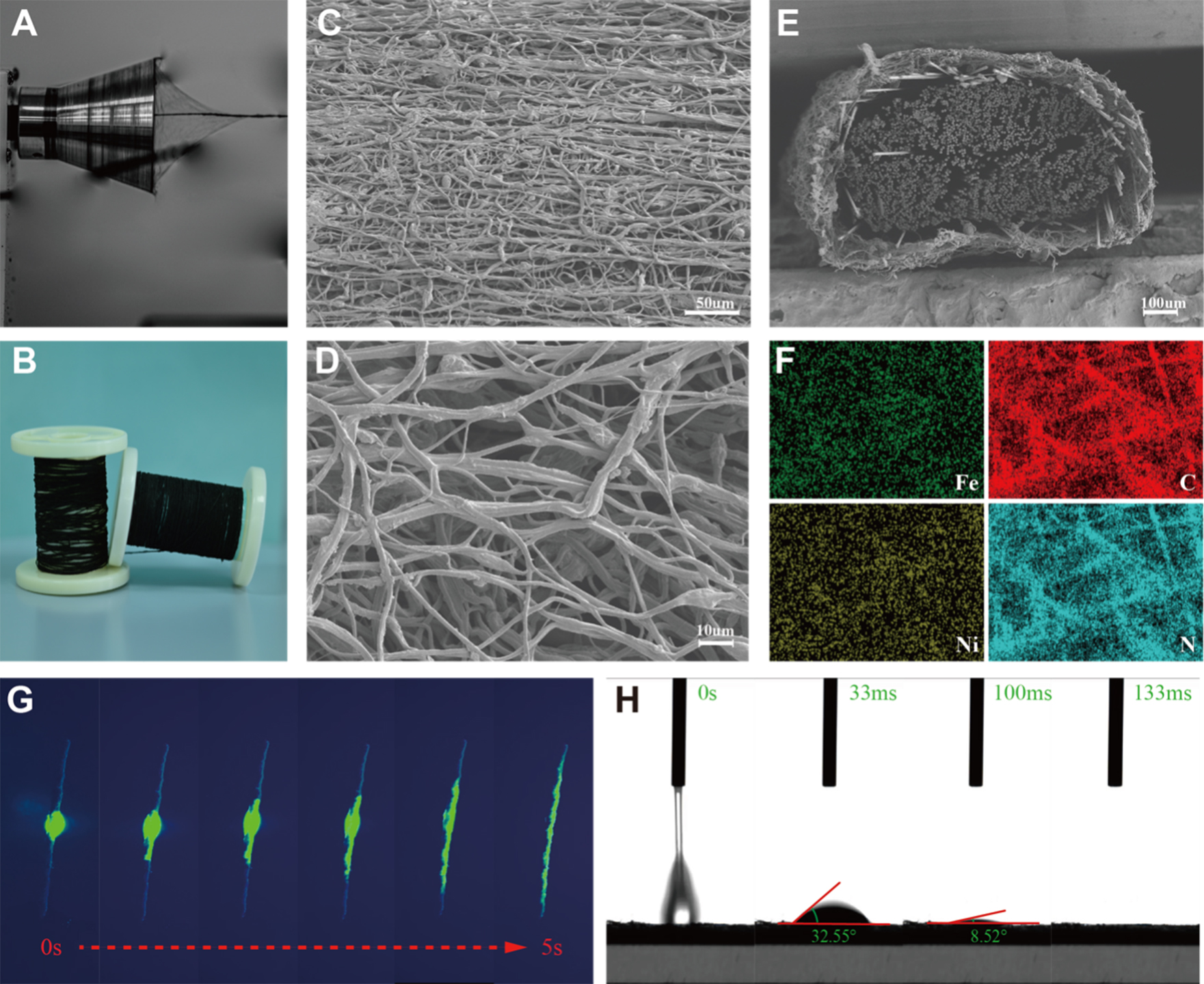
Figure 3. (A) Photo of conjugate electrospinning preparation of cortisol-sensing yarns; (B) Two rolls of cortisol-sensing yarns prepared through continuous conjugate electrospinning technology; (C and D) Surface morphology of cortisol-sensing yarns under low and high magnification; (E) Cross-section SEM image and (F) surface elemental mapping of a cortisol-sensing yarn; (G) Photos of dye infiltration into a cortisol-sensing yarn within 5 s; (H) WCA of electrospun sensing films within 133 ms. SEM: Scanning electron microscopy; WCA: water contact angle.


