


fig4
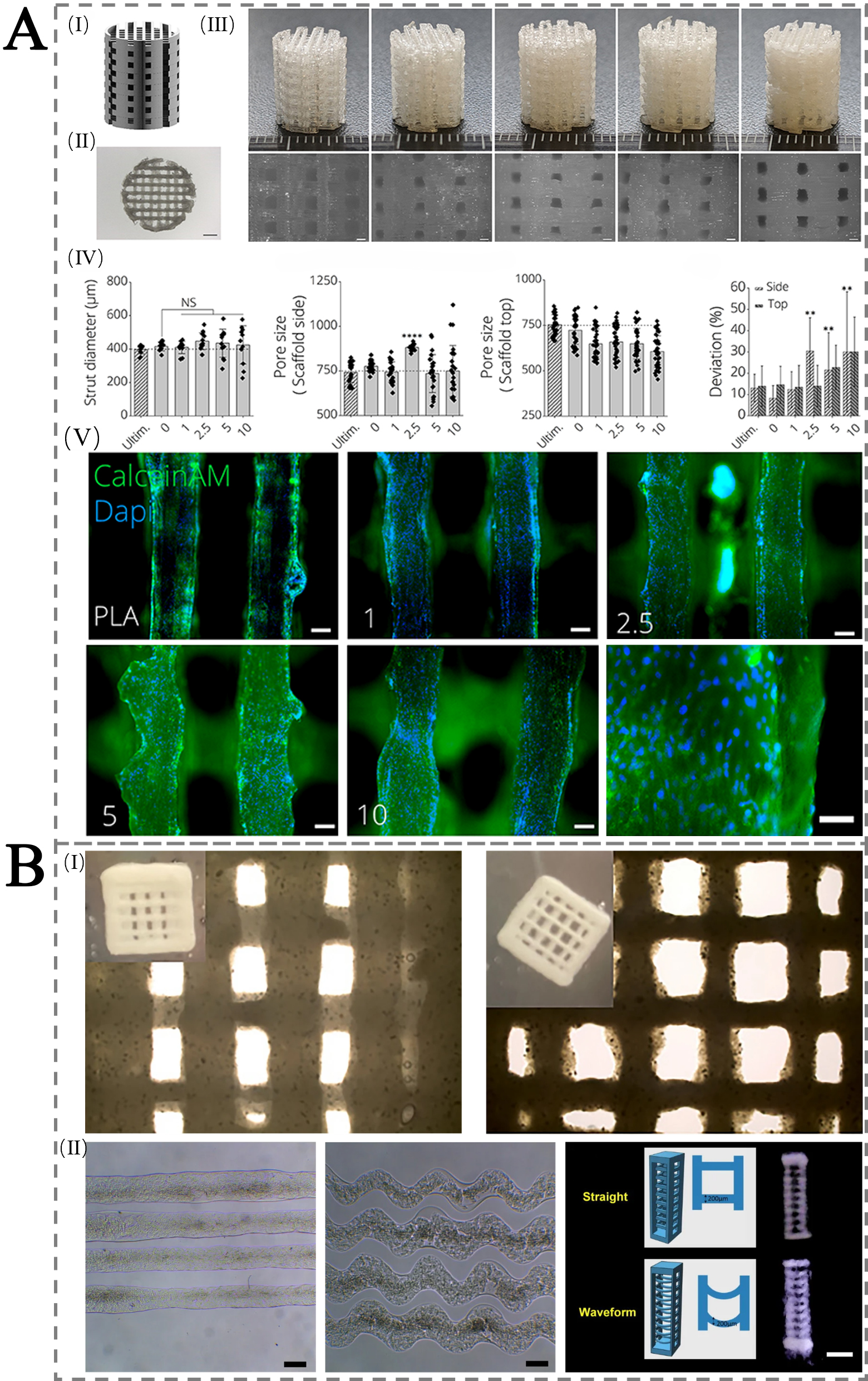
Figure 4. (A) Mechanical properties of 3D-printed PLA-45S5 BG scaffolds: (I) CAD design with 750 μm pores; (II) top-view (scale bar 2 mm); (III) FDM-printed scaffolds with 0-10 wt.% BG (left to right) and microscopy images (scale bar 500 μm); (IV-VII) printability/porosity analysis vs. Ultimaker PLA, showing strut diameter, side/top porosity, and pore area deviation. (VIII) Fluorescence images of MC3T3E1 cells on scaffolds after 24 h, stained with Calcein AM (green) and DAPI (blue), at 0-10 wt.% BG (scale bars 100 μm; 200 μm for 10 wt.%). (Bakhtiary et al.[116]. Copyright 2021 MDPI.); (B) Scaffolds for bone tissue engineering 3D printed with commercial inks. The above are optical images of 3D printed scaffolds using Ink-Bone from CELLINK. The bottom is microfibers obtained with Lifeink®200 as straight and waveform along with the design models and 3D printed scaffolds. (Reprinted from Chiticaru et al.[117]. Copyright 2024 ScienceDirect.). 3D: Three-dimensional; PLA: polylactic acid; BG: Bioglass; CAD: computer-aided design; AM: acetoxymethyl; DAPI:4',6-diamidino-2-phenylindole.


