fig3
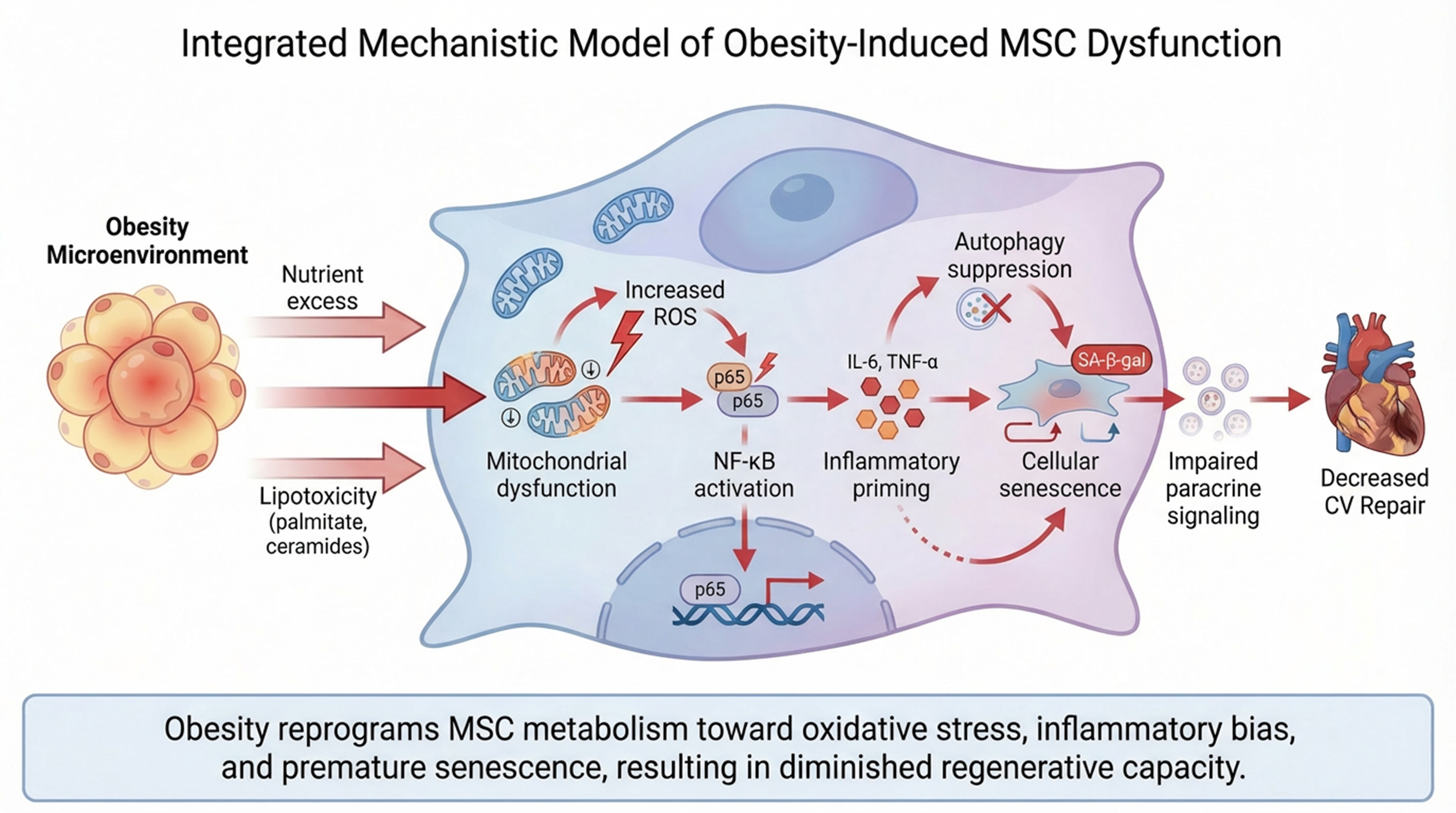
Figure 3. Integrated mechanistic model of obesity-induced MSC dysfunction. In the obese microenvironment, nutrient excess and lipotoxic mediators (e.g., palmitate and ceramides) induce mitochondrial dysfunction and increased ROS production in MSCs. Elevated oxidative stress activates NF-κB signaling, promoting inflammatory priming characterized by increased cytokine production (e.g., IL-6 and TNF-α). Concurrent suppression of autophagy contributes to the accumulation of damaged organelles and further oxidative stress. These processes drive cellular senescence, indicated by SA-β-gal activity, and impair MSC paracrine signaling capacity. Collectively, obesity reprograms MSCs toward a pro-inflammatory, senescent phenotype with diminished regenerative potential. Solid arrows denote experimentally supported relationships; the dashed arrow denotes an inferred link. The figure was created in BioRender [Elmaraezy, A. (2026) https://BioRender.com/mzjqngm] and refined using AI-assisted image-refinement tools (Figurelabs and Nano Banana Pro). MSC: Mesenchymal stem/stromal cell; ROS: reactive oxygen species; NF-κB: nuclear factor kappa-B; IL: interleukin; TNF-α: tumor necrosis factor-α; SA-β-gal: senescence-associated β-galactosidase; CV: cardiovascular.