


fig2
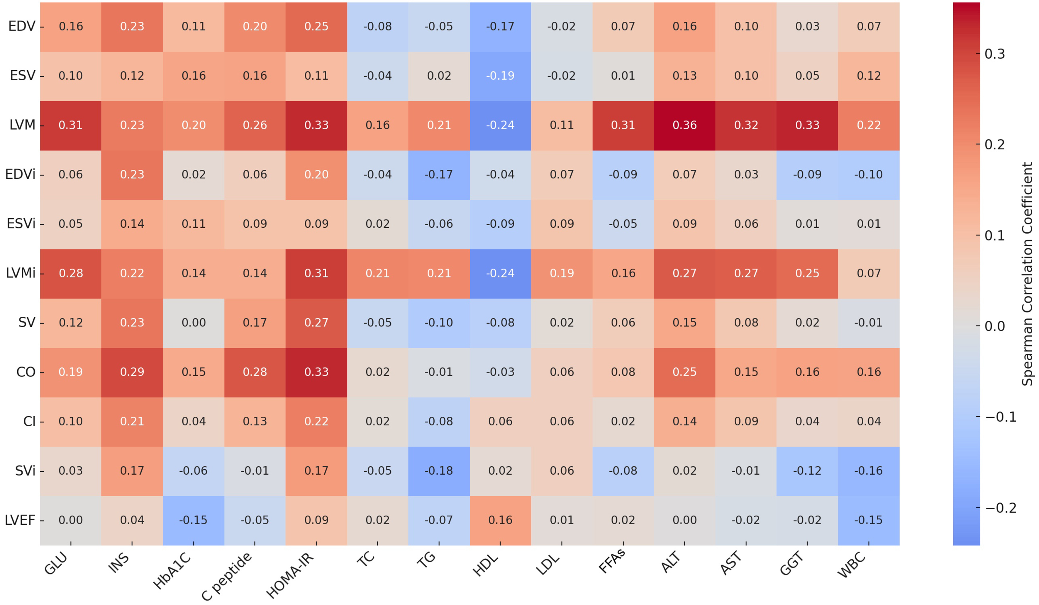
Figure 2. Spearman correlation heatmap between metabolic indicators (x-axis) and cardiac MRI parameters (y-axis). Color intensity represents correlation strength (red = positive, blue = negative). LVM and LVMi correlated positively with HOMA-IR, FFAs, ALT, and GGT, while other parameters showed mild-to-moderate associations. Detailed coefficients are listed in Supplementary Table 6. MRI: Magnetic resonance imaging; EDV: end-diastolic volume; ESV: end-systolic volume; LVM: left ventricular mass; EDVi: end-diastolic volume index; ESVi: end-systolic volume index; LVMi: left ventricular mass index; SV: stroke volume; CO: cardiac output; CI: cardiac index; SVi: stroke volume index; LVEF: left ventricular ejection fraction; GLU: fasting glucose; INS: insulin; HbA1c: hemoglobin A1c; C-peptide: connecting peptide; HOMA-IR: homeostasis model assessment of insulin resistance; TC: total cholesterol; TG: triglycerides; HDL: high-density lipoprotein; LDL: low-density lipoprotein; FFAs: free fatty acids; ALT: alanine aminotransferase; AST: aspartate aminotransferase; GGT: gamma-glutamyl transferase; WBC: white blood cell.


