fig1
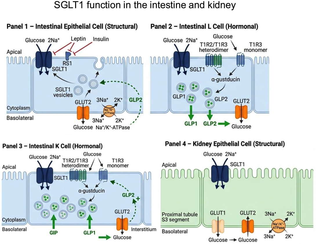
Figure 1. SGLT1 function in the intestine and kidney. Four cell types are depicted, separated into structural (glucose transport) and hormonal (incretin secretion) roles. Panel 1 - Intestinal epithelial cell (structural): SGLT1 mediates apical co-transport of glucose with 2Na+ from the intestinal lumen. RS1 protein inhibits SGLT1 activity at the apical membrane. Leptin and insulin exert inhibitory regulation of SGLT1 expression and trafficking. Intracellular SGLT1-containing vesicles are recruited to the apical membrane in response to luminal glucose, a process stimulated by GLP2 via a neuro-paracrine feedback loop (dashed arrow). GLUT2 on the basolateral membrane exports glucose to the interstitium. The Na+/K+-ATPase maintains the electrochemical sodium gradient driving apical SGLT1 activity. Panel 2 - Intestinal L cell (hormonal): SGLT1 on the apical membrane senses and transports luminal glucose. The T1R2/T1R3 heterodimer and T1R3 monomer sweet taste receptors activate α-gustducin, stimulating intracellular synthesis and basolateral secretion of GLP1 and GLP2. GLUT2 mediates basolateral glucose export. Panel 3 - Intestinal K cell (hormonal): Identical apical glucose-sensing machinery (SGLT1, T1R2/T1R3, T1R3, α-gustducin) drives secretion of GIP and GLP1 from the basolateral membrane. GLP2 arriving from L cells provides a positive feedback signal (+) stimulating SGLT1 activity. GLUT2 exports glucose basolaterally. Panel 4 - Kidney epithelial cell (structural): SGLT1 in the proximal tubule S3 segment recovers residual filtered glucose from the tubular lumen with 2Na+. The Na+/K+-ATPase (3Na+/2K+) maintains the driving gradient. GLUT1 and GLUT2 on the basolateral membrane return glucose to the interstitium. Figure created with FigureLabs. SGLT: sodium-glucose cotransporter; GLUT: facilitative glucose transporter; GIP: gastric inhibitory peptide; GLP: glucagon-like peptide.