

Fulminant myocarditis and cardiogenic shock in rheumatological disease: a case report
 0
Abstract
Background: Fulminant myocarditis (FM) is an infrequent life-threatening condition characterized by acute cardiac inflammation potentially leading to severe cardiac complications such as cardiogenic shock, ventricular arrhythmias, or multiorgan system failure and therefore necessitating prompt recognition and treatment. Case presentation: This case details the rapid progression in less than 24 h of FM to cardiogenic shock in a 58-year-old female with recently diagnosed systemic sclerosis and antiphospholipid syndrome, requiring urgent mechanical circulatory support. The strategy of promptly starting venoarterial extracorporeal membrane oxygenation in combination with an Impella device for left ventricular unloading, alongside empirical combined immunosuppressive therapy was crucial for the patient’s outcome. Despite the inability to confirm diagnosis by performing an endomyocardial biopsy or cardiac magnetic resonance due to the patient's critical state, the multidisciplinary approach, including aggressive mechanical circulatory support and combined immunosuppression, led to significant improvement in cardiac function and successful weaning from support. Conclusion: This case highlights the critical importance of rapid diagnosis in the context of suspected autoimmune fulminant myocarditis to enable early initiation of targeted treatment, such as mechanical circulatory support and empirical combined immunosuppression therapy. It also underscores the benefits of a combined EC-Pella approach for left ventricular unloading when required, and the pivotal role of multidisciplinary cooperation for enhancing patient outcomes.
Keywords


 Download PDF
INTRODUCTION
Myocarditis represents a life-threatening condition frequently triggered by immune-mediated or infectious insults. Its clinical spectrum is broad, ranging from mild symptoms to fulminant heart failure that requires mechanical support. The main etiologies have been identified as infectious (mainly viral), toxic or autoimmune diseases. Numerous autoimmune diseases have been related to myocarditis, with commonly known examples including sarcoidosis, systemic sclerosis (SSc), systemic lupus erythematosus and rheumatoid arthritis[1,2].
Although under normal conditions the myocardium expresses PD-L1, which blocks cardiac-specific T cells, this immune homeostasis may be disrupted by autoimmune diseases that facilitate the exposure of self-antigens to the immune system and erroneously activate myocardial cytotoxic T cells.
Independent of the initial trigger, the subsequent inflammatory response is responsible for myocardial injury, causing myocyte necrosis, interstitial edema, and inflammatory infiltrates. This myocardial damage can lead to three classic presentations: chest pain simulating an acute coronary syndrome, heart failure potentially evolving into cardiogenic shock and arrhythmic manifestations including malignant arrhythmias[1].
Patients with fulminant myocarditis (FM) experience significant morbidity and mortality because of a delay in diagnosis and targeted treatment including mechanical circulatory support[3].
Considering the rapidity and severity of progression, prompt diagnosis and treatment are crucial. In these cases, the pillars of treatment consist of circulatory support with inotropes or mechanical circulatory support devices (MCS) to prevent multiorgan system failure, and tailor disease-specific therapy as early as possible. We describe a case of FM in the setting of rheumatological disease requiring immediate mechanical circulatory support and combined immunosuppressive therapy.
CASE REPORT
Patient presentation
A 58-year-old female patient presented to the Emergency Department with persistent pain in her right foot despite analgesic therapy. She had a two-month history of bilateral toe necrosis. Her medical history included type II diabetes, dyslipidemia, venous thrombosis of the left leg (eight years prior), and Raynaud's phenomenon.
The condition, which had been evaluated in the months preceding admission, was initially suspected to represent a diabetic foot complication. However, following a comprehensive diagnostic work-up—including laboratory testing, Color Doppler ultrasound, and multidisciplinary specialist consultations—a rheumatologic or immunologic etiology was deemed more likely.
On examination, pitting scars were evident, together with necrosis of the second toe of the left foot and of the fourth and fifth toes of the right foot. Additional findings included acrocyanosis of the third toe of the right foot and right foot edema.
Her home medication regimen consisted of metformin (500 mg three times daily), rivaroxaban (20 mg once daily), omega-3 fatty acid supplements, tramadol, and paracetamol.
Initial workup
Upon admission, the patient was hemodynamically stable. Laboratory tests showed microcytic anemia, high count of white blood cells with neutrophilia, normal platelet count, normal renal and hepatic function, normal coagulation test and high inflammatory markers (exceeding the upper limit of normal by fourfold, Table 1). Vascular surgery evaluation was performed for digital necrosis; immediate amputation was not indicated, and infectious involvement was excluded. Medical treatment was optimized with intravenous morphine, and iloprost infusion was initiated.
Laboratory findings at admission
| Laboratory parameter | Value | Reference range |
| Complete blood count | ||
| Hemoglobin | 10.0 g/dL | 11.0-15.0 g/dL |
| White blood cell count | 11.0 × 10⁹/L | 4.0-10.0 × 10⁹/L |
| Neutrophil count | 9.00 × 10⁹/L | 2.0-7.0 × 10⁹/L |
| Platelet count | 230 × 109/L | 150-450 × 109/L |
| Biochemistry | ||
| C-reactive protein (CRP) | 20.0 mg/L | < 5.0 mg/L |
| Creatinine | 0.8 mg/dL | 0.5-0.9 mg/dL |
| Lactate | 0.9 mmol/L | < 2.0 mmol/L |
| Cardiac biomarkers | ||
| hs-Troponin T | 0.020 μg/L | < 0.014 μg/L |
| Immunology | ||
| Lupus anticoagulant (dRVVT ratio) | 2.4 | < 1.2 |
| Anti-cardiolipin IgG | 1,912 CU | < 20.0 CU |
| Anti-cardiolipin IgM | 3.6 CU | < 20.0 CU |
| Anti-β2 glycoprotein I IgG | 6,200 U/mL | < 20.0 U/mL |
| Anti-β2 glycoprotein I IgM | 5.7 U/mL | < 20.0 U/mL |
A thrombophilia work-up revealed triple-positive antiphospholipid antibodies (Anticardiolipin Antibodies, anti-β2 Glycoprotein I Antibodies and Lupus Anticoagulant, Table 1). Consequently, rivaroxaban was discontinued and replaced with low-molecular-weight heparin. Given the clinical and laboratory profile, SSc was strongly suspected. To assess SSc, a capillaroscopy (showing an early Scleroderma pattern, Figure 1A) and echocardiogram were performed. Echocardiography demonstrated a non-dilated left ventricle with severely reduced systolic function due to global hypokinesia (LVEF 35%), moderate-to-severe mitral regurgitation; no pericardial effusion was found.
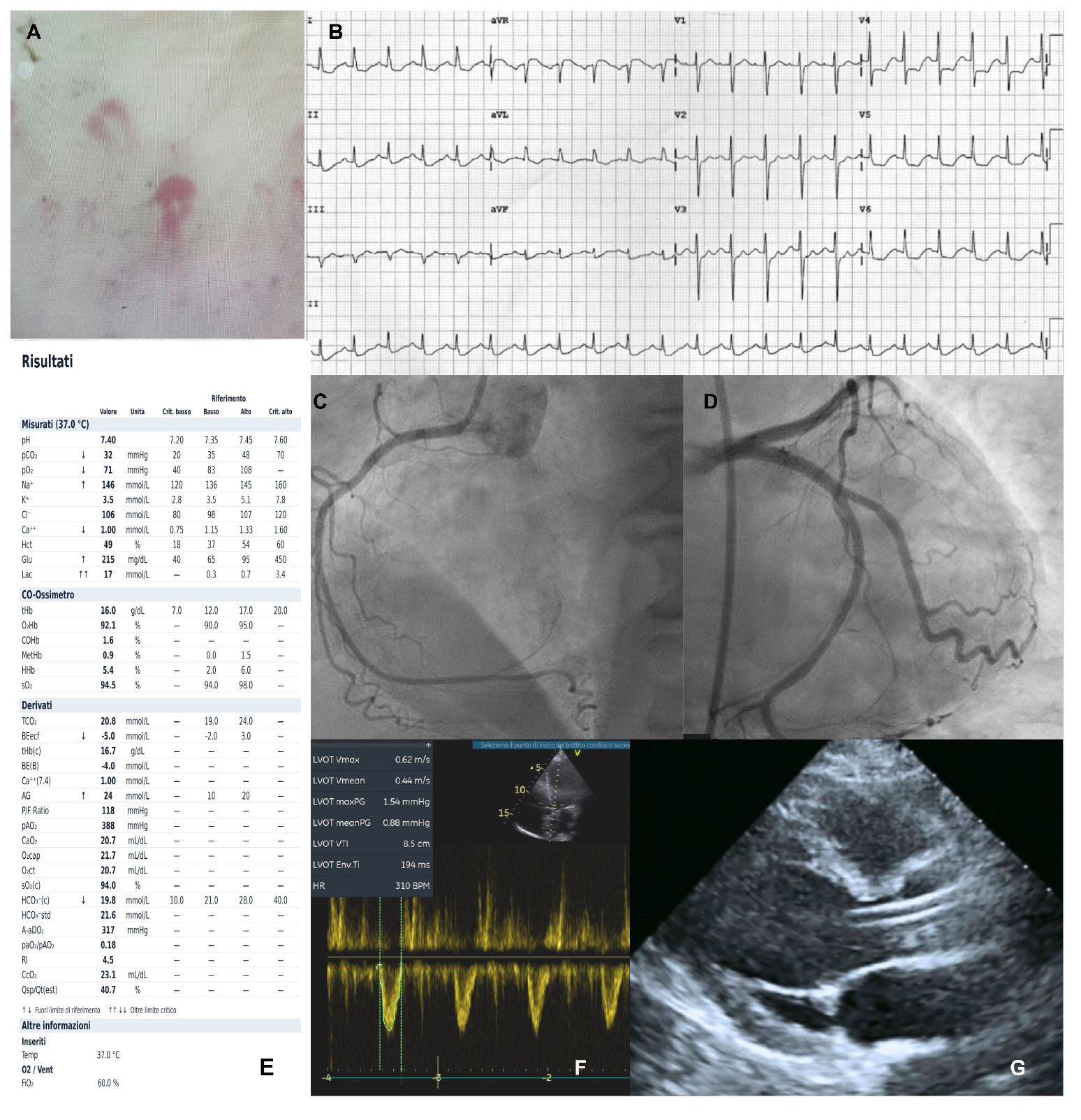
Figure 1. (A) Capillaroscopy showing a scleroderma-like pattern. (B) ECG demonstrating sinus tachycardia and ST-segment depression in the inferior and lateral leads. (C and D) Coronary angiography revealing no significant coronary artery disease. (E) Arterial blood gas (ABG) analysis during cardiogenic shock showing elevated lactate levels. (F) Pulsed-wave Doppler in the acute phase of cardiogenic shock, demonstrating a low cardiac output state with a LVOT VTI of 8 cm. (G) TTE parasternal long-axis view during mechanical circulatory support with ECMO and Impella.
By the following day, the patient reported dyspnea and chest pain, despite maintaining hemodynamic stability with preserved blood pressure and normal lactate levels. Electrocardiography revealed sinus tachycardia and ST-segment depression in the inferior and lateral leads [Figure 1B]. Laboratory assessments showed significant elevations in high-sensitivity troponin T (3.78 µg/L, peaking at 4.9 µg/L; reference
Diagnosis and management
Within 24 h of symptoms onset, a coronary angiography was performed confirming normal coronary arteries without evidence of significant stenosis [Figure 1C and D]. These findings excluded an acute coronary syndrome, a condition commonly associated with antiphospholipid syndrome, as the primary diagnosis. Concurrently, her hemodynamic status experienced a precipitous decline characterized by neurological impairment, refractory hypotension, and oliguria suggestive of compromised renal perfusion. Furthermore, arterial blood gas analysis revealed a critical rise in lactate levels (> 17 mmol/L, Figure 1E). Despite immediate initiation and titration of inotropic and vasopressor therapy with dobutamine and norepinephrine, the patient’s hemodynamic status remained refractory to pharmacological circulatory support.
A repeat echocardiogram showed severe deterioration in cardiac function, with LVEF reduced to 10% and Left Ventricular Outflow Tract Velocity-Time Integral (LVOT VTI) at 8 cm [Figure 1F]. Based on the clinical findings and the patient’s medical history, a suspected diagnosis of fulminant autoimmune myocarditis with cardiogenic shock was made, prompting urgent initiation of peripheral veno-arterial extracorporeal membrane oxygenation (VA-ECMO). Percutaneous cannulation was performed via the right femoral vein for drainage and the contralateral femoral artery for return. Extracorporeal support was provided using the Getinge Cardiohelp system, while respiratory support was managed through mechanical ventilation via an endotracheal tube.
Due to the urgent need for ECMO therapy, which required systemic anticoagulation with intravenous heparin, an endomyocardial biopsy could not be performed. Empirical immunosuppressive therapy was initiated based on the suspected autoimmune diagnosis (with pending lab results), as advised by Rheumatology. The regimen included high-dose methylprednisolone (14 mg/kg/day for 3 days, then tapered), plasmapheresis, and intravenous immunoglobulins (IVIg).
On day one of VA-ECMO, an Impella CP device was inserted to achieve left ventricular (LV) unloading (EC-Pella approach). This strategy was adopted due to the significant increase in LV afterload, severe systolic impairment with further reduction on stroke volume and emerging signs of ventricular dilatation on echocardiogram. Intravenous infusion with levosimendan was also administered to support LV function.
During the following days of mechanical support with EC-Pella, daily cardiological and rheumatological assessments were conducted. Serial echocardiograms showed progressive improvement in LV systolic function [Figure 1G]. As a result, mechanical circulatory support was gradually weaned, starting with the discontinuation of ECMO on day 7, followed by Impella removal on day 12. Therapeutic strategy is illustrated on Figure 2.
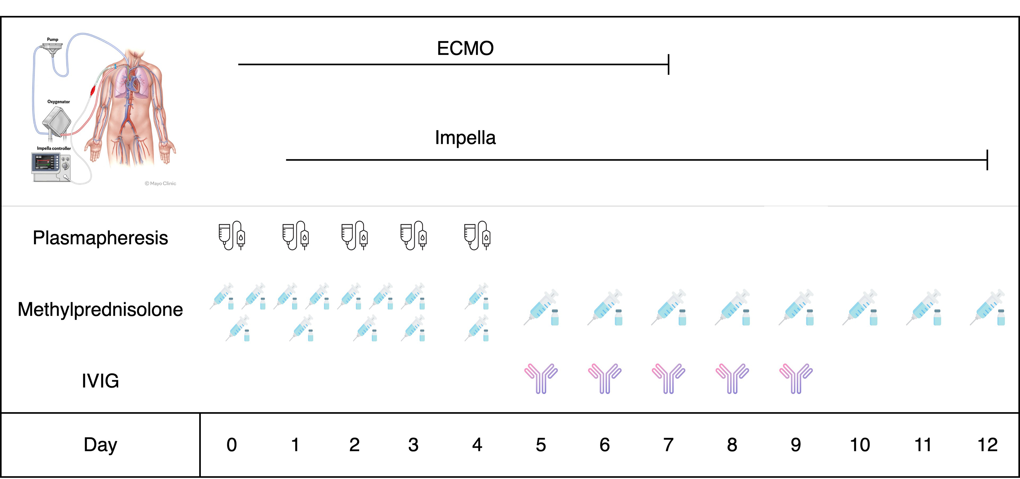
Figure 2. Timeline of therapeutic strategy during the acute phase. Temporal sequence of primary therapeutic interventions administered to the patient during the acute phase of fulminant myocarditis and cardiogenic shock. Key treatments highlighted include mechanical circulatory support with ECMO (Extracorporeal Membrane Oxygenation) and Impella device(“EC-pella approach”) and empirical immunosuppressive therapy with high-dose methylprednisolone (14 mg/kg/day for 3 days and then tapered), plasmapheresis and IVIG (intravenous immunoglobulins).
Due to clinical conditions and the need for mechanical ventilation, cardiac magnetic resonance (CMR) could not be performed during the acute phase.
Follow-up
After discontinuation of mechanical circulatory support, regular cardiologic and rheumatological assessments were continued. Echocardiograms performed in the absence of mechanical circulatory support revealed normal LV dimensions with improved systolic function (LVEF 35%, LVOT VTI 15 cm) and mild to moderate mitral regurgitation.
Heart failure therapy was optimized with sacubitril/valsartan, bisoprolol, gliflozin, and canrenone.
Mechanical ventilation was also progressively reduced, and the patient was transitioned to tracheostomy ventilation, showing gradual clinical improvement. Due to the priority of amputation of the right necrotic foot and II, III and IV necrotic toes of the left foot, following vascular surgery reassessment, and the ongoing need for tracheostomy ventilation, CMR could not yet be performed to confirm the diagnosis of myocarditis.
Meanwhile, autoimmune testing returned positive for antinuclear antibodies (centromeric pattern ANA) and considering the presence of Antiphospholipid antibodies, Raynaud Phenomenon, capillaroscopy findings (early scleroderma pattern) and the presence of pitting scars, an initial diagnosis of antiphospholipid syndrome and SSc was made. After induction therapy, the glucocorticoid was progressively tapered.
At one month follow-up, the patient was alive in a rehabilitation facility.
DISCUSSION
FM is an uncommon but critical syndrome characterized by sudden and profound hemodynamic compromise due to acute myocardial inflammation. Given its life-threatening presentation, it requires urgent and targeted management. The primary etiologies include infectious, toxic, and autoimmune causes. The underlying pathophysiology involves a cytokine storm and extensive immune cell infiltration into the myocardium[3]. While Endomyocardial Biopsy (EMB) remains the diagnostic gold standard for definitive histopathological confirmation, particularly in specific settings, and is, alongside Cardiac Magnetic Resonance (CMR), one of the two key diagnostic criteria in recent guidelines, the clinical approach in critically unstable patients often differs. The severity and rapid evolution toward cardiogenic shock can render both EMB and CMR unsafe or technically infeasible. In these highly unstable settings, a clinical diagnosis is often necessary, supported by non-invasive assessment (ECG, biomarkers, imaging) once coronary artery disease (CAD) has been ruled out[1,4,5].
This case describes a patient with newly diagnosed antiphospholipid syndrome and SSc who developed rapidly FM, evolving into cardiogenic shock within 24 h. Prompt diagnosis and immediate initiation of mechanical circulatory support with VA-ECMO combined with an Impella CP device for LV unloading, alongside combined empirical immunosuppressive therapy proved to be crucial for the patient’s outcome. Despite being unable to confirm the diagnosis via endomyocardial biopsy or cardiac magnetic resonance due to rapid hemodynamic deterioration of the patient requiring MCS and mechanical invasive ventilation, a multidisciplinary approach based on aggressive empirical treatment led to significant improvement in cardiac function and successful weaning from mechanical support.
Considering the excessive immune activation and intense cytokine release as pathophysiological mechanisms in FM, “immunomodulatory” therapy, comprising high-dose glucocorticoids and intravenous immunoglobulins, represents an essential component of the therapeutic regimen[6].
Although current guidelines recommend immunosuppressive therapy only in histologically confirmed virus-negative myocarditis, empirical immunosuppression may be considered for suspected cases with severe clinical manifestations, such as cardiogenic shock or malignant/refractory arrhythmias, when diagnostic procedures like EMB or CMR are not feasible. According to a Scientific Statement from the American Heart Association (AHA), if there is a strong suspicion of immune-mediated FM, a single 1g dose of methylprednisolone is often administered urgently, even before a biopsy confirms the diagnosis or other diagnostic tests are completed[7]. While such therapeutic choices are necessarily empirical and should be evaluated on a case-by-case basis, robust evidence suggests this approach should not become a routine, standardized practice[4]. In our case, the patient's strong autoimmune background and poor clinical condition led the multidisciplinary team to a combined empirical immunomodulatory therapy with high-dose glucocorticoids, intravenous immunoglobulins, and plasma exchange therapy. The decision to include plasmapheresis was based on evidence of a higher survival rate among patients with catastrophic anti-phospholipid syndrome, a finding that supported its use in this high-risk scenario[8].
In cases of refractory cardiogenic shock, mechanical circulatory support serves as a pivotal life-sustaining intervention. The choice of a MCS device depends on the clinical scenario, and timely intervention is critical in maximizing patient outcomes. Veno-arterial extracorporeal membrane oxygenation typically increases LV afterload, consequently causing LV distention, an increase in myocardial wall stress, and worsening of inflammation, leading to pathological cardiac remodeling.
These effects could be mitigated by combining Impella devices (EC-Pella approach), which improves clinical outcomes[6,9,10]. Effective ventricular unloading is thought to dampen cardiac mechanotransduction pathways, which translates into mitigation of the inflammatory cascade and a decrease in myocardial wall stress, thereby preventing adverse cardiac remodeling. The synergistic use of these devices is increasingly supported in the literature for refractory, FM cases that are unresponsive to medical therapy alone. Indeed, serial EMBs demonstrated a reduction of multiple markers of inflammation, including inflammatory cellular infiltrates in the heart, in the setting of LV unloading with Impella, in addition to immunosuppressive therapy[4].
LV unloading can be achieved through various strategies, including inotropic support, intra-aortic balloon pump (IABP), Impella devices, or percutaneous LV devices. Registry data suggest that IABP and inotropes may be suitable for lower degrees of increased afterload. However, in cases of a dilated, non-ejecting ventricle with significantly elevated filling pressures and clinical complications such as pulmonary edema, active and timely unloading—via an Impella device or surgical venting—is often required. Such strategies have demonstrated a reduction in mortality and increased rates of LV functional recovery in severe scenarios[11].
This case highlights the critical importance of rapid diagnosis in the context of FM to enable timely treatment.
In FM, early recognition and prompt initiation of tailored immunosuppressive therapy alongside mechanical circulatory support are essential to stabilize hemodynamics and improve outcomes. A multidisciplinary approach integrating rapid diagnosis, immunotherapy, and advanced mechanical support remains the cornerstone of management for this life-threatening condition.
Moreover, this case illustrates that integrated expertise among cardiologists, rheumatologists, immunologists, and critical care specialists is fundamental to tailoring therapeutic interventions, optimizing outcomes, and mitigating complications.
CONCLUSION
This case shows a rapidly progressive FM evolving in the lap of a few hours in cardiac shock, in a patient with a recent development of rheumatological disease.
Prompt diagnosis and prompt treatment with mechanical circulatory support and combined immunosuppressive therapy improve the clinical outcome of the patient, even in the absence of histological confirmation.
The combined use of ECMO with Impella to mitigate the excessive increase in left ventricular afterload that occurs with ECMO alone is crucial in achieving short-term improvement in left ventricular systolic function.
Moreover, last but not least, this case underscores the importance of multidisciplinary shared care and decision-making, which was the key to a patient's successful recovery.
DECLARATIONS
Acknowledgments
Illustrations on Figure 2 were created using icons from Flaticon.com.
Authors' contributions
Were responsible for clinical management, echocardiographic assessment, and follow-up: Birtolo LI, Scoccia G, Magliolo J, Badagliacca R, Vizza CD
Performed the rheumatological work-up and directed the immunosuppressive strategy: Caputo A
Managed the mechanical circulatory support (VA-ECMO): Pugliese F, Tosi A
Performed the coronary angiography and assisted in Impella placement: Mancone M
Wrote the original draft: Birtolo LI, Magliolo J
Contributed to writing - review and editing: Alessandri C, Caputo A, Mancone M, Pugliese F, Tosi A
Provided supervision and final review: Badagliacca R, Vizza CD
All authors have read and agreed to the published version of the manuscript.
Availability of data and materials
All data supporting the findings of this study are available from the corresponding authors upon reasonable request.
AI and AI-assisted tools statement
During the preparation of this manuscript, the AI tools ChatGPT (GPT-4o) and Grammarly were used solely for language editing and for assistance in the creation and modification of Figure 2 (EC-Pella strategy). The tools did not influence the study design, data collection, analysis, interpretation, or the scientific content of the work. All authors take full responsibility for the accuracy, integrity, and final content of the manuscript.
Financial support and sponsorship
Financial support for this study was provided by Sapienza University of Rome.
Conflicts of interest
All authors declared that there are no conflicts of interest.
Ethical approval and consent to participate
According to the policies of our Institution Sapienza university of Roma and Policlinico Umberto I and international publication standards, formal ethics committee approval was not required for this single-patient case report, as it did not involve experimental interventions or prospective research protocols. All procedures were conducted in accordance with the ethical standards of Sapienza university of Roma and Policlinico Umberto I and the principles of the Declaration of Helsinki. Written informed consent was obtained from the patient.
Consent for publication
Written informed consent was obtained from the patient for publication of this case report and any accompanying images.
Copyright
© The Author(s) 2026.
REFERENCES
1. Caforio ALP, Pankuweit S, Arbustini E, et al. Current state of knowledge on aetiology, diagnosis, management, and therapy of myocarditis: a position statement of the European Society of Cardiology Working Group on Myocardial and Pericardial Diseases. Eur Heart J. 2013;34:2636-48.
3. Montero S, Abrams D, Ammirati E, Huang F, et al. Fulminant myocarditis in adults: a narrative review. J Geriatr Cardiol. 2022;19:137-51.
4. Giordani AS, Baritussio A, Vicenzetto C, et al. Fulminant myocarditis: when one size does not fit all - a critical review of the literature. Eur Cardiol. 2023;18:e15.
5. Cipriani M, Merlo M, Gabrielli D, et al. ANMCO/SIC Consensus document on the management of myocarditis. G Ital Cardiol. 2020;21:969-89.
6. Jiang J, Shu H, Wang DW, et al. Chinese Society of Cardiology guidelines on the diagnosis and treatment of adult fulminant myocarditis. Sci China Life Sci. 2024;67:913-39.
7. Kociol RD, Cooper LT, Fang JC, et al. Recognition and initial management of fulminant myocarditis: a scientific statement from the American Heart Association. Circulation. 2020;141:6.
8. Rodríguez-Pintó I, Espinosa G, Erkan D, et al. The effect of triple therapy on the mortality of catastrophic anti-phospholipid syndrome patients. Rheumatology. 2018;57:1264-70.
9. Drazner MH, Bozkurt B, Cooper LT, et al. 2024 ACC expert consensus decision pathway on strategies and criteria for the diagnosis and management of myocarditis: A report of the American college of cardiology solution set oversight committee. J Am Coll Cardiol. 2025;85:391-431.
10. Meani P, Lorusso R, Pappalardo F. ECPella: concept, physiology and clinical applications. J Cardiothorac Vasc Anesth. 2022;36:557-66.
Cite This Article
 Open Access
How to Cite
Magliolo J, Birtolo LI, Caputo A, Scoccia G, Alessandri C, Pugliese F, Tosi A, Mancone M, Badagliacca R, Vizza CD. Fulminant myocarditis and cardiogenic shock in rheumatological disease: a case report. J Cardiovasc Aging. 2026;6:55. https://dx.doi.org/10.20517/jca.2025.76
Download Citation
If you have the appropriate software installed, you can download article citation data to the citation manager of your choice. Simply select your manager software from the list below and click on download.
Export Citation File
Type of Import
Tips on Downloading Citation
Citation Manager File Format
Type of Import
Direct Import: When the Direct Import option is selected (the default state), a dialogue box will give you the option to Save or Open the downloaded citation data. Choosing Open will either launch your citation manager or give you a choice of applications with which to use the metadata. The Save option saves the file locally for later use.
Indirect Import: When the Indirect Import option is selected, the metadata is displayed and may be copied and pasted as needed.
About This Article
Special Topic
Copyright

Data & Comments
Data
0











Comments
Comments must be written in English. Spam, offensive content, impersonation, and private information will not be permitted. If any comment is reported and identified as inappropriate content by OAE staff, the comment will be removed without notice. If you have any queries or need any help, please contact us at [email protected].