
fig3
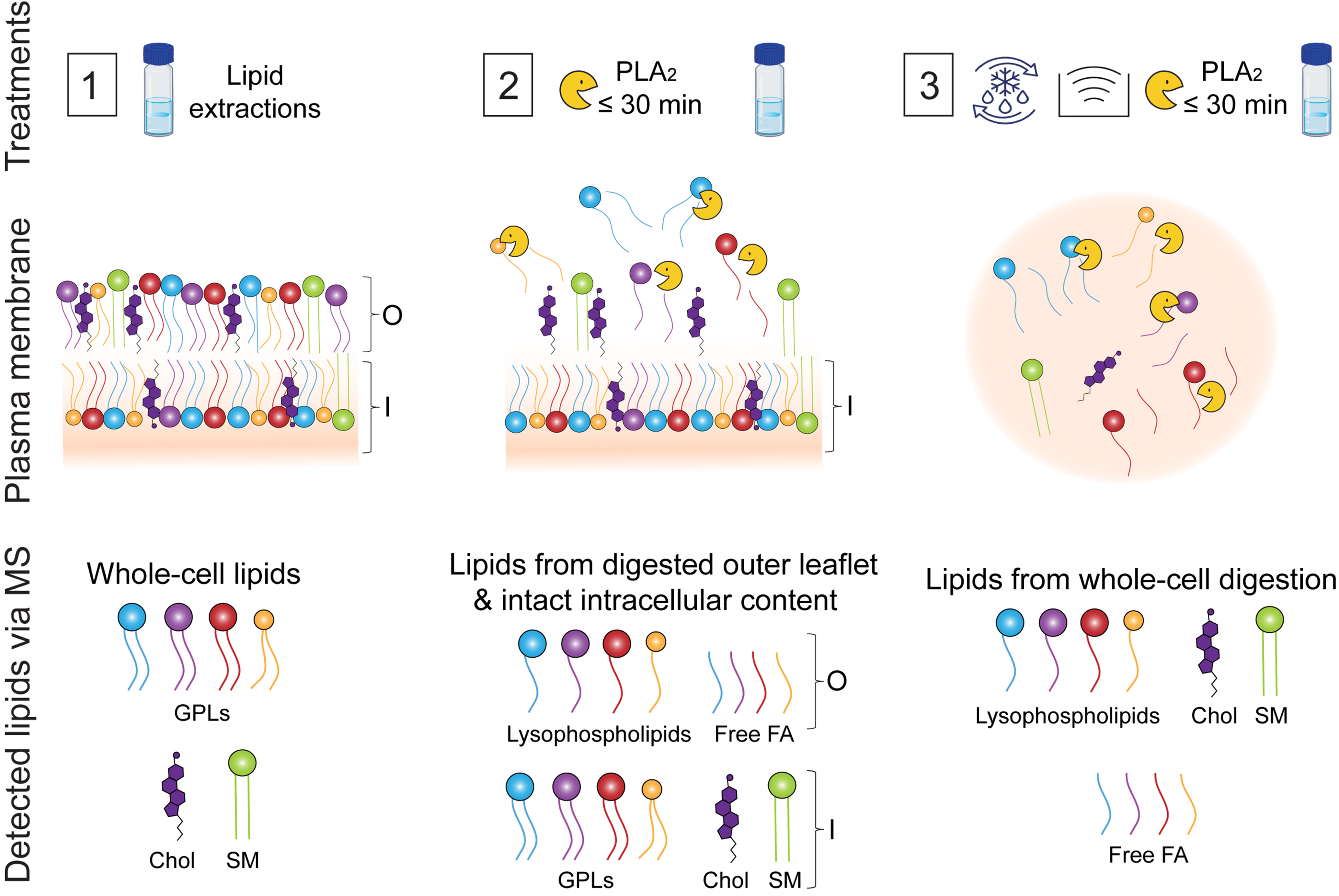
Figure 3. Schematic overview of treatments applied to melanoma cells prior to lipid extraction and mass spectrometry analysis to obtain outer leaflet lipid composition. Treatment 1: Phospholipids from intact/undigested cells were extracted and analysed to determine the total cellular lipidome. Treatment 2: Cells were incubated with PLA2 for ≤ 30 min at 37 °C to selectively digest phospholipids from the outer leaflet of the plasma membrane, while preserving the integrity of the inner leaflet, which was verified using a LDH assay (not illustrated). Extracts contain a mixture of GPLs from the plasma membrane inner leaflet and from cell organelles; lysophospholipids and free FAs from digested GPLs of the outer leaflet, and Chol and SM. Treatment 3: cells were subjected to freeze–thaw cycles, sonicated and fully digested with PLA2 to confirm enzymatic activity and ability to digest all the GPL classes. Treatment 3 contains lysophospholipids, FA and Chol and SM. The lipid composition of the outer leaflet (O) was obtained by subtracting the lipid profile of Treatment 2 from that of Treatment 1. This is an original figure created using Adobe Illustrator (Version 30.8). PLA2: Phospholipase A2; LDH: lactate dehydrogenase; GPLs: glycerophospholipids; FAs: fatty acids; Chol: cholesterol; SM: sphingomyelin.