
fig1
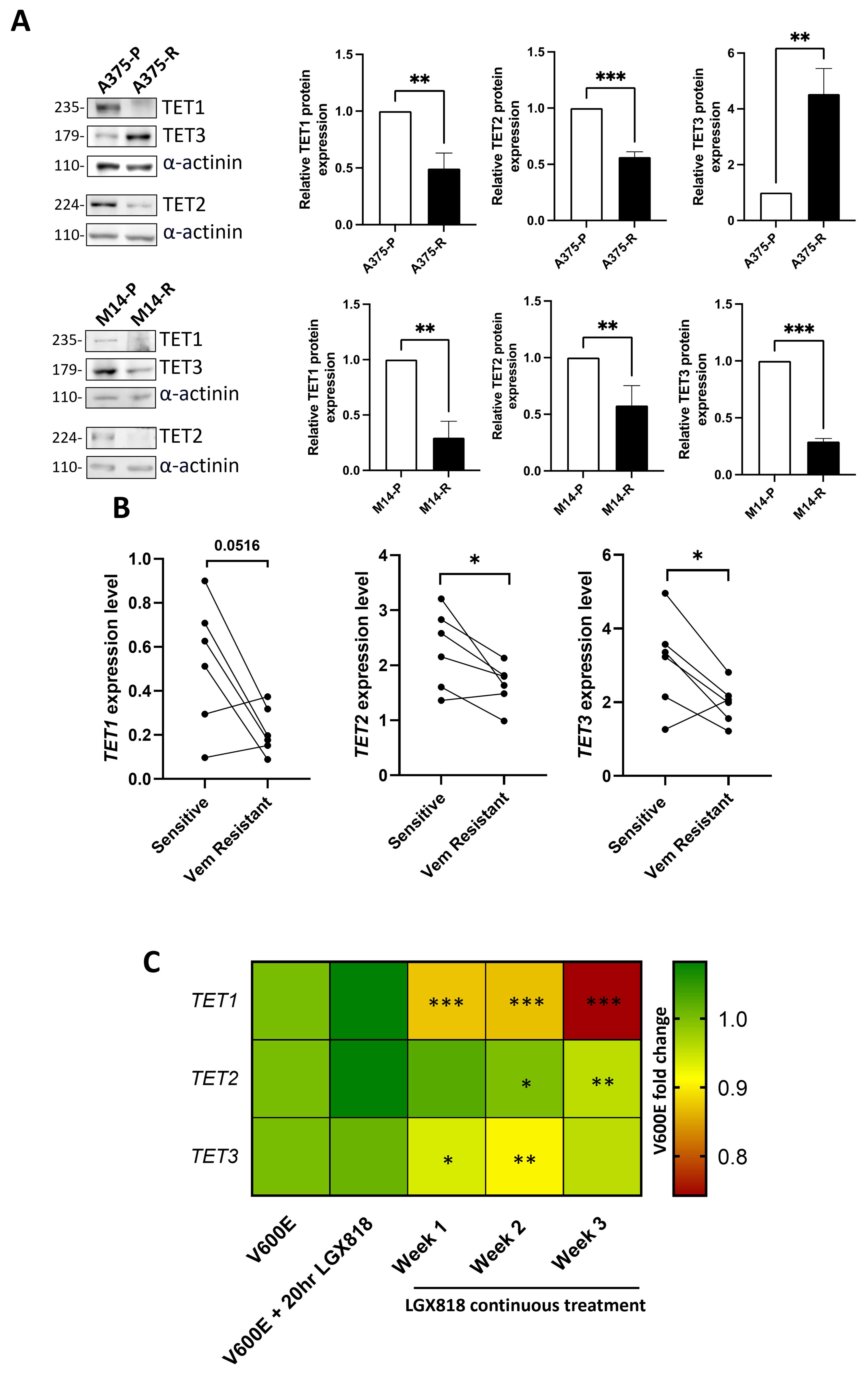
Figure 1. Analysis of TET expression levels in melanoma cells resistant to BRAFi. (A) Western blot of TET proteins in A375 and M14 dabrafenib-sensitive and -resistant cells; images are representative of three independent western blots that were quantified and shown in densitometric analysis. n = 3 independent biological replicates were performed. Statistical analysis was performed by unpaired t-test; (B) Expression levels of TET genes from RNA-seq of different melanoma cell lines resistant to vemurafenib; statistical analysis was performed by paired t-test; (C) Expression levels of TET genes from RNA-seq of a melanoma cell line overexpressing BRAFV600E and treated with encorafenib/LGX818; statistical significance was referred to V600E-overexpressing cells and assessed by one-way ANOVA. *P < 0.05; **P < 0.01; ***P < 0.001. TET: Ten-Eleven Translocation; BRAFi: BRAF inhibitors; ANOVA: analysis of variance.