


fig11
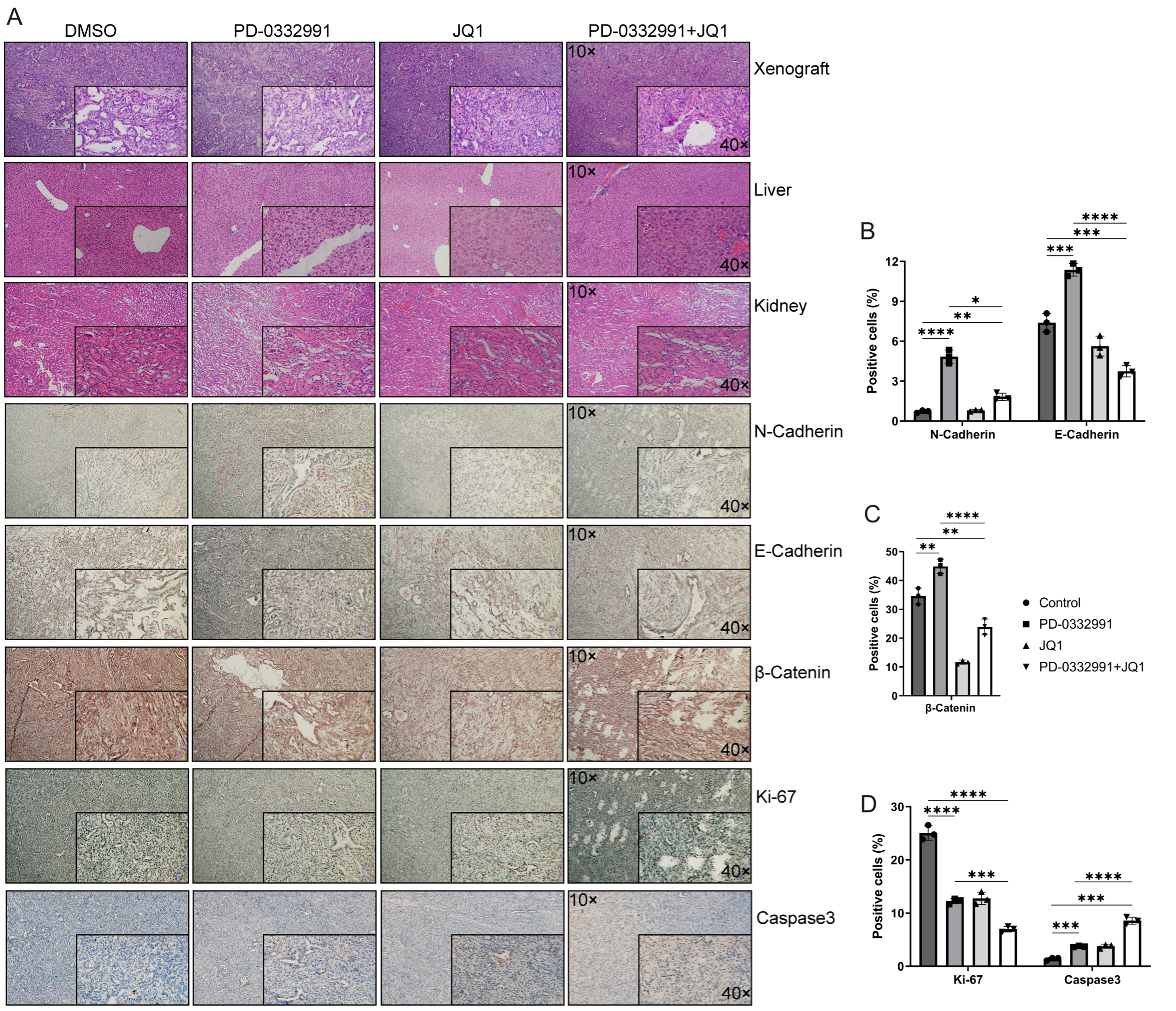
Figure 11. Morphological evaluation of xenograft tumors and liver and kidney tissues in nude mice using HE staining and IHC. (A) Nude mice were treated with PD-0332991 and JQ1. Primary antibodies against E-cadherin, N-cadherin, Ki-67, caspase-3, and active (non-phosphorylated) β-catenin were used for IHC analysis of xenograft tissues. Scale bars, 100 µm, 50 µm; (B-D) Quantitative analysis of E-cadherin, N-cadherin, active (non-phosphorylated) β-catenin, Ki-67, and Caspase-3 expression levels in xenograft tissues. Data are presented as mean ± SEM from three independent experiments. *P < 0.05, **P < 0.01, ***P < 0.001, ****P < 0.0001; ns: not significant. HE: Hematoxylin and eosin; IHC: immunohistochemistry; SEM: standard error of the mean.


