

Case Report: Sequential transcatheter treatment of coronary artery fistula, aneurysmal atrial septal artery and mitral regurgitation
 0
0 Graphical Abstract
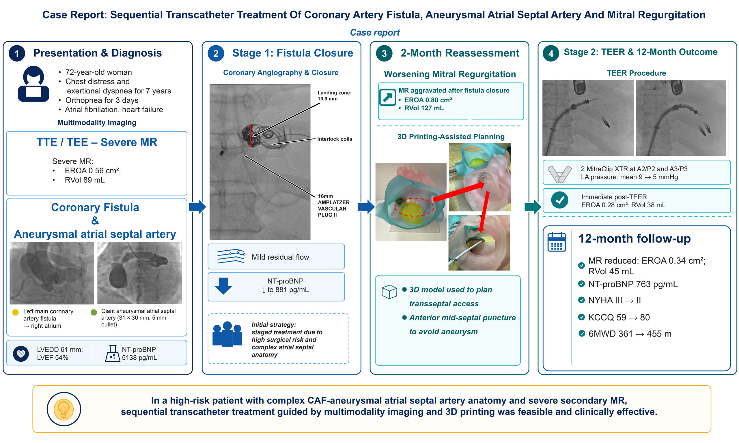
Abstract
Coronary artery fistulas are rare vascular anomalies that may cause chronic left-to-right shunting, cardiac chamber remodeling, and heart failure. We report a 72-year-old woman with a left main coronary artery-to-right atrium fistula draining through a giant aneurysmal atrial septal artery and accompanied by severe secondary mitral regurgitation. Echocardiography and cardiac computed tomography showed a 19 mm fistulous tract, a 31 × 30 mm aneurysmal vascular structure with a 5 mm outlet to the right atrium, and severe mitral regurgitation with an effective regurgitant orifice area of 0.56 cm2 and a regurgitant volume of
Keywords


 Download PDF
INTRODUCTION
Coronary artery fistulas are rare vascular anomalies characterized by abnormal communication between a coronary artery and a cardiac chamber or major vessel, with a reported prevalence of approximately 0.05%-0.25% on conventional coronary angiography and up to 0.9% on coronary computed tomography angiography[1]. Depending on the fistula size, drainage site, and shunt volume, untreated medium or large fistulas may lead to coronary steal, chronic volume overload, chamber enlargement, myocardial ischemia, pulmonary hypertension, arrhythmia, and heart failure[2]. Aneurysmal atrial septal artery is uncommon and may further complicate the anatomy when associated with coronary artery fistula. In this setting, long-standing left-to-right shunting and chamber remodeling may contribute to secondary mitral regurgitation even in the absence of primary mitral leaflet pathology[3].
Surgical repair remains an established treatment option for complex coronary artery fistulas; however, with advances in transcatheter equipment and techniques, percutaneous closure has become an alternative in selected patients, particularly when surgical risk is high and the anatomy is suitable for device closure[2]. In patients with concomitant severe mitral regurgitation, treatment sequencing requires careful assessment because mitral regurgitation may improve, persist, or worsen after correction of the primary shunt lesion. Here, we report a rare case of left coronary artery fistula associated with a giant aneurysmal atrial septal artery and severe mitral regurgitation, managed with sequential transcatheter fistula closure and transcatheter edge-to-edge repair supported by multimodality imaging and 3D printing.
CASE PRESENTATION
A 72-year-old female patient presented with a 7-year history of chest distress and exertional dyspnea. Over the preceding 3 days, her symptoms had worsened, with orthopnea and mild bilateral pitting edema. On admission, her blood pressure was 104/64 mm Hg, heart rate was 105 beats per minute, respiratory rate was 22 breaths per minute, and oxygen saturation was 99%. Physical examination revealed a continuous grade IV/VI murmur at the left sternal border and moist rales over both lower lung fields, consistent with heart failure. The hepatojugular reflux sign was negative. Electrocardiography showed atrial fibrillation with a ventricular rate of 90-130 bpm. The N-terminal pro-B-type natriuretic peptide (NT-proBNP) level was elevated at 5,138 pg/mL.
Transthoracic echocardiography revealed enlargement of the left atrium, right atrium, and left ventricle, with a left atrial diameter of 77 × 94 mm, right atrial diameter of 43 × 76 mm, left ventricular end-diastolic diameter of 61 mm, left ventricular end-systolic diameter of 48 mm, and left ventricular ejection fraction of 54% measured by the modified Simpson’s biplane method. Severe mitral regurgitation was mainly related to poor leaflet coaptation, with an effective regurgitant orifice area of 0.56 cm2 and a regurgitant volume of
Transesophageal echocardiography demonstrated an enlarged left main coronary artery with a 19 mm fistulous tract and a large interatrial lesion measuring 31 × 30 mm, which was identified as an aneurysmal atrial septal artery rather than an intracardiac tumor or thrombus. The aneurysm had a 5 mm outflow to the right atrium. No thrombus was detected in the left atrial appendage, and no pericardial effusion was observed. Three-dimensional reconstruction further confirmed the presence of a giant aneurysmal atrial septal artery [Figure 1].
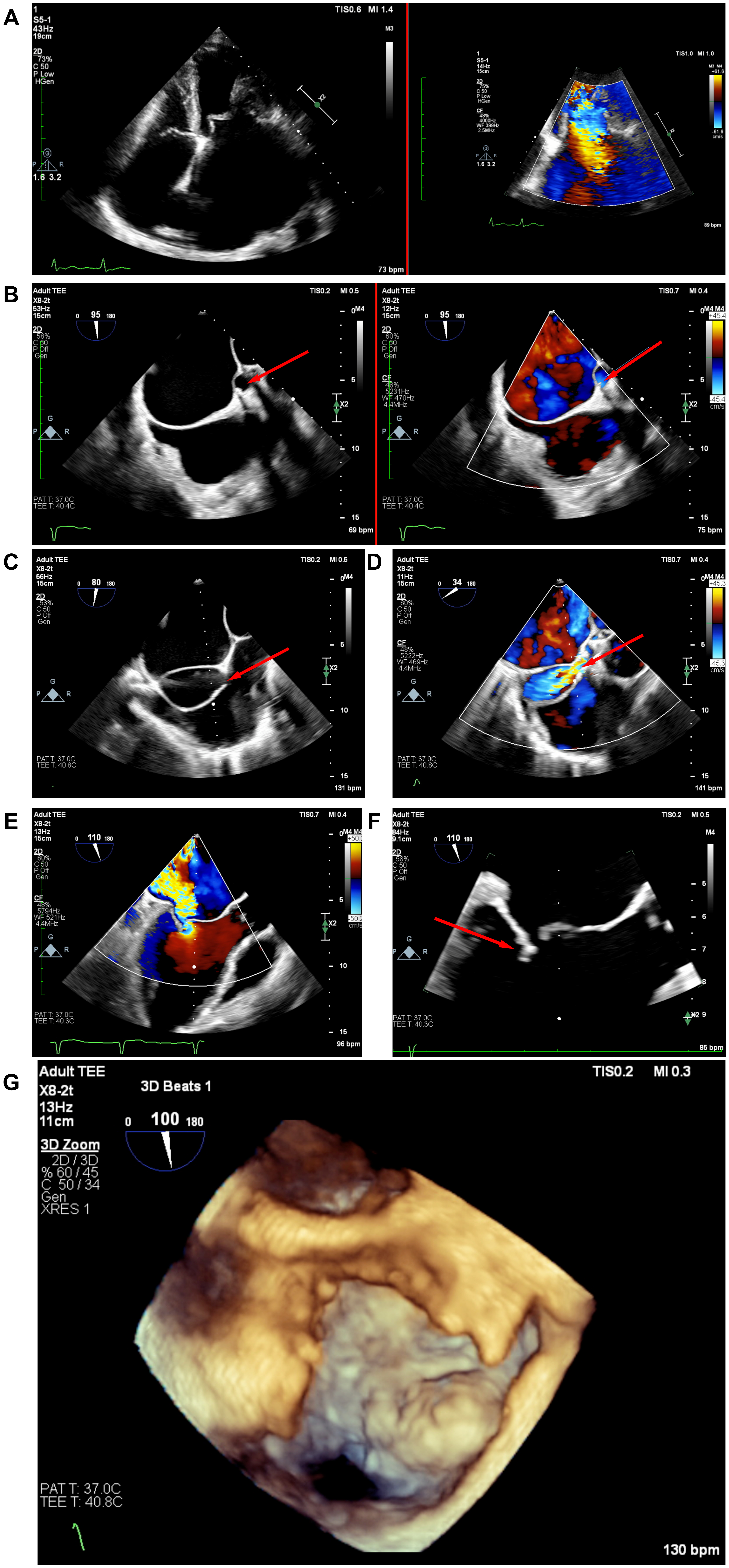
Figure 1. Baseline echocardiographic findings. (A) Apical four-chamber transthoracic echocardiography (TTE) and color Doppler demonstrate severe mitral regurgitation (MR); (B) Two-dimensional and color Doppler transesophageal echocardiography (TEE) delineate the enlarged left main coronary artery (LMCA) and the 19-mm fistulous tract leading to the aneurysmal atrial septal artery. The red arrow indicates the giant coronary artery fistula; (C) TEE measures the approximately 5-mm outflow from the aneurysmal sac to the right atrium (RA), as indicated by the red arrow; (D) Color Doppler confirms flow through the outflow into the RA; (E) TEE color Doppler shows severe MR; (F) TEE demonstrates posterior mitral leaflet tethering, resulting in incomplete mitral leaflet coaptation, as indicated by the red arrow; (G) Three-dimensional TEE depicts the 31 × 30-mm aneurysmal sac.
Cardiac computed tomography confirmed a tortuous and markedly dilated coronary artery fistula communicating with the right atrium through the giant aneurysmal atrial septal artery, which was located on the right lateral aspect of the atrial septum [Figure 2]. Coronary angiography showed no significant obstructive coronary artery disease and confirmed a left main coronary artery-to-right atrium fistula [Figure 3].
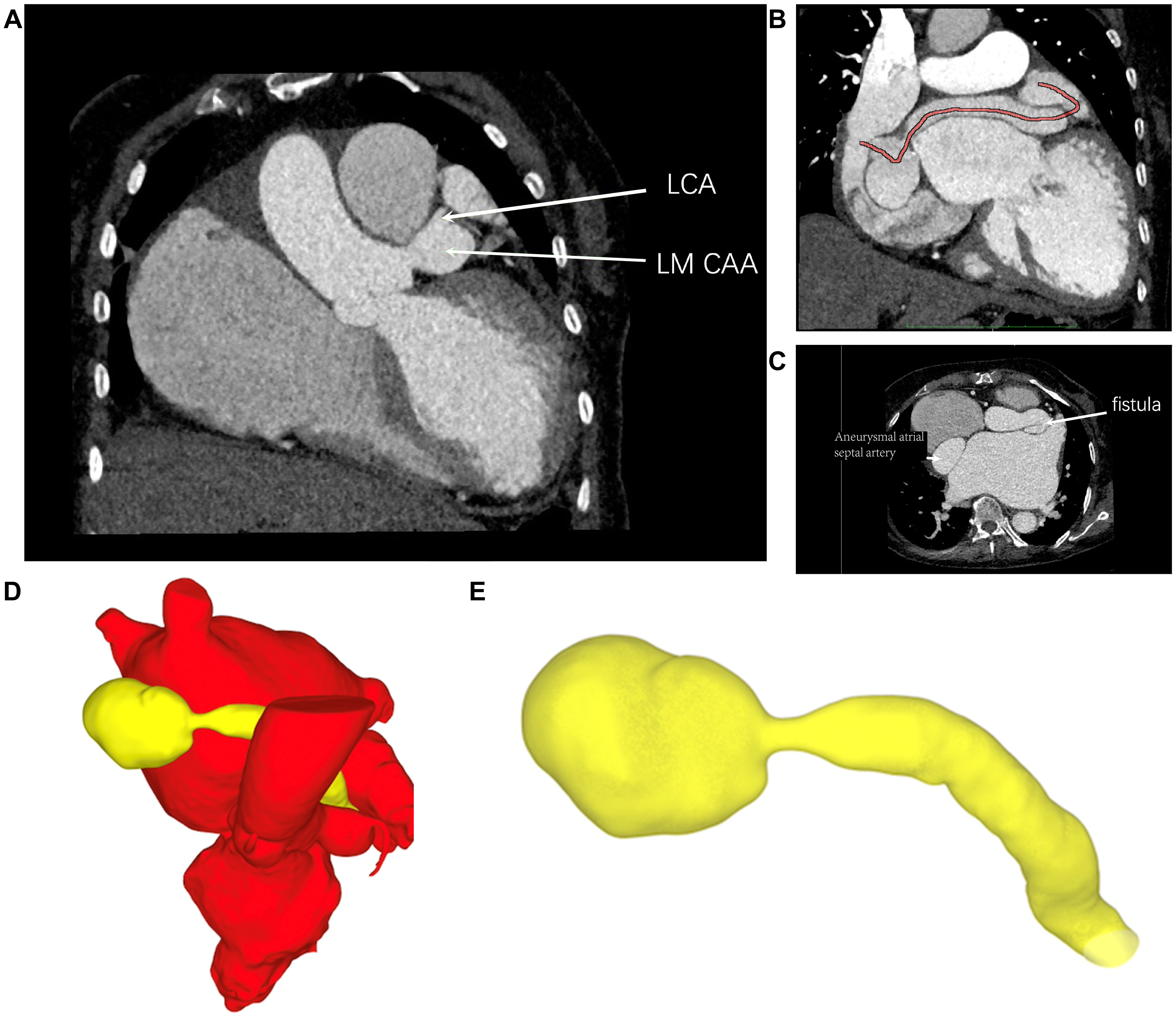
Figure 2. Cardiac computed tomography (CT) and three-dimensional reconstruction. (A) Cardiac CT identifies the dilated left coronary artery/left main coronary artery aneurysm and the proximal fistulous communication with the aneurysmal atrial septal artery; (B) Curved multiplanar CT reconstruction traces the tortuous course of the coronary artery fistula; (C) Axial CT delineates the fistulous tract and aneurysmal sac; (D) Three-dimensional reconstruction demonstrates the spatial relationship of the fistula and aneurysmal sac to the cardiac structures; (E) Isolated three-dimensional reconstruction shows the complete fistulous tract and giant aneurysmal sac. LCA: Left coronary artery; LM CAA: left main coronary artery aneurysm.
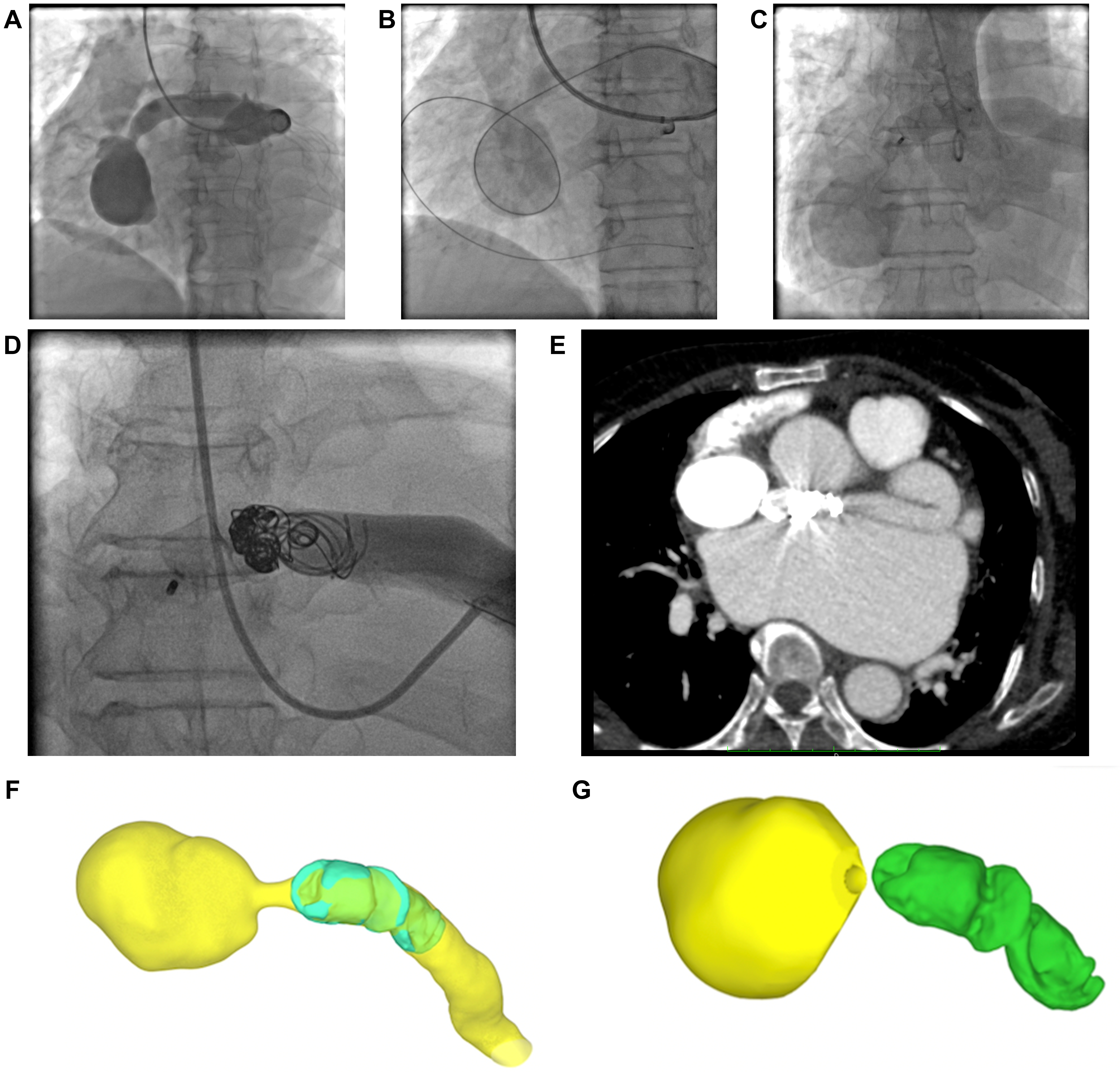
Figure 3. Transcatheter closure of the coronary artery fistula. (A) Coronary angiography demonstrates the LMCA-to-RA fistula through the aneurysmal sac; (B) A guidewire is advanced to establish the delivery rail; (C) Angiography after deployment of an AMPLATZER VASCULAR PLUG II shows residual shunting; (D) Final angiography after placement of multiple Interlock coils shows no obvious residual shunt in this projection; (E) Postprocedural CT demonstrates the occlusion devices in situ; (F and G) Three-dimensional renderings show the relationship of the devices to the fistulous tract and aneurysmal sac. CT: Computed tomography; LMCA: left main coronary artery; RA: right atrium.
Transcatheter closure of the fistula
Because of the patient’s high surgical risk and complex aneurysmal atrial septal anatomy, the multidisciplinary heart team selected a staged transcatheter strategy rather than simultaneous fistula closure and mitral valve intervention. The mitral regurgitation was considered predominantly secondary to long-standing volume overload and chamber remodeling; therefore, fistula closure followed by guideline-directed medical therapy and reassessment of mitral regurgitation was considered reasonable. In addition, immediate transcatheter edge-to-edge repair was avoided because the giant aneurysmal atrial septal artery increased the potential risk of transseptal puncture-related injury and acute left-to-right shunting.
Transcatheter closure was performed using an AMPLATZER VASCULAR PLUG II device (Abbott Medical Inc.) and multiple Interlock coils [Figure 3]. A 16 mm vascular plug was selected according to the angiographic landing zone diameter of 10.9 mm, allowing stable occlusion of the main fistulous tract while avoiding compromise of adjacent coronary branches. After plug deployment, coil embolization was performed to treat residual flow through small residual channels. Final angiography confirmed satisfactory closure of the main fistula with mild residual flow and no evidence of distal coronary flow compromise. The patient was discharged on postoperative day 7 with anticoagulation and guideline-directed heart failure therapy, including sacubitril/valsartan, metoprolol, dapagliflozin, spironolactone, and furosemide. Her symptoms improved, and NT-proBNP decreased to 881 pg/mL.
Follow-up
At 2 months after fistula closure, transthoracic echocardiography showed aggravation of mitral regurgitation, with an effective regurgitant orifice area of 0.80 cm2 and a regurgitant volume of 127 mL [Figure 4]. Given the persistent severe mitral regurgitation despite medical therapy, the heart team decided to proceed with transcatheter edge-to-edge repair.
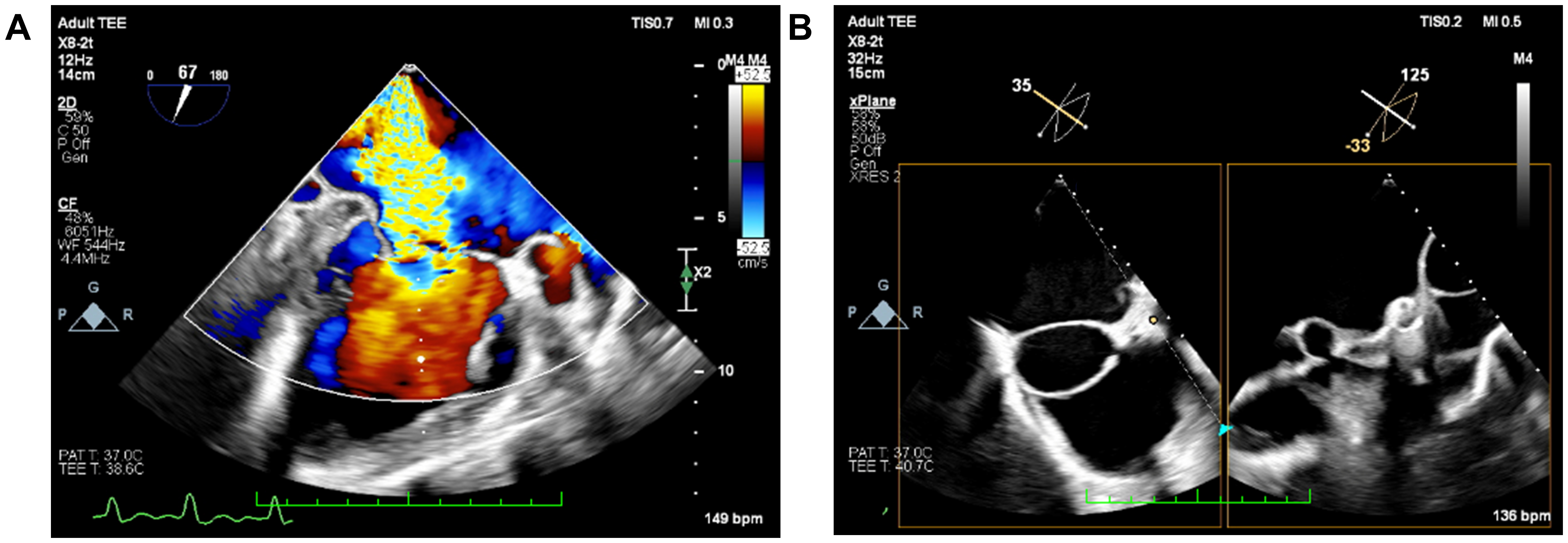
Figure 4. Two-month follow-up after fistula closure. (A) TEE color Doppler shows persistent severe MR, with an effective regurgitant orifice area of 0.80 cm2 and a regurgitant volume of 127 mL. (B) Biplane TEE demonstrates the persistent giant aneurysmal atrial septal artery. MR: Mitral regurgitation; TEE: transesophageal echocardiography.
Transcatheter edge-to-edge repair
To minimize the risk associated with transseptal access, a 3D-printed heart model was used for preprocedural planning. The model showed that the aneurysm was located posteriorly and superiorly along the atrial septum; therefore, an anterior and mid-septal puncture trajectory was selected to avoid the aneurysmal segment. Two MitraClip XTR devices were deployed at the A2/P2 and A3/P3 segments of the mitral valve. The XTR system was selected to facilitate leaflet grasping in the setting of severe secondary mitral regurgitation with poor leaflet coaptation. Immediately after clip implantation, mitral regurgitation was reduced, with an effective regurgitant orifice area of 0.28 cm2 and a regurgitant volume of 38 mL. The left atrial pressure decreased from 16/3 mm Hg, mean 9 mm Hg, to 8/2 mm Hg, mean 5 mm Hg [Figure 5].

Figure 5. Three-dimensional printing-guided transcatheter edge-to-edge repair (TEER). (A) The three-dimensional reconstruction, physical 3D-printed model, and bench simulation are used to select and verify a safe transseptal puncture region away from the aneurysmal atrial septal artery. The red arrow indicates the planned puncture region; (B) Biplane TEE confirms tenting at the selected puncture site; (C) Three-dimensional TEE confirms the transseptal access location relative to the aneurysmal sac; (D) Biplane TEE shows successful leaflet capture and two MitraClip XTR devices at A2/P2 and A3/P3; (E) Color Doppler after clip deployment shows mild residual MR; (F and G) En face three-dimensional TEE views demonstrate the two clips and the final double-orifice mitral valve configuration. 3D: Three-dimensional; MR: mitral regurgitation; TEE: transesophageal echocardiography.
Outcome and follow-up
At 12-month follow-up, transthoracic echocardiography showed sustained reduction of mitral regurgitation compared with the pre-transcatheter edge-to-edge repair (pre-TEER) status, with an effective regurgitant orifice area of 0.34 cm2 and a regurgitant volume of 45 mL [Figure 6]. NT-proBNP further decreased to 763 pg/mL. The patient’s New York Heart Association functional class improved to class II, Kansas City Cardiomyopathy Questionnaire score increased from 59 to 80, and 6-minute walk distance improved from 361 meters to 455 meters, reflecting enhanced functional capacity and quality of life.
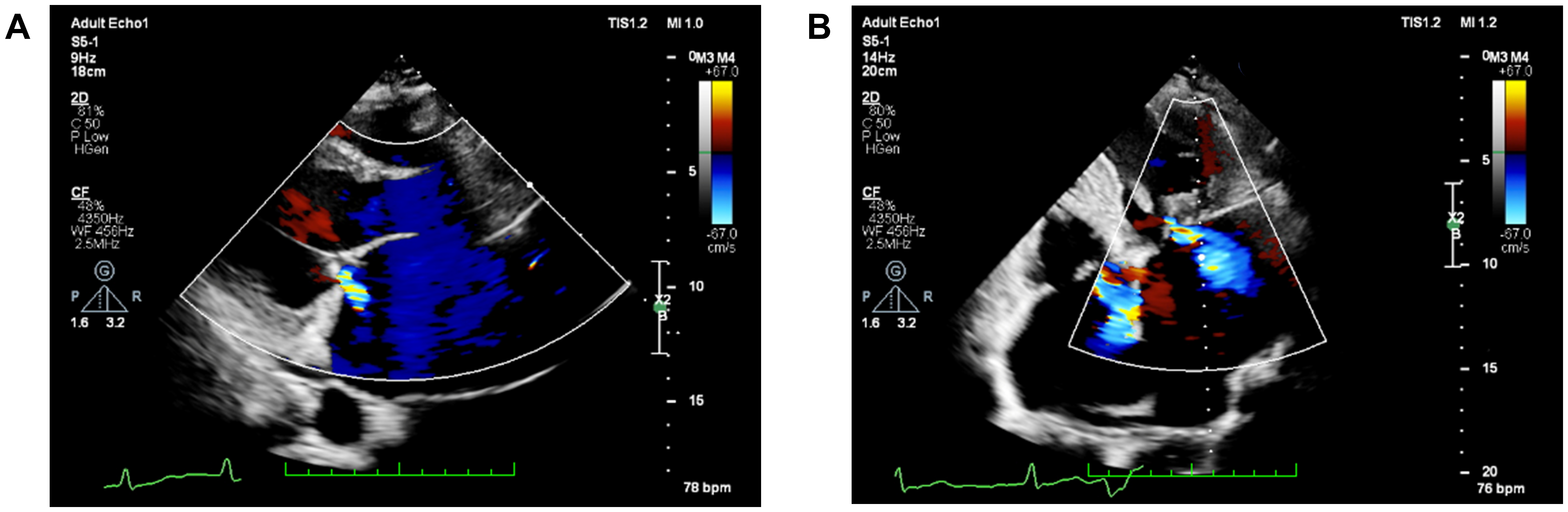
Figure 6. Twelve-month follow-up after TEER. (A) Parasternal long-axis and (B) apical four-chamber TTE color Doppler views show only mild residual MR, confirming sustained reduction compared with the preprocedural status. MR: Mitral regurgitation; TEER: transcatheter edge-to-edge repair; TTE: transthoracic echocardiography.
DISCUSSION
This case illustrates the staged transcatheter management of a rare and complex condition involving a left main coronary artery-to-right atrium fistula, a giant aneurysmal atrial septal artery, and severe secondary mitral regurgitation. The main value of this case lies in the combination of unusual anatomy, aggravation of mitral regurgitation after fistula closure, and subsequent transcatheter edge-to-edge repair guided by multimodality imaging and 3D printing.
Medium or large coronary artery fistulas may cause chronic left-to-right shunting, coronary steal, chamber enlargement, pulmonary hypertension, arrhythmia, and heart failure[2]. In the present case, long-standing left-to-right shunting was associated with marked enlargement of the left atrium, right atrium, and left ventricle, elevated pulmonary artery pressure, atrial fibrillation, elevated NT-proBNP, and severe mitral regurgitation due to poor leaflet coaptation. These findings supported a secondary mechanism of mitral regurgitation related to chronic chamber remodeling rather than isolated primary leaflet disease[3]. Severe heart failure related to large coronary cameral fistulas has been described in previous reports, although coexistence with a giant aneurysmal atrial septal artery and dynamic worsening of secondary mitral regurgitation after fistula closure remains uncommon[4,5].
The decision to adopt a staged strategy was based on both pathophysiological and procedural considerations. Because the mitral regurgitation was considered predominantly secondary to chronic volume overload and chamber remodeling, initial closure of the primary shunt lesion followed by guideline-directed medical therapy and reassessment was considered reasonable. In addition, simultaneous transcatheter edge-to-edge repair was not favored because the giant aneurysmal atrial septal artery increased the risk of transseptal puncture-related injury and acute left-to-right shunting. This rationale was particularly relevant in the present case, in which safe transseptal access was a major procedural concern.
The worsening of mitral regurgitation after fistula closure is an important observation in this case. One possible explanation is that elimination of the low-resistance shunt altered left-sided loading conditions and unmasked or aggravated pre-existing secondary mitral regurgitation. However, this interpretation remains inferential rather than definitive because right heart catheterization and Qp/Qs measurement were not performed. Therefore, the proposed mechanism was supported mainly by the temporal relationship, serial echocardiographic findings, and changes in clinical and biomarker status. This point is important because the case should be interpreted as an individualized treatment experience rather than definitive evidence of causality.
Precise imaging was essential for both stages of treatment. Echocardiography defined the severity and mechanism of mitral regurgitation, cardiac computed tomography delineated the fistula course and its relationship to the aneurysmal atrial septal artery, and coronary angiography guided device selection and closure. The 16 mm vascular plug was selected according to the 10.9 mm angiographic landing zone, with coil embolization used for residual small channels. Prior experience with device-based closure of giant high-flow vascular fistulas also supports the broader procedural principle that occluder selection should be individualized according to fistula anatomy and flow characteristics, although coronary fistulas require additional attention to preservation of distal coronary perfusion[6].
The 3D-printed model provided information beyond routine two-dimensional imaging by allowing direct assessment of the spatial relationship between the aneurysmal atrial septal artery and the planned transseptal route. In this case, the model showed that the aneurysm was located posteriorly and superiorly along the atrial septum, supporting an anterior and mid-septal puncture trajectory to avoid the aneurysmal segment. This helped reduce procedural uncertainty before transcatheter edge-to-edge repair. Similar applications of CT-based 3D modeling have been reported in complex fistula closure and transcatheter structural heart interventions[7-9].
Alternative strategies included surgical fistula ligation with aneurysm repair and mitral valve surgery, hybrid treatment, or simultaneous transcatheter intervention. Surgery may provide definitive anatomical correction in selected patients; however, it was not favored in this patient because of advanced age, heart failure, atrial fibrillation, recent clinical deterioration, and complex anatomy. Simultaneous transcatheter treatment was also avoided due to the anticipated risk of transseptal access in the presence of a giant aneurysmal atrial septal artery. The favorable 12-month clinical, echocardiographic, and biomarker outcomes suggest that sequential transcatheter treatment may be feasible in carefully selected high-risk patients.
This case has several limitations. First, it is a single case report and cannot establish generalizable treatment recommendations. Second, invasive hemodynamic assessment, including right heart catheterization and Qp/Qs measurement, was not performed. Finally, although 12-month follow-up showed sustained clinical improvement and reduced mitral regurgitation compared with the pre-TEER status, longer follow-up is needed to assess residual mitral regurgitation, residual fistula flow, and long-term cardiac remodeling.
CONCLUSION
In summary, this case demonstrates that sequential transcatheter treatment may be feasible for selected high-risk patients with coronary artery fistula, giant aneurysmal atrial septal artery, and severe secondary mitral regurgitation. Closure of the primary shunt lesion, followed by reassessment and transcatheter edge-to-edge repair, was associated with symptomatic, echocardiographic, and biomarker improvement at 12 months. Multimodality imaging and 3D printing were useful for defining complex anatomy, guiding device-based treatment, and reducing uncertainty during transseptal procedural planning.
DECLARATIONS
Authors’ contributions
Drafted the manuscript and contributed equally to this work: Huang Y, Chen Z
Contributed to data collection and imaging preparation: Zhu Z
Participated in clinical management and manuscript revision: Tang J
Supervised the case management, revised the manuscript critically, and approved the final version: Fang Z
All authors read and approved the final manuscript.
Availability of data and materials
The data supporting the findings of this case report are available from the corresponding author upon reasonable request, with appropriate protection of patient privacy.
AI and AI-assisted tools statement
During the preparation of this manuscript, the AI tool OpenAI ChatGPT powered by GPT-5.5 (version 5.5, released on 2026-04-23) was used solely for language editing. The tool did not influence the study design, data collection, analysis, interpretation, or the scientific content of the work. All authors take full responsibility for the accuracy, integrity, and final content of the manuscript.
Financial support and sponsorship
This work was supported by the Noncommunicable Chronic Diseases-National Science and Technology Major Project (2024ZD0527106)
Conflicts of interest
All authors declared that there are no conflicts of interest.
Ethical approval and consent to participate
The study was conducted in accordance with the Declaration of Helsinki and was approved by the Research Ethics Committee of The Second Xiangya Hospital, Central South University [Approval No. (2022)115]. Written informed consent for clinical management was obtained from the patient.
Consent for publication
Written informed consent for publication of the clinical details and accompanying images was obtained from the patient.
Copyright
© The Author(s) 2026.
REFERENCES
1. Lim JJ, Jung JI, Lee BY, Lee HG. Prevalence and types of coronary artery fistulas detected with coronary CT angiography. AJR Am J Roentgenol. 2014;203:W237-43.
2. Al-Hijji M, El Sabbagh A, El Hajj S, et al. Coronary artery fistulas: indications, techniques, outcomes, and complications of transcatheter fistula closure. JACC Cardiovasc Interv. 2021;14:1393-406.
3. Huang AL, Dal-Bianco JP, Levine RA, Hung JW. Secondary mitral regurgitation: cardiac remodeling, diagnosis, and management. Struct Heart. 2023;7:100129.
4. Chirumamilla Y, Brar A, Belal F, McDonald P. Large coronary cameral fistula to the left ventricle presenting as congestive heart failure. Eur J Case Rep Intern Med. 2024;11:004364.
5. Suri RM, Okoh AK, Espinosa RE, Breen JF, Rehfeldt KH, Nishimura RA. Left main coronary artery to biatrial fistula associated with severe functional mitral valve regurgitation. Circulation. 2011;124:2456-7.
6. Chen Z, Huang Y, Ye C, et al. Case Report: transcatheter closure of a giant post-traumatic femoral arteriovenous fistula using ventricular septal occluder. Front Surg. 2023;10:1295595.
7. Echarte-Morales J, Toribio-García I, Diéguez AR, de Prado AP, Fernández-Vázquez F. Applications of three-dimensional printing in percutaneous closure of aortic-to-right ventricle fistula after transcatheter aortic valve replacement: a case report. Eur Heart J Case Rep. 2024;8:ytae112.
8. Ørbæk Andersen M, Smerup MH, Munk K, et al. Computed tomographic-based three-dimensional printing of giant coronary artery fistulas to guide surgical strategy: a case series. Eur Heart J Case Rep. 2024;8:ytad413.
Cite This Article
 Open Access
How to Cite
Huang Y, Chen Z, Zhu Z, Tang J, Fang Z. Case Report: Sequential transcatheter treatment of coronary artery fistula, aneurysmal atrial septal artery and mitral regurgitation. Vessel Plus. 2026;10:52. https://dx.doi.org/10.20517/2574-1209.2026.17
Download Citation
If you have the appropriate software installed, you can download article citation data to the citation manager of your choice. Simply select your manager software from the list below and click on download.
Export Citation File
Type of Import
Tips on Downloading Citation
Citation Manager File Format
Type of Import
Direct Import: When the Direct Import option is selected (the default state), a dialogue box will give you the option to Save or Open the downloaded citation data. Choosing Open will either launch your citation manager or give you a choice of applications with which to use the metadata. The Save option saves the file locally for later use.
Indirect Import: When the Indirect Import option is selected, the metadata is displayed and may be copied and pasted as needed.
About This Article
Special Topic
Copyright

Data & Comments
Data
0









Comments
Comments must be written in English. Spam, offensive content, impersonation, and private information will not be permitted. If any comment is reported and identified as inappropriate content by OAE staff, the comment will be removed without notice. If you have any queries or need any help, please contact us at [email protected].