

fig2
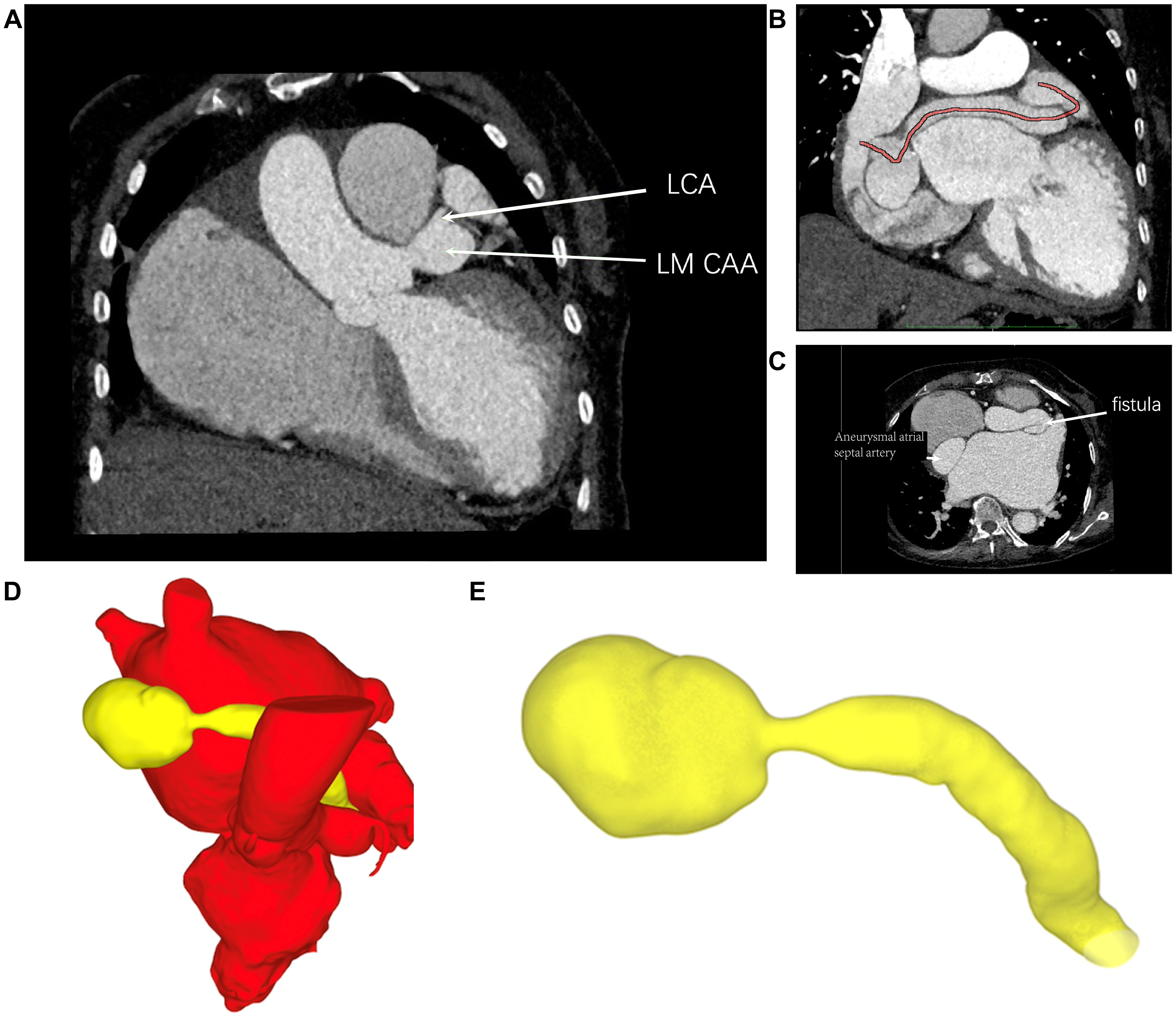
Figure 2. Cardiac computed tomography (CT) and three-dimensional reconstruction. (A) Cardiac CT identifies the dilated left coronary artery/left main coronary artery aneurysm and the proximal fistulous communication with the aneurysmal atrial septal artery; (B) Curved multiplanar CT reconstruction traces the tortuous course of the coronary artery fistula; (C) Axial CT delineates the fistulous tract and aneurysmal sac; (D) Three-dimensional reconstruction demonstrates the spatial relationship of the fistula and aneurysmal sac to the cardiac structures; (E) Isolated three-dimensional reconstruction shows the complete fistulous tract and giant aneurysmal sac. LCA: Left coronary artery; LM CAA: left main coronary artery aneurysm.



