

fig1
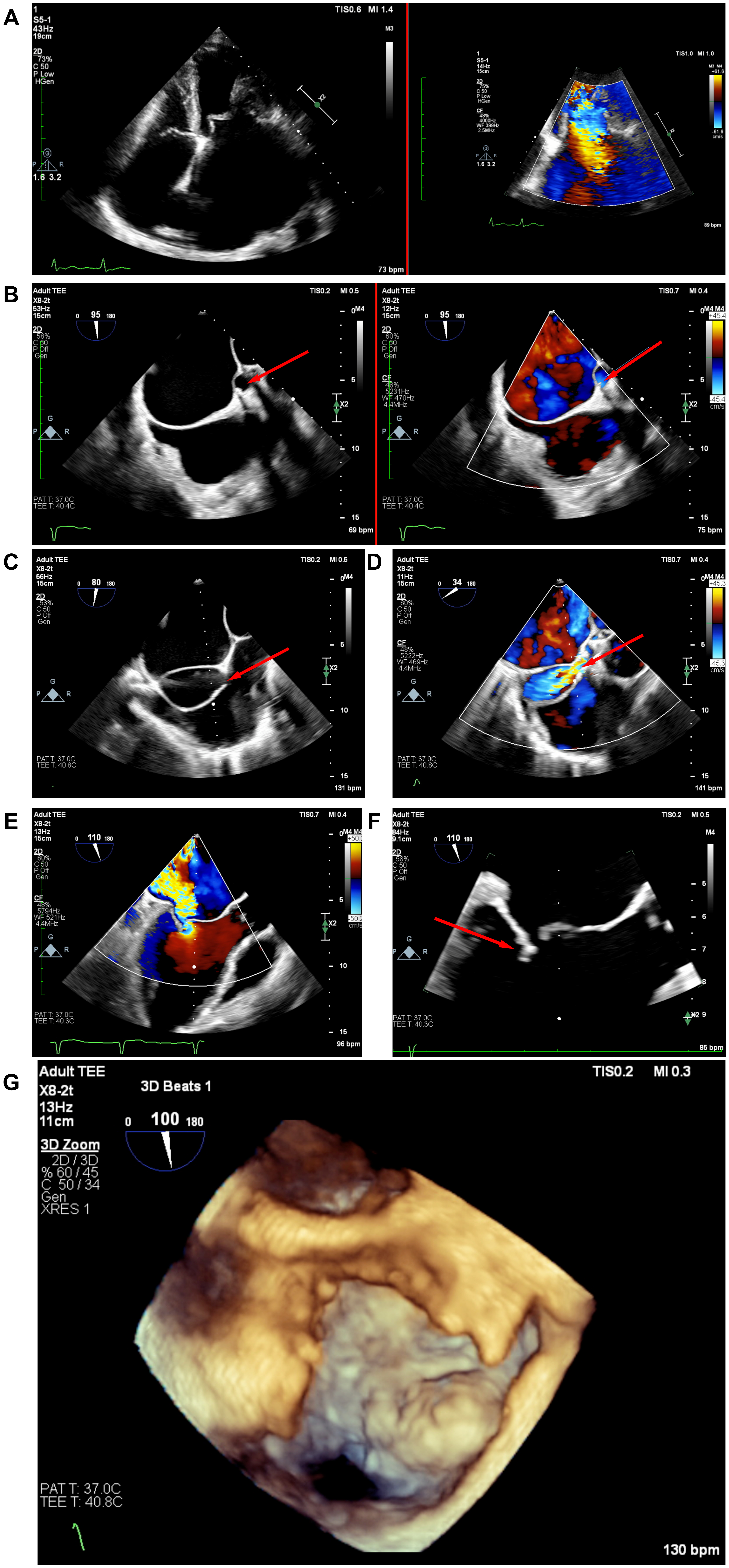
Figure 1. Baseline echocardiographic findings. (A) Apical four-chamber transthoracic echocardiography (TTE) and color Doppler demonstrate severe mitral regurgitation (MR); (B) Two-dimensional and color Doppler transesophageal echocardiography (TEE) delineate the enlarged left main coronary artery (LMCA) and the 19-mm fistulous tract leading to the aneurysmal atrial septal artery. The red arrow indicates the giant coronary artery fistula; (C) TEE measures the approximately 5-mm outflow from the aneurysmal sac to the right atrium (RA), as indicated by the red arrow; (D) Color Doppler confirms flow through the outflow into the RA; (E) TEE color Doppler shows severe MR; (F) TEE demonstrates posterior mitral leaflet tethering, resulting in incomplete mitral leaflet coaptation, as indicated by the red arrow; (G) Three-dimensional TEE depicts the 31 × 30-mm aneurysmal sac.



