

fig3
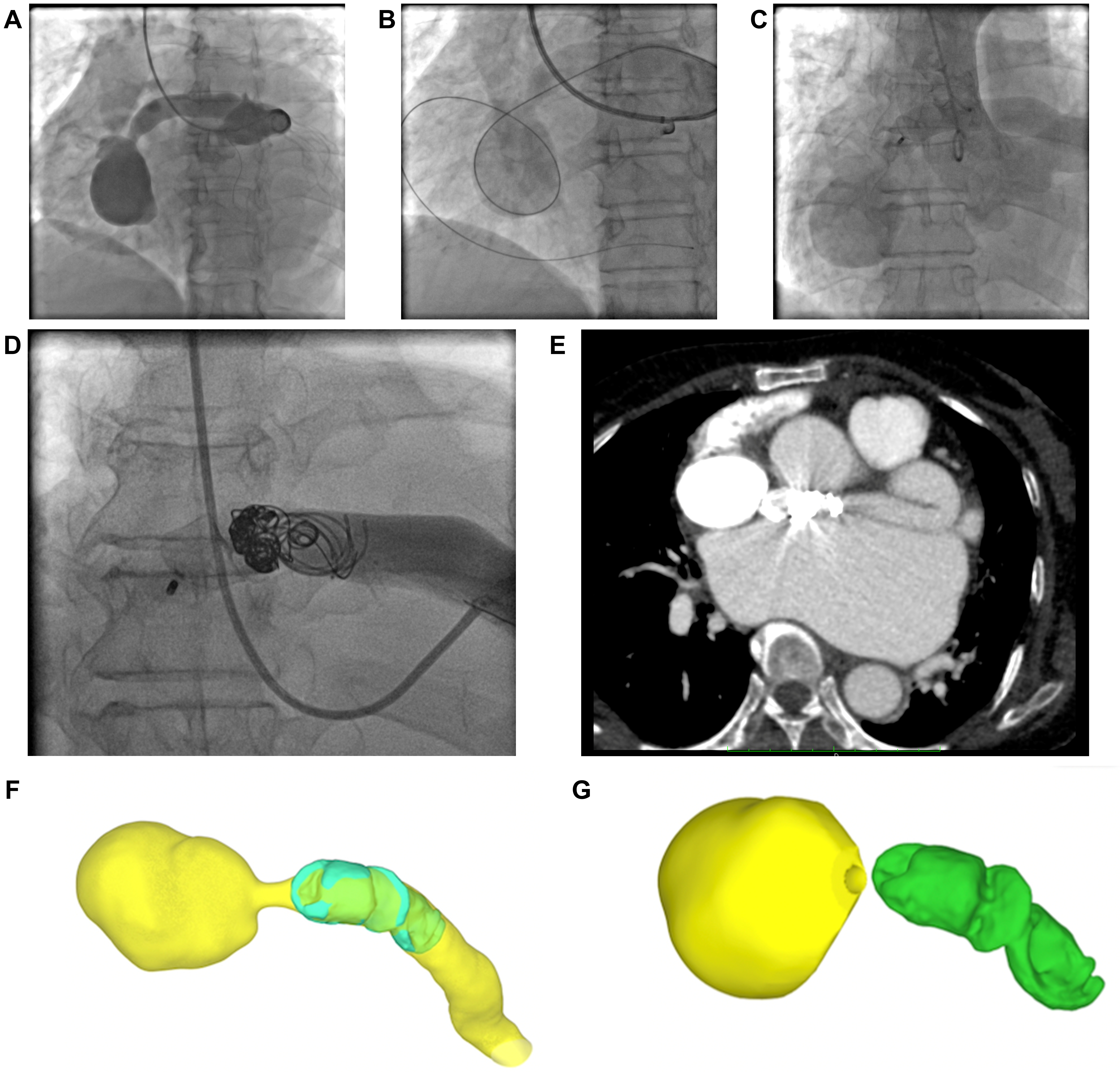
Figure 3. Transcatheter closure of the coronary artery fistula. (A) Coronary angiography demonstrates the LMCA-to-RA fistula through the aneurysmal sac; (B) A guidewire is advanced to establish the delivery rail; (C) Angiography after deployment of an AMPLATZER VASCULAR PLUG II shows residual shunting; (D) Final angiography after placement of multiple Interlock coils shows no obvious residual shunt in this projection; (E) Postprocedural CT demonstrates the occlusion devices in situ; (F and G) Three-dimensional renderings show the relationship of the devices to the fistulous tract and aneurysmal sac. CT: Computed tomography; LMCA: left main coronary artery; RA: right atrium.



