



ALDH2 modulates cardiac ischemic adaptation via mitochondrial DNMT1 translocation and dynamic mtDNA methylation remodeling
 0
0 Abstract
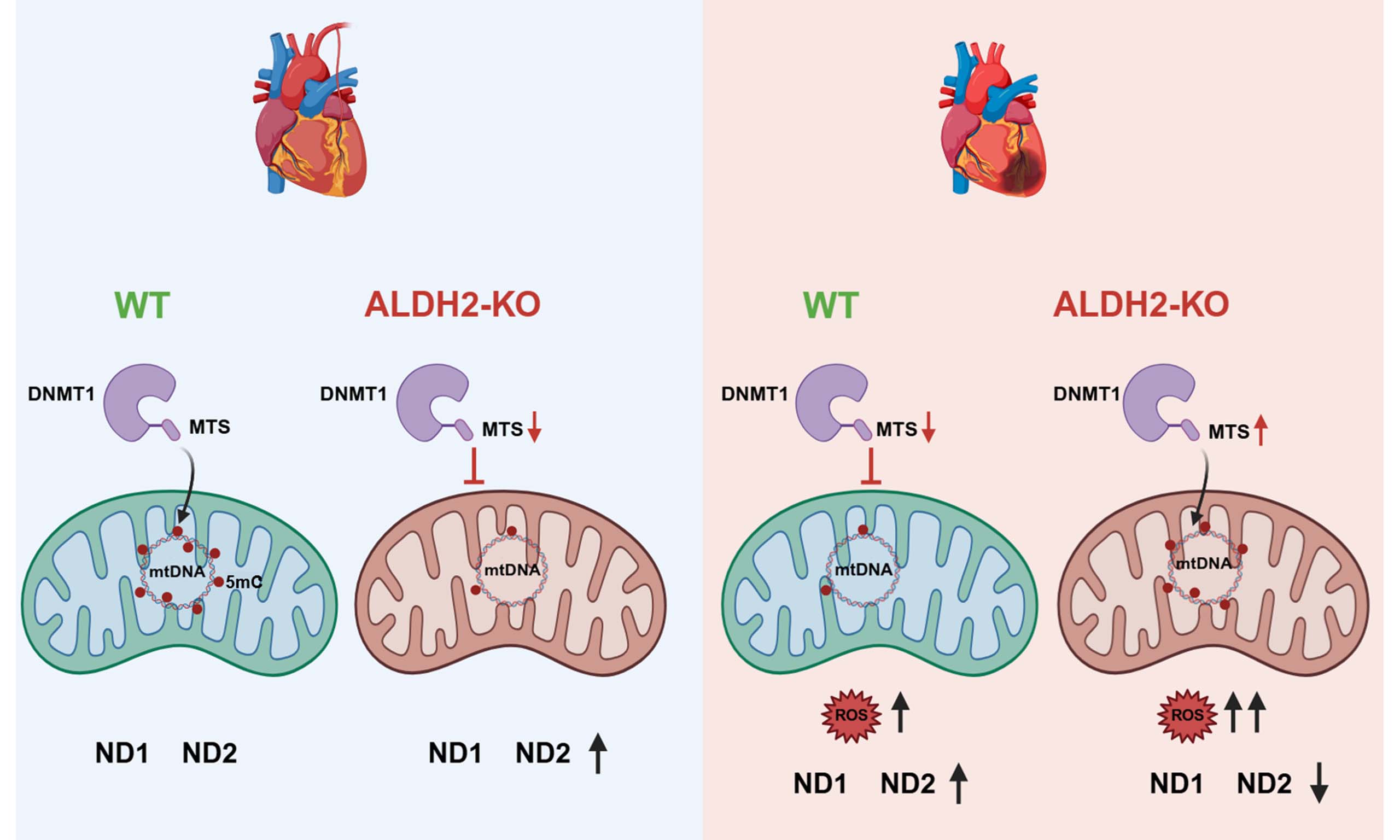
Aim: Mitochondrial DNA (mtDNA) methylation is an emerging epigenetic regulator of mitochondrial function, yet the mechanisms governing its dynamic regulation under cellular stress—particularly in the heart—remain poorly understood. This study aimed to investigate whether aldehyde dehydrogenase 2 (ALDH2) regulates mtDNA methylation via the mitochondrial targeting of DNA methyltransferase 1 (DNMT1) during myocardial ischemia, and to explore the functional consequences of this regulation in Ischemic heart disease (IHD).
Methods: We identified a specific DNMT1 isoform containing a mitochondrial targeting sequence (MTS) and confirmed its localization to cardiomyocyte mitochondria. Myocardial infarction (MI) was induced in wild-type and ALDH2-knockout mice. Mitochondrial DNMT1 levels, mtDNA 5-methylcytosine (5mC) content, and MTS expression were assessed at various post-MI time points. Site-specific mtDNA methylation at ND1 and ND2 loci was analyzed. Hypoxia experiments in cardiomyocytes were performed to evaluate ALDH2-dependent DNMT1 translocation, mitochondrial gene expression, and respiratory capacity. Post-MI cardiac function was assessed by echocardiography.
Results: We confirm that a specific DNMT1 isoform localizes to cardiomyocyte mitochondria via its MTS and modulates mtDNA 5mC levels. Following MI, mitochondrial DNMT1 levels and mtDNA methylation exhibited a biphasic response: an initial increase during the early ischemic phase (1 week post-MI), followed by a decline in the later remodeling phase. Genetic deletion of ALDH2 impaired mitochondrial translocation of DNMT1, associated with downregulated MTS expression and reduced global mtDNA 5mC. Site-specific analysis revealed that ALDH2 deficiency significantly altered methylation patterns at the ND1 and ND2 loci. Functionally, hypoxia inhibited DNMT1 mitochondrial translocation in an ALDH2-dependent manner, thereby modulating the expression of these mitochondrial-encoded genes and overall mitochondrial respiratory capacity. Importantly, ALDH2 deficiency exacerbated post-MI cardiac dysfunction.
Conclusion: Our findings uncover a novel regulatory axis in which ALDH2 fine-tunes the dynamic equilibrium of mtDNA methylation by governing DNMT1-MTS expression, offering mechanistic insight into mitochondrial epigenetic adaptation in IHD and a potential therapeutic target for ALDH2-deficient populations.
Keywords
INTRODUCTION
The mammalian mitochondrial genome is a compact, circular DNA molecule encoding 37 genes essential for oxidative phosphorylation (OXPHOS) and mitochondrial translation. Among these are 13 polypeptide subunits critical for the electron transport chain (ETC), alongside 2 ribosomal RNA and 22 transfer RNA genes[1]. While traditionally considered devoid of canonical epigenetic marks, compelling evidence now confirms the presence of 5-methylcytosine (5mC) at Cytosine-phosphate-Guanine (CpG) dinucleotides within mitochondrial DNA (mtDNA). This modification is implicated in the regulation of mitochondrial gene expression and bioenergetic adaptation[2]. DNA methyltransferase 1 (DNMT1), the principal enzyme responsible for maintaining DNA methylation patterns in the nucleus[3], harbors a mitochondrial targeting sequence (MTS) that facilitates its import into mitochondria[4]. Within this organelle, DNMT1 contributes to the establishment and maintenance of mtDNA methylation, although the functional consequences and regulatory mechanisms of this process are incompletely defined.
Ischemic heart disease (IHD) remains a leading cause of disability and mortality worldwide. It primarily arises from the pathological interplay between compromised coronary arteries and the myocardium, which depends on sufficient blood supply to maintain function. In IHD, increasing evidence highlights the mitochondrion as a central organelle mediating cardiomyocyte responses to ischemic injury. Maintaining a healthy mitochondrial population is crucial for cardiac homeostasis, as impaired mitochondria not only produce less adenosine triphosphate (ATP) but also generate excessive reactive oxygen species (ROS). During myocardial ischemia, substantial amounts of ROS are produced at the ubisemiquinone site of the mitochondrial electron transport chain in cardiomyocytes[5]. Accumulated ROS can inflict irreversible damage to mitochondrial components—including mtDNA, proteins, and lipids—ultimately triggering cell death[6]. Notably, mtDNA methylation influences genomic expression and may help mitigate ROS-induced mitochondrial damage. Therefore, elucidating the regulatory mechanisms of mtDNA methylation in response to mitochondrial stress is essential for understanding the progression of myocardial injury.
Previous studies have primarily focused on epigenetic regulation of the aldehyde dehydrogenase 2 (ALDH2) gene itself, rather than the role of ALDH2 protein in modulating epigenetic modifications. In a rat Myocardial infarction (MI) model, time-dependent hypermethylation of the ALDH2 promoter led to its downregulation[7]. Another study revealed that dapagliflozin reduces ALDH2 promoter methylation via the NHE1/ROS/DNMT1 pathway, directly implicating DNMT1 in the epigenetic control of ALDH2[8]. More broadly, mitochondrial epigenetics—particularly mtDNA methylation—is increasingly recognized as a key regulator of cardiac function, governing mtDNA transcription and mitochondrial respiration[9]; however, the underlying mechanisms remain unclear.
Our previous studies have demonstrated that ALDH2 plays a role in regulating the microenvironment and mitochondrial homeostasis, which is essential for myocardial adaptation and survival under ischemia or reperfusion conditions[10,11]. In the present study, we detected the translocation of DNMT1 into mitochondria and further elucidated its dynamic regulatory role in the 5mC modification of mtDNA in cardiomyocytes. Moreover, we investigated new evidence indicating that ALDH2 regulates mtDNA methylation in cardiomyocytes during different stages of ischemia. Given that a loss-of-function ALDH2 allele is present in nearly half of the East Asian population and approximately 8% of individuals worldwide, this study provides mechanistic insights and suggests potential therapeutic directions for patients carrying ALDH2 mutations.
METHODS AND MATERIALS
Animal model
Wild-type (WT) and global ALDH2-knockout (KO) mice on a C57BL/6J background (male, 8 weeks old) were housed under standard conditions (12-h light/dark cycle, 22 °C, ad libitum access to food and water). The MI model was induced by permanent ligation of the left anterior descending (LAD) coronary artery. Briefly, mice were anesthetized, and a left thoracotomy was performed to expose the heart, and the LAD was ligated at a point approximately one-third of the distance from its origin near the left atrial appendage with an 8-0 polypropylene suture. Successful occlusion was confirmed by visual blanching of the anterior left ventricular wall. Sham-operated control mice underwent identical surgical procedures, including thoracotomy and pericardial opening, without LAD ligation. Post-operative analgesia was administered, and animals were monitored until recovery. All animal experimental procedures were approved by the Animal Care and Use Committee of Zhongshan Hospital, Fudan University (approval No. ZSYY20170517), and were conducted in strict accordance with the National Institutes of Health Guide for the Care and Use of Laboratory Animals.
Primary adult cardiomyocyte isolation
For adult cardiomyocyte isolation, mice were anesthetized, the heart was rapidly excised, and the aorta was immediately cannulated on a blunted needle. It was then immediately perfused retrograde with warm, oxygenated buffer to minimize ischemia. The cannulated heart was suspended in a chamber. A syringe pump continuously perfused the heart sequentially with calcium-free perfusion buffer** (for ~4 min) to arrest the heart and remove blood. Then the heart was perfused with the digestion buffer containing a blend of Collagenase II (0.5 mg/mL) and Protease XIV (0.05 mg/mL) in calcium-free solution (for ~8-10 min) until the tissue softens. The softened ventricles were dissected away, minced, and gently triturated in a stop solution containing bovine calf serum to release individual cells. The cell suspension was filtered through a nylon mesh. Cells were pelleted and resuspended. Calcium concentration is gradually restored to physiological levels in steps to promote calcium tolerance in healthy cardiomyocytes. The rod-shaped, calcium-tolerant cardiomyocytes are then separated from rounded non-myocytes and debris by gravity sedimentation.
Mitochondrial isolation
Mice's heart tissue was enzymatically digested and subsequently homogenized in ice-cold mitochondrial isolation buffer. Mitochondria were purified by differential centrifugation: low-speed centrifugation (600× g, 10 min, 4 °C) to remove nuclei and debris, followed by high-speed centrifugation of the supernatant
Western blotting analysis
Tissues or cells were lysed in radioimmunoprecipitation assay buffer containing protease and phosphatase inhibitors. Protein concentration was determined using a bicinchoninic acid assay. Equal amounts of protein (20-40 µg) were separated by SDS-PAGE on 8%-15% gradient gels (depending on target protein molecular weight) and transferred onto polyvinylidene fluoride membranes. Membranes were blocked with 5% non-fat milk in TBST for 1 h at room temperature and incubated overnight at 4 °C with primary antibodies: anti-DNMT1 (1:1,000), anti-VDAC (1:2,000), anti-GAPDH (1:5,000), anti-transcription factor A (TFAM) (1:1,000), and anti-OXPHOS cocktail (1:5,000). After washing, membranes were incubated with appropriate HRP-conjugated secondary antibodies (1:5,000) for 1 h at room temperature. Protein bands were visualized using enhanced chemiluminescence (ECL) reagent and quantified by densitometry with ImageJ software.
Dot blot analysis for mtDNA methylation quantification
The global DNA methylation level was assessed by dot blot analysis using an anti-5mC antibody. Briefly, mtDNA was extracted from isolated mitochondria using a standard phenol-chloroform method or a commercial DNA extraction kit, with RNA removed by RNase A treatment. DNA concentration and purity were measured spectrophotometrically. For blotting, DNA samples (typically 200-400 ng per dot) were denatured by heating at 95 °C for 10 min in a solution containing 0.4 M NaOH and 10 mM EDTA, followed by immediate chilling on ice. The denatured DNA samples were then applied to a pre-wetted nitrocellulose membrane using a vacuum-driven dot-blot apparatus (Bio-Dot® Microfiltration Apparatus, Bio-Rad). Each well was washed with an equal volume of 0.4 M NaOH to ensure complete sample transfer. After sample application, the membrane was air-dried for 15 min. DNA was then cross-linked to the membrane by exposure to ultraviolet light (254 nm, 1,200 J/cm2) for 2 min. The membrane was subsequently blocked with 5% (w/v) non-fat dry milk in Tris-buffered saline containing 0.1% Tween-20 (TBST) for 1 h at room temperature with gentle agitation. Following blocking, the membrane was incubated overnight at 4 °C with a monoclonal mouse anti-5mC primary antibody (diluted 1:1,000 in blocking buffer). After three 10-min washes with TBST, the membrane was incubated with a horseradish peroxidase (HRP)-conjugated goat anti-mouse IgG secondary antibody (diluted 1:5,000 in blocking buffer) for 1 h at room temperature. Signal was developed using an ECL substrate kit and captured digitally with a chemiluminescence imaging system.
Immunofluorescence staining
Isolated cardiomyocytes were plated on laminin-coated coverslips, fixed with 4% paraformaldehyde for
mtDNA methylation analysis
mtDNA was extracted from purified mitochondrial fractions using a DNeasy Blood & Tissue Kit, with RNase A treatment. Global mtDNA methylation (5mC) levels were quantified using a commercial 5mC DNA ELISA kit according to the manufacturer’s instructions. Absorbance was measured at 450 nm, and 5mC levels were expressed as a percentage of total mtDNA.
Mitochondrial ROS detection
Isolated cardiomyocytes were loaded with the MitoSOX red fluorescent probe (5 µM) for 30 min at 37 °C. Fluorescence intensity, reflecting mitochondrial superoxide production, was measured via fluorescence microscopy.
Oxygen consumption rate (OCR)
Isolated mitochondria (10-20 µg protein/well) were seeded onto Seahorse XF96 plates by centrifugation (2,000× g, 20 min, 4 °C) in mitochondrial assay solution. Oxygen consumption rate (OCR) was measured using a Seahorse XF Analyzer under basal conditions and in response to sequential injections of oligomycin (1.5 µM, ATP synthase inhibitor), FCCP (1 µM, uncoupler), and rotenone/antimycin A (0.5 µM each, complex I/III inhibitors). Key parameters (basal respiration, ATP-linked respiration, maximal respiration, spare respiratory capacity) were calculated.
Transmission electron microscopy
Left ventricular tissue samples (1 mm3) were fixed in 2.5% glutaraldehyde, post-fixed in 1% osmium tetroxide, dehydrated, and embedded in epoxy resin. Ultrathin sections (70 nm) were stained with uranyl acetate and lead citrate. Mitochondrial ultrastructure (cristae density, matrix clarity, and swelling) was observed and imaged using a transmission electron microscope at 80 kV.
Data analysis
All data are presented as mean ± standard error (SEM) from at least three independent experiments. Normality of distribution was assessed using the Shapiro-Wilk test. Comparisons between two groups were performed using an unpaired, two-tailed Student’s t-test. Comparisons among multiple groups were analyzed by one-way analysis of variance (ANOVA), followed by Tukey’s post hoc test for multiple comparisons. Statistical analysis was performed using GraphPad Prism software (version 9.0). A
RESULTS
DNMT1 translocates to cardiomyocyte mitochondria via its MTS and associates with mtDNA methylation
mtDNA has been reported to contain 5mC at CpG dinucleotides. To examine the spatial distribution of 5mC, we isolated cardiomyocytes from WT C57BL/6J mice. Immunostaining results revealed positive 5mC signals in both the nucleus and the cytoplasm [Figure 1A]. To determine whether 5mC localizes within mitochondria, we isolated mitochondrial and cytosolic fractions from cardiomyocytes and analyzed the distribution of the methyltransferase DNMT1 by Western blotting. GAPDH and VDAC were used as markers for the cytosol and mitochondria, respectively. As shown in Figure 1B, GAPDH was scarcely detected in the mitochondrial fraction, and VDAC was scarcely detected in the cytosolic fraction, confirming high mitochondrial purity. DNMT1 was readily detected in the mitochondrial fraction, whereas its signal in the cytosolic fraction was hardly detectable under our experimental conditions. Notably, total cell lysates showed two distinct DNMT1 isoforms (~180 and ~130 kDa), with the smaller isoform being predominantly mitochondrial [Figure 1B], suggesting the presence of different splice variants in cardiomyocytes. Consistent with a previous report, DNMT1 contains a MTS that facilitates its import into mitochondria [Figure 1C]. Immunofluorescence staining further validated the subcellular distribution of DNMT1, showing predominant mitochondrial localization [Figure 1D]. Together, these results suggest that the MTS of DNMT1 promotes its translocation into mitochondria, implying a potential role for DNMT1 in mediating 5mC modification of mtDNA.

Figure 1. MTS of DNMT1 promoted its translocation into the mitochondria of cardiomyocytes. (A) Immunofluorescent staining of 5mC in cardiomyocytes. Scale bar: 50 μm. (B) Western blotting analysis for DNMT1 with different molecular weights. All loading wells were loaded with 20 μg protein. (C) Diagram of the MTS gene codons of DNMT1. (D) Immunofluorescent staining of DNMT1 in cardiomyocytes. Scale bar: 50 μm.
Dynamic characteristics of mtDNA methylation modification in myocardial ischemia
To investigate the dynamic features of mtDNA methylation modification during myocardial ischemia, we established a MI model by ligating the LAD coronary artery at the proximal one-third level in mice. Mitochondria were isolated from mice heart at 0, 7-, 14-, 21-, and 28-days post-MI. Western blot analysis revealed that DNMT1 levels were significantly increased one week after MI compared with sham-operated mice, followed by a gradual decline at weeks 2, 3, and 4 [Figure 2A]. To assess whether changes in DNMT1 levels correlated with alterations in mtDNA methylation, we extracted mtDNA from isolated mitochondria and measured 5mC content. As shown in Figure 2B, 5mC levels exhibited a trend consistent with DNMT1 expression, showing a significant increase one-week post-MI compared with sham control. Furthermore, 5mC levels markedly decreased at 2-, 3-, and 4-weeks post-MI compared with the first week. These results indicate that mtDNA methylation increases during early myocardial ischemia and subsequently declines in the later phase of myocardial repair. Given that mtDNA methylation can influence the expression of mitochondrial-encoded proteins, we examined the mRNA levels of representative mitochondrial genes, including ND1, Cytochrome b (Cyt b), and 16S rRNA. As illustrated in Figure 2C-E, the expression of these genes was suppressed at 1-week post-MI and then gradually upregulated during sustained ischemia. We also evaluated the protein expression of key subunits of the mitochondrial OXPHOS complexes: CI (NDUFB8), CII (SDHB), CIII (Core 2), CIV (MTCO1), and CV (ATP5A). Consistent with the transcriptional changes, the expression of these OXPHOS subunits was inhibited during the first week post-MI and progressively restored during the subsequent repair phase [Figure 2F]. 4-HNE is both a substrate of ALDH2 and a well-established marker of oxidative stress-induced lipid peroxidation. The expression of 4-HNE-conjugated protein was detected. We found that 4-HNE-conjugated protein levels increased post-MI at 1 week
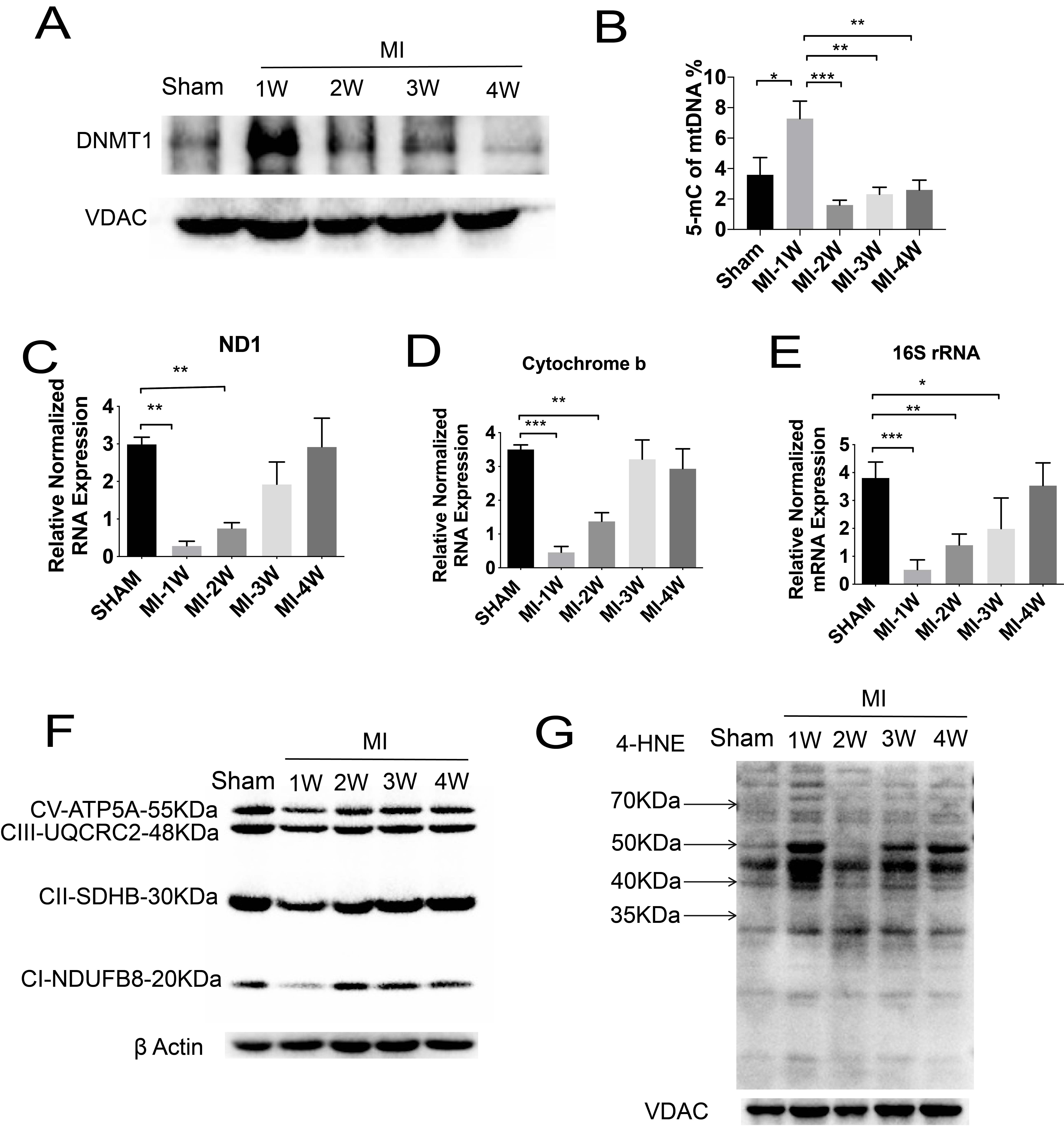
Figure 2. The characteristics of mtDNA modification in response to myocardial infarction. (A) Western blotting analysis for DNMT1 levels in mitochondria post-MI at different time points. (B) Detection of 5-mc levels in mtDNA in mitochondria post-MI at different time points. (C-E) Analysis of representative mRNA regulated by mtDNA in heart tissues post-MI. (F) Western blotting analysis of OXPHOS subunits in heart tissues post-MI at different time points. (G) Western blotting analysis of 4-HNE-conjugated protein levels in heart tissues at different time points post-MI. All results are presented as mean ± SEM. Statistical significance was determined by one-way ANOVA with Tukey’s post hoc test for multi-group comparisons. *P < 0.05, **P < 0.01, ***P < 0.001.
Methylation changes of mtDNA induce mitochondrial dysfunction post-MI
Given the established myocardial injury observed four weeks after MI, we assessed the corresponding alterations in mtDNA methylation at this time point. As shown in Figure 3A and B, mitochondrial DNMT1 levels were significantly reduced in MI mice compared with sham controls. Consistent with this finding, 5mC levels in mtDNA were also markedly decreased in the MI group [Figure 3C]. MI led to a disruption of redox homeostasis and enhanced mitochondrial ROS generation, as confirmed by MitoSOX staining in cardiomyocytes subjected to hypoxia [Figure 3D and E]. Transmission electron microscopy (TEM) further revealed ultrastructural abnormalities in mitochondria from MI cardiomyocytes, including swollen cristae and disrupted membrane integrity [Figure 3F], indicative of impaired mitochondrial respiration. To functionally assess mitochondrial OXPHOS, we measured the OCR in isolated mitochondria using a Seahorse analyzer. Mitochondria isolated from hearts of MI mice exhibited significantly reduced OCR compared with those from sham mice [Figure 3G]. Taken together, these data suggest that although reduced mtDNA 5mC levels—resulting from impaired DNMT1 translocation—may initially promote the expression of certain mitochondrial proteins, the accompanying accumulation of ROS ultimately damages mitochondrial structure and function, leading to the suppression of cardiac OXPHOS.
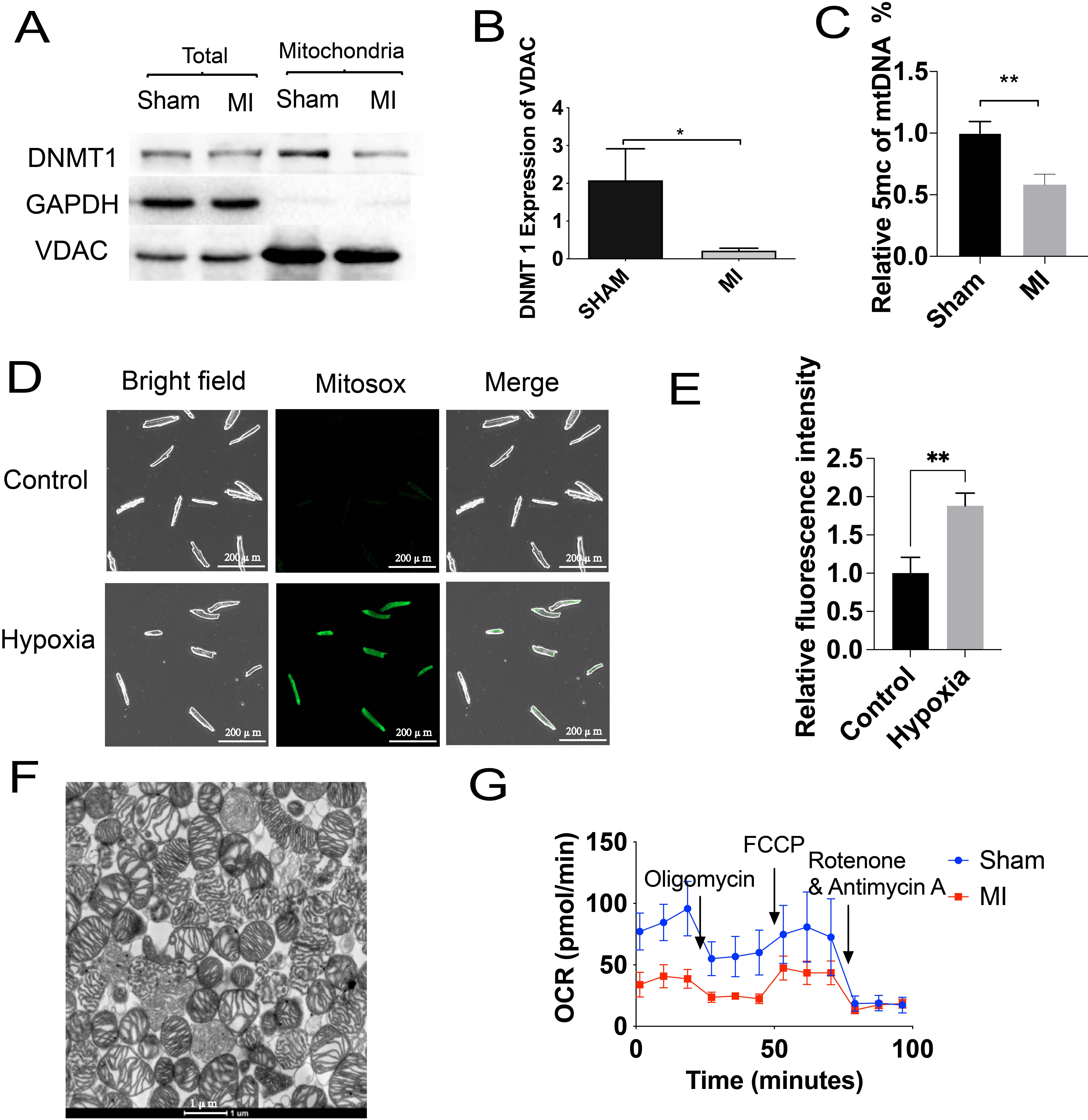
Figure 3. Methylation changes of mtDNA accompany mtROS accumulation in MI heart. (A) Western blotting analysis for DNMT1 levels in mitochondria post-MI for 4 weeks. (B) Quantitative analysis of mitochondrial DNMT1 expression in (A). (C) Detection of 5-mc levels in mtDNA in mitochondria post-MI for 4 weeks. (D and E) Mitochondrial ROS levels and statistical analysis in normal and hypoxic cardiomyocytes. (F) Representative TEM image of mitochondrial structure isolated from MI cardiomyocytes. Scale bar 1 μm. (G) Analysis of oxygen consumption rate (OCR) in mitochondria isolated from sham and MI hearts. All results are presented as mean ± SEM. Statistical significance for comparisons between two groups was determined by unpaired two-tailed Student’s t test. *P < 0.05, **P < 0.01.
ALDH2 deficiency impairs MTS-dependent mitochondrial localization of DNMT1 in cardiomyocytes
Previous work from our group has established the cardiac protective role of the mitochondrial enzyme ALDH2[12]. To identify potential regulators of DNMT1 mitochondrial translocation, we first examined the expression correlation between ALDH2 and DNMT1 using the genotype-tissue expression (GTEx) database. A weak but significant positive correlation was observed [Figure 4A], suggesting that ALDH2 may participate in regulating mtDNA methylation. To test this hypothesis, we isolated mitochondria from the hearts of WT and ALDH2-KO mice (C57BL/6J background). Western blot analysis of subcellular fractions showed that both the 180 and 130-kDa isoforms of DNMT1 were absent from the cytosolic fractions of WT and KO hearts. While the 180-kDa isoform was detected in whole-heart lysates of both genotypes, the

Figure 4. ALDH2 regulates MTS expression of DNMT1 in cardiomyocytes. (A) Genotype-Tissue Expression (GTEx) database establishes the expression correlation between ALDH2 and DNMT1. (B) Western blot analysis of the 180 and 130-kDa isoforms of DNMT1 subcellular location in WT and KO cardiomyocytes. (C) Immunofluorescent staining of DNMT1 and mitochondria in cardiomyocytes. Scale bar 50 μm. (D) Statistical analysis of MTS expression of DNMT1 in WT and KO cardiomyocytes. (E) Dot blot analysis of 5mc in isolated mtDNA of WT and KO mitochondria. (F) TFAM expression analysis in isolated WT and KO mitochondria. All results are presented as
ALDH2 deficiency delays mtDNA methylation signaling under hypoxic stress
To further dissect the role of ALDH2 in mtDNA methylation, we performed methylation sequencing on mtDNA from WT and ALDH2-KO mice, focusing on methylation differences that might arise from altered DNMT1 mitochondrial translocation. Sequencing analysis revealed an overall increase in mtDNA methylation in WT controls compared with KO mice, with particularly pronounced hypermethylation at the ND1 and ND2 loci [Figure 5A and B]. These results indicate that impaired mitochondrial translocation of DNMT1 in ALDH2-deficient cardiomyocytes decreases methylation levels at specific mtDNA regions, notably ND1 and ND2. Consistent with the repressive effect of DNA methylation on transcription, the expression of ND1 and ND2 was upregulated in KO mitochondria of cardiomyocytes [Figure 5C]. We next examined how hypoxia influences this regulatory relationship. In WT cardiomyocytes, hypoxia reduced mtDNA methylation and increased ND1 and ND2 expression, suggesting that stress-induced demethylation promotes the transcription of these genes. In contrast, hypoxia enhanced mtDNA methylation in ALDH2-KO cells and suppressed ND1 and ND2 expression [Figure 5C]. To confirm these observations at the level of global methylation, we quantified 5mC content in mtDNA. Under basal conditions, KO mtDNA showed significantly lower 5mC levels than WT mtDNA. Hypoxia markedly reduced 5mC in WT mtDNA, consistent with elevated expression of ND1 and ND2. Conversely, hypoxia increased total cellular 5mC levels in ALDH2-KO cardiomyocytes as shown by immunofluorescence [Figure 5D], which corresponded with the downregulation of ND1 and ND2. Then we detected the effect of ALDH2-induced mtDNA methylation on mice heart function. We established an MI model in WT and ALDH2-KO mice and found ALDH2 deficiency significantly impaired cardiac function of MI mice [Figure 5E]. In summary, these findings demonstrate that ALDH2 deficiency disrupts the timely modulation of mtDNA methylation under hypoxic stress by regulating DNMT1-MTS expression. This delay in methylation signaling alters the expression of key mitochondrial-encoded genes and may contribute to impaired cardiac function in ALDH2-deficient hearts.

Figure 5. Effect of ALDH2 on mitochondrial DNA methylation signaling under hypoxic stress. (A) Methylation sequencing analysis of mtDNA from WT and ALDH2-KO cardiomyocytes. (B) Different gene loci analysis in WT and ALDH2-KO mtDNA. (C) Western blot analysis of ND1 and ND2 expression changes in WT and ALDH2-KO mitochondria induced by hypoxic treatment. (D) Immunofluorescent staining of total cellular 5-methylcytosine (5mC) in WT and ALDH2-KO H9C2 cardiomyocytes under normoxic or hypoxic treatment. (E) Cardiac function analysis of WT and ALDH2-KO mice post-MI. All results are presented as mean ± SEM. For (A and B, E) (two-group comparisons), statistical significance was determined by unpaired two-tailed Student’s t test. For (C and D) (multi-group comparisons), one-way ANOVA with Tukey’s post hoc test was used. *P < 0.05, ***P < 0.001, ***P < 0.0001.
Limitation
However, this study has certain limitations. First, the functional sites of mtDNA methylation and their transcriptional effects require further validation. Second, the precise mechanism by which ALDH2 regulates DNMT1-MTS expression remains unclear, potentially involving post-transcriptional modifications or protein interactions. Third, this study used only male mice and global ALDH2-KO mice, which limits generalizability to females and precludes distinction of cardiomyocyte-autonomous effects from those in other cell types. Fourth, mitochondrial ROS were measured in intact cells rather than in isolated mitochondria, and ALDH2 activity or 4-HNE levels were not assessed across post-MI time points, leaving the temporal correlation with DNMT1 translocation unclear. Fifth, to compensate for the limited temporal resolution of the in vivo MI model in capturing early DNMT1 translocation, we employed an in vitro hypoxia model for precise control of hypoxic duration and mechanistic validation. However, under these conditions, we did not assess oxidative stress (e.g., by OxyBlot), measure ROS production in isolated mitochondria or heart tissue, or perform OXPHOS assays in isolated mitochondria. These analyses would provide more direct evidence for the ALDH2-DNMT1 axis.
DISCUSSION
Our present study reveals that ALDH2 regulates the dynamic process of mtDNA methylation by modulating DNMT1-MTS expression, thereby participating in the regulation of myocardial ischemic injury and repair. We found that the 130-kDa spliceosome of DNMT1was located in the mitochondria of cardiomyocytes and modified mtDNA methylation. During early myocardial ischemia, mitochondrial translocation of DNMT1 and mtDNA methylation were promoted. In the late ischemic process, DNMT1 escaped from mitochondria and inhibited mtDNA methylation. Then, cardiomyocytes showed significantly increased mtROS and decreased oxidative respiration. However, in the absence of ALDH2, mitochondrial localization of DNMT1 is impaired, leading to delayed mtDNA methylation signaling, while ischemia or hypoxia reactivated DNMT1, translocating into mitochondria, increasing mtDNA methylation modification, and inhibiting the expression of mitochondrial-encoded proteins, finally damaging cardiac function. In ALDH2-deficient cardiomyocytes, DNMT1 mitochondrial translocation is impaired under basal normoxia but is paradoxically reactivated upon acute hypoxia or ischemia.
Previous studies have shown that DNMT1 contains a MTS and can enter mitochondria to participate in mtDNA methylation[4]. Our results further confirm the functional presence of DNMT1 in cardiomyocyte mitochondria and reveal that its expression is dynamically regulated by myocardial ischemia[14]. The early ischemic increase in mtDNA methylation may represent an adaptive, transient silencing mechanism to reduce the transcription of ETC components, potentially conserving resources and limiting electron leak during acute stress. Subsequent demethylation in the later phase could then facilitate the recovery of mitochondrial biogenesis and function necessary for tissue repair[15]. This paradigm mirrors the dynamic DNA methylation changes observed in nuclear gene regulation during cellular adaptation.
The most significant finding is the identification of ALDH2 as a key upstream regulator of this process. The positive correlation between ALDH2 and DNMT1 expression in human tissues and our experimental data suggest that ALDH2 activity, likely through its role in clearing reactive lipid aldehydes such as 4-HNE, creates a permissive environment for the expression or stability of the DNMT1-MTS transcript/protein. 4-HNE is known to form adducts with proteins, altering their function and degradation. It is plausible that ALDH2 deficiency leads to 4-HNE accumulation, which modifies transcription factors or signaling kinases (e.g., PKC, JNK) involved in regulating DNMT1 expression or splicing, ultimately suppressing the MTS-containing variant. This hypothesis warrants future investigation. The consequence is a blunted mtDNA methylation response: ALDH2-KO hearts start from a lower basal methylation level and, upon hypoxia, exhibit a paradoxical increase in methylation at specific loci like ND1 and ND2, leading to their suppressed expression. This "epigenetic inflexibility" may underlie the inability of ALDH2-deficient mitochondria to appropriately adjust their transcriptional output in response to stress, contributing to bioenergetic failure.
Our work integrates two important fields: mitochondrial aldehyde metabolism and epigenetic regulation. While the cardioprotective effects of ALDH2 are well-documented and primarily attributed to its detoxification function[16], this study reveals an epigenetic dimension to its action. The impact of mtDNA methylation on transcription and respiration is gaining recognition, with studies showing lineage-specific patterns influencing OXPHOS[16] and its dysregulation in metabolic diseases like diabetic retinopathy[17]. We place ALDH2 as a critical mediator of this epigenetic landscape in the heart.
In summary, we propose a model where ALDH2 activity modulates the mitochondrial epigenetic landscape by regulating the availability of DNMT1 for mtDNA methylation. This regulation is essential for the dynamic reprogramming of mitochondrial gene expression required for adaptation to ischemic stress. Disruption of this axis, as seen in ALDH2 deficiency, leads to epigenetic maladaptation, worsening mitochondrial dysfunction and cardiac outcomes. Targeting the ALDH2-DNMT1-mtDNA methylation axis may offer a novel therapeutic strategy for IHD, especially in the large population carrying ALDH2 loss-of-function mutations.
DECLARATIONS
Acknowledgement
The graphical abstract was created with BioRender.com (https://BioRender.com/3lu0a6d).
Author contributions
Conceptualization, methodology, investigation, data curation, formal analysis, writing-original draft:
Methodology, software, data curation: Xia Y
Conceptualization, methodology, writing-review & editing, supervision, project administration: Abudupataer M, Xu L
Supervision, conceptualization, methodology, writing-review & editing, project administration, funding acquisition: Sun X
Availability of data and materials
The data that support the findings of this study are available from the corresponding author upon reasonable request.
AI and AI-assisted tools statement
Not applicable.
Financial support and sponsorship
This study was supported by the Noncommunicable Chronic Diseases-National Science and Technology Major Project (Grant No. 2024ZD0526900) and the National Natural Science Foundation of China (Grant Nos. 82270264, 82570302, 82200525 and 81900353).
Conflicts of interest
Xu L is an Editorial Board Member of the journal Vessel Plus. Xu L was not involved in any steps of editorial processing, notably including reviewers' selection, manuscript handling and decision making, while the other authors have declared that they have no conflicts of interest.
Ethical approval and consent to participate
All animal experiments were conducted in compliance with the National Institutes of Health Guide for the Care and Use of Laboratory Animals (8th edition, 2011) and approved by the Animal Ethics Committee of Zhongshan Hospital, Fudan University (approval No. ZSYY20170517).
Consent for publication
Not applicable.
Copyright
© The Author(s) 2026.
REFERENCES
1. Chocron ES, Munkácsy E, Pickering AM. Cause or casualty: the role of mitochondrial DNA in aging and age-associated disease. Biochim Biophys Acta Mol Basis Dis. 2019;1865:285-97.
2. Iacobazzi V, Castegna A, Infantino V, Andria G. Mitochondrial DNA methylation as a next-generation biomarker and diagnostic tool. Mol Genet Metab. 2013;110:25-34.
3. Jones PA, Liang G. Rethinking how DNA methylation patterns are maintained. Nat Rev Genet. 2009;10:805-11.
4. Shock LS, Thakkar PV, Peterson EJ, Moran RG, Taylor SM. DNA methyltransferase 1, cytosine methylation, and cytosine hydroxymethylation in mammalian mitochondria. Proc Natl Acad Sci USA. 2011;108:3630-5.
5. Becker LB, Vanden Hoek TL, Shao Z, Li C, Schumacker PT. Generation of superoxide in cardiomyocytes during ischemia before reperfusion. Am J Physiol Heart Circ Physiol. 1999;277:H2240-6.
6. Zorov DB, Juhaszova M, Sollott SJ. Mitochondrial reactive oxygen species (ROS) and ROS-induced ROS release. Physiol Rev. 2014;94:909-50.
7. Wang P, Shen C, Diao L, et al. Aberrant hypermethylation of aldehyde dehydrogenase 2 promoter upstream sequence in rats with experimental myocardial infarction. BioMed Res Int. 2015;2015:1-13.
8. Liu H, Jiang B, Hua R, et al. ALDH2 mediates the effects of sodium-glucose cotransporter 2 inhibitors (SGLT2i) on improving cardiac remodeling. Cardiovasc Diabetol. 2024;23:380.
9. Zhang F, Zhang L, Hu G, et al. Rectifying METTL4-mediated N6-methyladenine excess in mitochondrial DNA alleviates heart failure. Circulation. 2024;150:1441-58.
10. Yang K, Gao R, Chen H, et al. Myocardial reperfusion injury exacerbation due to ALDH2 deficiency is mediated by neutrophil extracellular traps and prevented by leukotriene C4 inhibition. Eur Heart J. 2024;45:1662-80.
11. Liu X, Sun A. Aldehyde dehydrogenase-2 roles in ischemic cardiovascular disease. Curr Drug Targets. 2017;18:1817-23.
12. Sun A, Zou Y, Wang P, et al. Mitochondrial aldehyde dehydrogenase 2 plays protective roles in heart failure after myocardial infarction via suppression of the cytosolic JNK/p53 pathway in mice. J Am Heart Assoc. 2014;3:e000779.
13. Nicholls TJ, Gustafsson CM. Separating and segregating the human mitochondrial genome. Trends Biochem Sci. 2018;43:869-81.
14. Boovarahan SR, Kurian GA. Investigating the role of DNMT1 gene expression on myocardial ischemia reperfusion injury in rat and associated changes in mitochondria. Biochim Biophys Acta. 2022;1863:148566.
15. Boovarahan SR, Kurian GA. Ischemic preconditioning modulates the DNA methylation process of the rat heart to provide tolerance to withstand ischemia reperfusion injury and its associated mitochondrial dysfunction. 3 Biotech. 2024;14:121.
16. Chen C, Budas GR, Churchill EN, Disatnik M, Hurley TD, Mochly-Rosen D. Activation of aldehyde dehydrogenase-2 reduces ischemic damage to the heart. Science. 2008;321:1493-5.
Cite This Article
 Open Access
How to Cite
Download Citation
Export Citation File:
Type of Import
Tips on Downloading Citation
Citation Manager File Format
Type of Import
Direct Import: When the Direct Import option is selected (the default state), a dialogue box will give you the option to Save or Open the downloaded citation data. Choosing Open will either launch your citation manager or give you a choice of applications with which to use the metadata. The Save option saves the file locally for later use.
Indirect Import: When the Indirect Import option is selected, the metadata is displayed and may be copied and pasted as needed.
About This Article
Copyright

Data & Comments
Data
0









Comments
Comments must be written in English. Spam, offensive content, impersonation, and private information will not be permitted. If any comment is reported and identified as inappropriate content by OAE staff, the comment will be removed without notice. If you have any queries or need any help, please contact us at [email protected].