


The roles of nuclear miRNAs in cardiovascular disease
 0
0 Abstract
MicroRNAs (miRNAs) represent a group of endogenous, single-stranded, small non-coding RNAs. Classically, mature miRNAs are in the cytoplasm where they mediate gene silencing by complementary pairing between their seed sequences (nucleotides 2-8 at the 5' end) and target mRNA 3' untranslated regions. However, many subsequent studies have documented the nuclear localization of miRNAs and unraveled their distinct biological roles beyond canonical cytoplasmic functions. In this review, we summarize the non-canonical roles of nuclear miRNAs in key cardiovascular pathologies to deepen our understanding of the miRNA regulatory network in cardiovascular disease and to provide a theoretical rationale for developing next-generation precision miRNA-targeted therapeutics.
Keywords
INTRODUCTION
MicroRNAs (miRNAs) represent a group of endogenous, single-stranded, small
In the context of cardiovascular diseases, dysregulated miRNA expression profiles have been closely linked to the pathogenesis of atherosclerosis, diabetic cardiomyopathy, heart failure, and so forth[8,10,11]. Based on the classical cytoplasmic functions of miRNAs, antisense oligonucleotides (ASOs) and miRNA mimics have been developed as therapeutic strategies, some of which have entered preclinical and early-phase clinical trials[12,13]. Nevertheless, these approaches largely overlook the existence and unique functions of nuclear miRNAs, raising a critical question: in cardiovascular diseases, are the regulatory roles of a given miRNA in the nucleus and cytoplasm complementary, antagonistic, or mutually interfering? For example, a nuclear miRNA may activate the transcription of protective genes, while the same miRNA suppresses the translation of target mRNA in the cytoplasm. Therefore, these two biological effects could be synergistic or conflicting. This functional duality introduces a potential confounding factor for miRNA-targeted therapies: targeting only the cytoplasmic fraction may fail to predict or counteract nuclear miRNA-mediated effects, potentially resulting in suboptimal efficacy or inconsistent therapeutic outcomes.
Therefore, a systematic dissection of the mechanisms through which nuclear miRNAs modulate the pathogenesis of cardiovascular diseases is indispensable for refining the current framework of miRNA-mediated gene regulation and optimizing the efficacy and specificity of miRNA-targeted therapeutic strategies. In this review, we summarize the non-canonical functions of nuclear miRNAs in key cardiovascular pathologies, deepen our understanding of the miRNA regulatory network in cardiovascular disease, and provide a theoretical rationale for the development of next-generation precision miRNA-targeted therapeutics.
CANONICAL BIOGENESIS AND FUNCTIONAL PARADIGM OF MIRNAS
miRNA biogenesis occurs in both the nucleus and cytoplasm[2,14,15]. In the nucleus, pri-miRNAs are cleaved by Drosha/DGCR8 to generate pre-miRNAs, which are subsequently exported to the cytoplasm via Exportin-5[14,16-18]. In the cytoplasm, Dicer further cleaves pre-miRNAs to produce mature double-stranded miRNA duplexes[19]. The guide strand is then loaded into the RNA-induced silencing complex (RISC) and pairs with the 3' UTR of target mRNAs through its seed sequence, leading to mRNA degradation or translational repression [Figure 1][20-22].
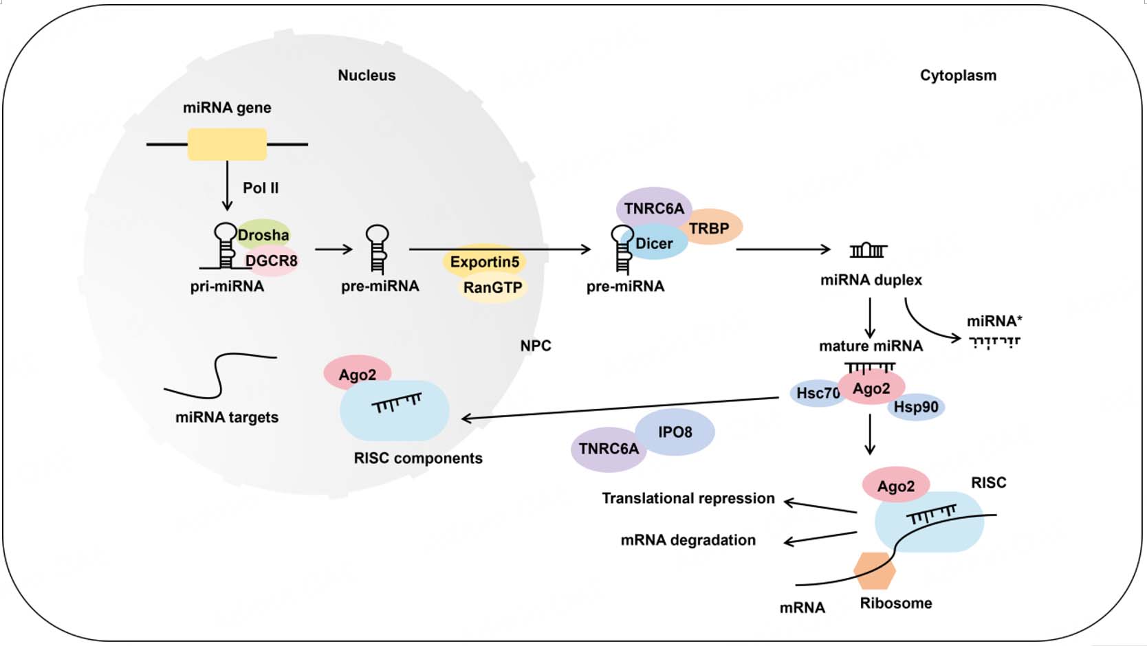
Figure 1. Canonical biogenesis and functional paradigm of miRNAs. Pri-miRNAs are transcribed from miRNA loci via Pol II, followed by cleavage mediated by the Drosha-DGCR8 Microprocessor complex to generate pre-miRNAs[16,17]. These ~70-nt pre-miRNAs are then exported to the cytoplasm via the NPC through association with Exportin-5[23]. In the cytoplasm, pre-miRNAs are processed by Dicer and TRBP to generate ~22 nt miRNA duplexes[19]. The guide strand, characterized by lower thermodynamic stability at its 5’ end, is subsequently loaded into RISC, while the passenger strand is typically degraded[20,21]. In the cytoplasm, miRNAs recognize target mRNAs primarily through sequence complementarity between their seed region and the 3' UTR of target transcripts, thereby mediating translational repression or mRNA degradation[2]. Notably, mature miRNAs can also be translocated into the nucleus via nuclear transport proteins, such as IPO8, where they exert distinct regulatory functions. Pol II: RNA polymerase II; DGCR8: DiGeorge syndrome critical region 8 protein; NPC: nuclear pore complex; TRBP: transactivation response RNA-binding protein; RISC: RNA-induced silencing complex; 3' UTR: 3' untranslated region; IPO8: importin 8; TNRC6A: trinucleotide repeat containing adaptor 6A.
NUCLEAR LOCALIZATION AND FUNCTION MECHANISM OF MIRNAS
Distribution and translocation of miRNAs in the nucleus
Early views held that mature miRNAs localize exclusively in the cytoplasm. However, in 2004, Meister et al. reported that ~20% of miR-21 resides in the nucleus of HeLa cells, challenging this classic notion[4]. Subsequent studies have further confirmed that various mature miRNAs can localize to the nucleus and exert distinct biological functions. For example, nucleolar miR-206 participates in rRNA processing, and high-throughput sequencing revealed consistent nuclear and cytoplasmic miRNA profiles[5,24,25]. Moreover, the existence of functional nuclear RISC has been validated, with nuclear let-7 cleaving a fully complementary target at 86% efficiency vs. 60% in the cytoplasm[26]. Collectively, these findings show that nuclear miRNAs are widespread and biologically active.
Research on the nuclear import mechanisms of miRNAs remains limited, which can be categorized into two types: sequence motif-dependent pathways and protein-mediated pathways. In the sequence motif-dependent pathway, the 3' terminal AGUGUU motif of miR-29b alone facilitates its nuclear localization[27]. Transferring this motif to the normally cytoplasmic miR-29a or to a small interfering RNA enriches them in the nucleus[27]. Another study identified a conserved 3' ASUS motif (where S denotes C or G) in nuclear-enriched miRNAs from neural precursor cells[28]. Regarding protein-mediated pathways, NRDE-3 in
The functional mechanism of nuclear miRNAs
Nuclear miRNAs have been confirmed to regulate gene expression at the transcription level either positively or negatively. They are increasingly recognized as key regulators of nuclear RNA metabolism, participating in autoregulatory feedback loops, mediating post-transcriptional silencing of nuclear-retained transcripts, and influencing pre-mRNA splicing decisions.
Mechanisms of nuclear miRNA-mediated gene regulation
Nuclear miRNAs regulate gene expression through multiple mechanisms, including binding to complementary sequences within promoters or to promoter-associated RNAs (paRNAs), altering chromatin states (e.g., H3K4me3, H3K27me3) and Pol II recruitment, thereby activating or repressing transcription [Figure 2A and B]. For example, miR-204 binds to a 100 bp upstream region of the CD36 promoter, displaces Ago2, reduces the enrichment of H3K27ac and H3K4me3, and consequently represses CD36 transcription[37]. In cardiomyocytes, miR-92a-3p interacts with ankyrin repeat domain-containing protein 1 (ANKRD1) sense promoter-proximal transcripts [sense promoter-associated transcripts (PATs)], recruits Ago2, and upregulates ANKRD1 expression[38].
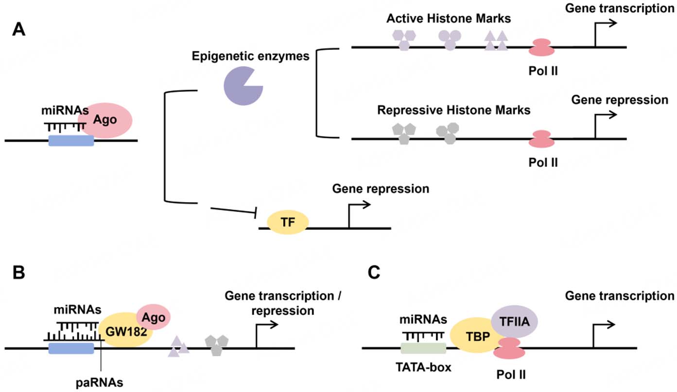
Figure 2. Mechanisms of nuclear miRNA-mediated gene regulation. (A) Ago-loaded miRNAs bind promoter sequences to recruit epigenetic enzymes, altering histone marks and Pol II occupancy to either activate or repress transcription. (B) Ago mediates miRNA binding to promoter-associated RNAs (paRNAs), altering histone marks and Pol II occupancy to regulate gene expression. (C) miRNAs interact with the TATA-box to facilitate PIC assembly and regulate transcription. Ago: Argonaute; paRNAs: promoter-associated RNAs; PIC: pre-initiation complex; TBP: TATA-box binding protein; TFIIA: transcription factor II A.
Some miRNAs can directly bind to the TATA box to facilitate pre-initiation complex (PIC) assembly
In summary, nuclear miRNAs regulate transcription via diverse mechanisms, including epigenetic modifications, paRNA-mediated scaffolding, and direct promoter binding. They primarily act as Argonaute-dependent molecular scaffolds that recruit transcriptional regulators. The RNA-RNA, RNA-DNA hybrid, and RNA-DNA triplex models proposed by Liu et al. (2018) offer a useful framework for understanding these modes of action[7]. Future studies on the context-specificity and hierarchy of these pathways will be key to fully elucidating nuclear miRNA biology.
Nuclear miRNA-mediated RNA regulation
Nuclear miRNAs have emerged as critical regulators of nuclear RNA metabolism. In C. elegans, nuclear let-7 binds a conserved sequence in pri-let-7, recruits ALG-1, and promotes its own processing, forming a positive feedback loop[42]. In mouse liver, nuclear miR-709 binds pri-miR-15a/16-1 to regulate mature miR-15a/16-1 expression[43]. miR-122 binds pri-miR-21, inhibits Drosha cleavage, and reduces mature miR-21 levels[44]. Functional RISCs are also present in the nucleus, supported by miRNA/siRNA-directed RNA cleavage activity detected in nuclear extracts[4,45]. Notably, nuclear let-7 outperforms cytoplasmic let-7 in cleaving fully matched targets, reaching an 86% cleavage efficiency vs. 60% in the cytoplasm[26]. Furthermore, nuclear miRNAs can directly influence pre-mRNA splicing. miR-10b interacts with U6 snRNA to disrupt spliceosome assembly, alters CDC42 alternative splicing, and promotes glioma cell survival[46]. Overall, only a subset of nuclear miRNAs regulates nuclear RNA, and research is still limited. Their precise mechanisms and physiological functions remain to be elucidated. Unraveling this regulatory network represents a key frontier in RNA biology, which promises to deepen our understanding of gene regulation and accelerate the development of optimized targeted therapeutics.
THE ROLE OF NUCLEAR MIRNAS IN CARDIOVASCULAR DISEASE
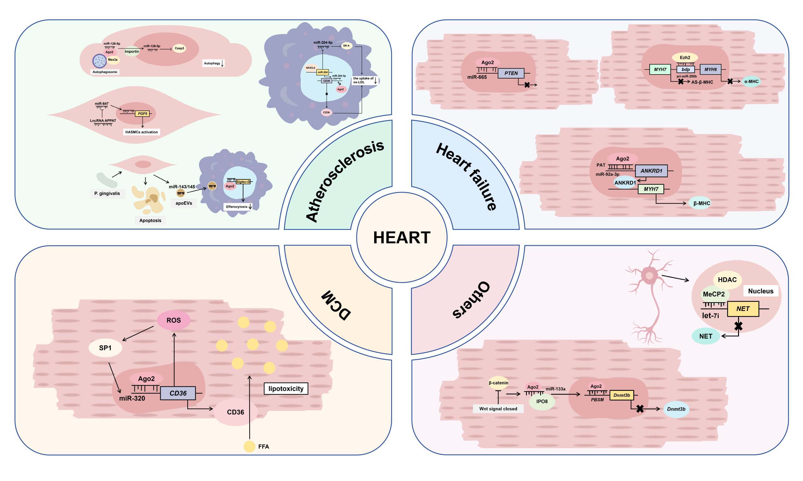
In contrast to the well-established roles of cytoplasmic miRNAs in cardiovascular diseases, the functions of nuclear miRNAs are poorly understood. This disparity not only highlights a critical gap in our knowledge but also presents a major opportunity to uncover previously unrecognized layers of gene regulation with potential therapeutic implications. The following is an overview of the reported functions of nuclear miRNAs in cardiovascular diseases [Table 1, Figure 3].
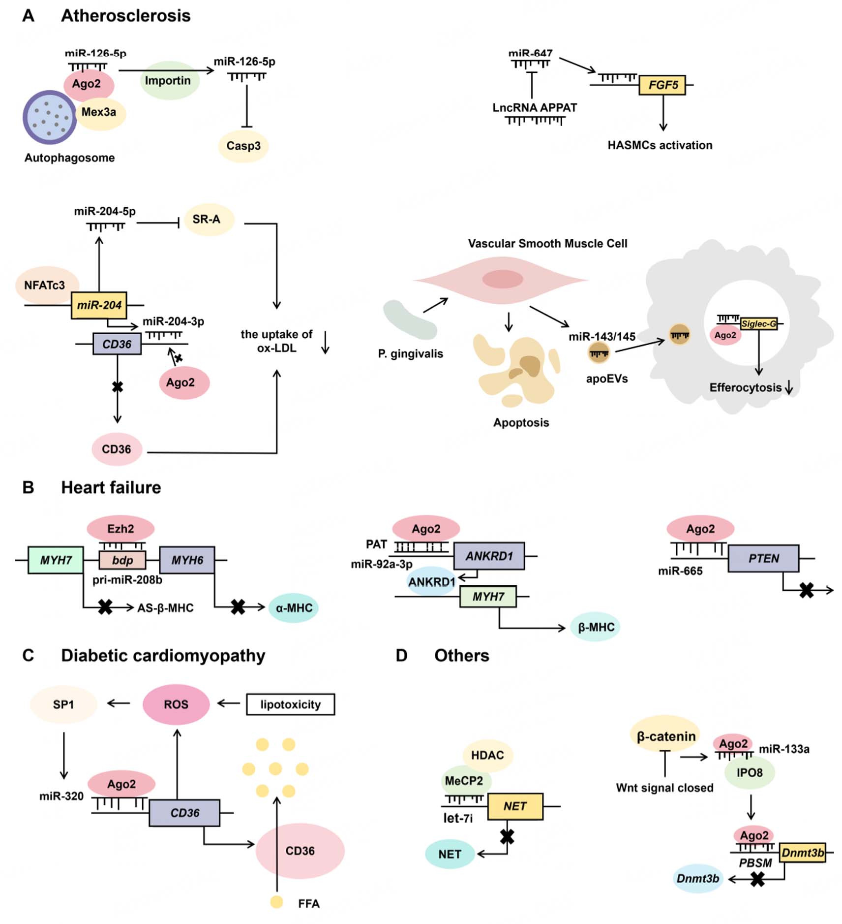
Figure 3. The role of nuclear miRNAs in cardiovascular diseases. (A) In atherosclerosis, various miRNAs translocate to the nucleus to exert either athero-protective or pro-atherogenic effects by regulating target gene expression. (B) In heart failure, miR-92a-3p and pri-miR-208b promote pathological cardiac remodeling via MHC regulation, while nuclear miR-665 drives this process by targeting PTEN. (C) In diabetic cardiomyopathy, miR-320 promotes CD36 transcription to induce lipotoxicity, which in turn upregulates miR-320 expression, forming a positive feedback loop. (D) let-7i and miR-133a target NET and Dnmt3b, respectively, contributing to the pathogenesis of other cardiovascular diseases. FGF5: Fibroblast growth factor 5; MHC: myosin heavy chain; ANKRD1: ankyrin repeat domain 1; PTEN: phosphatase and tensin homolog; NET: norepinephrine transporter; Dnmt3b: DNA methyltransferase 3 beta; IPO8: importin 8.
Nuclear functions of miRNAs in cardiovascular diseases
| Diseases | Cell types | miRNAs | Targets | Modulation pattern | Pathology | Reference |
| Atherosclerosis | Endothelial | miR-126-5p | Caspase-3* | Inhibition# | Reduction of endothelial apoptosis | [47] |
| Macrophage | miR-204-3p | CD36 | Downregulation | Inhibition of foam cell formation | [37] | |
| Macrophage | miR-143/145 | Siglec-G | Upregulation | Impairment of macrophage efferocytosis | [48] | |
| HASMC | miR-647 | FGF5 | Upregulation | Promotion of oxLDL-induced HASMCs migration and proliferation | [49] | |
| Heart failure | Cardiomyocyte | miR-92a-3p | ANKRD1 sense PATs | Upregulation | Cardiomyocyte hypertrophy and fibrosis | [38] |
| Cardiomyocyte | pri-miR-208b | bdP of MHC | Downregulation | Cardiomyocyte hypertrophy and fibrosis | [50] | |
| Cardiomyocyte | miR-665 | PTEN | Downregulation | Cardiac hypertrophy | [51] | |
| Diabetic cardiomyopathy | Cardiomyocyte | miR-320 | CD36 | Upregulation | Myocardial lipotoxicity and diastolic dysfunction | [52,53] |
| POTS | Leukocyte | let-7i | NET | Downregulation | NET deficiency and sympathetic dyshomeostasis | [54] |
| ICF syndrome | Cardiomyocyte | miR-133a | Dnmt3b | Downregulation | Aberrant cardiomyocyte differentiation of hESCs | [55] |
Atherosclerosis
Atherosclerosis constitutes a chronic inflammatory disorder of the arterial wall driven by multiple risk factors, including hyperlipidemia, hypertension, smoking, diabetes, and genetic susceptibility[56,57]. Lesions often localize to regions of disturbed blood flow (such as arterial bifurcations), where turbulent shear stress can induce endothelial dysfunction[58]. Endothelial cell proliferation and regenerative capacity are essential for maintaining vascular integrity and preventing lesion formation. Among miRNAs enriched in endothelial cells, miR-126 plays a central role in angiogenesis and vascular homeostasis[59-61]. Derived from pre-miR-126, miR-126-5p plays a well-established role in angiogenesis and has been shown to exert atheroprotective effects through spatially distinct mechanisms[62]. The expression of miR-126-5p is modulated by hemodynamics[62]. Researchers divided the aorta of high-fat diet-fed Apoe-/- mice into atherosclerosis-resistant regions (laminar flow regions, straight segments of the thoracic and abdominal aorta) and atherosclerosis-susceptible regions (turbulent flow regions, branch points of the aortic arch, aortic root, and carotid arteries)[62]. Quantitative real-time PCR (qRT-PCR) results revealed that the laminar flow-dominated resistant regions exhibited high expression of miR-126-5p, accompanied by low mRNA and protein levels of Dlk1, whereas the opposite pattern was observed in turbulent flow-prone susceptible regions[62]. No significant difference in miR-126-3p expression was detected between the two types of regions, indicating that blood flow specifically regulates miR-126-5p alone[62]. Functionally, miR-126-5p targets Dlk1 mRNA via the RISC in the cytoplasm, relieving the inhibition of Notch1 and promoting endothelial cell regeneration[62]. Beyond this canonical cytoplasmic role, miR-126-5p can also be imported into the nucleus under high shear stress (HSS) or rapamycin-induced autophagy via an Ago2:Mex3a-containing complex, as validated in HUVECs[47]. In vitro, nuclear miR-126-5p dissociates to target monomeric caspase-3, inhibiting its activity and reducing endothelial apoptosis[47]. Consistently, in ATG5EC-KO mice and Mex3a-/- mice, the nuclear enrichment ability of miR-126-5p is significantly reduced, leading to exacerbated endothelial apoptosis and aggravated atherosclerotic lesions[47]. Notably, in human carotid artery samples, miR-126-5p and caspase-3 exhibit markedly stronger co-localization signals in non-lesioned regions than in atherosclerotic regions[47]. Collectively, these findings establish that miR-126-5p orchestrates endothelial protection against atherosclerosis through a spatially coordinated, dual-compartment strategy.
Foam cell formation derived from macrophages is a central pathological feature of atherosclerosis[63,64]. Scavenger receptors (SR-A and CD36) mediate the cellular uptake of oxidized low-density lipoprotein (oxLDL), a critical step in foam cell development[56,65]. As a member of the nuclear factor of activated T-cells (NFAT) protein family, NFATc3 functions as a transcription factor triggered by the Ca2+/calcineurin signaling pathway in T cells[66-68]. Liu et al. (2021) found that the transcription factor NFATc3 in macrophages acts as a negative regulator of atherosclerosis[37]. The expression of NFATc3 is markedly reduced in human carotid plaques and in aortic lesions of hyperlipidemic Apoe-/- mice, and lower NFATc3 mRNA levels in peripheral blood mononuclear cells correlate significantly with symptomatic plaque progression[37]. Consistent with a protective role, after high-fat diet treatment, NFATc3MKO mice display larger atherosclerotic plaques, increased necrotic cores, and reduced plaque stability compared with control mice of the same genotype[37]. In contrast, NFATc3MTG mice presented markedly alleviated aortic lesions and suppressed local vascular inflammation upon high-fat diet challenge[37]. Meanwhile, the proportions of SR-A- and CD36-positive macrophages within plaques and foam cell formation are increased in NFATc3MKO mice, whereas the opposite phenotype is observed in NFATc3MTG mice[37]. Mechanistically, NFATc3 deficiency upregulates the scavenger receptors SR-A and CD36, promoting oxLDL uptake and foam cell formation[37]. NFATc3 exerts its atheroprotective effect by directly binding to the promoter of miR-204 and driving its transcription[37]. This activation initiates a spatially organized regulatory circuit: In the cytoplasm, the miR-204-5p strand post-transcriptionally silences SR-A via canonical 3' UTR targeting[37]. Simultaneously, the miR-204-3p strand translocates to the nucleus, where it binds to the CD36 promoter, displaces Ago2, and reduces active histone marks (H3K27ac and H3K4me3), ultimately repressing CD36 transcription[37]. Adeno-associated viruses (AAV)-mediated pre-miR-204 treatment for 8 weeks effectively reversed the upregulated expression of SR-A and CD36 in peritoneal macrophages of high-fat diet-fed NFATc3MKO mice[37]. This intervention also reduced aortic atherosclerotic plaque area and enhanced plaque stability in these mice[37]. In contrast, inhibition of miR-204 in NFATc3MTG mice abolished the protective effects of NFATc3[37]. Consequently, the NFATc3/miR-204 axis represents a coordinated defense mechanism against foam cell formation, simultaneously engaging post-transcriptional and chromatin-based silencing to downregulate two critical scavenger receptors, revealing a multifaceted transcriptional epigenetic barrier against atherosclerotic progression.
In addition to anti-atherogenic function, nuclear miRNAs can also exert pro-atherogenic effects, as exemplified by a pathogenic cascade initiated by the periodontal bacterium Porphyromonas gingivalis (P. gingivalis)[48,69-71]. In clinical samples, P. gingivalis can be found in more than 80% of atherosclerotic plaques[70,72,73]. Apoe-/- mice infected with P. gingivalis exhibited increased aortic plaque area, accompanied by elevated levels of miR-143/145 and increased apoptotic cells within the lesions[48]. In vitro, vascular smooth muscle cells (SMCs) are more susceptible to P. gingivalis-induced apoptosis than endothelial cells[48].
The lncRNA atherosclerotic plaque pathogenesis-associated transcript (APPAT) is detectable in circulation, and its levels correlate with atherosclerotic progression[74,75]. Functionally, APPAT overexpression suppresses the oxLDL-induced migratory and proliferative capacity of HASMCs, whereas overexpression of miR-647 promotes these pro-atherogenic phenotypes[49]. Bioinformatic analysis and dual-luciferase reporter assays validate the direct reciprocal suppression between APPAT and miR-647[49]. Dual-luciferase reporter assays confirm that miR-647 binds to the 3'-UTR of FGF5 and upregulates its mRNA and protein levels, while APPAT overexpression reverses ox-LDL-induced elevation of FGF5[49]. Notably, subcellular fractionation shows that miR-647 localizes to both the nucleus and cytoplasm of HASMCs, with a relatively lower abundance in the cytoplasmic compartment[49]. This distribution intriguingly suggests that the positive regulation of FGF5 by miR-647 may occur via a non-canonical nuclear pathway, potentially involving direct gene activation[49]. However, given its detectable cytoplasmic presence, a complementary canonical mechanism, in which miR-647 indirectly elevates FGF5 by repressing its suppressors via the RISC pathway, cannot be excluded[49]. Thus, the APPAT/miR-647/FGF5 axis orchestrates a multi-tiered regulatory circuit that fine-tunes smooth muscle cell phenotypes in atherosclerosis by integrating a lncRNA-mediated checkpoint with the dual-compartment actions of a miRNA, underscoring the sophisticated layering of RNA-based control in vascular disease.
Heart failure
Myocardial remodeling, a key pathophysiological process in heart failure, involves extensive transcriptomic reprogramming, most notably the pathological switch from the adult α-myosin heavy chain (MYH6) to the fetal β-myosin heavy chain (MYH7)[76-78]. Existing research demonstrates that pressure overload significantly upregulates nucleus-localized Ago2 in cardiomyocytes, as observed in human dilated cardiomyopathy patients and transverse aortic constriction (TAC) mouse models[38]. Within the nucleus, Ago2 functions as a transcriptional co-activator. It is recruited to the promoter region of ANKRD1 through an interaction mediated by nuclear miR-92a-3p[38]. This miRNA guides Ago2 by binding at the ANKRD1 locus, thereby enhancing ANKRD1 transcription[38]. The interactions between miR-92a-3p, Ago2, and ANKRD1 are observed in HL-1 cells[38]. The upregulated ANKRD1 protein, in turn, functions as a transcriptional co-activator that binds specifically to the MYH7 promoter, selectively increasing MHC7 expression without affecting MHC6[38]. The consequent rise in the MHC7/MHC6 ratio impairs myocardial contractility, promotes hypertrophy and fibrosis, and accelerates the transition from adaptive cardiac hypertrophy to decompensated heart failure[38]. A recombinant adeno-associated virus serotype 9 (rAAV9)-mediated knockdown of Ago2 or ANKRD1 effectively reverses the heart failure phenotype induced by TAC[38]. Intervention with ivermectin (which inhibits the nuclear import of Ago2 and ANKRD1) or synthetic ANKPep (a peptide mimicking the nuclear localization signal of ANKRD1 that specifically blocks ANKRD1 nuclear translocation) in TAC mice both ameliorated cardiac function and myocardial hypertrophy[38].
Correspondingly, nuclear primary miRNAs also participate in transcriptional reprogramming during cardiac hypertrophy. A prominent example is pri-miR-208b, which modulates pathological cardiac remodeling through direct chromatin interactions[50]. During TAC-induced cardiac hypertrophy, Ezh2, a Polycomb-group protein, binds to the bidirectional promoter (bdP) of the α-myosin Heavy Chain (α-MHC) and β-MHC (antisense), facilitating H3K27me3 to suppress α-MHC and β-MHC expression[50]. pri-miR-208b reinforces this repression by interacting with both Ezh2 and the bdP, as in vitro experiments confirm direct binding of pri-miR-208b to the -1227 to -1330 region of bdP, and this interaction can be disrupted by RNase H treatment[50]. The histone deacetylase inhibitor Trichostatin A (TSA) disrupts the interaction between Ezh2 and bdP, which diminishes chromatin occupancy of pri-miR-208b. Consequently, TSA treatment reverses the aberrant gene expression associated with pathological cardiac hypertrophy in TAC mice[50]. Notably, antisense β-MHC (AS β-MHC) can associate with chromatin independently of Ezh2 and pri-miR-208b, and its knockdown does not significantly alter Ezh2-promoter interaction[50]. Interestingly, a recent study reveals a sex-specific regulatory mechanism in cardiac tissue. Female mouse left ventricles exhibit substantially higher nuclear pri-miR-208b abundance relative to male counterparts[79]. This sex difference is independent of DNA methylation but relies on sex-specific methylation of the lncRNA Mhrt[79]. In female mice, methylation of the CG18 locus within Mhrt facilitates its binding to methyl-CpG-binding protein MeCP2[79]. Through an RNA-dependent mechanism, the MeCP2 complex subsequently recruits corepressors (Rest and HDAC2) to the pri-miR-208b promoter, ultimately alleviating transcriptional repression[79]. Given the established role of pri-miR-208b in regulating the pathological Myh6/Myh7 switch, these observations suggest that nuclear miRNA-guided epigenetic mechanisms may contribute to sex-based differences in cardiovascular disease susceptibility[50,79]. Nevertheless, this study exhibits distinct limitations. In particular, the hormonal regulation of Mhrt CG18 methylation remains undefined, and its stability in the context of hypertrophy and other pathological conditions has yet to be validated.
Phosphatase and tensin homolog (PTEN) is widely recognized as a tumor suppressor[80,81]. It is also abundantly expressed in various cardiovascular cell types, including cardiomyocytes, fibroblasts, vascular smooth muscle cells, and endothelial cells, and functions as a potent negative regulator of cardiac hypertrophy[82,83]. PTEN maintains cardiomyocyte homeostasis and modulates myocardial energy metabolism via inhibiting the PI3K/Akt/mTOR signaling axis[82-84]. While cytoplasmic miRNAs such as miR-217 are known to post-transcriptionally suppress PTEN to facilitate cardiac hypertrophy, a recent study reveals that nuclear miRNAs can also target PTEN through a distinct regulatory mechanism[51,84]. miR-665 represents a typical example of such nuclear regulatory miRNAs. In TAC mice, nuclear-enriched miR-665 is markedly upregulated and drives pathological cardiac remodeling by directly repressing PTEN in cardiomyocytes[51]. Elevated miR-665 exacerbates cardiomyocyte hypertrophy, promotes myocardial fibrosis, and aggravates cardiac dysfunction, as validated in both cellular models and TAC-induced mouse models[51]. Mechanistically, miR-665 represses PTEN transcription independent of the canonical 3' UTR pathway[51]. Instead, it binds two evolutionarily conserved regions within the PTEN promoter: site 1 (-217/-211) and site 2 (-835/-827), and recruits Ago2 to inhibit PTEN expression at the transcriptional level. These regulatory effects are validated by dual-luciferase reporter assays and ChIP assays[51]. Restoration of PTEN expression reverses miR-665-induced cardiac dysfunction, cardiomyocyte hypertrophy and myocardial fibrosis in TAC mice, highlighting the pivotal role of this axis in pathological cardiac progression[51].
Diabetic cardiomyopathy
Diabetic cardiomyopathy represents a common form of secondary cardiomyopathy, characterized by the synergistic pathophysiological effects of hyperglycemia-induced glucotoxicity and dyslipidemia-associated lipotoxicity[85]. These pathological processes collectively induce myocardial structural and functional abnormalities of the myocardium via oxidative stress and mitochondrial dysfunction[85]. CD36, a critical transmembrane transporter for long-chain fatty acids (FAs), facilitates free fatty acids (FFAs) uptake in cardiomyocytes and provides substrates for fatty acid oxidation[86,87]. Cardiomyocyte FFA uptake is markedly reduced in MHC-PPARα/CD36-/- mice[86]. Consistently, CD36 mRNA and protein levels are elevated in insulin-resistant primary cardiomyocytes[86]. shRNA-mediated CD36 knockout decreases cellular uptake of long-chain free fatty acids by approximately 50% and greatly alleviates intracellular lipid accumulation, while silencing other fatty acid transporters fails to produce comparable improvements in lipotoxicity[86]. A key factor driving this pathological CD36 upregulation is nuclear-enriched miR-320, which acts as a small activating RNA (saRNA) that directly binds to the CD36 promoter and enhances its transcription[52]. This effect promotes fatty acid uptake and exacerbates lipotoxicity, thereby accelerating diabetic cardiomyopathy progression. Therapeutic intervention with a rAAV9-delivered miR-320 inhibitor (TuD) effectively restores cardiac function in db/db mice[52]. Interestingly, a positive feedback loop exists between CD36 and miR-320[53]. Elevated CD36 promotes reactive oxygen species (ROS) generation, and further activates the transcription factor SP1[53]. SP1 in turn binds to the miR-320 promoter, further increasing miR-320 expression and thereby reinforcing CD36 transcription[53]. This CD36-ROS-SP1-miR-320 axis forms a self-sustaining cycle that exacerbates lipotoxicity[53]. CD36 serves as the initiator of this feedback loop. In STZ-induced diabetic mice, CD36 protein levels are significantly increased within 2 weeks of hyperglycemia onset, while miR-320 upregulation is delayed until four weeks[53]. Correspondingly, high-glucose-treated AC16 cardiomyocytes exhibit CD36 protein accumulation prior to elevated CD36 mRNA levels[53]. This early CD36 protein elevation is closely associated with high glucose-induced extracellular Ago2 secretion, which reduces intracellular Ago2 availability[53]. Consequently, diminished Ago2 binding to CD36 mRNA alleviates translational repression, thereby enhancing CD36 protein synthesis[53].
Other cardiovascular system-related diseases
Chronic orthostatic intolerance syndromes, including the classic subtype postural orthostatic tachycardia syndrome (POTS), are characterized by persistent dizziness, palpitations, and fatigue upon prolonged standing, which arises from cardiac sympathetic dysautonomia[88,89]. A hallmark of POTS is impaired synaptic clearance of norepinephrine (NE), leading to excessive sympathetic tone in the upright position[88]. Genetic investigations have identified specific molecular alterations responsible for disrupted NE homeostasis[90]. A genetic analysis of a family with orthostatic intolerance identified a G237C missense mutation (Ala457Pro) in exon 9 of the norepinephrine transporter (NET) gene (SLC6A2)[90]. This mutation abolishes over 98% of NET uptake activity, resulting in severely reduced NE clearance, elevated upright plasma NE levels, and an altered DHPG/NE ratio, directly linking defective NET function to familial orthostatic tachycardia and NE dysregulation[90]. Beyond genetic defects, epigenetic mechanisms further contribute to NET deficiency in POTS. In patients’ leukocytes, the nuclear microRNA let-7i forms a complex with MeCP2 that binds to the NET promoter, where it facilitates histone deacetylation and represses NET transcription[54]. Although this regulatory axis has been corroborated in HeLa cells, its direct role in modulating NET expression in patient-specific sympathetic tissues awaits further validation[54]. Consistent with these findings, immunohistochemical analyses of peripheral sympathetic nerves from POTS patients reveal reduced NET protein expression and blunted sympathetic responsiveness to postural challenge, even without elevated systemic NE spillover[91]. This dissociation between nerve activity and NE release indicates that NET dysfunction, whether inherited or epigenetically acquired, is a core pathophysiological disturbance that disrupts sympathetic homeostasis and triggers the clinical presentation of orthostatic intolerance.
DNA methyltransferase Dnmt3b is essential for mammalian embryogenesis[92]. Loss of Dnmt3b leads to Immunodeficiency, Centromeric instability, and Facial anomalies syndrome (ICF syndrome)[93]. Dnmt3b-knockout mice exhibit embryonic lethality at E14.5-E16.5 with typical ventricular septal defects and multi-organ hypoplasia, highlighting its non-redundant role in cardiac morphogenesis[93]. In HL-1 cells, inhibition of canonical Wnt signaling (e.g., via IWR-1) triggers IPO8/Ago2-mediated nuclear translocation of miR-133a[55]. Nuclear miR-133a binds to a specific promoter-associated sequence (PBSM) of Dnmt3b and epigenetically represses its expression through H3K27 trimethylation and disruption of a Dnmt3b autoregulatory feedback loop[55]. This regulatory pathway has been validated in human embryonic stem cell-derived cardiomyocytes, where nuclear miR-133a-mediated Dnmt3b suppression alters the expression of key maturation genes, including contractile components (Myh6, Tnni2) and calcium-handling elements (Cacna1c), and affects the subcellular localization of actin and calcium channels, thereby modulating cardiomyocyte structural and functional maturation[55]. Taken together, these results define a pathogenic cascade whereby Wnt signaling suppression triggers nuclear miR-133a-mediated epigenetic silencing of Dnmt3b, resulting in defective cardiomyocyte gene expression[55]. Since Dnmt3b is indispensable for ventricular septation, dysregulation of this axis thus identifies miR-133a-mediated transcriptional control as a potential etiological contributor to congenital cardiac malformations.
Integrated discussion of nuclear miRNAs in cardiovascular diseases
In conclusion, nuclear miRNAs play key regulatory roles across a spectrum of cardiovascular diseases, including atherosclerosis, heart failure, diabetic cardiomyopathy, POTS, and so forth. A common feature is that most nuclear miRNAs assemble sequence-specific guiding complexes with Ago2 as the core scaffold protein. Through direct binding to gene promoters or physical interactions with chromatin-associated factors, such miRNAs engage chromatin and modulate the transcription of target genes in either an activating or repressive manner. Despite sharing similar molecular mechanisms, nuclear miRNAs exhibit strong context-dependent regulatory effects. The regulatory effects of these nuclear miRNAs on target genes, molecular functions, and downstream biological consequences are fully determined by disease categories, cellular phenotypes, and upstream signaling cascades [Table 1]. Such discrepancies arise from divergent co-regulatory molecules recruited to miRNA-loaded complexes. Even the identical target gene may exhibit opposing regulatory outputs under different pathological conditions. CD36 serves as a typical example. In atherosclerosis, nuclear-localized miR-204-3p binds to the CD36 promoter and suppresses its transcription, thereby reducing macrophage foam cell formation and slowing plaque progression[37]. In contrast, miR-320 functions as a saRNA to upregulate CD36 expression in diabetic cardiomyopathy, exacerbating myocardial lipotoxic injury[52]. These findings firmly prove that nuclear miRNAs exhibit disease-specific regulatory patterns matching the corresponding pathophysiological traits.
Instead of modulating individual molecular targets in isolation, nuclear miRNAs achieve precise transcriptional control via multi-layered, multi-compartmental coordinated regulatory networks, which can be categorized into three canonical regulatory modes. The first pattern refers to dual compartmental functions of a single miRNA molecule. miR-126-5p exerts distinct activities in the cytoplasm and nucleus. In the cytoplasm, it targets and represses Dlk1 via RISC to maintain endothelial proliferation and mitigate plaque formation[62]. In the nucleus, it directly binds and inhibits caspase-3, thereby suppressing endothelial apoptosis[47]. As such, one miRNA molecule concurrently modulates two signaling cascades, markedly improving regulatory efficiency. The second pattern lies in functional differentiation between two strands of one miRNA. For example, the two strands of miR-204 establish a complementary regulatory system. Cytoplasmic miR-204-5p silences SR-A through canonical post-transcriptional pathways, while nuclear miR-204-3p represses CD36 at the transcriptional level, jointly restraining foam cell generation[37]. Such strand-specific functional divergence is also observed in the miR-126 family. Cytoplasmic miR-126-3p binds Ago2 to target SPRED1 and PIK3R2, relieving the inhibition of the VEGF pathway to facilitate vascular repair and alleviate endothelial inflammation, which complements the anti-apoptotic function of nuclear miR-126-5p[47,60]. These findings imply that strand-specific functional differentiation may represent a widespread regulatory strategy among miRNA families. The third pattern involves pathological positive feedback loops that amplify tissue damage. The CD36-ROS-SP1-miR-320 positive-feedback loop in diabetic cardiomyopathy serves as a representative example[53]. Collectively, these spatiotemporally coordinated pathways render cardiovascular transcriptional networks highly robust. Single molecules concurrently operate at multiple regulatory nodes and hierarchical layers to enable efficient pathological signal transduction.
On this basis, nuclear miRNAs interact with lncRNAs and transcription factors to form intricate regulatory networks, such as the APPAT/miR-647/FGF5 axis and the CD36-ROS-SP1-miR-320 feedback loop, which collectively mediate pathological phenotypic switching of vascular smooth muscle cells and cardiomyocytes[49,53]. Notably, functional diversification arises from disease-specific targeting and regulatory orientation. In atherosclerosis, nuclear miRNAs display bidirectional effects. miR-126-5p and miR-204 attenuate endothelial injury and foam cell formation, whereas miR-143/145 accelerate plaque progression via intercellular signaling, with targets centered on vascular-cell genes like Dlk1, CD36, and Siglec-G[37,47,48]. In heart failure, nuclear miRNAs predominantly drive maladaptive remodeling by regulating myosin isoform switching (e.g., miR-92a-3p-mediated transcription activation of ANKRD1 induces MYH7 upregulation) and modulating hypertrophic signaling pathways (e.g., miR-665-mediated PTEN repression)[38,51]. In diabetic cardiomyopathy, nuclear miRNAs primarily disrupt lipid metabolic dysregulation, as exemplified by miR-320-mediated CD36 activation that exacerbates myocardial lipotoxic injury[52]. In POTS, nuclear let-7i uniquely targets NET, disrupting sympathetic homeostasis[54]. An additional layer of regulatory complexity arises from sex-specific regulation. In heart failure, female-specific methylation of the lncRNA Mhrt modulates the expression of pri-miR-208b, representing a unique regulatory trait that has not been widely observed in other diseases[79]. Thus, despite the conserved transcriptional mechanism of nuclear miRNAs, their functional outputs are precisely adapted to the cellular and molecular landscape of each cardiovascular pathology.
Overall, nuclear miRNAs function via conserved chromatin-based transcriptional regulatory mechanisms. Through cytoplasmic-nuclear compartmentalized division of labor, strand-specific functional differentiation, pathological positive feedback loops, and crosstalk with lncRNAs and transcription factors, they form multi-layered coordinated regulatory networks. Their biological functions shift dynamically in response to disease microenvironments to accommodate the pathological progression of diverse cardiovascular disorders, providing multi-dimensional theoretical foundations for developing precise cardiovascular therapeutic strategies targeting nuclear miRNAs.
CLINICAL POTENTIAL OF NUCLEAR MIRNAS IN CARDIOVASCULAR DISEASES
MiRNA-targeted therapy
miRNA-targeted therapy represents a pivotal frontier in the development of non-coding RNA-based therapeutics, demonstrating unique and irreplaceable advantages in disease treatment and offering innovative strategies for the clinical intervention of complex disorders. As endogenous regulatory molecules, miRNAs can directly harness intrinsic biogenesis and functional mechanisms without the need for exogenous regulatory systems, thereby minimizing off-target risks. Furthermore, chemical modifications such as 2'-O-methylation and LNA significantly reduce the immunogenicity of miRNAs, making them safer than certain exogenous synthetic drugs[13,94]. A distinctive strength of miRNA-based therapeutics lies in their capacity to simultaneously regulate multiple key genes within a shared pathological pathway via a single seed sequence. This multi-targeting modality enables miRNA-based therapies to overcome the limitations of traditional single-target approaches, making them particularly suitable for treating polygenic disorders. The inherent flexibility of this approach further permits the therapeutic restoration of protective miRNAs via mimics, or the suppression of pathogenic miRNAs using anti-miRNA oligonucleotides (AMOs). When integrated with advanced delivery platforms, such as AAV or GalNAc conjugates, this strategy facilitates precise targeting of disease-driving pathways, aligning with the principles of precision medicine[95,96]. Consequently, miRNA-targeted therapy has rapidly evolved into a promising frontier for the diagnosis and treatment of cardiovascular diseases[95,97,98]. Several cardiovascular-relevant miRNAs have shown encouraging efficacy in preclinical models, and a number of candidates (e.g., CDR132L, Remlarsen) have progressed to Phase II clinical trials[99,100], underscoring their substantial translational potential[13,97].
Among the nuclear miRNAs previously discussed in relation to cardiovascular disease mechanisms, miR-92a-3p and miR-126-3p exemplify successful transitions from mechanistic insight to clinical exploration. In large-animal (porcine) ischemia-reperfusion models, anti-miR-92a-3p agents, such as LNA-modified inhibitors, significantly reduce myocardial infarction size while attenuating cardiomyocyte apoptosis and inflammation[101]. Mechanistically, these protective effects are attributed to the repression of pro-survival genes, including BCL-2, and activation of the PI3K/Akt signaling pathway[101,102]. The anti-miR-92a-3p candidate MRG-110 has advanced into clinical trials, where it is being evaluated for safety and efficacy in promoting angiogenesis and accelerating tissue repair in wound healing[13]. Preclinical and early-phase clinical data indicate that subcutaneous delivery achieves target tissue distribution, robustly suppresses miR-92a-3p activity, and demonstrates a favorable safety profile, with no significant immune-related adverse events reported[103]. miR-126-3p, predominantly expressed in vascular endothelial cells, promotes angiogenesis and maintains endothelial integrity by inhibiting SPRED1 and PI3KR2, thereby activating the VEGF pathway[104]. Its downregulation in conditions such as myocardial infarction and atherosclerosis leads to impaired vascular repair[104]. Notably, patients with advanced heart failure complicated by pulmonary hypertension (PH) display markedly diminished circulating miR-126-3p concentrations, a finding that correlates with higher pulmonary artery pressure and worse right ventricular function[104]. This suggests a potential role for miR-126-3p in mitigating pulmonary hypertension-associated vascular remodeling, highlighting it as a novel therapeutic target for heart failure complications. Preclinical research is currently exploring the targeted delivery of miR-126 mimics via exosomes or aptamers (e.g., transferrin receptor aptamers) to promote angiogenesis in ischemic myocardium, demonstrating potential in improving vascular remodeling in PH[104,105].
Despite the compelling promise of miRNA-based therapy evident from basic research, translating it into clinical practice encounters considerable obstacles. The ability of miRNAs to target multiple genes, often viewed as a therapeutic advantage, also represents a dual-edged sword. Because a single miRNA can modulate numerous downstream mRNAs, altering its expression may simultaneously influence several signaling pathways. While effectively suppressing or activating disease-driving genes, this could inadvertently dysregulate genes essential for normal physiological functions, potentially leading to severe side effects or toxicity. Furthermore, existing therapeutic designs predominantly center on the canonical cytoplasmic roles of miRNAs, often overlooking the distinct roles of miRNAs localized to different subcellular compartments, which may lead to unintended consequences. For instance, in a porcine model of myocardial infarction, prolonged overexpression of miR-92a was found to induce aberrant cardiomyocyte proliferation and arrhythmia[106]. As discussed earlier, nuclear-enriched miR-92a-3p enhances ANKRD1 transcription, leading to increased ANKRD1 protein, which in turn selectively upregulates MHC7 expression. This shift elevates the MHC7/MHC6 (β/α) ratio, ultimately impairing myocardial contractility and promoting hypertrophy and fibrosis[38]. Given the faster contraction kinetics of MYH7 compared to MYH6, the physiological balance of these isoforms is essential for synchronous cardiac contraction[107]. Disruption of this balance can cause regional mechanical asynchrony (e.g., regions contracting too rapidly or too slowly), triggering electrical instability and increasing arrhythmic risk[107-109]. Thus, the arrhythmias observed following sustained miR-92a overexpression in pigs may reflect, at least in part, its nuclear activity, although the intrinsic pro-arrhythmic milieu of myocardial infarction cannot be ruled out as a contributing factor. Collectively, these findings highlight the necessity of considering miRNAs' subcellular localization and compartment-specific functions in the development of miRNA-targeted therapies and underscore the critical need to strictly control the duration and dosage of miRNA expression for clinical safety.
Circulating miRNAs as a biomarker
Biomarker-guided therapy facilitates therapeutic selection, efficacy assessment, dosage optimization, and the avoidance of side effects. Circulating miRNAs have emerged as a promising class of non-invasive biomarkers due to their unique physicochemical properties. Extracellular miRNAs, encapsulated in lipid vesicles or bound to carrier proteins/lipoproteins, are highly stable in biofluids, resistant to RNase degradation, and amenable to sensitive quantification via RT-qPCR and next-generation sequencing (NGS), offering practical advantages over conventional peptide markers[110-114]. The expression of miRNAs is highly cell-specific and dynamically regulated by physiological states and pathological stimuli. These fluctuations provide a real-time molecular readout of cardiovascular status, underpinning their diagnostic and prognostic utility in cardiovascular diseases[97,115,116]. For instance, circulating miR-126 levels correlate with platelet activation and are suppressed by aspirin, suggesting its role as a marker of antiplatelet efficacy[117,118]. Patients with heart failure exhibit characteristic downregulation of 24 circulating miRNAs. Among these candidates, miR-26b-5p, miR-145-5p, miR-92a-3p, miR-30e-5p, and miR-29a-3p correlate with cardiac functional markers including LVEF and NT-proBNP, and their expression levels rise markedly in patients who achieve favorable responses to cardiac resynchronization therapy (CRT)[119]. Levels of miR-1 and miR-133a directly reflect myocardial steatosis and are elevated in type 2 diabetes, consistent with altered myocardial lipid metabolism[120]. In mouse models of acute myocardial infarction, cardiac-enriched miRNAs (miR-1, miR-133a, miR-208a, miR-499-5p) increase sharply in circulation and are trafficked to distant organs such as bone marrow, participating in systemic injury responses[121]. In LVAD-supported patients, persistently elevated miR-483-3p tracks treatment response over time (at 3, 6, and 9 months post-LVAD support), while baseline miR-1202 outperforms NT-proBNP and INTERMACS scores in distinguishing responders from non-responders[122]. Collectively, these findings highlight the great clinical potential of circulating miRNAs as biomarkers to track disease progression and predict therapeutic response as well as patients’ prognosis. However, these conclusions necessitate validation through large-scale studies with expanded cohorts.
CONCLUSION AND PERSPECTIVES
Although extensive studies have focused on miRNAs in cardiovascular diseases, the majority of literature primarily emphasizes their canonical cytoplasmic functions, including mediating target mRNA degradation and translational repression. Previous reviews have systematically summarized the biological functions of cytoplasmic miRNAs in various cardiovascular diseases[10,12]. In contrast, systematic investigations into nuclear-localized miRNAs remain scarce. Moreover, the spectrum of cardiovascular diseases covered by current studies is relatively limited, lacking high-incidence clinical disorders such as hypertension, ischemic heart disease, and myocardial ischemia-reperfusion injury, which hinders the construction of a comprehensive and global research framework for miRNA regulatory networks in cardiovascular systems.
Distinct from the canonical cytoplasmic modes of action, miRNAs can translocate into the nucleus relying on conserved sequence motifs and transport protein complexes. In the nucleus, miRNAs participate in pathological processes via non-canonical pathways such as transcriptional regulation, chromatin epigenetic remodeling, and nuclear RNA interactions, exerting vital regulatory effects in cardiac diseases, including atherosclerosis, heart failure, diabetic cardiomyopathy, and so forth. For instance, laminar shear stress drives the nuclear translocation of endothelial miR-126-5p, which directly binds and inhibits nuclear caspase-3 activation to reduce endothelial apoptosis and counteract atherosclerosis[47]. Under pressure overload, miR-665 accumulates in the cardiomyocyte nucleus, binds to the PTEN gene promoter to repress its transcription, and facilitates pathological cardiac remodeling[51]. Under diabetic high-glucose conditions, miR-320 translocates into the nucleus and acts as a saRNA to upregulate CD36 transcription, triggering the CD36-ROS-SP1-miR-320 positive feedback loop and aggravating myocardial lipotoxicity[53].
However, most regulatory mechanisms of nuclear miRNAs have only been validated in vitro in cell lines (e.g., HeLa, AC16, and 293T cells), with no evidence from precise gene-edited animal models such as tissue-specific knockout or overexpression and target promoter site-directed mutation. Findings obtained from cellular experiments cannot always perfectly recapitulate the complex in vivo microenvironment. For example, the nuclear let-7i/MeCP2 complex that regulates NET is only verified in HeLa cells and peripheral leukocytes from patients with POTS, and its authentic regulatory role in native sympathetic nerve tissues lacks direct in vivo evidence. Whether the epigenetic silencing of CD36 mediated by NFATc3 downstream miR-204-3p via Ago2 displacement exists in vascular tissues of hyperlipidemic Apoe-/- mice still requires further validation in animal models. In addition, the regulatory axis that miR-133a silences Dnmt3b to modulate cardiomyocyte maturation and cardiac development needs more embryo-specific intervention experiments to supplement in vivo data. The disconnect between in vitro and in vivo experimental evidence greatly reduces the translational potential of existing nuclear miRNA regulatory pathways, constituting a core challenge to be addressed in this field.
Furthermore, the upstream regulatory networks governing miRNAs nuclear translocation remain largely uncharacterized. Current studies have identified only a limited set of stimuli capable of triggering miRNAs nuclear import, including blood flow shear stress, rapamycin-induced autophagy, canonical Wnt pathway inhibition, high glucose stimulation, and P. gingivalis infection. Representative examples include shear stress-mediated nuclear transport of endothelial miR-126-5p, Wnt suppression-induced nuclear translocation of cardiomyocyte miR-133a, and high glucose-triggered nuclear enrichment of cardiac miR-320[47,52,55]. However, the complete molecular cascades mediating miRNAs shuttling under diverse pathological stimuli have not been systematically elucidated, and universal nuclear localization signals or regulatory elements controlling miRNA nuclear trafficking are yet to be identified. Meanwhile, it remains unclear whether nuclear-localized miRNAs possess specific structural or sequence features, and their recognition patterns for genomic target loci have not yet been elucidated. This greatly impedes high-throughput screening and functional prediction of nuclear miRNAs and their target genes at the genome-wide level, restricting in-depth exploration of nuclear miRNAs regulatory networks in cardiovascular diseases.
From the perspective of clinical translation, current miRNA intervention strategies face notable technical barriers. The primary therapeutic agents, miRNA ASOs and miRNA mimics, are incapable of targeting specific subcellular compartments. Following administration, these agents may simultaneously bind to homologous target miRNAs in both the cytoplasm and nucleus. Although these reagents can suppress pathological signaling cascades, they may perturb the physiological roles of miRNAs in separate subcellular compartments. This triggers non-specific off-target responses and latent toxic adverse effects, which substantially narrow the therapeutic safety window. Moving forward, it is necessary to further deepen the research on the molecular mechanisms of nuclear miRNAs transport and regulation, clarify the core regulatory network underlying the functional differences between nuclear and cytoplasmic miRNAs in cardiovascular diseases, and verify their diagnostic and therapeutic value through multi-center, large-sample clinical trials. Such efforts will provide a more robust scientific foundation and innovative strategies for the precise prevention and treatment of cardiovascular diseases.
DECLARATIONS
Authors’ contributions
Conceptualization: Jin K, Chen C
Writing: Han L
Searching literature: Han L, Jin K
All authors have read and approved the article.
Availability of data and materials
Not applicable.
AI and AI-assisted tools statement
During the preparation of this manuscript, the AI tool DeepSeek (version V3.2, released 2025-12-01) was used solely for language editing. The tool did not influence the study design, data collection, analysis, interpretation, or the scientific content of the work. All authors take full responsibility for the accuracy, integrity, and final content of the manuscript.
Financial support and sponsorship
This work was supported by grants from the Noncommunicable Chronic Diseases-National Science and Technology Major Project (grant numbers 2025ZD0547302, 2025ZD0547100); the National Natural Science Foundation of China (grant numbers 82525005, U22A20266, 82270363, 825B2007); the Natural Science Foundation of Hubei Province (grant number 2025EHA037); and the Basic Research Program of Huazhong University of Science and Technology (grant numbers 2024BRA020). The funders had no role in study design, data collection and analysis, manuscript preparation, or decision to publish.
Conflicts of interest
Chen C is the Guest Editor of the special issue "Current Knowledge and Future Applications of Non-Coding RNAs in Cardiomyopathies and Heart Failure" in the journal Vessel Plus, and is also a member of the journal’s Editorial Board. Chen C was not involved in any steps of the editorial process for this manuscript, including reviewer selection, manuscript handling, or decision-making, while the other authors have declared that they have no conflicts of interest.
Ethical approval and consent to participate
Not applicable.
Consent for publication
Not applicable.
Copyright
© The Author(s) 2026.
REFERENCES
2. Shang R, Lee S, Senavirathne G, Lai EC. microRNAs in action: biogenesis, function and regulation. Nat Rev Genet. 2023;24:816-33.
3. Lee RC, Feinbaum RL, Ambros V. The C. elegans heterochronic gene lin-4 encodes small RNAs with antisense complementarity to lin-14. Cell. 1993;75:843-54.
4. Meister G, Landthaler M, Patkaniowska A, Dorsett Y, Teng G, Tuschl T. Human Argonaute2 mediates RNA cleavage targeted by miRNAs and siRNAs. Mol Cell. 2004;15:185-97.
5. Liao J, Ma L, Guo Y, et al. Deep sequencing of human nuclear and cytoplasmic small RNAs reveals an unexpectedly complex subcellular distribution of miRNAs and tRNA 3′ trailers. PLoS ONE. 2010;5:e10563.
6. Billi M, De Marinis E, Gentile M, Nervi C, Grignani F. Nuclear miRNAs: gene regulation activities. Int J Mol Sci. 2024;25:6066.
7. Liu H, Lei C, He Q, Pan Z, Xiao D, Tao Y. Nuclear functions of mammalian MicroRNAs in gene regulation, immunity and cancer. Mol Cancer. 2018;17:64.
8. Santovito D, Weber C. Non-canonical features of microRNAs: paradigms emerging from cardiovascular disease. Nat Rev Cardiol. 2022;19:620-38.
9. Costa S, La Rocca G, Cavalieri V. Epigenetic regulation of chromatin functions by MicroRNAs and long noncoding RNAs and implications in human diseases. Biomedicines. 2025;13:725.
11. Sessa F, Salerno M, Esposito M, Cocimano G, Pomara C. miRNA dysregulation in cardiovascular diseases: current opinion and future perspectives. Int J Mol Sci. 2023;24:5192.
12. Zhou S, Jin J, Wang J, et al. miRNAS in cardiovascular diseases: potential biomarkers, therapeutic targets and challenges. Acta Pharmacol Sin. 2018;39:1073-84.
13. Nappi F. Non-coding RNA-targeted therapy: a state-of-the-art review. Int J Mol Sci. 2024;25:3630.
14. Kim H, Lee Y, Kim VN. The biogenesis and regulation of animal microRNAs. Nat Rev Mol Cell Biol. 2024;26:276-96.
15. Komatsu S, Kitai H, Suzuki HI. Network regulation of microRNA biogenesis and target interaction. Cells. 2023;12:306.
16. Zeng Y, Yi R, Cullen BR. Recognition and cleavage of primary microRNA precursors by the nuclear processing enzyme Drosha. EMBO J. 2004;24:138-48.
17. Lee Y, Ahn C, Han J, et al. The nuclear RNase III Drosha initiates microRNA processing. Nature. 2003;425:415-9.
18. Lee Y, Jeon K, Lee JT, Kim S, Kim VN. MicroRNA maturation: stepwise processing and subcellular localization. EMBO J. 2002;21:4663-70.
19. Chendrimada TP, Gregory RI, Kumaraswamy E, et al. TRBP recruits the dicer complex to Ago2 for microRNA processing and gene silencing. Nature. 2005;436:740-4.
20. Suzuki HI, Katsura A, Yasuda T, et al. Small-RNA asymmetry is directly driven by mammalian Argonautes. Nat Struct Mol Biol. 2015;22:512-21.
21. Iwasaki S, Kobayashi M, Yoda M, et al. Hsc70/Hsp90 chaperone machinery mediates ATP-dependent RISC loading of small RNA duplexes. Mol Cell. 2010;39:292-9.
22. Abel Y, Charron C, Virciglio C, et al. The interaction between RPAP3 and TRBP reveals a possible involvement of the HSP90/R2TP chaperone complex in the regulation of miRNA activity. Nucleic Acids Res. 2022;50:2172-89.
23. Lund E, Güttinger S, Calado A, Dahlberg JE, Kutay U. Nuclear export of MicroRNA precursors. Science. 2004;303:95-8.
24. Ritland Politz JC, Zhang F, Pederson T. MicroRNA-206 colocalizes with ribosome-rich regions in both the nucleolus and cytoplasm of rat myogenic cells. Proc Natl Acad Sci USA. 2006;103:18957-62.
25. Ritland Politz JC, Hogan EM, Pederson T. MicroRNAs with a nucleolar location. RNA. 2009;15:1705-15.
26. Robb GB, Brown KM, Khurana J, Rana TM. Specific and potent RNAi in the nucleus of human cells. Nat Struct Mol Biol. 2005;12:133-7.
27. Hwang H, Wentzel EA, Mendell JT. A hexanucleotide element directs MicroRNA nuclear import. Science. 2007;315:97-100.
28. Jeffries CD, Fried HM, Perkins DO. Nuclear and cytoplasmic localization of neural stem cell microRNAs. RNA. 2011;17:675-86.
29. Guang S, Bochner AF, Pavelec DM, et al. An argonaute transports siRNAs from the cytoplasm to the nucleus. Science. 2008;321:537-41.
30. Wei Y, Li L, Wang D, Zhang C, Zen K. Importin 8 regulates the transport of mature MicroRNAs into the cell nucleus. J Biol Chem. 2014;289:10270-5.
31. Weinmann L, Höck J, Ivacevic T, et al. Importin 8 is a gene silencing factor that targets argonaute proteins to distinct mRNAs. Cell. 2009;136:496-507.
32. Schraivogel D, Schindler SG, Danner J, et al. Importin-β facilitates nuclear import of human GW proteins and balances cytoplasmic gene silencing protein levels. Nucleic Acids Res. 2015;43:7447-61.
33. Nishi K, Nishi A, Nagasawa T, Ui-tei K. Human TNRC6A is an Argonaute-navigator protein for microRNA-mediated gene silencing in the nucleus. RNA. 2013;19:17-35.
34. Sobti M, Mead BJ, Stewart AG, Igreja C, Christie M. Molecular basis for GIGYF-TNRC6 complex assembly. RNA. 2023;29:724-34.
35. Johnson KC, Kilikevicius A, Hofman C, et al. Nuclear localization of Argonaute 2 is affected by cell density and may relieve repression by microRNAs. Nucleic Acids Res. 2024;52:1930-52.
36. Pitchiaya S, Heinicke LA, Park JI, Cameron EL, Walter NG. Resolving subcellular miRNA trafficking and turnover at single-molecule resolution. Cell Rep. 2017;19:630-42.
37. Liu X, Guo J, Lin X, et al. Macrophage NFATc3 prevents foam cell formation and atherosclerosis: evidence and mechanisms. Eur Heart J. 2021;42:4847-61.
38. Xie R, Yuan S, Hu G, et al. Nuclear AGO2 promotes myocardial remodeling by activating ANKRD1 transcription in failing hearts. Mol Ther. 2024;32:1578-94.
39. Zhang Y, Fan M, Zhang X, et al. Cellular microRNAs up-regulate transcription via interaction with promoter TATA-box motifs. RNA. 2014;20:1878-89.
40. Bai Y, Pan B, Zhan X, Silver H, Li J. MicroRNA 195-5p targets Foxo3 promoter region to regulate its expression in granulosa cells. Int J Mol Sci. 2021;22:6721.
41. Wan D, Liang X, Liang F, Zhu M, Zhang C, Zhang S. The role of miR-3188-mediated transcriptional activation of IL-7Rα in tumor immunotherapy research. Mol Immunol. 2026;189:142-52.
42. Zisoulis DG, Kai ZS, Chang RK, Pasquinelli AE. Autoregulation of microRNA biogenesis by let-7 and Argonaute. Nature. 2012;486:541-4.
43. Tang R, Li L, Zhu D, et al. Mouse miRNA-709 directly regulates miRNA-15a/16-1 biogenesis at the posttranscriptional level in the nucleus: evidence for a microRNA hierarchy system. Cell Res. 2011;22:504-15.
44. Wang D, Sun X, Wei Y, et al. Nuclear miR-122 directly regulates the biogenesis of cell survival oncomiR miR-21 at the posttranscriptional level. Nucleic Acids Res. 2018;46:2012-29.
45. La Rocca G, Cavalieri V. Roles of the core components of the mammalian miRISC in chromatin biology. Genes. 2022;13:414.
46. El Fatimy R, Zhang Y, Deforzh E, et al. A nuclear function for an oncogenic microRNA as a modulator of snRNA and splicing. Mol Cancer. 2022;21:17.
47. Santovito D, Egea V, Bidzhekov K, et al. Noncanonical inhibition of caspase-3 by a nuclear microRNA confers endothelial protection by autophagy in atherosclerosis. Sci Transl Med. 2020;12:eaaz2294.
48. Xie H, Qin Z, Ling Z, et al. Oral pathogen aggravates atherosclerosis by inducing smooth muscle cell apoptosis and repressing macrophage efferocytosis. Int J Oral Sci. 2023;15:26.
49. Meng F, Han L, Liang Q, Lu S, Huang Y, Liu J. The Lnc-RNA APPAT suppresses human aortic smooth muscle cell proliferation and migration by interacting with MiR-647 and FGF5 in Atherosclerosis. J Endovasc Ther. 2022;30:937-50.
50. Mathiyalagan P, Okabe J, Chang L, Su Y, Du X, El-Osta A. The primary microRNA-208b interacts with polycomb-group protein, Ezh2, to regulate gene expression in the heart. Nucleic Acids Res. 2013;42:790-803.
51. Fan J, Zhang X, Nie X, et al. Nuclear miR-665 aggravates heart failure via suppressing phosphatase and tensin homolog transcription. Sci China Life Sci. 2019;63:724-36.
52. Li H, Fan J, Zhao Y, et al. Nuclear miR-320 mediates diabetes-induced cardiac dysfunction by activating transcription of fatty acid metabolic genes to cause lipotoxicity in the heart. Circ Res. 2019;125:1106-20.
53. Zhan J, Jin K, Ding N, et al. Positive feedback loop of miR-320 and CD36 regulates the hyperglycemic memory-induced diabetic diastolic cardiac dysfunction. Mol Ther Nucleic Acids. 2023;31:122-38.
54. Khan AW, Ziemann M, Corcoran SJ, et al. NET silencing by let-7i in postural tachycardia syndrome. JCI Insight. 2017;2:e90183.
55. Di Mauro V, Crasto S, Colombo FS, Di Pasquale E, Catalucci D. Wnt signalling mediates miR-133a nuclear re-localization for the transcriptional control of Dnmt3b in cardiac cells. Sci Rep. 2019;9:9320.
58. Souilhol C, Serbanovic-Canic J, Fragiadaki M, et al. Endothelial responses to shear stress in atherosclerosis: a novel role for developmental genes. Nat Rev Cardiol. 2019;17:52-63.
59. Wei Y, Nazari-Jahantigh M, Neth P, Weber C, Schober A. MicroRNA-126, -145, and -155: a therapeutic triad in atherosclerosis? Arterioscler Thromb Vasc Biol. 2013;33:449-54.
60. Fish JE, Santoro MM, Morton SU, et al. miR-126 regulates angiogenic signaling and vascular integrity. Dev Cell. 2008;15:272-84.
61. Theofilis P, Oikonomou E, Vogiatzi G, et al. The role of MicroRNA-126 in atherosclerotic cardiovasculardiseases. Curr Med Chem. 2023;30:1902-21.
62. Schober A, Nazari-Jahantigh M, Wei Y, et al. MicroRNA-126-5p promotes endothelial proliferation and limits atherosclerosis by suppressing Dlk1. Nat Med. 2014;20:368-76.
63. Liu X, Wu J, Tian R, Su S, Deng S, Meng X. Targeting foam cell formation and macrophage polarization in atherosclerosis: the therapeutic potential of rhubarb. Biomed Pharmacother. 2020;129:110433.
64. Ma C, Li Y, Tian M, et al. Gsα regulates macrophage foam cell formation during atherosclerosis. Circ Res. 2024;134:e34-e51.
65. Tian K, Xu Y, Sahebkar A, Xu S. CD36 in Atherosclerosis: pathophysiological mechanisms and therapeutic implications. Curr Atheroscler Rep. 2020;22:59.
66. Crabtree GR, Olson EN. NFAT signaling: choreographing the social lives of cells. Cell. 2002;109:S67-79.
67. Mozafari M, Md Hashim SN, Ahmad Amin Noordin KB, Zainal SA, Azlina A. Nuclear factor of activated T cells (NFAT) proteins as targeted molecules in diseases: a narrative review. Cureus. 2024;16:e75844.
68. Park Y, Yoo S, Kim M, Kim W. The role of calcium-calcineurin-NFAT signaling pathway in health and Autoimmune diseases. Front Immunol. 2020;11:195.
69. Haraszthy V, Zambon J, Trevisan M, Zeid M, Genco R. Identification of periodontal pathogens in Atheromatous plaques. J Periodontol. 2000;71:1554-60.
70. Xie M, Tang Q, Nie J, et al. BMAL1-downregulation aggravates Porphyromonas gingivalis-induced atherosclerosis by encouraging oxidative stress. Circ Res. 2020:126.
71. Park S, Kim I, Han SJ, et al. Oral Porphyromonas gingivalis infection affects intestinal microbiota and promotes atherosclerosis. J Clin Periodontol. 2023;50:1553-67.
72. Rao A, D’souza C, Subramanyam K, et al. Molecular analysis shows the presence of periodontal bacterial DNA in atherosclerotic plaques from patients with coronary artery disease. Indian Heart J. 2021;73:218-20.
73. Šubarić A, Arsić Z, Mihailović Đ, et al. Detecting oral bacteria in abdominal aorta atherosclerotic plaques—how far can they go? Medicina. 2025;61:1976.
74. Meng F, Yan J, Ma Q, et al. Expression status and clinical significance of lncRNA APPAT in the progression of atherosclerosis. PeerJ. 2018;6:e4246.
75. Lu S, Liang Q, Huang Y, Meng F, Liu J. Definition and review on a category of long non-coding RNA: atherosclerosis-associated circulating lncRNA (ASCLncRNA). PeerJ. 2020;8:e10001.
76. Hang CT, Yang J, Han P, et al. Chromatin regulation by Brg1 underlies heart muscle development and disease. Nature. 2010;466:62-7.
77. Guo S, Hu Y, Ling L, et al. Molecular mechanisms and intervention approaches of heart failure (Review). Int J Mol Med. 2025;56:1-15.
78. Yue P, Xia S, Wu G, et al. Attenuation of cardiomyocyte hypertrophy via depletion Myh7 using CASAAV. Cardiovasc Toxicol. 2020;21:255-64.
79. K N H, Okabe J, Mathiyalagan P, et al. Sex-based Mhrt methylation chromatinizes MeCP2 in the heart. iScience. 2019;17:288-301.
80. Yim E, Peng G, Dai H, et al. Rak functions as a tumor suppressor by regulating PTEN protein stability and function. Cancer Cell. 2009;15:304-14.
81. Jha M, Bhatt LK. Exploring phosphatase and tensin homologue deleted on chromosome ten (PTEN) as a potential therapeutic target for cardiac hypertrophy. ACS Pharmacol Transl Sci. 2025;8:2312-36.
82. Oudit G, Sun H, Kerfant BG, Crackower MA, Penninger JM, Backx PH. The role of phosphoinositide-3 kinase and PTEN in cardiovascular physiology and disease. J Mol Cell Cardiol. 2004;37:449-71.
83. Oudit GY, Penninger JM. Cardiac regulation by phosphoinositide 3-kinases and PTEN. Cardiovasc Res. 2008;82:250-60.
84. Nie X, Fan J, Li H, et al. miR-217 promotes cardiac hypertrophy and dysfunction by targeting PTEN. Mol Ther Nucl Acids. 2018;12:254-66.
85. Huo J, Feng Q, Pan S, Fu W, Liu Z, Liu Z. Diabetic cardiomyopathy: early diagnostic biomarkers, pathogenetic mechanisms, and therapeutic interventions. Cell Death Discov. 2023;9:256.
86. Yang J, Sambandam N, Han X, et al. CD36 deficiency rescues lipotoxic cardiomyopathy. Circ Res. 2007;100:1208-17.
87. Rada P, González-Rodríguez Á, García-Monzón C, Valverde ÁM. Understanding lipotoxicity in NAFLD pathogenesis: is CD36 a key driver? Cell Death Dis. 2020;11:802.
88. Goldstein DS, Holmes C, Frank SM, et al. Cardiac sympathetic dysautonomia in chronic orthostatic intolerance syndromes. Circulation. 2002;106:2358-65.
89. Mar PL, Raj SR. Postural orthostatic tachycardia syndrome: mechanisms and new therapies. Annu Rev Med. 2020;71:235-48.
90. Shannon JR, Flattem NL, Jordan J, et al. Orthostatic intolerance and tachycardia associated with norepinephrine-transporter deficiency. N Engl J Med. 2000;342:541-9.
91. Lambert E, Eikelis N, Esler M, et al. Altered sympathetic nervous reactivity and norepinephrine transporter expression in patients with postural tachycardia syndrome. Circ Arrhythm Electrophysiol. 2008;1:103-9.
92. Okano M, Bell DW, Haber DA, Li E. DNA methyltransferases Dnmt3a and Dnmt3b are essential for de novo methylation and mammalian development. Cell. 1999;99:247-57.
93. Ueda Y, Okano M, Williams C, Chen T, Georgopoulos K, Li E. Roles for Dnmt3b in mammalian development: a mouse model for the ICF syndrome. Development. 2006;133:1183-92.
94. Khvorova A, Watts JK. The chemical evolution of oligonucleotide therapies of clinical utility. Nat Biotechnol. 2017;35:238-48.
95. Brillante S, Volpe M, Indrieri A. Advances in MicroRNA therapeutics: from preclinical to clinical studies. Hum Gene Ther. 2024;35:628-48.
96. Kara G, Calin GA, Ozpolat B. RNAi-based therapeutics and tumor targeted delivery in cancer. Adv Drug Delivery Rev. 2022;182:114113.
97. Nappi F, Avtaar Singh SS, Jitendra V, Alzamil A, Schoell T. The roles of microRNAs in the cardiovascular system. Int J Mol Sci. 2023;24:14277.
98. Chakraborty C, Sharma AR, Sharma G, Lee S. Therapeutic advances of miRNAs: a preclinical and clinical update. J Adv Res. 2021;28:127-38.
99. Bauersachs J, Solomon SD, Anker SD, et al. Efficacy and safety of CDR132L in patients with reduced left ventricular ejection fraction after myocardial infarction: rationale and design of the HF-REVERT trial. Eur J Heart Fail. 2024;26:674-82.
100. Chioccioli M, Roy S, Newell R, et al. A lung targeted miR-29 mimic as a therapy for pulmonary fibrosis. eBioMedicine. 2022;85:104304.
101. Hinkel R, Penzkofer D, Zühlke S, et al. Inhibition of MicroRNA-92a protects against ischemia/reperfusion injury in a large-animal model. Circulation. 2013;128:1066-75.
102. Bonauer A, Carmona G, Iwasaki M, et al. MicroRNA-92a controls angiogenesis and functional recovery of ischemic tissues in mice. Science. 2009;324:1710-3.
103. Abplanalp WT, Fischer A, John D, et al. Efficiency and target derepression of anti-miR-92a: results of a first in human study. Nucleic Acid Ther. 2020;30:335-45.
104. Gallo A, Agnese V, Sciacca S, et al. MicroRNA-30d and -483-3p for Bi-ventricular remodelling and miR-126-3p for pulmonary hypertension in advanced heart failure. ESC Heart Fail. 2024;11:155-66.
105. Zernecke A, Bidzhekov K, Noels H, et al. Delivery of MicroRNA-126 by apoptotic bodies induces CXCL12-dependent vascular protection. Sci Signal. 2009;2:ra81.
106. Gabisonia K, Prosdocimo G, Aquaro GD, et al. MicroRNA therapy stimulates uncontrolled cardiac repair after myocardial infarction in pigs. Nature. 2019;569:418-22.
107. Locher MR, Razumova MV, Stelzer JE, Norman HS, Patel JR, Moss RL. Determination of rate constants for turnover of myosin isoforms in rat myocardium: implications for in vivo contractile kinetics. Am J Physiol Heart Circ Physiol. 2009;297:H247-56.
108. Hsieh J, Becklin KL, Givens S, et al. Myosin heavy chain converter domain mutations drive early-stage changes in extracellular matrix dynamics in hypertrophic cardiomyopathy. Front Cell Dev Biol. 2022;10:894635.
109. Mosqueira D, Mannhardt I, Bhagwan JR, et al. CRISPR/Cas9 editing in human pluripotent stem cell-cardiomyocytes highlights arrhythmias, hypocontractility, and energy depletion as potential therapeutic targets for hypertrophic cardiomyopathy. Eur Heart J. 2018;39:3879-92.
110. Mitchell PS, Parkin RK, Kroh EM, et al. Circulating microRNAs as stable blood-based markers for cancer detection. Proc Natl Acad Sci USA. 2008;105:10513-8.
111. Weber JA, Baxter DH, Zhang S, et al. The MicroRNA spectrum in 12 body fluids. Clin Chem. 2010;56:1733-41.
112. Arroyo JD, Chevillet JR, Kroh EM, et al. Argonaute2 complexes carry a population of circulating microRNAs independent of vesicles in human plasma. Proc Natl Acad Sci USA. 2011;108:5003-8.
113. Mayr M, Zampetaki A, Willeit P, Willeit J, Kiechl S. MicroRNAs within the continuum of postgenomics biomarker discovery. Arterioscler Thromb Vasc Biol. 2013;33:206-14.
114. Vanhaverbeke M, Attard R, Bartekova M, et al. Peripheral blood RNA biomarkers for cardiovascular disease from bench to bedside: a position paper from the EU-CardioRNA COST action CA17129. Cardiovasc Res. 2022;118:3183-97.
115. De Gonzalo-Calvo D, Vea A, Bär C, et al. Circulating non-coding RNAs in biomarker-guided cardiovascular therapy: a novel tool for personalized medicine? Eur Heart J. 2019;40:1643-50.
116. Viereck J, Thum T. Circulating noncoding RNAs as biomarkers of cardiovascular disease and injury. Circ Res. 2017;120:381-99.
117. De Boer HC, Van Solingen C, Prins J, et al. Aspirin treatment hampers the use of plasma microRNA-126 as a biomarker for the progression of vascular disease. Eur Heart J. 2013;34:3451-7.
118. Willeit P, Zampetaki A, Dudek K, et al. Circulating MicroRNAs as novel biomarkers for platelet activation. Circ Res. 2013;112:595-600.
119. Marfella R, Di Filippo C, Potenza N, et al. Circulating microRNA changes in heart failure patients treated with cardiac resynchronization therapy: responders vs. non-responders. Eur J Heart Fail. 2013;15:1277-88.
120. De Gonzalo-Calvo D, Van Der Meer RW, Rijzewijk LJ, et al. Serum microRNA-1 and microRNA-133a levels reflect myocardial steatosis in uncomplicated type 2 diabetes. Sci Rep. 2017;7:47.
121. Cheng M, Yang J, Zhao X, et al. Circulating myocardial microRNAs from infarcted hearts are carried in exosomes and mobilise bone marrow progenitor cells. Nat Commun. 2019;10:959.
Cite This Article
 Open Access
How to Cite
Download Citation
Export Citation File:
Type of Import
Tips on Downloading Citation
Citation Manager File Format
Type of Import
Direct Import: When the Direct Import option is selected (the default state), a dialogue box will give you the option to Save or Open the downloaded citation data. Choosing Open will either launch your citation manager or give you a choice of applications with which to use the metadata. The Save option saves the file locally for later use.
Indirect Import: When the Indirect Import option is selected, the metadata is displayed and may be copied and pasted as needed.
About This Article
Special Topic
Copyright

Data & Comments
Data
0









Comments
Comments must be written in English. Spam, offensive content, impersonation, and private information will not be permitted. If any comment is reported and identified as inappropriate content by OAE staff, the comment will be removed without notice. If you have any queries or need any help, please contact us at [email protected].