


Updates on the management of neuroendocrine liver metastasis
 0
0  ,
,
Abstract
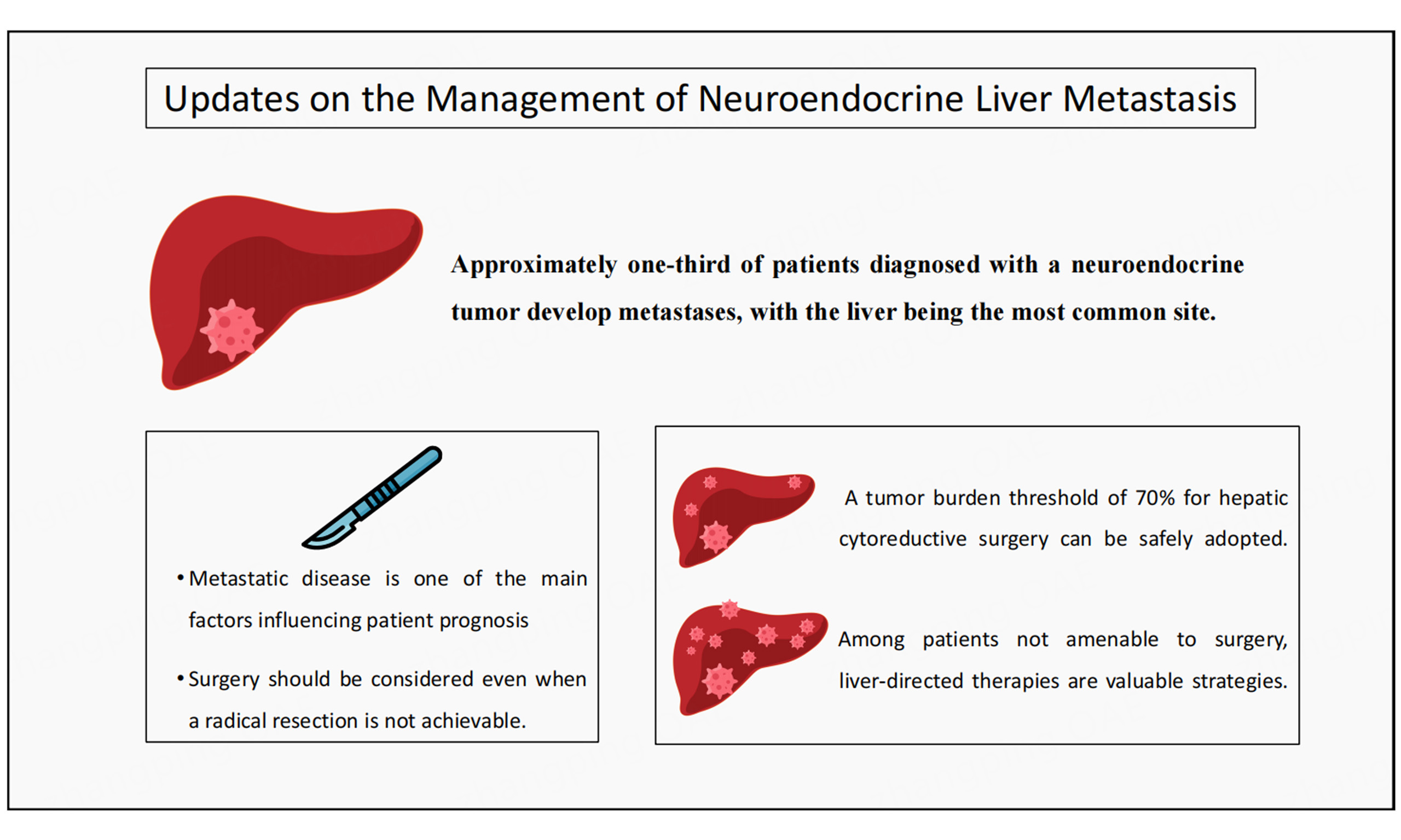
Approximately one-third of patients diagnosed with a neuroendocrine tumor (NET) develop distant metastases, with the liver being the most common site. Therefore, the management of patients with neuroendocrine liver metastasis (NELM) is particularly important, as metastatic disease is often one of the main factors influencing patient prognosis. When patients are amenable to surgery, liver resection is associated with improved long-term outcomes and relief from potential tumor-related symptoms. NELM resection should be considered even when a radical resection is not achievable. Moreover, a tumor burden threshold of 70% for hepatic cytoreductive surgery can be safely adopted with favorable long-term outcomes. For patients with NELM who are not candidates for surgical resection, liver-directed therapies provide a valuable treatment strategy, enabling optimal disease control while preserving liver parenchyma. Furthermore, liver transplantation has emerged as a potential therapy for patients with NELM. Although significant progress has been made in managing NELM, the heterogeneity of NETs poses substantial challenges to research due to the variability in tumor characteristics. Therefore, devising an optimal therapeutic strategy requires a multidisciplinary approach to develop individualized treatment plans and optimize patient outcomes.
Keywords
INTRODUCTION
Neuroendocrine tumors (NETs) are a group of rare neoplasms arising from neuroendocrine cells that can have heterogeneous biological behaviors. Although the incidence of NETs has increased over the last decades, overall survival appears to be improving, possibly as a result of improved imaging and therapeutic strategies[1]. Overall, NETs are classified as functional or non-functional tumors, with the former secreting bioactive amines such as serotonin, histamine, and kallikrein, possibly leading to diarrhea, flushing, wheezing, and cardiac valvular abnormalities, which characterize carcinoid syndrome[2]. Similarly, pancreatic NETs (PNETs) can produce other hormones, including: gastrin, which can result in
Grade and morphology classification of GEP-NET
| Grade | Morphology | Mitotic count | Ki-67 | |
| Low grade | 1 | Well differentiated | < 2 | < 3% |
| Intermediate grade | 2 | Well differentiated | 2-20 | 3%-20% |
| High grade | 3 | Well differentiated | > 20 | > 20% |
| NEC | 3 | Poorly differentiated | > 20 | > 20% |
The majority of NETs originate from the small intestine, the lungs, and the colon-rectum, followed by PNET, gastric, and appendiceal NETs[5]. Although NETs are mostly indolent, approximately a third of patients diagnosed with NET develop distant metastasis, with the liver being the most common site[6,7]. Specifically, the prevalence of liver metastasis ranges from 205-90%, with 12-74% of patients having synchronous neuroendocrine liver metastasis (NELM)[7-9]. Patients who develop metastatic disease have a markedly reduced survival compared with patients who only have localized disease, with 5-year survival ranging from 13%-54% among patients with NELM compared with 75%-99% for patients with
Diagnosis of NELM
Although most patients with NET undergo a contrast-enhanced computed tomography (CT) at the time of diagnosis, contrast-enhanced magnetic resonance imaging (MRI) scans have a higher sensitivity to detect hepatic metastases[14-16]. NELMs typically demonstrate bright enhancement on arterial-phase CT and
Classification of NELM
The most used classification scheme of NELMs describes three possible distributions of liver metastases: a single metastasis is classified as Type 1 regardless of its size; Type II includes isolated metastatic bulk with smaller deposits that involve both lobes; a disseminated metastatic spread to both lobes or a single lesion with no normal liver parenchyma is classified as Type III[24]. A more recent approach, suggested by Mahuron and Singh, describes four categories based on the ability to reduce tumor burden through surgical resection. According to this classification, patients classified as Type 1 have only one isolated metastasis that can be surgically removed, patients classified as Type 2 have multiple bilobar lesions that can be cytoreduced for 70% or more of total tumor burden by using parenchyma-sparing techniques, whereas patients classified as Type 3 have extensive bilobar hepatic involvement in which a 70% tumor burden debulking cannot be achieved. Additionally, patients classified as Type 4 cannot achieve the 70% debulking threshold but are significantly symptomatic; therefore, they may benefit from surgical cytoreduction

Figure 1. Classification of neuroendocrine tumor liver metastases based on the ability to reduce tumor burden through surgical resection.
MANAGEMENT OF NELM
Surgical management
Liver resection
Among patients with NELM, liver resection is the only curative option when patients are amenable to surgery, as it is associated with improved long-term outcomes and immediate relief from possible symptoms caused by the tumor[6,10]. Despite excellent survival following surgical resection of NELM, most patients will experience disease recurrence with long-term recurrence rates approaching 95%. Nonetheless, surgical resection currently represents the best option for patients with NELMs, enabling repeat hepatectomy after disease recurrence with favorable long-term outcomes and optimal palliation of symptoms. A recent meta-analysis of 1,108 patients reported improved survival for patients undergoing liver resection versus systemic therapy alone[26]. Similar findings were reported in a previous systematic review on patients with pan-NETs who underwent liver resection for NELMs, with longer overall survival (OS) and increased symptom relief among patients who underwent NELM resection[27]. Liver resection for NELM can be performed either via laparotomy or in a minimally invasive fashion (i.e., via a laparoscopic or robotic approach). Nonetheless, although patients who undergo minimally invasive resection have less postoperative pain, shorter hospital stays, and lower blood loss, minimally invasive surgeries for NETs are often uncommon, given the limited ability to fully evaluate disease extent[28,29]. In fact, the most recent North American Neuroendocrine Tumor Society (NANETS) guidelines recommend exploratory laparotomy with manual small bowel palpation as the preferred operative approach for small bowel NETs[30].
Primary tumor resection is overwhelmingly recommended when patients are amenable to curative-intent resection, especially for G1 and G2 NETs, with substantial survival benefits for patients who also undergo concurrent resection of NELM[31]. Specifically, surgical resection of NELM without resection of the primary lesion has been associated with worse long-term outcomes versus concurrent resection of primary tumor and metastatic lesions[32-35]. A recent study on 536 PNET patients with liver metastases from the Surveillance, Epidemiology, and End Results Program (SEER) database noted that patients who underwent primary tumor resection had a better 5-year OS compared with patients who did not undergo primary tumor resection (67.9% vs. 22.3%); age <65 years and good or moderate tumor differentiation were associated with longer OS[36]. Similarly, another study on 1,547 patients with gastro-entero-pancreatic NET (GEP-NET) and liver metastases reported that primary tumor resection was associated with prolonged survival (5-year OS: 57.0% vs. 15.4%); patients with a colorectal NET or with a primary tumor ≥ 4 cm had a higher risk of death[34]. Surgical resection of NELM is associated with improved OS compared with other treatment modalities, with 5-year OS ranging from 50%-76%[37-41]. Nonetheless, even after curative-intent surgery, the majority of patients develop disease recurrence. Repeat hepatic resection in selected patients has been associated with good long-term outcomes[42,43]. Factors associated with disease recurrence include the maximum diameter of the primary tumor, NELM tumor burden score, and the presence of bilateral liver involvement[44]. In a recent series from the Mayo Clinic Rochester, in a cohort of 546 patients with NELMs from 2000 to 2020, 75% achieved complete resection, whereas 20% achieved tumor debulking greater than 90%; median progression-free survival (PFS) was 17 months and median OS was 122 months. In this series, Ki-67, primary tumor site, number and size of NELM, and presence of distant metastases were associated with OS[45]. Even when complete resection of the hepatic tumor burden is not technically feasible, optimal cytoreduction (resection of more than 90% of tumor burden in the liver) may improve symptoms and, perhaps, improve survival compared with systemic treatment alone[14,46]. A recent systematic review by Muttillo et al. involving 32 studies on surgery, transplant, and non-surgical treatments for NELM reported that up-front surgical resection was mainly performed for well-differentiated metastases, whereas systemic therapies were preferred for G3 NETs or NECs. Furthermore, 5-year OS was 67%, with 30.2% of patients achieving 5-year disease free survival (DFS). Recurrence was reported in 24.5% of patients and was mainly intrahepatic (58%). Among patients who underwent transplant, 5-year OS and DFR were 60% and 20%, respectively. The majority of patients who received locoregional treatments had invasive bilateral disease:
Early studies on tumor debulking for NELM established a 90% cytoreduction threshold as the optimal goal to maximize both patient survival and symptom control[46,48]. However, recent evidence has highlighted how even surgical resection of a decreased proportion of tumor burden may be associated with improved symptoms and prolonged survival. Specifically, in a series by Graff-Baker et al., the percentage of gross disease resected (i.e., 70% to 89%, 90% to 99%, and 100%) did not correlate with PFS[49]. Similarly, in a cohort of patients who underwent NELM debulking, those who had 70% or more of the gross disease resected experienced improved PFS and OS compared to those who underwent cytoreduction of less than 70%[50]. In a secondary analysis, after stratifying patients according to the number of NELMs resected (i.e., 1-5, 6-10, and > 10), median OS and PFS did not differ between groups, whereas achieving a cytoreduction greater than 70% was associated with a better OS versus a cytoreduction < 70% (134 vs. 38 months, respectively)[51]. Even among patients with an unknown primary tumor, surgical debulking of NELMs was associated with favorable OS versus patients with small intestine or pancreatic NELMs, according to a retrospective analysis of the National Cancer Database[52]. More recently, experts have debated the prognostic value of residual tumor volume as opposed to percentage cytoreduction. In fact, assessing only percentage cytoreduction results in a wide range of residual tumor volume depending on the initial extent of disease[53]. Therefore, recently published studies have advocated that the goal of cytoreductive surgery should be to resect as much tumor as possible, rather than merely achieving a predetermined cytoreduction threshold. Thus, the lower the residual tumor volume after surgery, the better the prognosis. Residual tumor volume has been more strongly linked to survival than percentage cytoreduction[54]. However, the best threshold for acceptable residual tumor volume above which cytoreductive surgery does not provide a significant survival benefit remains unclear; furthermore, no definitive consensus has been reached on how to account for ablation sites when calculating the volume of residual disease. This topic and the routine application of tumor volumetric measurements in the preoperative evaluation of patients should be the focus of future prospective studies evaluating surgical outcomes of patients with NELMs.
Furthermore, although historically radical/complete (i.e., R0) resection was the aim of liver resection for NELM, more recent series have reported similar outcomes among patients who underwent R0 or R1 resection[42,46,55,56]. These data likely reflect unrecognized disease that was not resected among patients who are believed to be disease-free after R0 surgical resection[42,46,55,56]. The NANETS guidelines recommend performing surgical cytoreduction even when a R0 resection cannot be achieved, as removal of the majority of gross disease has been associated with improved symptoms and long-term outcomes[30,57]. Additionally, resection is recommended when a debulking threshold of at least 70% can be achieved based on preoperative imaging evaluation. Parenchyma-sparing procedures, such as enucleation of superficial metastases and ablation of deeper lesions, are recommended as possible approaches to achieve optimal cytoreduction without sacrificing large volumes of normal liver[30,57]. In fact, enucleation of even large NELMs abutting major vascular structures is often preferred over major or extended liver resections. Recently published series have noted how enucleation of NELMs is effective at achieving tumor control while preserving the future liver remnant by avoiding major hepatectomy. This approach is particularly important among patients with NELMs given the high recurrence of this disease in the liver after hepatectomy, potentially allowing the patients to undergo repeat resection or other potential locoregional treatments after disease recurrence[54,58].
Among patients with extensive liver metastases in which a major liver resection cannot be avoided, possibly leading to post-hepatectomy liver failure (PHLF) as a result of inadequate future liver remnant (FLR), procedures such as portal vein embolization (PVE) can be used to induce parenchymal hypertrophy in the contralateral lobe[59-61]. Nonetheless, these techniques are rarely considered for NELMs, and
Liver transplantation
Liver transplantation (LT) has emerged as a potential treatment strategy for patients with neuroendocrine liver metastases, particularly when the tumor burden is greater than the functional liver reserve can support[13]. Among some patients who would otherwise be deemed unresectable, LT could offer the potential for a cure. Nonetheless, LT is a complex and resource-intensive treatment option reserved for a small subset of patients with extensive, unresectable disease confined to the liver. In fact, NELMs are still an infrequent indication for LT, as only a small percentage of transplants worldwide are performed for NELM. No randomized trial has been carried out comparing LT with other treatment strategies for NELM[14]. Although several selection criteria for patients with NELM who are suitable for LT have been published, there is no universal agreement upon the best performance criteria [Table 2]. A systematic review by Palaniappan et al. reported how indications and selection criteria for NELM patients undergoing LT are still poorly defined. In this study, the median 5-year OS was 65%, whereas the median 10-year OS was 50.0%[65]. A multi-institutional series comprising 455 undergoing either liver resection (LR) (230) or LT (225) with a median follow-up of 97 months demonstrated that LT outside Milan criteria (HR 2.40, 95%CI: 1.16-4.92, P = 0.018) was a negative prognostic factor among transplanted patients. Furthermore, after propensity score match (PSM), patients had a 73.0% and 52.8% 5-year OS after LT and LR, respectively. Nonetheless, no survival benefit was observed after LT among patients transplanted outside Milan criteria[66].
Selection criteria for liver transplantation for patients with NELM
| Milan NET criteria | UNOS | EASL | ENETS | |
| Primary tumor drainage | Portal system | Portal system | Portal system | - |
| Age | < 60 years | < 60 years | - | < 60 years |
| NET type | - | GEP | GEP | - |
| Tumor burden | < 50% liver parenchyma | < 50% liver parenchyma | Non-resectable metastases | - |
| Tumor grade | G1-G2 | G1-G2 | G1-G2 | G1-G2 |
| Ki-67 | - | - | - | ≤ 10% |
| Time interval | Stable disease > 6 months after resection of the primary tumor and all extrahepatic metastasis | No evidence of recurrence > 6 months after resection of the primary tumor and all extrahepatic metastasis | Stable disease for >6 months after resection of the primary tumor | - |
| Other | No other metastases | No other metastases | Early refractory to multiple systemic treatments; No extrahepatic disease; Low bilirubin; Carcinoid syndrome or functional NETs |
The Milan NET criteria, initially devised in 2007, consist of 6 characteristics: age < 60 years; low grade on histology (i.e., G1 or G2); primary tumor drained by the portal system; primary tumor and all deposits radically resected before consideration for transplant; metastatic liver involvement of less than 50% of liver volume; stable disease for at least 6 months prior to listing for transplant[67,68]. According to the most recent European Association for the Study of the Liver (EASL) guidelines, selected patients with G1 or G2
Comparison of liver resection and transplantation for neuroendocrine liver metastases
| Liver resection | Liver transplantation | |
| Indications | Surgical removal of liver lesions | Unresectable, diffuse NELM confined primarily to the liver |
| Liver tumor burden | Localized or multifocal disease if resection or optimal debulking (> 70%-90%) is achievable with adequate FRLV | Extensive, bilobar disease not amenable to resection due to tumor volume or distribution |
| Primary tumor | Resected or stable, or concurrently resectable | Primary tumor must be resected prior to LT, with no evidence of active primary disease |
| Extrahepatic disease | Generally contraindicated if extensive or unresectable | Strict contraindication |
| Patient status | Good PS, limited EHD, capable of tolerating a major hepatectomy | Young patients (< 60-65 years), good PS, liver-limited disease, favorable tumor biology, no significant comorbidities |
| Outcomes | 5-year OS 50.0%-76.0% | 5-year OS 60.0%-73.0% (only in highly selected patients) |
Liver-directed therapies
Ablation
Tumor ablation using radiofrequencies, microwaves, cryotherapy, or electroporation is an alternative strategy to surgical resection when liver lesions are not amenable to surgical resection. Ablation can be performed either percutaneously or during laparotomy or minimally invasive surgery. Additionally, ablation techniques can be used as an adjunct to surgical resection to achieve greater cytoreduction while minimizing the loss of normal liver tissue in a parenchyma-sparing fashion. Smaller lesions (< 3 cm in diameter) are usually more amenable to ablation, whereas tumors near vascular structures or tumors greater than 5 cm in diameter have a higher risk of complication and recurrence[78,79]. Additionally, injury to major bile ducts is another major concern after tumor ablation; in particular, bulky tumors located in liver segments 4b and 5 are at risk of bilateral bile duct injury and subsequent liver failure. Complication rates for NELM ablation include hemorrhage, pneumothorax, and local symptoms, with morbidity rates ranging around 6%-52% and with negligible mortality[80,81].
Radiofrequency ablation (RFA) heats the target tissue at temperatures exceeding 60 °C via either percutaneous or intraoperative needle placement. In a series by Huang et al. on patients with NELMs who underwent RFA, the technical efficacy rate was 91.3% without major complications, although new liver lesions were noted in up to 50% of patients after treatment with a median PFS of 15 months, which was longer among patients with Ki-67 < 5%[82]. In another study on 129 NELM patients undergoing laparoscopic ablation, the incidence of local recurrence and extrahepatic recurrence was 22% and 33%, respectively, after a median follow-up of 73 months; tumor size, grade, and resection status of the primary tumor were associated with OS[32].
Microwave ablation (MWA) is now more often used for liver metastases with both curative and cytoreductive intents. In a retrospective analysis of 50 patients, Pickens et al. reported a 5-year OS of 70% for patients who underwent curative-intent MWA, whereas patients who underwent cytoreductive MWA had a 5-year OS of 69%[83]. In a recently published single-center retrospective series of 94 patients who underwent surgical MWA between 2007 and 2022, the rate of incomplete ablation was 0.3% per tumor; 2.8% of tumors developed local recurrence, with a 5-year survival probability of 70.2%[84].
Embolization
Transarterial embolization (TAE) achieves tumor reduction by inducing ischemia through intravascular administration of embolic beads, thus obstructing the arterial blood flow to the tumor, whereas transarterial chemoembolization (TACE) is based on the administration of cytotoxic chemotherapy into the tumor’s vascular supply[26]. In patients with NELM, tumor embolization is associated with symptom improvement and prolonged long-term survival. In a cohort of 91 NELM patients undergoing TACE, 5-year OS and PFS were 40.8% and 20.3%, respectively, whereas 54% of patients with carcinoid syndrome experienced symptom relief[31,85]. A meta-analysis comparing 504 patients who underwent TACE or TAE for NELM reported no differences in OS and PFS between the two treatment strategies[86]. Similarly, a recent retrospective series by Ruff et al. noted that among 412 patients with NELM, TACE was associated with similar short-term outcomes (23.3% vs. 29.3%) and improved PFS (21.8 vs. 10.7 months), but no difference in OS compared with TAE after matching[87]. The RETNET multi-institutional trial (Randomized Embolization Trial for NeuroEndocrine Tumor Metastases to the Liver) recently released its first preliminary data. The study randomized patients with liver-dominant NET 1:1:1 to TAE with 40-500μm microspheres, TACE with lipiodol-docorubicin 40-500μm microspheres, and drug-eluting bead doxorubicin (DEBDOX) delivery. Notably, the DEBDOX arm was halted early due to a 40%
Transarterial radioembolization (TARE) has been widely used for the treatment of NELMs to debulk large tumor burdens while preserving normal liver parenchyma. Through TARE, the vascular supply of the tumor is embolized using Yttrium-90-coated microspheres[89]. In a series of 91 patients undergoing either TARE or TACE for NELMs, the number of patients who experienced intrahepatic progression of disease was higher among patients undergoing TACE versus patients undergoing TARE (75% vs. 43%, respectively)[90]. Similarly, in a recent meta-analysis that included a total of 643 patients with NELMs, OS ranged from 16.8-81.9 months after TACE and from 14.5-66.8 months after TARE, with patients treated with TACE having improved OS (OR 1.92, 95%CI 1.14-3.22). Furthermore, morbidity was comparable between the two groups, with only 6.9% and 8.5% of patients developing major complications after TACE and TARE, respectively[91]. A recent systematic review by Garrou et al. on the use of TARE for NELM patients demonstrated that 28.6% of patients had a complete or partial response on imaging, while 13.6% had disease progression; median OS was 33 months, whereas median hepatic PFS was 24 months, supporting TARE as a viable treatment option in selected patients[92]. Overall complication rates for embolization procedures range from 15-90%, with the most common complication being post-embolization syndrome (PES), an inflammatory clinical syndrome characterized by fever and right upper quadrant abdominal pain, often with nausea, vomiting, leukocytosis, thrombopenia, and elevation of transaminases and lactate dehydrogenase. Such symptoms can often limit the use of these techniques as a long-term strategy to achieve disease control[31,93,94].
Peptide receptor radionuclide therapy
Peptide receptor radionuclide therapy (PRRT) is a technique based on the intravascular administration of a somatostatin receptor (SSTR) ligand (usually DOTATATE) attached to a chelator and a radionuclide such as Yttrium-90 or Lutetium-177, which bind to somatostatin receptors on the tumor surface, inducing tumor necrosis[26,31]. Currently, low-grade NELMs and extrahepatic metastases with ample SSTR expression represent the main indication for PRRT[23]. In the NETTER-1 trial, undergoing PRRT was associated with increased PFS compared with somatostatin analog (SSA) therapy alone, although no patients with NELM were included in the study[95]. In a series assessing long-term outcomes among NELM patients undergoing PRRT, OS and PFS were 33.5 and 28.5 months, respectively, with patients who underwent liver resection prior to PRRT showing improved PFS[96]. PRRT can also be used in conjunction with systemic therapies to improve its efficacy, as well as after other treatment strategies, such as liver embolization, with adequate safety and efficacy[97,98]. Similarly, in the NETTER-2 randomized trial on 230 treatment-naive patients with SSTR-positive G2 or G3 NET, the association of PRRT with Lu-177 DOTATATE and octreotide was found to improve PFS compared to high-dose octreotide (23 vs. 9 months, respectively), with an acceptable toxicity profile, suggesting that this combination could represent an appropriate treatment strategy for carefully selected patients[99]. Moreover, PRRT has demonstrated promising results in reducing hepatic tumor burden prior to liver resection in patients who present with advanced disease[100]. The most common complications of PRRT are kidney, bone marrow, and liver toxicity[31,98].
Medical management
Systemic medical therapy is used in patients with NELM to both control symptoms and manage tumor burden. Furthermore, patients with poor performance status, significant comorbidities, or high-grade NELMs with extensive liver involvement may not be candidates for surgical management and may instead be managed with systemic approaches[30,101]. Commonly used treatments include SSA, targeted therapy, and cytotoxic chemotherapy.
SSAs are currently the primary treatment for symptom control in patients with carcinoid syndrome and also act as antiproliferative agents. SSAs such as octreotide and lanreotide bind to SSTRs on the cell surface of most gastro-entero-pancreatic NETs, thus inhibiting peptide secretion[31]. A PET-CT scan using gallium Ga-68 DOTATATE, Ga-68 DOTATOC, or copper Cu-64 DOTATATE is often needed to detect SSTR expression, as SSTR-positive tumors are most likely to benefit from SSΑ therapy[102]. In patients with locally advanced or metastatic NETs with high tumor burden or significant symptoms, therapy with a long-acting SSA has been demonstrated to halt tumor growth and reduce symptom intensity. Specifically, in the PROMID trial, Octreotide long-acting release was associated with improved PFS among patients with metastatic NET compared with a placebo control group (HR 0.34, 95%CI 0.20-0.59)[103]. Similarly, in the CLARINET trial, patients receiving lanreotide had a markedly prolonged PFS versus a placebo group, with a reduced risk of death (HR 0.47, 95%CI 0.30-0.73), although with a higher rate of adverse events[104]. Although limited data are available on the effect of SSAs on OS, longer PFS seems to be associated with an improved OS among patients with advanced NET treated with SSΑs[105].
For high-grade NETs and NECs, systemic cytotoxic chemotherapies are generally indicated[6,57].
CONCLUSIONS
About one-half of patients with GEP-NET will develop NELM during follow-up. While the characteristics of the primary tumor are key prognostic factors, the presence and extent of NELM are important determinants of prognosis, as the primary tumor is often indolent in behavior. Primary tumor resection and resection of NELM should always be considered, even when a R0 resection cannot be achieved. Furthermore, in light of recent evidence, a 70% tumor burden threshold for liver cytoreductive surgery should be considered for surgical management. For patients with NELM who are not amenable to surgical resection, liver-directed therapies offer a valid treatment strategy, achieving optimal disease control in a parenchyma-sparing fashion. Although significant progress has been made in the management of NELMs, the heterogeneity of NETs poses significant challenges due to variability in tumor characteristics. Therefore, a multidisciplinary approach is required to develop individualized treatment strategies to improve patient outcomes.
DECLARATIONS
Authors’ contributions
Contributed to the conception and design of the study: Catalano G, Alaimo L, Ruzzenente A, Pawlik TM
Conducted the literature review: Catalano G, Alaimo L
Drafted the manuscript: Catalano G, Alaimo L, Ruzzenente A, Pawlik TM
All authors critically revised the manuscript for important intellectual content and approved the final version for submission.
Availability of data and materials
Not applicable.
Financial support and sponsorship
None.
Conflicts of interest
Pawlik TM is an Advisory Editor of Journal of Cancer Metastasis and Treatment. Pawlik TM was not involved in any steps of editorial processing, notably including reviewer selection, manuscript handling, or decision making. The other authors declared that there are no conflicts of interest.
Ethical approval and consent to participate
Not applicable.
Consent for publication
Not applicable.
Copyright
© The Author(s) 2025.
REFERENCES
1. Das S, Dasari A. Epidemiology, incidence, and prevalence of neuroendocrine neoplasms: are there global differences? Curr Oncol Rep. 2021;23:43.
2. Modlin IM, Kidd M, Latich I, Zikusoka MN, Shapiro MD. Current status of gastrointestinal carcinoids. Gastroenterology. 2005;128:1717-51.
3. Scott AT, Howe JR. Evaluation and management of neuroendocrine tumors of the pancreas. Surg Clin North Am. 2019;99:793-814.
4. Rindi G, Mete O, Uccella S, et al. Overview of the 2022 WHO classification of neuroendocrine neoplasms. Endocr Pathol. 2022;33:115-54.
5. Yao JC, Hassan M, Phan A, et al. One hundred years after “carcinoid”: epidemiology of and prognostic factors for neuroendocrine tumors in 35,825 cases in the United States. J Clin Oncol. 2008;26:3063-72.
6. Pavel M, Baudin E, Couvelard A, et al. Barcelona Consensus Conference participants. ENETS Consensus Guidelines for the management of patients with liver and other distant metastases from neuroendocrine neoplasms of foregut, midgut, hindgut, and unknown primary. Neuroendocrinology. 2012;95:157-76.
7. Basturk O, Tang L, Hruban RH, et al. Poorly differentiated neuroendocrine carcinomas of the pancreas: a clinicopathologic analysis of 44 cases. Am J Surg Pathol. 2014;38:437-47.
8. Egger ME, Armstrong E, Martin RC II, et al. Transarterial chemoembolization vs radioembolization for neuroendocrine liver metastases: a multi-institutional analysis. J Am Coll Surg. 2020;230:363-70.
9. Levy S, Verbeek WHM, Eskens FALM, et al. First-line everolimus and cisplatin in patients with advanced extrapulmonary neuroendocrine carcinoma: a nationwide phase 2 single-arm clinical trial. Ther Adv Med Oncol. 2022;14:17588359221077088.
10. Zhang XF, Beal EW, Chakedis J, et al. Early recurrence of neuroendocrine liver metastasis after curative hepatectomy: risk factors, prognosis, and treatment. J Gastrointest Surg. 2017;21:1821-30.
11. Dasari A, Shen C, Halperin D, et al. Trends in the incidence, prevalence, and survival outcomes in patients with neuroendocrine tumors in the United States. JAMA Oncol. 2017;3:1335-42.
12. Rindi G, D’Adda T, Froio E, Fellegara G, Bordi C. Prognostic factors in gastrointestinal endocrine tumors. Endocr Pathol. 2007;18:145-9.
13. Fan ST, Le Treut YP, Mazzaferro V, et al. Liver transplantation for neuroendocrine tumour liver metastases. HPB. 2015;17:23-8.
14. Sharma A, Muralitharan M, Ramage J, et al. Current management of neuroendocrine tumour liver metastases. Curr Oncol Rep. 2024;26:1070-84.
15. Pisegna JR, Doppman JL, Norton JA, Metz DC, Jensen RT. Prospective comparative study of ability of MR imaging and other imaging modalities to localize tumors in patients with Zollinger-Ellison syndrome. Dig Dis Sci. 1993;38:1318-28.
16. Chambers AJ, Pasieka JL, Dixon E, Rorstad O. Role of imaging in the preoperative staging of small bowel neuroendocrine tumors. J Am Coll Surg. 2010;211:620-7.
17. Horton KM, Kamel I, Hofmann L, Fishman EK. Carcinoid tumors of the small bowel: a multitechnique imaging approach. AJR. 2004;182:559-67.
18. Jackson T, Darwish M, Cho E, Nagatomo K, Osman H, Jeyarajah DR. 68Ga-DOTATATE PET/CT compared to standard imaging in metastatic neuroendocrine tumors: a more sensitive test to detect liver metastasis? Abdom Radiol. 2021;46:3179-83.
19. Antunes P, Ginj M, Zhang H, et al. Are radiogallium-labelled DOTA-conjugated somatostatin analogues superior to those labelled with other radiometals? Eur J Nucl Med Mol Imaging. 2007;34:982-93.
20. Klimstra DS, Modlin IR, Adsay NV, et al. Pathology reporting of neuroendocrine tumors: application of the Delphic consensus process to the development of a minimum pathology data set. Am J Surg Pathol. 2010;34:300-13.
21. Armutlu A, Saeed O, Saxena R. Metastatic liver tumors in surgical pathology: impact of contemporary diagnostic and therapeutic paradigms in a tertiary care center. Int J Surg Pathol. 2022;30:138-44.
22. Sherman SK, Maxwell JE, Carr JC, et al. Gene expression accurately distinguishes liver metastases of small bowel and pancreas neuroendocrine tumors. Clin Exp Metastasis. 2014;31:935-44.
23. Hicks RJ, Kwekkeboom DJ, Krenning E, et al. Antibes Consensus Conference participants. ENETS Consensus Guidelines for the standards of care in neuroendocrine neoplasia: peptide receptor radionuclide therapy with radiolabeled somatostatin analogues. Neuroendocrinology. 2017;105:295-309.
24. Frilling A, Li J, Malamutmann E, Schmid KW, Bockisch A, Broelsch CE. Treatment of liver metastases from neuroendocrine tumours in relation to the extent of hepatic disease. Br J Surg. 2009;96:175-84.
25. Mahuron KM, Singh G. Defining a new classification system for the surgical management of neuroendocrine tumor liver metastases. J Clin Med. 2023;12:2456.
26. Machairas N, Daskalakis K, Felekouras E, Alexandraki KI, Kaltsas G, Sotiropoulos GC. Currently available treatment options for neuroendocrine liver metastases. Ann Gastroenterol. 2021;34:1-16.
27. Yu X, Gu J, Wu H, Fu D, Li J, Jin C. Resection of liver metastases: a treatment provides a long-term survival benefit for patients with advanced pancreatic neuroendocrine tumors: a systematic review and meta-analysis. J Oncol. 2018;2018:6273947.
28. Kandil E, Noureldine SI, Koffron A, Yao L, Saggi B, Buell JF. Outcomes of laparoscopic and open resection for neuroendocrine liver metastases. Surgery. 2012;152:1225-31.
29. Thomaschewski M, Neeff H, Keck T, Neumann HPH, Strate T, von Dobschuetz E. Is there any role for minimally invasive surgery in NET? Rev Endocr Metab Disord. 2017;18:443-57.
30. Howe JR, Cardona K, Fraker DL, et al. The surgical management of small bowel neuroendocrine tumors: Consensus Guidelines of the North American Neuroendocrine Tumor Society. Pancreas. 2017;46:715-31.
31. Harrelson A, Wang R, Stewart A, et al. Management of neuroendocrine tumor liver metastases. Am J Surg. 2023;226:623-30.
32. Kose E, Kahramangil B, Aydin H, et al. Outcomes of laparoscopic tumor ablation for neuroendocrine liver metastases: a 20-year experience. Surg Endosc. 2020;34:249-56.
33. Gangi A, Manguso N, Gong J, et al. Midgut neuroendocrine tumors with liver-only metastases: benefit of primary tumor resection. Ann Surg Oncol. 2020;27:4525-32.
34. Zheng M, Li Y, Li T, Zhang L, Zhou L. Resection of the primary tumor improves survival in patients with gastro-entero-pancreatic neuroendocrine neoplasms with liver metastases: a SEER-based analysis. Cancer Med. 2019;8:5128-36.
35. Lin C, Dai H, Hong X, et al. The prognostic impact of primary tumor resection in pancreatic neuroendocrine tumors with synchronous multifocal liver metastases. Pancreatology. 2018;18:608-14.
36. Mou Y, Wang ZY, Tan CL, Chen YH, Liu XB, Ke NW. The role of primary tumor resection in patients with pancreatic neuroendocrine tumors with liver metastases. Front Oncol. 2022;12:838103.
37. Chamberlain RS, Canes D, Brown KT, et al. Hepatic neuroendocrine metastases: does intervention alter outcomes? J Am Coll Surg. 2000;190:432-45.
38. Chen H, Hardacre JM, Uzar A, Cameron JL, Choti MA. Isolated liver metastases from neuroendocrine tumors: does resection prolong survival? J Am Coll Surg. 1998;187:88-92.
39. Coppa J, Pulvirenti A, Schiavo M, et al. Resection versus transplantation for liver metastases from neuroendocrine tumors. Transplant Proc. 2001;33:1537-9.
40. Dousset B, Saint-Marc O, Pitre J, Soubrane O, Houssin D, Chapuis Y. Metastatic endocrine tumors: medical treatment, surgical resection, or liver transplantation. World J Surg. 1996;20:908-14; discussion 914.
41. Du S, Wang Z, Sang X, et al. Surgical resection improves the outcome of the patients with neuroendocrine tumor liver metastases: large data from Asia. Medicine. 2015;94:e388.
42. Mayo SC, de Jong MC, Pulitano C, et al. Surgical management of hepatic neuroendocrine tumor metastasis: results from an international multi-institutional analysis. Ann Surg Oncol. 2010;17:3129-36.
43. Spolverato G, Bagante F, Aldrighetti L, et al. Management and outcomes of patients with recurrent neuroendocrine liver metastasis after curative surgery: an international multi-institutional analysis. J Surg Oncol. 2017;116:298-306.
44. Altaf A, Munir MM, Endo Y, et al. Development of an artificial intelligence-based model to predict early recurrence of neuroendocrine liver metastasis after resection. J Gastrointest Surg. 2024;28:1828-37.
45. Gudmundsdottir H, Habermann EB, Vierkant RA, et al. Survival and symptomatic relief after cytoreductive hepatectomy for neuroendocrine tumor liver metastases: long-term follow-up evaluation of more than 500 patients. Ann Surg Oncol. 2023;30:4840-51.
46. Sarmiento JM, Heywood G, Rubin J, Ilstrup DM, Nagorney DM, Que FG. Surgical treatment of neuroendocrine metastases to the liver: a plea for resection to increase survival. J Am Coll Surg. 2003;197:29-37.
47. Muttillo EM, Mazzarella G, Picardi B, et al. Treatment strategies for neuroendocrine liver metastases: a systematic review. HPB. 2022;24:1832-43.
48. McEntee GP, Nagorney DM, Kvols LK, Moertel CG, Grant CS. Cytoreductive hepatic surgery for neuroendocrine tumors. Surgery. 1990;108:1091-6.
49. Graff-Baker AN, Sauer DA, Pommier SJ, Pommier RF. Expanded criteria for carcinoid liver debulking: maintaining survival and increasing the number of eligible patients. Surgery. 2014;156:1369-76; discussion 1376.
50. Maxwell JE, Sherman SK, O’Dorisio TM, Bellizzi AM, Howe JR. Liver-directed surgery of neuroendocrine metastases: what is the optimal strategy? Surgery. 2016;159:320-33.
51. Scott AT, Breheny PJ, Keck KJ, et al. Effective cytoreduction can be achieved in patients with numerous neuroendocrine tumor liver metastases (NETLMs). Surgery. 2019;165:166-75.
52. Ruff SM, Thompson DA, Lad NL, et al. Surgical debulking is associated with improved survival for patients with neuroendocrine liver metastases of unknown primary. HPB. 2023;25:1074-82.
53. Gudmundsdottir H, Cleary SP, Halfdanarson TR. Reply to “residual tumor volume, not percent cytoreduction, matters for surgery of neuroendocrine liver metastasis”. Ann Surg Oncol. 2023;30:5459-60.
54. Kasai Y, Hirose K, Corvera CU, et al. Residual tumor volume discriminates prognosis after surgery for neuroendocrine liver metastasis. J Surg Oncol. 2020;121:330-6.
55. Glazer ES, Tseng JF, Al-Refaie W, et al. Long-term survival after surgical management of neuroendocrine hepatic metastases. HPB. 2010;12:427-33.
56. Frilling A, Sotiropoulos GC, Li J, Kornasiewicz O, Plöckinger U. Multimodal management of neuroendocrine liver metastases. HPB. 2010;12:361-79.
57. Kunz PL, Reidy-Lagunes D, Anthony LB, et al. North American Neuroendocrine Tumor Society. Consensus Guidelines for the management and treatment of neuroendocrine tumors. Pancreas. 2013;42:557-77.
58. Yogo A, Kasai Y, Nakakura EK. Enucleation of neuroendocrine liver metastases. J Gastrointest Surg. 2024;28:1735-7.
59. van Lienden KP, van den Esschert JW, de Graaf W, et al. Portal vein embolization before liver resection: a systematic review. Cardiovasc Intervent Radiol. 2013;36:25-34.
60. Madoff DC, Gupta S, Ahrar K, Murthy R, Yao JC. Update on the management of neuroendocrine hepatic metastases. J Vasc Interv Radiol. 2006;17:1235-50.
61. Loffroy R, Favelier S, Chevallier O, et al. Preoperative portal vein embolization in liver cancer: indications, techniques and outcomes. Quant Imaging Med Surg. 2015;5:730-9.
62. Abulkhir A, Limongelli P, Healey AJ, et al. Preoperative portal vein embolization for major liver resection: a meta-analysis. Ann Surg. 2008;247:49-57.
63. Rassam F, Olthof PB, van Lienden KP, et al. Comparison of functional and volumetric increase of the future remnant liver and postoperative outcomes after portal vein embolization and complete or partial associating liver partition and portal vein ligation for staged hepatectomy (ALPPS). Ann Transl Med. 2020;8:436.
64. Linecker M, Kambakamba P, Raptis DA, et al. ALPPS in neuroendocrine liver metastases not amenable for conventional resection - lessons learned from an interim analysis of the International ALPPS registry. HPB. 2020;22:537-44.
65. Palaniappan V, Li CH, Frilling A, Clift AK. Long-term outcomes of liver transplantation for the management of neuroendocrine neoplasms: a systematic review. J Pers Med. 2023;13:1428.
66. Eshmuminov D, Studer DJ, Lopez Lopez V, et al. Controversy over liver transplantation or resection for neuroendocrine liver metastasis: tumor biology cuts the deal. Ann Surg. 2023;277:e1063-71.
67. Mazzaferro V, Pulvirenti A, Coppa J. Neuroendocrine tumors metastatic to the liver: how to select patients for liver transplantation? J Hepatol. 2007;47:460-6.
68. Mazzaferro V, Sposito C, Coppa J, et al. The long-term benefit of liver transplantation for hepatic metastases from neuroendocrine tumors. Am J Transplant. 2016;16:2892-902.
69. Association for the Study of the Liver. EASL Clinical Practice Guidelines on liver transplantation. J Hepatol. 2024;81:1040-86.
70. Fernandes ESM, Kyt CVG, de Mello FPT, et al. Liver transplantation in gastroenteropancreatic neuroendocrine tumors. Front Oncol. 2023;12:1001163.
71. Shimata K, Sugawara Y, Hibi T. Liver transplantation for unresectable pancreatic neuroendocrine tumors with liver metastases in an era of transplant oncology. Gland Surg. 2018;7:42-6.
72. Nobel YR, Goldberg DS. Variable use of model for end-stage liver disease exception points in patients with neuroendocrine tumors metastatic to the liver and its impact on patient outcomes. Transplantation. 2015;99:2341-6.
73. Le Treut YP, Grégoire E, Klempnauer J, et al. For ELITA. Liver transplantation for neuroendocrine tumors in Europe-results and trends in patient selection: a 213-case European liver transplant registry study. Ann Surg. 2013;257:807-15.
74. Le Treut YP, Grégoire E, Belghiti J, et al. Predictors of long-term survival after liver transplantation for metastatic endocrine tumors: an 85-case French multicentric report. Am J Transplant. 2008;8:1205-13.
75. Line PD, Dueland S. Liver transplantation for secondary liver tumours: the difficult balance between survival and recurrence. J Hepatol. 2020;73:1557-62.
76. Moris D, Tsilimigras DI, Ntanasis-Stathopoulos I, et al. Liver transplantation in patients with liver metastases from neuroendocrine tumors: a systematic review. Surgery. 2017;162:525-36.
77. Clift AK, Hagness M, Lehmann K, et al. Transplantation for metastatic liver disease. J Hepatol. 2023;78:1137-46.
78. N’Kontchou G, Mahamoudi A, Aout M, et al. Radiofrequency ablation of hepatocellular carcinoma: long-term results and prognostic factors in 235 Western patients with cirrhosis. Hepatology. 2009;50:1475-83.
79. Elias D, Baton O, Sideris L, et al. Hepatectomy plus intraoperative radiofrequency ablation and chemotherapy to treat technically unresectable multiple colorectal liver metastases. J Surg Oncol. 2005;90:36-42.
80. Vogl TJ, Naguib NN, Zangos S, Eichler K, Hedayati A, Nour-Eldin NE. Liver metastases of neuroendocrine carcinomas: interventional treatment via transarterial embolization, chemoembolization and thermal ablation. Eur J Radiol. 2009;72:517-28.
81. Akyildiz HY, Mitchell J, Milas M, Siperstein A, Berber E. Laparoscopic radiofrequency thermal ablation of neuroendocrine hepatic metastases: long-term follow-up. Surgery. 2010;148:1288-93.
82. Huang J, Liu B, Lin M, et al. Ultrasound-guided percutaneous radiofrequency ablation in treatment of neuroendocrine tumor liver metastases:a single-center experience. Int J Hyperthermia. 2022;39:497-503.
83. Pickens RC, Sulzer JK, Passeri MJ, et al. Operative microwave ablation for the multimodal treatment of neuroendocrine liver metastases. J Laparoendosc Adv Surg Tech A. 2021;31:917-25.
84. Wells A, Butano V, Phillips M, et al. Surgical microwave ablation of 397 neuroendocrine liver metastases: a retrospective cohort analysis of 16 years of experience. Surg Endosc. 2024;38:6743-52.
85. Dhir M, Shrestha R, Steel JL, et al. Initial treatment of unresectable neuroendocrine tumor liver metastases with transarterial chemoembolization using streptozotocin: a 20-year experience. Ann Surg Oncol. 2017;24:450-9.
86. Tai E, Kennedy S, Farrell A, Jaberi A, Kachura J, Beecroft R. Comparison of transarterial bland and chemoembolization for neuroendocrine tumours: a systematic review and meta-analysis. Curr Oncol. 2020;27:e537-46.
87. Ruff SM, Chang JY, Xu M, et al. Trans-arterial embolization versus chemoembolization for neuroendocrine liver metastases: a propensity matched analysis. HPB. 2024;26:1505-14.
88. First release RETNET data show no difference in progression-free survival between TAE and TACE for neuroendocrine liver tumours. Interventional News. Available from: https://interventionalnews.com/first-release-retnet-data-show-no-difference-in-progression-free-survival-between-tae-and-tace-for-neuroendocrine-liver-tumours/. [Last accessed on 12 June 2025].
89. Tomozawa Y, Jahangiri Y, Pathak P, et al. Long-term toxicity after transarterial radioembolization with yttrium-90 using resin microspheres for neuroendocrine tumor liver metastases. J Vasc Interv Radiol. 2018;29:858-65.
90. Currie BM, Nadolski G, Mondschein J, et al. Chronic hepatotoxicity in patients with metastatic neuroendocrine tumor: transarterial chemoembolization versus transarterial radioembolization. J Vasc Interv Radiol. 2020;31:1627-35.
91. Ngo L, Elnahla A, Attia AS, et al. Chemoembolization versus radioembolization for neuroendocrine liver metastases: a meta-analysis comparing clinical outcomes. Ann Surg Oncol. 2021;28:1950-8.
92. Garrou F, Sacchetti GM, Leva L, et al. Transarterial radioembolization in neuroendocrine liver metastases 25 years later: a systematic review. Crit Rev Oncol Hematol. 2025;210:104697.
93. Lee HN, Hyun D. Complications related to transarterial treatment of hepatocellular carcinoma: a comprehensive review. Korean J Radiol. 2023;24:204-23.
94. Mason MC, Massarweh NN, Salami A, Sultenfuss MA, Anaya DA. Post-embolization syndrome as an early predictor of overall survival after transarterial chemoembolization for hepatocellular carcinoma. HPB. 2015;17:1137-44.
95. Strosberg JR, Caplin ME, Kunz PL, et al. NETTER-1 investigators. 177Lu-Dotatate plus long-acting octreotide versus highdose long-acting octreotide in patients with midgut neuroendocrine tumours (NETTER-1): final overall survival and long-term safety results from an open-label, randomised, controlled, phase 3 trial. Lancet Oncol. 2021;22:1752-63.
96. Yalchin M, Oliveira A, Theocharidou E, et al. The impact of radiological response to peptide receptor radionuclide therapy on overall survival in patients with metastatic midgut neuroendocrine tumors. Clin Nucl Med. 2017;42:e135-41.
97. Alayli A, Ngo H, Sikaria D, et al. Safety and efficacy of peptide receptor radionuclide therapy (PRRT) following bland embolization for metastatic neuroendocrine tumors. Cancers. 2024;16:2703.
98. Yordanova A, Ahrens H, Feldmann G, et al. Peptide receptor radionuclide therapy combined with chemotherapy in patients with neuroendocrine tumors. Clin Nucl Med. 2019;44:e329-35.
99. Singh S, Halperin D, Myrehaug S, et al. all the NETTER-2 Trial Investigators. [177Lu]Lu-DOTA-TATE plus long-acting octreotide versus highdose long-acting octreotide for the treatment of newly diagnosed, advanced grade 2-3, well-differentiated, gastroenteropancreatic neuroendocrine tumours (NETTER-2): an open-label, randomised, phase 3 study. Lancet. 2024;403:2807-17.
100. Chiapponi C, Lürssen N, Cremer B, et al. Peptide receptor radionuclide therapy as a two-step strategy for initially unresectable liver disease from neuroendocrine tumors: a single-center experience. Endocrine. 2020;70:187-93.
101. Howe JR, Merchant NB, Conrad C, et al. The North American Neuroendocrine Tumor Society consensus paper on the surgical management of pancreatic neuroendocrine tumors. Pancreas. 2020;49:1-33.
102. Paganelli G, Matteucci F. Importance of PET with 68Ga-labeled somatostatin analogs. J Nucl Med. 2020;61:187S-8S.
103. Rinke A, Wittenberg M, Schade-Brittinger C, et al. PROMID Study Group. Placebo-controlled, double-blind, prospective, randomized study on the effect of octreotide LAR in the control of tumor growth in patients with metastatic neuroendocrine midgut tumors (PROMID): results of long-term survival. Neuroendocrinology. 2017;104:26-32.
104. Caplin ME, Pavel M, Ćwikła JB, et al. CLARINET Investigators. Lanreotide in metastatic enteropancreatic neuroendocrine tumors. N Engl J Med. 2014;371:224-33.
105. Ter-Minassian M, Zhang S, Brooks NV, et al. Association between tumor progression endpoints and overall survival in patients with advanced neuroendocrine tumors. Oncologist. 2017;22:165-72.
106. Sorbye H, Welin S, Langer SW, et al. Predictive and prognostic factors for treatment and survival in 305 patients with advanced gastrointestinal neuroendocrine carcinoma (WHO G3): the NORDIC NEC study. Ann Oncol. 2013;24:152-60.
107. Kouvaraki MA, Ajani JA, Hoff P, et al. Fluorouracil, doxorubicin, and streptozocin in the treatment of patients with locally advanced and metastatic pancreatic endocrine carcinomas. JCO. 2004;22:4762-71.
108. Kunz PL, Graham NT, Catalano PJ, et al. Randomized study of temozolomide or temozolomide and capecitabine in patients with advanced pancreatic neuroendocrine tumors (ECOG-ACRIN E2211). JCO. 2023;41:1359-69.
109. de Mestier L, Walter T, Brixi H, et al. Comparison of temozolomide-capecitabine to 5-fluorouracile-dacarbazine in 247 patients with advanced digestive neuroendocrine tumors using propensity score analyses. Neuroendocrinology. 2019;108:343-53.
110. Raymond E, Dahan L, Raoul JL, et al. Sunitinib malate for the treatment of pancreatic neuroendocrine tumors. N Engl J Med. 2011;364:501-13.
111. Jiao Y, Shi C, Edil BH, et al. DAXX/ATRX, MEN1, and mTOR pathway genes are frequently altered in pancreatic neuroendocrine tumors. Science. 2011;331:1199-203.
112. Yao JC, Shah MH, Ito T, et al. RAD001 in Advanced Neuroendocrine Tumors; Third Trial (RADIANT-3) Study Group. Everolimus for advanced pancreatic neuroendocrine tumors. N Engl J Med. 2011;364:514-23.
113. Yao JC, Fazio N, Singh S, et al. RAD001 in Advanced Neuroendocrine Tumours; Fourth Trial (RADIANT-4) Study Group. Everolimus for the treatment of advanced, non-functional neuroendocrine tumours of the lung or gastrointestinal tract (RADIANT-4): a randomised, placebo-controlled, phase 3 study. Lancet. 2016;387:968-77.
114. Yao JC, Lombard-Bohas C, Baudin E, et al. Daily oral everolimus activity in patients with metastatic pancreatic neuroendocrine tumors after failure of cytotoxic chemotherapy: a phase II trial. J Clin Oncol. 2010;28:69-76.
115. Pavel ME, Hainsworth JD, Baudin E, et al. RADIANT-2 Study Group. Everolimus plus octreotide long-acting repeatable for the treatment of advanced neuroendocrine tumours associated with carcinoid syndrome (RADIANT-2): a randomised, placebo-controlled, phase 3 study. Lancet. 2011;378:2005-12.
116. Halperin DM, Liu S, Dasari A, et al. Assessment of clinical response following atezolizumab and bevacizumab treatment in patients with neuroendocrine tumors: a nonrandomized clinical trial. JAMA Oncol. 2022;8:904-9.
117. Strosberg J, Mizuno N, Doi T, et al. Efficacy and safety of pembrolizumab in previously treated advanced neuroendocrine tumors: results from the phase II KEYNOTE-158 Study. Clin Cancer Res. 2020;26:2124-30.
118. Mehnert JM, Bergsland E, O'Neil BH, et al. Pembrolizumab for the treatment of programmed death-ligand 1-positive advanced carcinoid or pancreatic neuroendocrine tumors: Results from the KEYNOTE-028 study. Cancer. 2020;126:3021-30.
119. Capdevila J, Fazio N, Lopez C, et al. Lenvatinib in patients with advanced grade 1/2 pancreatic and gastrointestinal neuroendocrine tumors: results of the phase II TALENT Trial (GETNE1509). JCO. 2021;39:2304-12.
Cite This Article
 , ... Timothy M. Pawlik
How to Cite
Download Citation
Export Citation File:
Type of Import
Tips on Downloading Citation
Citation Manager File Format
Type of Import
Direct Import: When the Direct Import option is selected (the default state), a dialogue box will give you the option to Save or Open the downloaded citation data. Choosing Open will either launch your citation manager or give you a choice of applications with which to use the metadata. The Save option saves the file locally for later use.
Indirect Import: When the Indirect Import option is selected, the metadata is displayed and may be copied and pasted as needed.
About This Article
Copyright

Data & Comments
Data
0


Comments
Comments must be written in English. Spam, offensive content, impersonation, and private information will not be permitted. If any comment is reported and identified as inappropriate content by OAE staff, the comment will be removed without notice. If you have any queries or need any help, please contact us at [email protected].